Ophthalmic Products Dr. M. Wazaify, R Abu- Dahab University of Jordan.
Upload
brett-shepherdCategory
view
223download
1
Ophthalmic Products
Dr. M. Wazaify
University of Jordan

Why study ophthalmic products?
Many common conditions cause ocular discomfort are minor and self-limiting.
However, relatively minor symptoms may be associated with severe, potentially blinding conditions
Thus, pharmacists should provide best possible guidance to patients who seek assistance in choosing between self-treatment or professional medical care

Eye Anatomy & Physiology
External Eye Location: susceptible to
environmental and microbial contamination
Many natural defence mechanisms to protect against such contamination:
1. Eyelids2. Eye-lashes3. Tears

1. Eyelids
a multi-layer tissue, covered externally by the skin and internally by a thin, mucucutaneous epithelial layer (conjuctiva). It contains 5 glands: meibomian, Zeis & Moll (sebaceous) + Krause & Wolfring’s (lacrimal)
The eyelids play important roles in the eye

1. Eyelids The functions of eyelid: (1) protect
front surface of the eye neural reflex mechanism blocks contaminants from reaching ocular surface(2) spread the tears produced by lacrimal glands(3) close by zipperlike manner force the tears towards the nose drainage

2. Eye-lashes Collect debris before it encounters the
eye
N.B. The lacrimal drainage system is lined by a highly vascularised epithelium and & absorption into systemic circulation along this pathway gives rise to potential systemic effects of topically administered eye medication
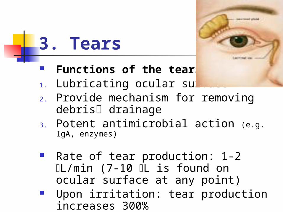
3. Tears Functions of the tears:1. Lubricating ocular surface2. Provide mechanism for removing
debris drainage3. Potent antimicrobial action (e.g. IgA,
enzymes)
Rate of tear production: 1-2 L/min (7-10 L is found on ocular surface at any point)
Upon irritation: tear production increases 300%

3. Tears The tear film is a tri-layer film: Outer (by sebaceous glands): lipid, prevent
the evaporation of the tears, Middle (by lacrimal lands): the largest layer,
aqueous, responsible for wetting properties
Inner (by goblet cells): mucinous, allows the lipid and aqueous layers to maintain adhesion across the cornea and conjuctivaAbnormalities in any of these layers causes ocular
discomfort

External Eye The visible external eye consists of the
sclera + cornea 1. Sclera: a tough, collagenous layer that
gives the eye rigidity & encases internal eye structures
Covered by 2 epithelial layers: epi-sclera + bulbar conjuctiva, contain the vascular & lymphatic systems of the anterior eye surface the source for visible eye redness during irritation & inflammation

External eye
2. Cornea: aspherical, avascular tissue that is the principal refractive element of the eye
Consists of 5 layers: epithelium, Bowman’s layer, the stroma, Descemet’s membrane & endothelium
The unique anatomic structure of the eye affects drug absorption. HOW?
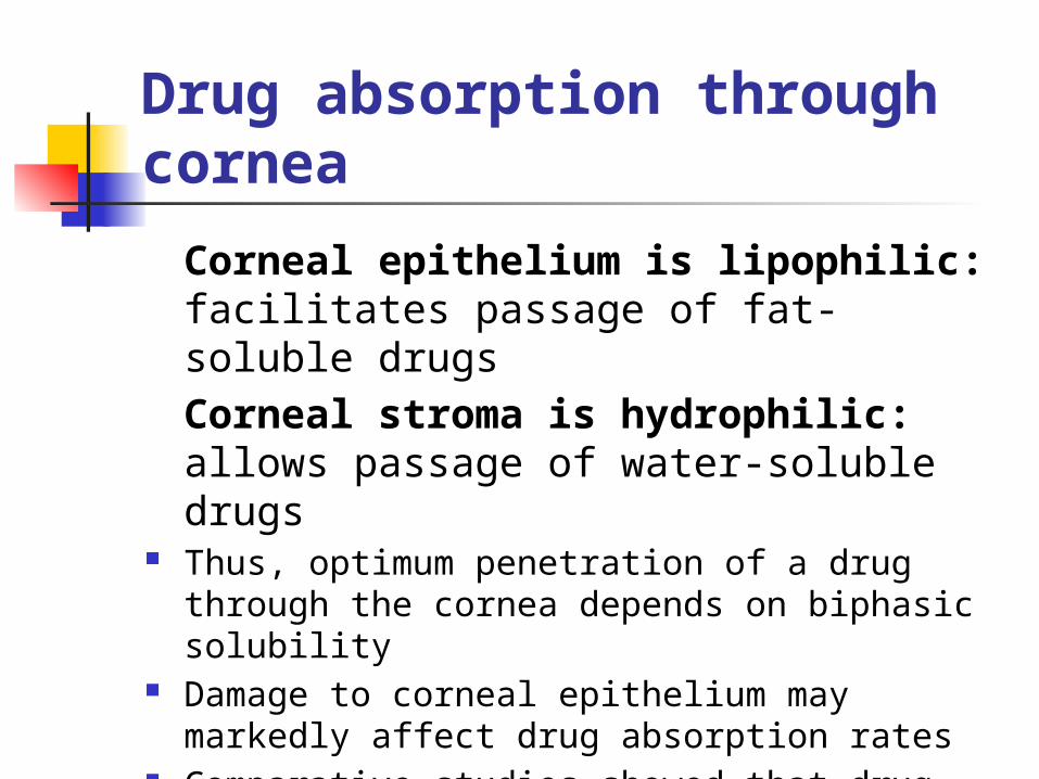
Drug absorption through cornea
Corneal epithelium is lipophilic: facilitates passage of fat-soluble drugsCorneal stroma is hydrophilic: allows passage of water-soluble drugs
Thus, optimum penetration of a drug through the cornea depends on biphasic solubility
Damage to corneal epithelium may markedly affect drug absorption rates
Comparative studies showed that drug penetration to the aqueous layer increased to threefold in compromised epithelium


Factors that may compromise epithelium:
Trauma Routine contact lens wear Topical ophthalmic anaesthetics Thermal or UV light exposure
It was found that: as much as 90% of the dose instilled in the eye may be lost. WHY IS THAT????

Factors that may hinder efforts to maintain therapeutic drug level in the eye
1. Difficulty of drug to penetrate through the cornea
2. Dilution of the drug by reflex tearing (e.g. 2.5% NaCl 0.9-0.95% within 1-2 minutes)
3. Rapid removal of drug through the tear system


Internal Eye1. Anterior chamber (behind cornea): contains
aqueous humour a. maintains normal internal eye pressure; b. provides nutritional support for lens & cornea
2. Iris: visible coloured portion of the eye regulates light striking retina, contains 2 types of muscle (sphincter & dilator)
3. Pupil: central opening of iris, Prostaglandins secreted by iris during an inflammation stimulates sphincter muscles reduces the pupil diameter
This helps to distinguish simple external eye irritation from severe internal inflammation

Internal Eye4. Crystalline lens: avascular, biconvex
structure that alters its shape to focus light on retina “accommodation”
5. Ciliary body= ciliary muscle + epithelium
- the contraction of ciliary muscle helps in “accomodation”
- during inflammation spasm of ciliary muscle fluctuating vision and pain. Thus, inhibition of ciliary muscle (cycloplegia) by anticholinergic drugs frequent treatment drug of internal ocular inflammation

Internal Eye
6. Vitreous Body: (80% of total eye volume),
filled with vitreous humour, a gel like, fluid collagen matrix that helps maintain eye volume
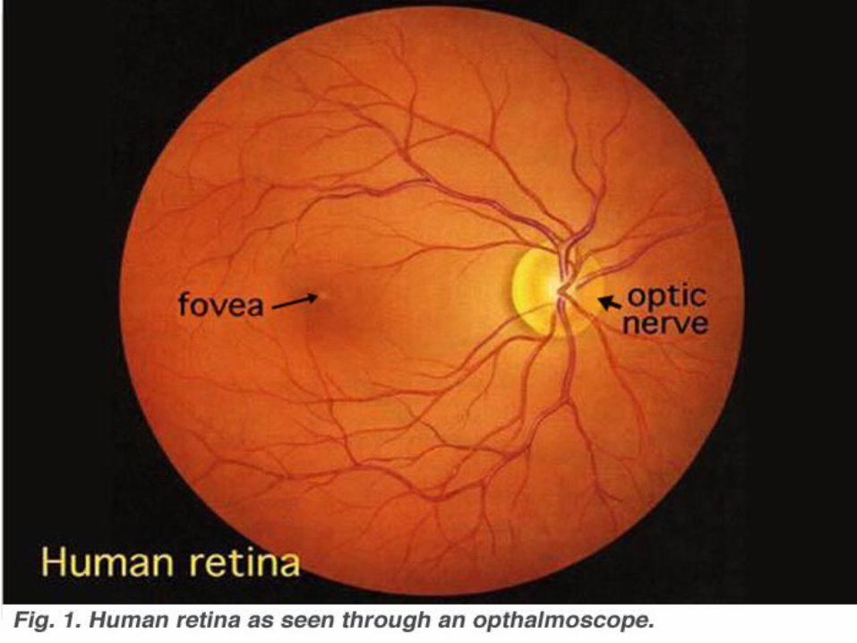
7. Retina: multi-layer neural tissue that begins the visual pathways (capture light, rods & cons, photic message optic nerve to posterior cerebral cortex visual information decoded)
Common Ocular Disorders
• Eyelid Disorders
• Ocular Surface Disorders
• Internal Eye Conditions

Common Ocular Disorders Ocular inflammation and irritation can be
caused by many conditions, some of which can be treated safely and effectively by OTC drugs
OTC drugs relieve minor symptoms: burning, itching, stinging and watering
FDA suggested self-treatment indications:- tear insufficiency - blepharitis- corneal edema - conjuctivitis- external inflammation & irritation- Hordeolum (stye)

Common Ocular Disorders
Referral is mandatory: Embedded foreign body Uveitis; ”uvea: middle coat of the eyeball, the
choroid, ciliary body & iris as a whole”
Flash burns Chemical burns Tear duct infection Corneal ulcer

Chemical eye burn
Thermal burn

1. Eyelid Disorders:
Blunt Trauma: Highly vascularised eyelid blunt
traumableeding swelling and ocular discomfort
Mostly, blunt trauma does not result in internal damage & treatment is usually supportive: A. cold compresses; B. Oral OTC analgesics
However,all individuals should be evaluated by an optometrist or ophthalmologist ASAP after the event
Complications: internal eye bleeding, secondary glaucoma, retinal detachment

1. Eyelid Disorders:Belpharitis (Very common) Almost always due to Staphylococcus
epidermidis or Staphylococcus aureus Typical: red, scaly, thickened eyelids,
often with loss of eyelashes, itching & burning (most common)
Acute or Chronic (patients aware of diagnosis)
Treatment: hot compress, topical antibiotic & eyelid scrub using an OTC eyelid hygienic preparation (baby-shampoo)

1. Eyelid Disorders:
Lice Infestation Phthirius Pubis (crab louse) & Pediculus
humanus capitis (head louse) May cause symptoms similar to those of
blepharitis Peduculicides (=lice shampoos: NIX,
RID, A-200) should not be used around the eye hypersensitivity reaction

Lice InfestationTreatment:
1. A bland ophthalmic ointment (e.g. petrolatum) used for 10 days effective because it suffocates the louse and deprives its eggs of oxygen
2. Hygienic measures: wash clothing & bedding that may contain unhatched eggs

Crab louse eyelid

1. Eyelid DisordersContact Dermatitis Cause: changing make-up , soap,
exposure to some foreign substance Symptoms: swelling, scaling of eyelid
with profuse itching Usually both eyes are involved
suggests allergy (ask patient about newly used products)
Treatment: (1) best treatment is removal of offending substances; (2) OTC oral antihistamines; (3) cold compresses

1. Eyelid DisordersHordeolum (stye) Inflammation of either Palapable, tender nodule Cause: Staphylococcus epidermidis or
Staphylococcus aureus Treatment: (1) Hot compresses 3-4
times/day (5-10 minutes; clearing within 1 week); (2) External hordeolum topical antibiotic; Internal hordeolum oral antibiotic; (3) if resistant surgical drainage
Mebomien gland (internal hordeolum)
Zeis & Moll glands (external hordeolum)
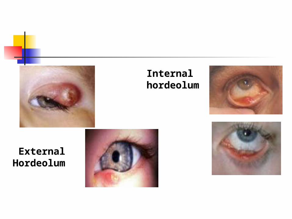
External Hordeolum
Internal hordeolum
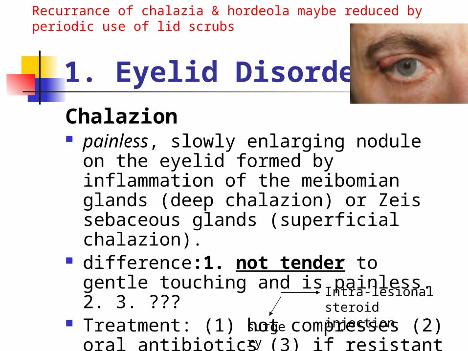
1. Eyelid DisordersChalazion painless, slowly enlarging nodule on the
eyelid formed by inflammation of the meibomian glands (deep chalazion) or Zeis sebaceous glands (superficial chalazion).
difference:1. not tender to gentle touching and is painless. 2. 3. ???
Treatment: (1) hot compresses (2) oral antibiotics (3) if resistant
Intra-lesional steroid injection
surgery
Recurrance of chalazia & hordeola maybe reduced by periodic use of lid scrubs

2. Ocular Surface Disorders
Foreign substance contact If reflex tearing does not remove the foreign
substance, the eye may need to be flushed with sterile saline or specific eye wash preparations (irrigants: witch hazel, baby shampoo)
If trapped up the eye lid may not go with flushing optometrist or ophthalmologist
Metallic foreign body NOT SELF-IRRIGATION (abrasion, scarring, chronic red eye)
Immediate medical referral

2. Ocular Surface Disorders
Abrasions “superficial injury to a skin or mucous
membrane, in this case, the cornea or conjuctiva”
Causes: Scratches by fingernails or by foreign bodies
Such injuries cause partial or total loss of the epithelium & are painful especially if cornea is involved
Self –treatment NOT RECOMMENDED because of the risk of bacteria or fungi contaminating and infecting the eye


2. Ocular Surface Disorders
Chemical Exposure Splash injury, fumes or solid chemicals
serious problem Medical Emergency immediate referral to A&E department
Initial treatment: flushing eye with sterile saline or water for at least 10 minutes
Chemical burn of the cornea

2. Ocular Surface Disorders
Thermal Damage Ranges
Minor Severee.g. exposure to UV
radiation during skiing
e.g. welder's arc (acute UV-keratoconjuctivitis)
Refer to Doctor
Treatment: artificial tears, ointments

Minor thermal burn Major thermal burn
(Welder’s arc)

2. Ocular Surface Disorders
Conjuctivitis: inflammation of bulbar conjuctiva
Viral conjuctivitis Allergic conjuctivitis Bacterial conjuctivitis Chlamydial conjuctivitis

Viral conjunctivitis The most common conjunctivitis Causes: recent cold, sore throat,
exposure to someone with pink eye (acute contagious conjunctivitis)
Symptoms: “pink-eye” with copious amounts of watery discharge; nondescript ocular discomfort; mild to moderate foreign body sensation; occasionally blurred vision; low grade fever, swollen lymph nodes
Treatment: relief major symptoms using artificial tears & ocular decongestants.
Certain forms are extremely contagious: wash hands, do not share towels, properly dispose of tissues used to blot the eye
Usually self-limiting, with symptoms resolving 1-3 weeks
Pinkeye

Allergic conjunctivitis Caused by many antigens (Ag): pollen
grains, animal dander & topical eye preparations
Symptoms: red eye with watery discharge Hallmark symptom: itching Afflicted people often report seasonal
allergic rhinitis Ask patient about recent exposure to Ag Treatment: removal of cause (best);
ocular decongestants and antihistamines; oral antihistamines; cold compresses

Bacterial conjuctivitis Staphylococcus epidermidis,
Staphylococcus aureus, Heamophilus influenza & Streptococcus pneumoniae
Symptoms: red eye with purulent discharge
Key symptom: eyelids sticking together on awakening
Self-limiting within 2 weeks, but topical antibiotics may clear the symptoms more quickly

Chlamydial conjuctivitis Classified as sexually transmitted disease
(STD) Often initially misdiagnosed with bacterial
& viral conjunctivitis, because of common signs and symptoms
Misdiagnosis is a problem: (1) as such individuals may harbor other STDs & (2) bacterial and viral conjunctivitis are self-limiting
Discourage self-treatment if symptoms are vague

Keratitis Inflammation of the cornea Either separate or with other kind of
conjunctivitis Potentially vision threatening problem Symptoms: those of conjunctivitis+1 or
more additional symptom (pain, photophobia, blurred vision)
Complications: corneal ulceration or even loss of eye (especially in those who sleep with contact lens overnight) WHY?
Pseudomonas aureginosa “the most common cause of corneal ulceration among contact lens wearers” produces collagenase that can destroy the cornea within 24 hours

Corneal Oedema Oedematous area of cornea: epithelium Causes: over-wearing of contact lens,
surgical damage to cornea, inherited cornea dystrophies
Hallmark symptom: halos or starbursts around lights, with or without reduced visionbecause: accumulation of fluid distorts optical properties of cornea
Treatment: apply hypertonic saline solution or ointment (2-5%) to dehydrate cornea
WHY?

Corneal Edema

Dry Eye Very common eye disorder Symptoms: white or mildly red eye,
sandy or gritty feeling & excess tearing Any abnormality in tear layers less
lubrication to ocular surface leads to production of more inadequate tears “vicious cycle”
Contrary to what the name suggests
What are the causes of dry eye?

Dry EyeTreatment: OTC lubricants and artificial tears (drops or
ointments)
Vitamin A: greatest benefit in treatment of severe dry eye associated with glandular tissue destruction
If very severe dry eye:
Occlusion of lacrimal drainage system to increase available tear pool
Ocular inserts of Na hyaluronate

How does artificial tears work?
Compromised Tear FilmFor dry eye sufferers, dry spots on the surface of the eye cause irriation, and may create the potential for more serious damage to the surface of the eye.

Artificial Tears
drop of artificial tears

The artificial tear solution is quickly absorbed and key ingredients rapidly work to help restore the tear film.

All layers of the normal tear film are restored

Lubricants solutions/ointments that help to alleviate
dryness of ocular surface MOA: increase viscosity of existing tears, retard
drainage and increase retention time. However, although viscous agents enhance the
ocular retention time of tear substitutes, high viscosity itself does not provide relief for all dry eye conditions (Pharmaceutical Journal; 264 (7082):212-218; 2000)
A. Artificial Tear Solutions (Demulcents) water-soluble polymers Administered 3-4 times daily
solutionsointments

1. Cellulose ethers:- HPMC (hypromellose) ; HPC, HEC,
methylcellulose, carboxymethylcellulose- Colourless and vary in viscosity- Methylcellulose 0.2-1.0%, if >2%
ointment- HEC & HPC solutions: are less viscous but
have greater emollient (cohesive, film-making) properties than methylcellulose
- The most important property of cellulose ethers: stabilize tear film (surface active properties) and prevent evaporation
- Lack toxicity & irritation

Other less viscous hydrophilic substances, such as polyvinyl alcohol (PVA) and polyvinyl pyrrolidone (povidone or PVP), have been included as the polymeric ingredients of many artificial tear formulations.
The tears of patients with dry eyes due to aqueous deficiency have been shown to have a higher osmolarity than normal subjects, a factor which may be responsible for the ocular surface disease in this condition.
In such patients, hypotonic solutions such as polyvinyl alcohol 1 per cent with an osmolarity of 150 mOsm/L have been shown to be superior to an isotonic solution of 300 mOsm/L in providing symptom relief.

2. Polyvinyl Alcohol Important: avoid using PVA with
ophthalmic products that contain: NaHCO3, Na-Borate, Na/K/Zn sulphate…..
Cause: it will react and form a thick gele.g. PVA-containing contact lens wetting
solution & irrigants containing Na-Borate
3. Povidone (polyvinyl pyrrolidone; PVP)
Exerts surface active properties similar to those of cellulose ethers forms hydrophilic layer on corneal surface, mimicking conjunctival mucin promotes wetting of ocular surface
Patients with mild dry eye may benefit from instillation of one of these artificial tear drops up to four times a day.
However, in moderate to severe cases, these preparations need to be instilled more frequently.
To overcome this problem, preparations containing a longer-acting polymer, polyacrylic acid, also known as carbomer 940, have been introduced. Such preparations have a much longer retention time in the eye and symptom relief is obtained with significantly fewer instillations.

In Jordan:
CMC
PVA+PVP PVA+1% polyethylene glycol

4. Retinol Solution An alcohol form of vitamin A What is the mechanism of action? Retinol palmitate aqueous ophthalmic
solution is used for the treatment of dry eye failing to respond to the conventional therapy with artificial tears;
The benefits of vitamin A in treatment of dry eye are speculative (lack controlled trials)

Note: (Benzalkonium Cl)
Benzalkonium chloride (BAK) is a poor choice preservative for artificial tear solution, because it has toxic effects on tear film & corneal endothelium*
A single drop BAK can break the lipid superficial layer of tear film into numerous oil droplets
Alternative preservatives: chlorhexidine, chlorbutanol, EDTA
* Reference: Lemp MA, Zimmerman LE. Toxic endothelial degeneration in ocular surface disease treated with topical medications containing benzalkonium chloride. Am J Ophthalmol 1988;105:670-3.

5. Preservative-Free Formulations For patients who are sensitive to
preservatives like benzalkonium chloride (BAK) & thimerosal*
Formed as unit-dose dispensers More expensive than products with
preservative. Requires strictly hygienic procedure:
easy to get contaminated Discard any unused solution after 12
hours* Reference: Lee-Wong M, Resnick D, Chong K.A generalized reaction to thimerosal from an influenza vaccine. Ann Allergy Asthma Immunol. 2005 Jan;94(1):90-4.

Göbbels and Spitznas have shown a decrease in epithelial permeability in patients treated with unpreserved PVP 2%, while those receiving the same preparation preserved with BAK 0.005% showed an increase in permeability.
The authors suggest that, in dry eyes, treatment with unpreserved artificial tears may lead to an objective improvement in corneal surface disease, with this effect being counteracted by preservation of tear substitutes with BAK.
Reference: Göbbels M, Spitznas M. Corneal epithelial permeability of dry
eyes before and after treatment with artificial tears. Ophthalmol 1992;99:873-8.
Evidence Based!

However,…… It is unlikely that patients purchasing
dry eye products OTC would wish to bear the cost of unit dose preparations unless they fall into the category of patients in whom preserved eye-drops are contraindicated.
WHO ARE THEY??1. patients allergic to, or intolerant of, preservative
and
2. patients who wear soft contact lenses.

What gives the cats eyes the characteristic night-time glow when they are caught in a beam of light?
A feature, which enables the amount of light hitting the retina to be increased, is the tapetum lucidum. This is positioned at the back of the eye, behind the retina and acts like a mirror, reflecting light back onto the light sensor cells in the retina.
BREAK

B. non-medicated ointments (Emollients)
Main advantage: melt at the temperature of the ocular tissue and are retained longer than other ophthalmic vehicles enhance integrity of tear film
Preferably instilled at bedtime: 1. To keep eyes moist during sleep &
improve morning symptoms of dry eye2. Because they cause blurred vision
e.g. white soft paraffin, lanolin and liquid paraffin.

Decision Making Algorithm
Clinical presentation Management
With painWith blurred visionWith photophobia (light sensitivity)With history of traumaWith contact lens wear (??)
IMMEDIATE REFERRAL
RED EYE

Decision Making Algorithm
Clinical presentation Management
With history of pink eye exposure, cold, flu,
and watery discharge
and mucous discharge
Self-treatment
Referral
RED EYE

Decision Making Algorithm
Clinical presentation Management
With known allergies and itching, watery discharge
and mucous discharge
Self-treatment
Referral
RED EYE

Decision Making Algorithm
Clinical presentation Management
With foreign body sensation
and possible contamination
and is elderly
Immediate Referral Self-treatment
RED EYE

disorders that you don’t treat in your pharmacy!
OTC
In addition to keratitis and the previous algorithms

3. Internal Eye Conditions
Uveitis: inflammation of the uvea tract (iris, ciliary body or choroid)
Causes: trauma, systemic disease, idiopathic
Symptoms: v similar to viral conjuctivitis or keratitis (mildly red eyes, pain, photophobia, blurred vision)
Complications: secondary glaucoma, destruction of iris or even blindness
Treatment: topical, depot or systemic oral steroids depending on the cause. Self-treatment is not recommended

3. Internal Eye Conditions
Angle-closure glaucoma Caused by blockage of “trabecular meshwork”
drainage system of aqueous humour increase the intraocular pressure
Occurs usually as pupil is returning to normal size after the use of a mydriatic
Symptoms: painful eye, brow ache, headache, nausea & vomiting
It is vision-threatening see doctor

OTC ophthalmic products Lubricants: 1. Artificial tear solutions
(demulcents); 2. Non-medicated ointments (emollients)
Decongestants Antihistamines Irrigants Hyperosmotics Antiseptics Eyelid scrubs Multivitamins
Decongestants Phenylphrine & Imidazoles (naphazoline,
tetrahydrozoline & oxymetazoline) -adrenoceptor agonists vasoconstriction
of conjuctival vessels If instilled to irritant/damaged cornea:
dilate pupil may precipitate angle-closure glaucoma
Systemic S.E: very rare at OTC dose Caution in patients with CVD, HTN or DM

Decongestants Most common S.E if chronic use:
rebound congestion “hyperameia” Rebound congestion is less with
(naphazoline, tetrahydrozoline) than with phenylephrine and oxymetazoline
In some patients “Xerosis” (abnormal dryness) with prolonged topical instillation of local decongestants

DecongestantsNapahzoline (0.02%): - The ocular decongestant of choice:
higher efficacy and relative lack of S.E
in addition to constricting conjuctival vessels, it reduces pain & tearing associated with ocular inflammation
Patients with lightly pigmented irides (blue or green eyes) are more sensitive to the mydriatic effects of naphazoline

AntihistaminesPheniramine maleate & Antazoline
phosphate Indication: rapid relief of symptoms
associated with seasonal allergic conjunctivitis
Almost always used along with naphazoline: much more effective than if used individually
Dose: 1-2 drops applied to each eye 3-4 times daily
May cause mydriasis, because of anticholinergic
C/I: sensitivity to one of the components or known risk to angle-closure glaucoma

Irrigants Cleanse ocular tissues while maintaining
their moisture Must be physiologically balanced: pH &
osmolality Uses: (1) after certain clinical procedures
to wash away mucus & debris from eye (2) to clean eyes in between changes of ocular dressings (3) wash out eyes after wearing contact lens (4) initial ocular lavage after chemical injuries to eye before seeing doctor

Hyperosmotics Promote movement of fluid from cornea NaCl: solution and ointment (2-5%) 5% is more effective, but causes stinging,
burning but 2% is preferable for long term use
1-2 drops instilled 3-4 times daily Several instillations in the 1st few waking
hours are helpful as vision associated with corneal edema is worse on awakening
Non toxic and very rarely to cause allergy

Antiseptics To reduce bacterial population on ocular
surface including eyelid margins May be recommended for patients with
minor conjuctival or eyelid inflammation that is possibly associated with infectious organisms
Examples: silver protein, Boric acid & zinc sulphate, distilled witch hazel

Silver protein: A colloidal preparation of silver oxide and protein, usually gelatin or albumin, used as an antibacterial agent.
Treatment of ocular infections and preoperative use in ocular surgery
At low doses: antimicrobial activity against gram +ve and gram –ve bacteria
Preoperative: 2-3 drops instilled then rinse with sterile irrigating solution
In mild infections: several drops instilled every 3-4 hours for several days
Avoid frequent topical application for prolonged periods of time may cause permanent discoloration of eyelid skin or conjunctiva “argyria”
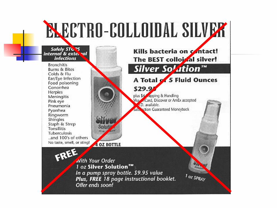

Argyria

Boric acid Treatment of irritated, inflamed eyelids Applied in small quantity on the inner
surface of the lower eyelid once or twice daily
Zn sulphate Mild astringent for temporary relief of
minor ocular irritation Also effective in infections cause by
moraxella (uncommon gram –ve bacteria, member of the URT normal flora, occasionally cause infections)

Eyelid scrubs Best treatment for blepharitis is
maintain eyelid hygiene This is best done by hot compresses 15-
20 minutes 2-4 times daily followed by eye lid scrubs using baby shampoo with cotton pad or a gauze
Application Technique