Open access Original research Crocodylus porosus: a ...
22
1 Jeyamogan S, et al. BMJ Open Science 2020;4:e100040. doi:10.1136/bmjos-2019-100040 Open access Crocodylus porosus: a potential source of anticancer molecules Shareni Jeyamogan, 1 Naveed Ahmed Khan , 2 K Sagathevan, 3 Ruqaiyyah Siddiqui 2 This article has received OSF badges for Open data. To cite: Jeyamogan S, Khan NA, Sagathevan K, et al. Crocodylus porosus: a potential source of anticancer molecules. BMJ Open Science 2020;4:e100040. doi:10.1136/ bmjos-2019-100040 ► Prepublication history for this paper is available online. To view these files, please visit the journal online (http://dx.doi.org/ 10.1136/bmjos-2019-100040). Received 07 August 2019 Revised 09 February 2020 Accepted 19 March 2020 1 Department of Biological Sciences, Sunway University, Bandar Sunway, Selangor, Malaysia 2 Department of Biology, Chemistry and Environmental Sciences, American University of Sharjah, University City, Sharjah, United Arab Emirates 3 Science and Technology, Sunway College, Bandar Sunway, Selangor, Malaysia Correspondence to Professor Naveed Ahmed Khan; [email protected] Original research © Author(s) (or their employer(s)) 2020. Re-use permitted under CC BY. Published by BMJ. Strengths and limitations of this study ► To our knowledge, this is the first detailed study performed to investigate the presence of anticancer activity, anticancer molecules and peptides from the serum of Crocodylus porosus as well as the differ- ential gene expression of cancer cells treated with crocodile serum. ► Adequate steps were taken to limit the risk of bias: the primary end point was prespecified, and the number of samples to be tested was determined beforehand to ensure sufficient clarity. ► Experiments were substantiated by repetition, under a range of conditions that demonstrate the robust- ness of the effects observed. The data are repre- sentative as the mean±SE of three independent experiments. ► The present study involved serum collection from one crocodile and data obtained from subsequent experiments conducted, are hypothesis-confirming experiments and should be interpreted as such, being in the exploratory phase of research. Further validation of results is needed. ABSTRACT Background Cancer remains a global threat resulting in significant morbidity and mortality despite advances in therapeutic interventions, suggesting urgency for identification of anticancer agents. Crocodiles thrive in polluted habitat, feed on germ-infested meat, are exposed to carcinogenic heavy metals, are the very few species to survive the catastrophic Cretaceous–Paleogene extinction event, yet have a prolonged lifespan and rarely been reported to develop cancer. Therefore, we hypothesised that animals living in polluted environments such as crocodiles possess anticancer molecules/mechanisms. Methods Crocodylus porosus was procured, blood collected, dissected and lysates prepared from internal organs. Organ lysates and sera were tested for growth inhibition, cytotoxic effects and cell survival against HeLa, PC3 and MCF7 cells and subjected to liquid chromatography mass spectrometry. RNA transcriptome analysis and differential gene analysis were performed using Galaxy Bioinformatics. Results Sera exhibited potent growth inhibition and cytotoxic effects against cancer cells. 80 molecules were detected from C. porosus and 19 molecules were putatively identified. Additionally, more than 100 potential anticancer peptides were identified from sera using bioinformatics based on peptide amino acid composition, binary profile, dipeptide composition and pseudo-amino acid composition. Following transcriptome analysis, 14 genes in treated HeLa cells, 51 genes in treated MCF7 cells and 2 genes in treated PC3 cells, were found to be expressed, compared with untreated controls. Conclusion Animals residing in polluted milieus are an unexploited source for prospective pharmaceutical drugs, and could lead to identification of novel antitumour compound(s) and/or further understanding of the mechanisms of cancer resistance. INTRODUCTION An significant increase in the number of cancer cases and cancer deaths was observed from 2000 to 2018 despite advances in ther- apeutic interventions and supportive care. 1–3 GLOBOCAN reports demonstrated an increase in the number of cancer cases from 10.1 million in 2000 to 18.1 million in 2018 while the number of deaths has increased from 6.2 million in 2000 to 9.6 million cases in 2018. 1–3 Therefore, there is an urgent need for the discovery and development of new and efficient anticancer agents. Animals such as crocodiles inhabit unsan- itary and polluted environments, feed on rotten meat which is present with numerous pathogenic microbes, are continuously exposed to heavy metals that are genotoxic and carcinogenic such as arsenic, nickel, zinc, cadmium, cobalt, mercury, selenium, lead and chromium 4–7 and are also the very few species to survive the Cretaceous–Paleo- gene mass extinction despite being exposed to extreme levels of radiation. 4 8–10 Despite all of the above, these animals have prolonged lifespan and rarely develop cancer. Our lab has recently hypothesised that animals living in polluted environments such as crocodiles possess mechanisms or molecules against cancer development. In support, our previous studies showed that organ lysates of Crocodylus palustris inhibited the growth and demonstrated killing effects against PC3 cells. 4 Moreover, previous studies on March 13, 2022 by guest. Protected by copyright. http://openscience.bmj.com/ BMJ Open Science: first published as 10.1136/bmjos-2019-100040 on 27 October 2020. Downloaded from
Transcript of Open access Original research Crocodylus porosus: a ...

1Jeyamogan S, et al. BMJ Open Science 2020;4:e100040. doi:10.1136/bmjos-2019-100040
Open access
Crocodylus porosus: a potential source of anticancer molecules
Shareni Jeyamogan,1 Naveed Ahmed Khan ,2 K Sagathevan,3 Ruqaiyyah Siddiqui2
This article has received OSF badges for Open data.
To cite: Jeyamogan S, Khan NA, Sagathevan K, et al. Crocodylus porosus: a potential source of anticancer molecules. BMJ Open Science 2020;4:e100040. doi:10.1136/bmjos-2019-100040
► Prepublication history for this paper is available online. To view these files, please visit the journal online (http:// dx. doi. org/ 10. 1136/ bmjos- 2019- 100040).
Received 07 August 2019Revised 09 February 2020Accepted 19 March 2020
1Department of Biological Sciences, Sunway University, Bandar Sunway, Selangor, Malaysia2Department of Biology, Chemistry and Environmental Sciences, American University of Sharjah, University City, Sharjah, United Arab Emirates3Science and Technology, Sunway College, Bandar Sunway, Selangor, Malaysia
Correspondence toProfessor Naveed Ahmed Khan; naveed5438@ gmail. com
Original research
© Author(s) (or their employer(s)) 2020. Re- use permitted under CC BY. Published by BMJ.
Strengths and limitations of this study
► To our knowledge, this is the first detailed study performed to investigate the presence of anticancer activity, anticancer molecules and peptides from the serum of Crocodylus porosus as well as the differ-ential gene expression of cancer cells treated with crocodile serum.
► Adequate steps were taken to limit the risk of bias: the primary end point was prespecified, and the number of samples to be tested was determined beforehand to ensure sufficient clarity.
► Experiments were substantiated by repetition, under a range of conditions that demonstrate the robust-ness of the effects observed. The data are repre-sentative as the mean±SE of three independent experiments.
► The present study involved serum collection from one crocodile and data obtained from subsequent experiments conducted, are hypothesis- confirming experiments and should be interpreted as such, being in the exploratory phase of research. Further validation of results is needed.
AbStrACtbackground Cancer remains a global threat resulting in significant morbidity and mortality despite advances in therapeutic interventions, suggesting urgency for identification of anticancer agents. Crocodiles thrive in polluted habitat, feed on germ- infested meat, are exposed to carcinogenic heavy metals, are the very few species to survive the catastrophic Cretaceous–Paleogene extinction event, yet have a prolonged lifespan and rarely been reported to develop cancer. Therefore, we hypothesised that animals living in polluted environments such as crocodiles possess anticancer molecules/mechanisms.Methods Crocodylus porosus was procured, blood collected, dissected and lysates prepared from internal organs. Organ lysates and sera were tested for growth inhibition, cytotoxic effects and cell survival against HeLa, PC3 and MCF7 cells and subjected to liquid chromatography mass spectrometry. RNA transcriptome analysis and differential gene analysis were performed using Galaxy Bioinformatics.results Sera exhibited potent growth inhibition and cytotoxic effects against cancer cells. 80 molecules were detected from C. porosus and 19 molecules were putatively identified. Additionally, more than 100 potential anticancer peptides were identified from sera using bioinformatics based on peptide amino acid composition, binary profile, dipeptide composition and pseudo- amino acid composition. Following transcriptome analysis, 14 genes in treated HeLa cells, 51 genes in treated MCF7 cells and 2 genes in treated PC3 cells, were found to be expressed, compared with untreated controls.Conclusion Animals residing in polluted milieus are an unexploited source for prospective pharmaceutical drugs, and could lead to identification of novel antitumour compound(s) and/or further understanding of the mechanisms of cancer resistance.
InTroducTIonAn significant increase in the number of cancer cases and cancer deaths was observed from 2000 to 2018 despite advances in ther-apeutic interventions and supportive care.1–3 GLOBOCAN reports demonstrated an increase in the number of cancer cases from 10.1 million in 2000 to 18.1 million in 2018 while the number of deaths has increased from 6.2 million in 2000 to 9.6 million cases in 2018.1–3 Therefore, there is an urgent need
for the discovery and development of new and efficient anticancer agents.
Animals such as crocodiles inhabit unsan-itary and polluted environments, feed on rotten meat which is present with numerous pathogenic microbes, are continuously exposed to heavy metals that are genotoxic and carcinogenic such as arsenic, nickel, zinc, cadmium, cobalt, mercury, selenium, lead and chromium4–7 and are also the very few species to survive the Cretaceous–Paleo-gene mass extinction despite being exposed to extreme levels of radiation.4 8–10 Despite all of the above, these animals have prolonged lifespan and rarely develop cancer.
Our lab has recently hypothesised that animals living in polluted environments such as crocodiles possess mechanisms or molecules against cancer development. In support, our previous studies showed that organ lysates of Crocodylus palustris inhibited the growth and demonstrated killing effects against PC3 cells.4 Moreover, previous studies
on March 13, 2022 by guest. P
rotected by copyright.http://openscience.bm
j.com/
BM
J Open S
cience: first published as 10.1136/bmjos-2019-100040 on 27 O
ctober 2020. Dow
nloaded from

2 Jeyamogan S, et al. BMJ Open Science 2020;4:e100040. doi:10.1136/bmjos-2019-100040
Open access
Figure 1 The species, scientific classification, habitat and diet of crocodile (Crocodylus porosus).
have shown that bile products of crocodile (Crocodylus siamensis) inhibited the growth of cancer cells such as human cholangiocarcinoma cells that include Mz- ChA-1 cells, QBC939 cells, Sk- ChA-1 cells,11 12 A2780 human ovarian cancer cells13 and human gastric adenocarci-noma BGC823 cells.14Furthermore, it was shown that leucrocin I, peptide isolated from the blood extracts of C. siamensis, was also capable of inducing cell death.15 Here we have undertaken a detailed study to investigate the presence of anticancer activity, potential anticancer mole-cules and peptides from the serum of Crocodylus porosus. Furthermore, we also investigated the differential gene expression of cancer cells treated with crocodile serum. The discovery of anticancer molecule/mechanism from crocodile can pave the way for the discovery and develop-ment of therapeutic interventions.
MAteriAlS And MethodSEthics committee consent and procurement of crocodileThe saltwater crocodile, C. porosus was donated by a Convention on International Trade in Endangered Species of Wild Fauna and Flora (CITES)- registered croc-odile farm (figure 1). Handling of the animal, anaesthesia and dissection of the internal organs were carried out by the pathologist at the farm. Personnel at the crocodile farm routinely perform such procedures.
Chemicals and reagentsThe chemicals and reagents used for the assays in this manuscript comprise of lactate dehydrogenase (LDH) cytotoxicity kit (Roche Diagnostics, Indianapolis, USA), Trypan blue (Merck Millipore, Germany), pronase enzyme from Streptomyces griseus (Cat. No. 10165921001; Roche Applied Science, Switzerland), Quick Start Brad-ford dye and Quick Start bovine serum albumin stand-ards (Bio- Rad Laboratories, Hercules, California, USA), protease inhibitor (Problock Gold Mammalian, St Louis, Missouri, USA), Roswell Park Memorial Institute 1640, L- glutamine solution, fetal bovine serum (FBS) and trypsin 2.5% solution (Serana, Pessin, Germany), peni-cillin streptomycin antibiotic solution (Life Technologies, Carlsbad, California,USA), minimum essential medium non- essential amino acid (MEM NEAA) solution (Sigma Aldrich, St Louis, Missouri, USA, high- performance liquid chromatography (HPLC)- graded methanol, HPLC- graded formic acid and HPLC- graded acetonitrile (Merck Group, Darmstadt, Germany), ultra- pure deion-ised MiLi- Q water (EMD Millipore, Burlington, USA), ammonium bicarbonate, trifluoroethanol, dithiothreitol and iodoacetamide (Nacalai Tesque, Kyoto, Japan) unless stated otherwise.
Sample collectionBriefly, crocodile blood was collected in sterile ethylen-ediaminetetraacetic acid (K2EDTA) vacutainers (Becton Dickinson, Franklin Lakes, New Jersey, USA) and the internal organs were dissected out using aseptic tech-niques by a pathologist of CITES- registered crocodile farm. A female crocodile, weighing 25 kg, measuring 193 cm in length and 43 cm in width was caught, phys-ically restrained and the blood was collected using a 26- gauge needle from the supravertebral vein,16 post- occipital venous sinus17 on the head and from the ventral coccygeal vein that is located on the tail region.18 Addi-tionally, internal organs were collected by making an inci-sion along the ventral midline, from the cloaca until the lower jaw.19 The internal organs were collected in sterile containers (figure 2). These procedures are routinely carried out by the personnel at the farm, and were conducted by these professionals.
Preparation of organ lysates and serumBriefly, crocodile blood was centrifuged at 3000 × g for 15 min at 37°C.20 21 Next, the supernatant was collected and stored at −80°C in aliquots until tested further.4 For the preparation of crude organ lysates, the internal organs were cut to small pieces and homogenised using mortar and pestle22 with sterile distilled water as the solvent. Of note, 10 µL/mL of protease inhibitors and EDTA was then added to the lysates. Subsequently, lysates were subjected to 10 rounds of freeze- thaw, sonicated on ice for approx-imately 3 min at 20 kHz using a Cole–Parmer Ultrasonic Processor and centrifugated at 20 000 × g for 80 min at 4°C.4 23 Next, the supernatants were collected, filtered using sterile 0.2 µm pore- sized syringe filters (Sartorius,
on March 13, 2022 by guest. P
rotected by copyright.http://openscience.bm
j.com/
BM
J Open S
cience: first published as 10.1136/bmjos-2019-100040 on 27 O
ctober 2020. Dow
nloaded from

3Jeyamogan S, et al. BMJ Open Science 2020;4:e100040. doi:10.1136/bmjos-2019-100040
Open access
Figure 2 Dissection of crocodile (Crocodylus porosus). A female crocodile, of species C. porosus was dissected (A). The various body organs of the crocodile were dissected out, including (B) large intestines and caecum, (C) kidneys, (D) gall bladder, (E) fat tissue, (F) eyes, (G) muscles, (H) thyroid, (I) trachea, (J) stomach and gastroliths, (K) lungs, (L) liver, (M) heart and (N) small intestines.
Germany) and stored at −80°C in aliquots until tested further.4 The protein concentration of crude organ lysates and serum was determined via Bradford assay.4
Culture of cell linesHuman cervical adenocarcinoma cells (HeLa) (American Type Cell Culture (ATCC)CCL-2), human breast adeno-carcinoma cells (MCF7) (ATCCHTB-22) and human prostate adenocarcinoma cells (PC3) (ATCCCRL-1435) were obtained from ATCC. Human keratinocyte skin cells (Hacat) (CVCL_0038 and Cell Lines Service (CLS): 300493) were procured from CLS, Germany. Briefly, cells were cultivated in Roswell Park Memorial Institute-1640 supplemented with 10% (v/v) FBS, 1% L- glutamine, 1% MEM NEAA and 1% of antibiotic solution containing 10 000 U/mL of penicillin and 10 000 µg/mL strepto-mycin. The cells were incubated at 37°C in a 95% humid-ified incubator with 5% carbon dioxide (CO2).4 24
Growth inhibition assayA growth inhibition assay was done to investigate the ability of organ lysates and serum to inhibit the growth of cancer cells. Briefly, 3×10⁴ cells were cultured onto 96- well plates and incubated at 37°C in a 95% humidi-fied incubator with 5% CO2 until an approximately 50% semiconfluent monolayer of cells was achieved. Next, media were removed and cells were treated with 100 µg/mL of organ lysates and 10% (v/v) of sera for 24 hours. An initial cell count was performed on the control well to determine the number of cells present in the monolayer of the 50% semiconfluent well. Once a 100% confluent monolayer of cells were achieved in the control well, the media was discarded and the cells were detached by incu-bation with 2.5 g/L trypsin solution for 5 min at 37°C. Next, fresh media supplemented with 10% (v/v) FBS was
added to stop trypsin activity and cells were centrifuged at 3000 × g for 5 min. Supernatant was then discarded, the cell pellet was resuspended in fresh media and viable cells were counted using Trypan blue exclusion assay. The membrane of cells treated with organ lysates and serum/haemolymph, if damaged, enabled penetration of Trypan blue dye. As a result, these damaged and non- viable cells were stained blue. In contrast, intact membrane of viable cells prevented the penetration of Trypan blue dye, resulting in unstained cells.25 The percentage of cell growth was calculated by comparing the number of viable cells present in treated wells and control wells containing 10% (v/v) FBS (control for serum). The growth inhibi-tion was calculated as follows: Total cells per mL=Total cells counted × [dilution factor/number of counted squares] × 10 000 cells per mL. The data are representa-tive of the mean±SE of at least three independent experi-ments performed in duplicate.
Cell cytotoxicity assayCell cytotoxicity assay was done to investigate the cytotoxic effects of organ lysates and serum against cancer cells. Briefly, 4×10⁵ cells were cultivated in 96- well plates and incubated overnight to achieve confluent monolayers. Next, media was replaced and cells were treated with 100 µg/mL of organ lysates and 10% of serum for 24 hours at 37°C in a 5% CO2 incubator. The negative control wells were treated with media alone and FBS (control for serum). After 24 hours, the supernatant of each well was collected and percentage cell death was determined using LDH cytotoxicity detection kit.4 LDH is a soluble cytosolic enzyme which is present in all viable cells. Cells with affected and damaged membrane permeability lead to the release of LDH enzymes from the cytoplasm to the surrounding matrix. The cell supernatant containing LDH enzyme catalyses the conversion of lactate to pyruvate, resulting in the generation of NADH and H+. Following that, the diaphorase enzyme (catalyst solution from the kit) transfers the H and H+ from NADH and H+ to the tetrazolium salt p- iodonitrotetrazolium violet (solu-tion in the kit), resulting in the reduction of this colour-less salt to the red formazan dye. The absorbance of each well was then measured via a microplate reader at 490 nm. The positive control well that represents total cell death was prepared by incubating the cells with 0.1% Triton X-100 at 37°C for 60 min. The percentage cell death was determined as follows: test value – control value/total LDH release – control value × 100 = %cytotoxicity. The data are representative of the mean±SE of at least three independent experiments performed in duplicate.
Cell survival assayCell survival assay was done to investigate the revival potential of cancer cells treated with organ lysates and serum. Briefly, cells were grown to confluency in 96 plates. Next, media was removed and the cells were treated with 100 µg/mL and 10% of serum for 24 hours. The nega-tive control wells were treated with media alone and FBS
on March 13, 2022 by guest. P
rotected by copyright.http://openscience.bm
j.com/
BM
J Open S
cience: first published as 10.1136/bmjos-2019-100040 on 27 O
ctober 2020. Dow
nloaded from

4 Jeyamogan S, et al. BMJ Open Science 2020;4:e100040. doi:10.1136/bmjos-2019-100040
Open access
Table 1 The growth and cytotoxic effects of serum and organ lysates of crocodile (Crocodylus porosus) against human cancer and normal cells
Animal species Lysates/ serum
Growth (%) Cytotoxicity (%)
HeLa PC3 MCF7 HeLa PC3 MCF7
Crocodile(C. porosus)
Fat water 100 98±2.39 100 1±1.25 18±1.69 0
Fat 100 100 97±2.84 0 17±2.22 0
Liver 87±8.68 100 88±12.30 40±1.51* 28±0.29* 0
Tail muscles 100 100 96±0.39 12±1.47 17±1.08 0
Stomach 100 100 94±0.58 19±1.04* 0 0
Thyroid 0* 30±9.55 61±8.80 29±0.86* 17±5.26 0
Eyes 100 100 98±1.89 36±7.61 19±0.32 0
Heart 100 100 96±3.78 37±0.03* 27±1.59* 0
Trachea 100 100 100 0 3±2.71 0
Lungs 100 99±0.80 100 13±2.87 6±0.35 0
Large intestines 100 100 100 1±1.26 6±0.32 0
Gall bladder 0* 0* 0* 49±0.38* 21±1.71* 62±2.90*
Small intestines 100 100 100 0 0 0
Kidneys 0* 0* 70±13.78 0 0 0
Serum 0* 0* 0* 55±0.55* 56±5.83 50±1.57*
Briefly, semiconfluent cells were incubated with 100 µg/mL of lysates and 10% serum and growth inhibitory effects were determined. For cytotoxicity assessment, confluent cells were incubated with lysates and serum. Note that serum, gall bladder and thyroid of C. porosus showed growth inhibition as well as cytotoxicity against cancer cells (p<0.05 using independent t- test, two- tailed distribution). Asterisk and bold denote significant (p<0.05) difference. Data are presented as the mean±SE of at least three independent experiments performed in duplicates.
(control for serum). Next, the supernatant was discarded and the cells were detached using 2.5 g/L trypsin for 5 min. Media supplemented with 10% (v/v) FBS was then added to stop the activity of trypsin and the cell suspen-sion was subjected to centrifugation at 3000 × g for 5 min. The cell pellet was then resuspended with fresh media and regrown in 96- well plates. After 24 hours, the number of cells were enumerated to determine cell growth. The data are representative of the mean±SE of at least three independent experiments performed in duplicate.
heat inactivation of crocodile serumTo investigate the nature of potential anticancer mole-cules from crocodile serum, the serum was heat inacti-vated by boiling at 56°C and 65°C for 30 min and 99°C for 5 min26–28 and cooling at 4°C. HeLa cells were then incu-bated with 10% (v/v) heat- treated serum for 24 hours at 37°C in a 5% CO2 incubator and subjected to LDH cyto-toxicity assay as described previously.4 The data are repre-sentative of the mean±SE of at least three independent experiments performed in duplicate.
Crocodile serum protein digestion using pronase enzymeCrocodile serum was treated with 7U of pronase enzyme that was prepared as a stock solution of 1 mg/mL for 1 hour at 37°C in a 5% CO2 incubator. Next, HeLa cells were treated with 10% (v/v) of pronase- treated serum at 37°C for 24 hours in a 5% CO2 incubator. These cells were then subjected to LDH cytotoxicity assay as previously
described.4 Negative control wells consisted of HeLa cells treated with fresh media alone and pronase- treated FBS. The data are representative of the mean±SE of at least three independent experiments performed in duplicate.
liquid chromatography mass spectrometry analysis of small molecules in sera samplesLiquid–liquid extraction method using a mixture of ice- cold methanol and ultra- pure deionised MiLi- Q water was performed on the samples.29 Briefly, 800 µL of ice- cold mixture containing HPLC- grade methanol and ultra- pure deionised MiLi- Q water at a ratio of (8:1) (v/v) was mixed with 100 µL of serum, vortexed for 2 min and stored at 4°C. After 30 min, sample was subjected to centrifuga-tion at 7000 × g for 8 min, and supernatant was collected, filtered and stored at −80°C. Liquid chromatography mass spectrometry (LC- MS) analyses were done using Agilent 1290 infinity liquid chromatography (LC) system that was linked to Agilent 6520 accurate- mass quadrupole–time of flight (Q- TOF) mass spectrometer with dual electro-spray ionisation (ESI) source (Agilent Technologies, Santa Clara, California, USA). Briefly, 1.0 µL of sample was injected and the chromatographic separation was achieved using Agilent Zorbax Eclipse XDB- C18, narrow bore 2.1×150 mm, 3.5 µm (P/N: 9 30 990–902) column. The column temperature was maintained at 25°C and the samples were housed in an autosampler at 4°C. Solvent A (0.1% formic acid in water) and solvent B (0.1% formic
on March 13, 2022 by guest. P
rotected by copyright.http://openscience.bm
j.com/
BM
J Open S
cience: first published as 10.1136/bmjos-2019-100040 on 27 O
ctober 2020. Dow
nloaded from

5Jeyamogan S, et al. BMJ Open Science 2020;4:e100040. doi:10.1136/bmjos-2019-100040
Open access
Figure 3 (A–R) Cell survival ability of HeLa cells treated with lysates of crocodile (Crocodylus porosus). Representative images of HeLa cells with and without crude lysates (A: control; B: bovine serum albumin; C: fetal bovine serum; D: fat water; E: fat; F: liver; G: tail muscles; H: stomach; I: thyroid; J: eyes; K: heart; L: trachea; M: lungs; N: large intestines; O: gall bladder; P: small intestines; Q: kidneys and R: serum).
Figure 4 (A–B) The cytotoxic effects of heat- inactivated crocodile (Crocodylus porosus) serum (100× magnification). Briefly, HeLa cells were incubated with 10% crude unboiled, 56°C and 65°C boiled serum at 37°C for 24 hours. Next day, cell cytotoxicity assay was performed by determining the concentration of lactate dehydrogenase enzyme released by the affected cells at 490 nm. Representative images of HeLa cells treated with boiled and unboiled serum and FBS control. (i: FBS control; ii: unboiled serum; iii: serum boiled at 56°C; iv: serum boiled at 65°C). FBS, fetal bovine serum.
acid in acetonitrile) with a flow rate of 0.5 mL/min were used for the mobile phase. Nitrogen gas (carrier gas) had a flow rate of 10 L/min and temperature that was set at 300°C. For mass spectrometry (MS), LC eluents were analysed with an Agilent 6520 Accurate- Mass Q- TOF mass spectrometer with ESI source in positive and negative ionisation mode. The mass range was set to 100–3200 m/z using both negative and positive ionisation mode. The fragmentation voltage was set at 125 V and the capillary voltage was set at 4000 V for positive ionisation mode and 3500 V for negative ionisation mode. The raw LC- MS data were acquired and processed using the Molecular Feature Extraction in Agilent Mass Hunter Qualitative Analysis
B.05.00 software. Only peaks with abundance value of above 5000 counts with a relative height of more than 2.5% were selected. The small molecules were identified using the METLIN Personal Metabolite Database and Molecular Formula Generation software (Agilent Tech-nologies). The data are representative of the mean±SE of at least three independent experiments performed in duplicate.
identification of potential anticancer peptidesProtein samples were digested as described previously using In- solution digestion method.30 Approximately 1 mg/mL of serum protein was mixed with 50 µL of 100 mM ammonium bicarbonate, 50 µL of trifluoroeth-anol and 2 µL of 200 mM dithiothreitol, vortexed and incubated for 1 hour at 60°C. To alkylate the proteins, 8 µL of 200 mM iodoacetamide was added and incubated for 1 hour in the dark at room temperature. Next, 2 µL of 200 mM dithiothreitol was added into the mixture and incubated for 1 hour in the dark for the removal of excess
on March 13, 2022 by guest. P
rotected by copyright.http://openscience.bm
j.com/
BM
J Open S
cience: first published as 10.1136/bmjos-2019-100040 on 27 O
ctober 2020. Dow
nloaded from

6 Jeyamogan S, et al. BMJ Open Science 2020;4:e100040. doi:10.1136/bmjos-2019-100040
Open access
Figure 5 (A–B) The cytotoxic effects of pronase- treated crocodile (Crocodylus porosus) serum (100× magnification). Briefly, HeLa cells were incubated with serum and pronase- treated serum at 37°C for 24 hours. Next day, cell cytotoxicity assay was performed by determining the concentration of lactate dehydrogenase enzyme released by the affected cells at 490 nm. Representative images of HeLa cells treated with serum and pronase- treated serum (i: FBS control; ii: serum; iii: pronase- treated serum). FBS, fetal bovine serum.
iodoacetamide. The pH of the mixture was adjusted to pH 7–9 using 600 µL of distilled water and 200 µL of 100 mM ammonium bicarbonate. Next, 50 µL of 2.5 g/L trypsin was added to the protein mixture and incubated overnight at 37°C. To deactivate trypsin activity, 1 µL of formic acid was added and the mixture was stored at −80°C before analysis. The samples were subjected to LC- MS/MS (iquid chromatography–mass spectrometry (LC- MS)) analyses using Agilent 1200 HPLC- Chip/MS Interface, coupled with Agilent 6550 iFunnel Q- TOF LC/MS. Briefly, 1.0 µL of digested sample was injected and separation of molecules was achieved using Agilent Large Capacity Chip, 300 Å, C18, 160 nL enrichment column and 75 µm × 150 mm analytical column (P/N: G4240-62010) which was equilibrated with 0.1% formic acid in water (solvent A). The samples were housed in an autosampler at 4°C and eluted via an increasing gradient of 90% acetonitrile in 0.1% formic acid in water (solvent B) as follows: 5%–75% solution B from 0 to 30 min and 75% from 30 to 39 min with a flow rate of 4 µL/min from Agilent 1200 Series capillary pump and 0.5 µL/min from
Agilent 1200 Series nano pump. Nitrogen gas (carrier gas) had a flow rate of 5 L/min and temperature that was set at 325°C. For MS, LC eluents were analysed with an Agilent 6550 iFunnel Q- TOF LC- MS/MS in positive ioni-sation mode. The protein spectrum mass range was set to 110–3000 m/z for MS and 50–3000 m/z for MS/MS scan. The fragmentation voltage was set at 360 V and the capil-lary voltage was set at 2050 V. The mass spectrum analysis was achieved with Agilent Mass Hunter data acquisition software and PEAKS V.7.0 software.
Serum peptide detection and protein identification by automated de novo sequencingFrom the list of peptides detected, automated de novo sequencing was performed using PEAKS Studio V.7.0 (Bioinformatics Solution, Waterloo, Canada). For protein identification and homology search by de novo sequence comparison, SwissProt.Crocodylia (January 2019) data-base was used for crocodile (C. porosus). Enzyme used for protein digestion was set as trypsin and the false discovery rate of 1% and ≥2 unique peptides were set as parameters to exclude inaccurate proteins. A −10lgP score of greater than 20 was set to indicate proteins with high confidence scores. The presence of potential anticancer peptides (ACP) was predicted using the Machine- Learning- Based Prediction of Anticancer Peptides (MLACP) tool.31
differential gene expression analysis of cancer cells treated with crocodile seraBriefly, cells grown to confluency were treated with 10% of serum for 40 min. The control well cells were treated with FBS. Next, the supernatant was discarded and the cells were detached using 2.5 g/L trypsin for 5 min. Phosphate- buffered saline supplemented with 10% (v/v) FBS was then added to stop trypsin activity and cell suspension was subjected to centrifugation at 300 × g for 5 min. The supernatant was discarded and RNA was extracted from the treated cells as per manufacturer’s guidelines. The cell pellet was resuspended with 700 µL of Qiazol cell lysis buffer, vortexed, homogenised using syringe and needle and incubated at room temperature for 5 min. Next, 140 µL chloroform were added and the sample was mixed by inverting the tubes for 15 s followed by incuba-tion at room temperature for 3 min for the separation of aqueous and organic phase. The sample was then centri-fuged at 12 000×g for 15 min at 4°C and the RNAs on the uppermost layer (aqueous phase) were collected. The sample was then washed, purified and eluted using the buffers provided in the kit as per manufacturer’s guide-lines (Qiagen miRNeasy Kit, Canada). RNA concentra-tion and quality were assessed using BioDrop Duo Micro- Volume UV–Vis Spectrophotometer. Pure RNA samples should comprise of a ratio between 1.8 and 2.0 when measured at the spectrophotometric relative absorbance ratio at (260 nm/280 nm). RNA quality was then further assessed using Agilent Bioanalyzer for RNA integrity number (≥ 6.5) score.32 Sequencing libraries were then prepared using the NEBNext Ultra RNA Library Prep Kit
on March 13, 2022 by guest. P
rotected by copyright.http://openscience.bm
j.com/
BM
J Open S
cience: first published as 10.1136/bmjos-2019-100040 on 27 O
ctober 2020. Dow
nloaded from

7Jeyamogan S, et al. BMJ Open Science 2020;4:e100040. doi:10.1136/bmjos-2019-100040
Open access
Table 2 Compounds identified from the serum of crocodile (Crocodylus porosus) via liquid chromatography mass spectrometry
Compound Mass m/z Reported activity
Compounds detected: 80; compounds identified: 19
1 2- Amino-3- methyl-1- butanolMolecular formula: C5 H13 N OCAS: 473-75-6
103.10 104.11 ► Derivatives of imidazolines- oxazolines prepared from acid derivatives and enantiomerically pure (S)−2- amino-3- methyl-1- butanol exhibited antibacterial and antifungal activity49
2 4- MethylaminobutyrateMolecular formula: C5 H11 N O2CAS: 1119-48-8
117.08 118.09 ► No reported activity
3 Isoamyl nitriteMolecular formula: C5 H11 N O2CAS: NA
117.08 118.09 ► No reported activity
4 PurineMolecular formula: C5 H4 N4CAS: 120-73-0
120.04 121.05 ► Purine derivatives such as 2,6- dipropynylthio-7- methylpurine 4, 2- chloro-6,8- dipropynylthio-7- methylpurine 14, and 2- chloro-6,8- di(N- morpholinylbutynylthio)-7- methylpurine exhibited anticancer activity against glioblastoma SNB-19, melanoma C-32 and adenocarcinoma MDA- MB-23150
► Novel derivatives of purine analogues were found to possess anticancer activities against various cancer cell lines41
Continued
on March 13, 2022 by guest. P
rotected by copyright.http://openscience.bm
j.com/
BM
J Open S
cience: first published as 10.1136/bmjos-2019-100040 on 27 O
ctober 2020. Dow
nloaded from

8 Jeyamogan S, et al. BMJ Open Science 2020;4:e100040. doi:10.1136/bmjos-2019-100040
Open access
Compound Mass m/z Reported activity
5 CreatineMolecular formula: C4 H9 N3 O2CAS: 57-00-1
131.07 132.08 ► Used as a supplement along with potential anticancer compounds to inhibit and slow down the proliferation rate or growth of Walker 256 tumour cells in rats with no toxicity51
► Reduces the degree of nephrotoxicity effect due to anticancer drugs such as cisplatin52
► (Boc)2- creatine compound that has creatine exhibits anticancer activity against cancer cells via blocking creatine kinase activity53
6 Aminocaproic acidMolecular formula: C6 H13 N O2CAS: 60-32-2
131.09 132.10 NA
7 N- AcryloylglycineMolecular formula: C5 H7 N O3CAS: 24599-25-5
129.04 147.08 ► Drug carrier54
8 L- MethionineMolecular formula: C5 H11 N O2 SCAS: 63-68-3
149.05 150.06 ► Inhibits growth of cancer cells by disrupting the cell cycle of BXPC-3 (mutated p53) and HPAC (wild- type p53) pancreatic cancer cells besides triggering apoptosis mechanism in BXPC-3 pancreatic cancer cells55
Table 2 Continued
Continued
on March 13, 2022 by guest. P
rotected by copyright.http://openscience.bm
j.com/
BM
J Open S
cience: first published as 10.1136/bmjos-2019-100040 on 27 O
ctober 2020. Dow
nloaded from

9Jeyamogan S, et al. BMJ Open Science 2020;4:e100040. doi:10.1136/bmjos-2019-100040
Open access
Compound Mass m/z Reported activity
9 Erythronic acidMolecular formula: C4 H8 O5CAS: NA
136.04 159.03 ► Urinary metabolites such as erythronic acid, 5- oxoprolinate and N- acetylaspartic acid was highly elevated in Human papillomavirus (HPV) patients as compared with negative controls suggesting that this metabolite can be used as a marker for high- risk HPV- infected patients56
10 BenzocaineMolecular formula: C9 H11 N O2CAS: 94-09-7
165.08 166.09 ► A type of local anaesthetic that can induce methaemoglobinaemia57
11 N- alpha- MethylhistidineMolecular formula: C7 H11 N3 O2CAS: 24886-03-1
169.08 170.09 NA
12 Corchorifatty acid AMolecular formula: C18 H28 O4CAS: NA
308.20 309.20 ► A type of corchorifatty acid known as corchorifatty acid B that was isolated from ethanol extracts of the aerial parts of Melissa officinalis Linne’ (Labiatae) exhibited inhibitory effects on cellular pigmentation/ melanogenesis58
Table 2 Continued
Continued
on March 13, 2022 by guest. P
rotected by copyright.http://openscience.bm
j.com/
BM
J Open S
cience: first published as 10.1136/bmjos-2019-100040 on 27 O
ctober 2020. Dow
nloaded from
10 Jeyamogan S, et al. BMJ Open Science 2020;4:e100040. doi:10.1136/bmjos-2019-100040
Open access
Compound Mass m/z Reported activity
13 ArtocarpinMolecular formula: C26 H28 O6CAS: NA
436.19 437.19 ► Artocarpin exhibits would healing activity59
► Artocarpin selectively was cytotoxic against human colon cancer cells via resulting in cell cycle arrest and inducing apoptosis60
14 Uric acidMolecular formula: C5 H4 N4 O3CAS: 69-93-2
168.03 167.02 ► Demonstrates antioxidative effects in terms of being a strong peroxyl radical, hydroxyl radicals and singlet oxygen scavenger that may have a positive effect against cancer and ageing process61
► Uric acid acts as an antioxidant, provides neuroprotection and activates immune and inflammatory responses62
► Uric acid has oxidative and antioxidative properties63
► Uric acid plays a role in the prolonged lifespans of termites64
► Uric acid has antioxidative effects against neurodegenerative disease65
15 2- HydroxyethanesulfonateMolecular formula: C2 H6 O4 SCAS: 107-36-8
126.00 185.01 ► 2- Hydroxyethanesulfonate, a component in the drug NSC 370147, exhibited more effective anticancer activity against multidrug- resistant cells as compared with vincristine66
► 2- Hydroxyethanesulfonate, a component in the drug NSC 370147, prevents drug resistance murine P388 murine tumour cells when treated in combination with doxorubicin, melphalan, cisplatin or methotrexaten67
Table 2 Continued
Continued
on March 13, 2022 by guest. P
rotected by copyright.http://openscience.bm
j.com/
BM
J Open S
cience: first published as 10.1136/bmjos-2019-100040 on 27 O
ctober 2020. Dow
nloaded from

11Jeyamogan S, et al. BMJ Open Science 2020;4:e100040. doi:10.1136/bmjos-2019-100040
Open access
Compound Mass m/z Reported activity
16 2- (Fluoromethoxy)-1,1,3,3,3- pentafluoro-1- propene (compound A)Molecular formula: C4 H2 F6 OCAS: 58109-34-5
180.00 225.00 ► No reported activity
17 ValdecoxibMolecular formula: C16 H14 N2 O3 SCAS: 181695-72-7
314.07 315.08 ► COX-2 inhibitor to treat inflammation in rheumatoid arthritis patients40
► Used to treat moderate to severe osteoarthritis of the knee68
► Pain killer for menstrual pain due to primary dysmenorrhoea (cramps in the lower abdomen before or during menstruation)69
► Altered the lipid composition of cell membrane which resulted in anti- inflammatory activity in cancer cells38
18 RofecoxibMolecular formula: C17 H14 O4 SCAS: 162011-90-7
314.06 313.05 ► Rofecoxib, a COX-2 inhibitor exhibited anticancer effects against BGC-823 independently and in combination with other anticancer drugs70
► Combination of cyclophosphamide, vinblastine and rofecoxib exhibited anticancer activity in patients with advanced solid tumours with minimal side effects71
► Rofecoxib protects against UVB- induced DNA damage via mechanisms not related to the inhibition COX-272
19 Pseudobaptigenin 7- O- laminaribiosideMolecular formula: C28 H30 O15CAS: NA
606.16 605.15 NA
COX-2, cyclooxygenase 2.
Table 2 Continued
for Illumina (New England Biolabs, Ipswich, Massachu-setts, USA) following manufacturer’s instructions. The size quality of libraries was assessed using the Bioanalyzer
2100 High Sensitivity DNA chip (Agilent Technologies, Waldbronn, Germany) and subjected to Next Generation Sequencing using Miseq System (Illumina, San Diego,
on March 13, 2022 by guest. P
rotected by copyright.http://openscience.bm
j.com/
BM
J Open S
cience: first published as 10.1136/bmjos-2019-100040 on 27 O
ctober 2020. Dow
nloaded from

12 Jeyamogan S, et al. BMJ Open Science 2020;4:e100040. doi:10.1136/bmjos-2019-100040
Open access
Table 3 List of detected proteins from in- solution digested serum of crocodile (Crocodylus porosus) via LC- MS/MS
Accession ID −10lgPCoverage (%) #Peptide
#Unique peptide Description
A0A1U7S0T0|A0A1U7S0T0_ALLSI 115.10 4 8 7 alpha-2- macroglobulin isoform X2 (Alligator sinensis)
A0A286T2Q9|A0A286T2Q9_CROSI 106.55 4 5 5 Transferrin (Crocodylus siamensis)
A0A1U7S0C0|A0A1U7S0C0_ALLSI 98.45 3 7 6 Complement C3 (A. sinensis)
A0A1U7SP96|A0A1U7SP96_ALLSI 91.00 7 3 3 Fibrinogen beta chain (A. sinensis)
P86919|HBB_CROSI 80.86 12 2 2 Haemoglobin subunit beta (C. siamensis)
A0A1U8CYA2|A0A1U8CYA2_ALLSI 69.59 6 3 3 Serum albumin isoform X2 (A. sinensis)
California, USA). The differential gene expression of treated cells was then analysed using Galaxy online tool (Galaxy, Baltimore, Maryland, USA). Briefly, the gener-ated sequences were trimmed using the Trimmomatic tool to remove the adapters and noisy background. Next, sequences were aligned with the human reference genome (Homosappien; GRCh38) using RNAstar followed by the quantification of gene expression using the FeatureCounts tool from Galaxy online tool. The differ-ential gene expression was then done using DeSeq2 which calculated the significance of the expressed genes using Wald statistical method (Galaxy, USA). DESeq2 performs normalisation for each gene across samples to correct for any library size and RNA composition bias such that a small number of genes are very highly expressed in one experiment condition but not in the other. DESeq2 uses shrinkage estimation for dispersions and fold changes. DESeq2 uses the Wald test for significance and Benja-mini–Hochberg adjustment for multiple testing prob-lems.33 The data are representative of the mean±SE of at least three independent experiments performed in dupli-cate.
reSultSSera and organ lysates of crocodile demonstrated irreversible growth inhibition and cytotoxic activity against cancer cellsAmong various lysates, gall bladder lysate and sera of C. porosus but not bovine exhibited growth inhibition and cytotoxic effects against cancer cells (p<0.05 using inde-pendent t- test, two- tailed distribution) (table 1). Among other lysates, growth inhibition and cytotoxic effects are selective against HeLa cells, PC3 cells and MCF7 cells. C. porosus gall bladder lysates containing gall fluid and 10% serum inhibited more than 99% growth of HeLa cells, PC3 cells and MCF7 cells (p<0.05), whereas the gall bladder lysate was cytotoxic against HeLa cells, PC3 cells and MCF7 cells (p<0.05) (table 1). The serum of C. porosus was cytotoxic against HeLa cells and MCF7 cells (p<0.05) (table 1). Most importantly, cell survival ability of HeLa cervical adenocarcinoma cells treated with 100 µg/mL of crude lysate and 10% (v/v) of serum revealed that serum and gall bladder lysate of C. porosus exhibited irreversible killing effect against HeLa cells as
compared with the control (figure 3). Heat- treated C. porosus serum demonstrated reduced cytotoxic effects against HeLa cells (figure 4) whereas pronase- digested serum did not demonstrate cytotoxic effects against HeLa cells (figure 5).
Potential anticancer small molecules and potential ACP were identified from crocodile seraSerum is made up of approximately 95% of water content, therefore a polar extraction solvent (methanol) was used for the extraction of small molecules29 and analysed using LC- MS. A total of 80 small molecules were detected and a total of 19 small molecules were putatively iden-tified from the serum of the saltwater C. porosus when compared and matched against the METLIN metabolo-mics database (table 2). It was reported that lesser than 24 small molecules are normally detected and identi-fied at a time due to the limitation in the availability of metabolite databases.29 Using LC- MS/MS approach as described earlier, the serum of C. porosus demonstrated seven peptides belonging to the alpha-2- macroglobulin isoform X2 (Accession ID: A0A1U7S0T0) protein family, five peptides belonging to the transferrin (Accession ID: A0A286T2Q9) protein family, six peptides belonging to the Ccmplement C3 (Accession ID: A0A1U7S0C0) protein family, three peptides belonging to the fibrinogen beta chain (Accession ID: A0A1U7SP96) protein family, two peptides belonging to the haemoglobin subunit beta (Accession ID: P86919) protein family and three peptides belonging to the serum albumin isoform X2 (Accession ID: A0A1U8CYA2) protein family (table 3). A total of 207 peptides were predicted to be potential ACP from a total of 749 detected using LC- MS/MS (table 4).
Cancer cells treated with crocodile sera demonstrated difference in gene expression compared with the controlGene expression analysis revealed the presence of 14 genes in treated HeLa cells, 51 genes in treated MCF7 cells and 2 genes in treated PC3 cells that were differen-tially expressed, as compared with untreated control cells (p<0.05) (figures 6–8). Treated HeLa cells demonstrated 14 genes that were upregulated as compared with the control (p<0.05) (figure 6), whereas treated MCF7 cells demonstrated 26 genes that were downregulated and 25 genes that were upregulated as compared with the
on March 13, 2022 by guest. P
rotected by copyright.http://openscience.bm
j.com/
BM
J Open S
cience: first published as 10.1136/bmjos-2019-100040 on 27 O
ctober 2020. Dow
nloaded from

13Jeyamogan S, et al. BMJ Open Science 2020;4:e100040. doi:10.1136/bmjos-2019-100040
Open access
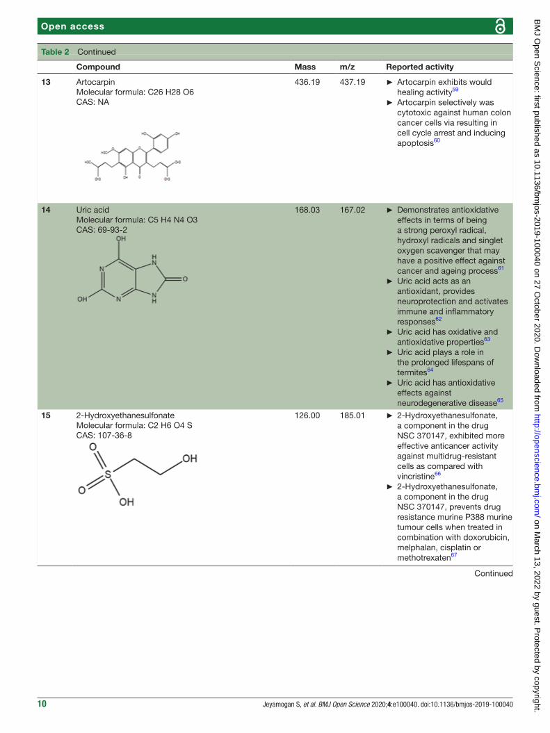
Table 4 Potential anticancer peptides from the serum of crocodile (Crocodylus porosus) predicted using MLACP tool
No. Peptide sequence Length RFACP (probability) SVMACP (probability)
1 FKMWPSSPAVPLAPK 15 ACP 0.5416 ACP 0.5518
2 WFDDKHFGLPPKER 14 ACP 0.5583 Non- ACP 0.4127
3 LLNPMLPDPPLPK 13 Non- ACP 0.4048 ACP 0.5497
4 QVLQGLVFVGAHK 13 Non- ACP 0.4766 ACP 0.631
5 WRPELPPPDLPK 12 Non- ACP 0.4891 ACP 0.5159
6 HWVQMPPSGMFK 12 ACP 0.5225 Non- ACP 0.3798
7 MLPPGGYYWDR 11 ACP 0.5557 Non- ACP 0.4891
8 HFSLLMGSLFK 11 ACP 0.5077 ACP 0.6218
9 LHPDFSSSLLK 11 Non- ACP 0.3752 ACP 0.5794
10 MAMLWDPRDDR 11 Non- ACP 0.4538 ACP 0.5609
11 VVLLPLGGPAR 11 Non- ACP 0.4822 ACP 0.6966
12 DLLLNHLHPWK 11 Non- ACP 0.4363 ACP 0.5711
13 TLPDTLTEWK 10 Non- ACP 0.4323 ACP 0.5552
14 LTPDTLTEWK 10 Non- ACP 0.4259 ACP 0.6
15 SDPLLLPLLK 10 ACP 0.515 ACP 0.8213
16 LYVPQAYRWK 10 ACP 0.5612 ACP 0.6028
17 AMPLLLLPLK 10 ACP 0.5454 ACP 0.8253
18 GFQVVQPARK 10 Non- ACP 0.3925 ACP 0.5233
19 ATATNAEMYR 10 Non- ACP 0.3802 ACP 0.5855
20 WVLFGFFPGR 10 ACP 0.6034 ACP 0.7262
21 HFFPDELWK 9 ACP 0.522 ACP 0.5942
22 WLGNFPEPR 9 ACP 0.5073 Non- ACP 0.5081
23 TFHDTATPR 9 Non- ACP 0.4784 ACP 0.6241
24 DLFVLVMMR 9 ACP 0.5432 Non- ACP 0.4574
25 LLGPHRGVR 9 Non- ACP 0.4037 ACP 0.5145
26 GFVVGDHVR 9 Non- ACP 0.4493 ACP 0.6179
27 TFGPYTNAR 9 Non- ACP 0.3669 ACP 0.5372
28 YSEHAYPSK 9 Non- ACP 0.4497 ACP 0.5481
29 LVPLGSLLK 9 ACP 0.5523 ACP 0.8112
30 VFVSPGLEK 9 Non- ACP 0.4142 ACP 0.5631
31 NSLDLLHWR 9 Non- ACP 0.4464 ACP 0.5299
32 ELGPVLLLR 9 Non- ACP 0.4478 ACP 0.6011
33 LDSPLQMWK 9 ACP 0.5121 ACP 0.6523
34 VLPEVFEHK 9 Non- ACP 0.4625 ACP 0.5347
35 RFLAAVAPK 9 ACP 0.5159 Non- ACP 0.4542
36 FMWAAMYSR 9 ACP 0.6187 ACP 0.6
37 SATPYTYSK 9 Non- ACP 0.4879 ACP 0.7018
38 TYMWPPANR 9 ACP 0.5722 ACP 0.6616
39 WLGPAATPR 9 Non- ACP 0.4897 ACP 0.6688
40 FGVLLQAPK 9 Non- ACP 0.4581 ACP 0.5811
41 EVVVPLKK 8 ACP 0.5258 ACP 0.6353
42 VEVVLPQK 8 Non- ACP 0.4164 ACP 0.6362
43 FPEPVLVK 8 ACP 0.5002 ACP 0.6295
44 TDTFFNHR 8 Non- ACP 0.4429 ACP 0.5917
45 AVLGPLLK 8 ACP 0.531 ACP 0.871
Continued
on March 13, 2022 by guest. P
rotected by copyright.http://openscience.bm
j.com/
BM
J Open S
cience: first published as 10.1136/bmjos-2019-100040 on 27 O
ctober 2020. Dow
nloaded from

14 Jeyamogan S, et al. BMJ Open Science 2020;4:e100040. doi:10.1136/bmjos-2019-100040
Open access
No. Peptide sequence Length RFACP (probability) SVMACP (probability)
46 KAEQVPWK 8 Non- ACP 0.4466 ACP 0.6226
47 WVMHLEPK 8 ACP 0.5021 Non- ACP 0.4553
48 DLALHVHK 8 Non- ACP 0.3524 ACP 0.5127
49 FFPEDLWK 8 ACP 0.5271 ACP 0.6959
50 VEALHVHK 8 Non- ACP 0.4108 ACP 0.6439
51 MFVQFTLK 8 Non- ACP 0.4481 ACP 0.5337
52 AVLGPLLK 8 ACP 0.531 ACP 0.871
53 DGWLPVPK 8 Non- ACP 0.4731 ACP 0.5618
54 NNAHVLHK 8 Non- ACP 0.3853 ACP 0.5884
55 TDTFFNHR 8 Non- ACP 0.4429 ACP 0.5917
56 KGSLLDPK 8 Non- ACP 0.3294 ACP 0.523
57 MLVVRPLR 8 ACP 0.5123 ACP 0.5661
58 MVVLEMMR 8 Non- ACP 0.4746 ACP 0.553
59 LPLLPLLK 8 ACP 0.5675 ACP 0.7105
60 VDTVLPLK 8 Non- ACP 0.4101 ACP 0.53
61 MDPPLLWR 8 ACP 0.6041 ACP 0.6961
62 TYNAKFYK 8 ACP 0.5178 ACP 0.6208
63 FGLVSVPR 8 Non- ACP 0.4853 ACP 0.6416
64 LTVGPLTK 8 Non- ACP 0.3892 ACP 0.5232
65 FFPEDLWK 8 ACP 0.5271 ACP 0.6959
66 VVMLPFFR 8 ACP 0.5845 ACP 0.5499
67 WVMHLEPK 8 ACP 0.5021 Non- ACP 0.4553
68 FFPENNWK 8 ACP 0.5797 ACP 0.6316
69 LWDLVKPR 8 ACP 0.5051 ACP 0.6331
70 QFAPLFLK 8 ACP 0.5929 ACP 0.7371
71 RANMPRAK 8 Non- ACP 0.3497 ACP 0.5257
72 MFAFDFHK 8 Non- ACP 0.4741 ACP 0.5742
73 MLSASGSK 8 Non- ACP 0.3128 ACP 0.6286
74 LWDLVQPR 8 Non- ACP 0.4407 ACP 0.5506
75 LLNLLPR 7 Non- ACP 0.4294 ACP 0.6567
76 MLLELAR 7 Non- ACP 0.406 ACP 0.5744
77 MLLELAR 7 Non- ACP 0.406 ACP 0.5744
78 LALLSQK 7 Non- ACP 0.3908 ACP 0.6906
79 LLDDLLK 7 Non- ACP 0.331 ACP 0.675
80 LLDDLLK 7 Non- ACP 0.331 ACP 0.675
81 LPPVLPR 7 ACP 0.5395 ACP 0.5947
82 HHVPVAK 7 ACP 0.5579 ACP 0.7224
83 DLVVPLK 7 ACP 0.5172 ACP 0.5848
84 VTTPPLK 7 Non- ACP 0.4781 ACP 0.5423
85 ALLPSMK 7 Non- ACP 0.4951 ACP 0.6904
86 DLVVPLK 7 ACP 0.5172 ACP 0.5848
87 EPNLLPR 7 Non- ACP 0.3636 ACP 0.5232
88 LVYSVPK 7 Non- ACP 0.4665 ACP 0.5228
89 LSADTWK 7 Non- ACP 0.4728 ACP 0.6968
90 WLSVVPR 7 ACP 0.5624 ACP 0.5675
Table 4 Continued
Continued
on March 13, 2022 by guest. P
rotected by copyright.http://openscience.bm
j.com/
BM
J Open S
cience: first published as 10.1136/bmjos-2019-100040 on 27 O
ctober 2020. Dow
nloaded from

15Jeyamogan S, et al. BMJ Open Science 2020;4:e100040. doi:10.1136/bmjos-2019-100040
Open access
No. Peptide sequence Length RFACP (probability) SVMACP (probability)
91 LALQFVR 7 Non- ACP 0.478 ACP 0.5234
92 LALLQSK 7 Non- ACP 0.4606 ACP 0.7293
93 QPVYPWK 7 Non- ACP 0.4994 ACP 0.6283
94 MFMVTYR 7 ACP 0.5219 ACP 0.6157
95 MLEMSSK 7 Non- ACP 0.4204 ACP 0.5768
96 LVYPVSK 7 Non- ACP 0.4945 ACP 0.547
97 MLEMSSK 7 Non- ACP 0.4204 ACP 0.5768
98 LPPLVPR 7 Non- ACP 0.4929 ACP 0.5448
99 DLLPLLR 7 Non- ACP 0.496 ACP 0.6599
100 LLTHVMK 7 Non- ACP 0.4609 ACP 0.5138
101 NMMYHWK 7 ACP 0.5915 ACP 0.5983
102 ELQLALK 7 Non- ACP 0.4174 ACP 0.6298
103 LTSQFYK 7 Non- ACP 0.4452 ACP 0.5353
104 ALGYNNK 7 Non- ACP 0.3125 ACP 0.562
105 YFTWLHK 7 ACP 0.5759 ACP 0.5697
106 QQLALLR 7 Non- ACP 0.4272 ACP 0.5309
107 YDMVTYR 7 Non- ACP 0.3997 ACP 0.6572
108 QMWVPNK 7 Non- ACP 0.4776 ACP 0.5299
109 YDLVFYK 7 Non- ACP 0.4436 ACP 0.5672
110 VTEWDYK 7 Non- ACP 0.4635 ACP 0.5493
111 DLLLHTR 7 Non- ACP 0.3646 ACP 0.5372
112 LYEWSLK 7 ACP 0.5251 ACP 0.6027
113 EPPEPRR 7 Non- ACP 0.4117 ACP 0.6196
114 MWVFPER 7 ACP 0.5971 ACP 0.6294
115 MLLLHSR 7 Non- ACP 0.4436 ACP 0.5846
116 LTVSRPR 7 Non- ACP 0.3673 ACP 0.5351
117 ANAVAVR 7 Non- ACP 0.2906 ACP 0.5421
118 YDLVFYK 7 Non- ACP 0.4436 ACP 0.5672
119 LVAATLK 7 Non- ACP 0.424 ACP 0.5182
120 DLTVVVK 7 Non- ACP 0.4075 ACP 0.5722
121 LYLDLK 6 Non- ACP 0.4018 ACP 0.558
122 FFYPGK 6 ACP 0.5371 ACP 0.5578
123 WAFPLK 6 ACP 0.7036 ACP 0.7724
124 FFYPGK 6 ACP 0.5371 ACP 0.5578
125 LYLDLK 6 Non- ACP 0.4018 ACP 0.558
126 FQVLVK 6 ACP 0.5909 ACP 0.6439
127 WVDLDK 6 Non- ACP 0.4744 ACP 0.595
128 WAFPLK 6 ACP 0.7036 ACP 0.7724
129 LLPFPR 6 ACP 0.6473 ACP 0.7133
130 WLLLTR 6 ACP 0.5993 ACP 0.6604
131 LLPFPR 6 ACP 0.6473 ACP 0.7133
132 TWDMAK 6 Non- ACP 0.4816 ACP 0.5691
133 NFLMAR 6 ACP 0.505 ACP 0.5337
134 TFPLPK 6 ACP 0.5892 ACP 0.6088
135 TFPPLK 6 ACP 0.5434 ACP 0.6122
Table 4 Continued
Continued
on March 13, 2022 by guest. P
rotected by copyright.http://openscience.bm
j.com/
BM
J Open S
cience: first published as 10.1136/bmjos-2019-100040 on 27 O
ctober 2020. Dow
nloaded from

16 Jeyamogan S, et al. BMJ Open Science 2020;4:e100040. doi:10.1136/bmjos-2019-100040
Open access
No. Peptide sequence Length RFACP (probability) SVMACP (probability)
136 MEMMFK 6 ACP 0.6083 ACP 0.6372
137 LLVSHK 6 Non- ACP 0.4603 ACP 0.6162
138 LVQDLK 6 Non- ACP 0.3525 ACP 0.5831
139 TWSETK 6 Non- ACP 0.4565 ACP 0.5924
140 LFDVYK 6 ACP 0.5263 ACP 0.558
141 MWDAPR 6 ACP 0.5559 ACP 0.6512
142 AVLDLK 6 Non- ACP 0.3807 ACP 0.6063
143 MEMMFK 6 ACP 0.6083 ACP 0.6372
144 MDLFVR 6 Non- ACP 0.4711 ACP 0.5242
145 LLVSHK 6 Non- ACP 0.4603 ACP 0.6162
146 TFPPLK 6 ACP 0.5434 ACP 0.6122
147 DEVLVK 6 Non- ACP 0.3553 ACP 0.5543
148 GFWESR 6 ACP 0.5612 ACP 0.6225
149 LYPSAK 6 Non- ACP 0.4521 ACP 0.5554
150 FMVGEK 6 Non- ACP 0.367 ACP 0.5215
151 LFEYGR 6 ACP 0.566 ACP 0.5336
152 LMMDNK 6 Non- ACP 0.391 ACP 0.5885
153 WLLLEK 6 ACP 0.601 ACP 0.7139
154 VTLPLK 6 Non- ACP 0.4849 ACP 0.6107
155 DFTDNK 6 Non- ACP 0.3698 ACP 0.5505
156 LVDKLK 6 ACP 0.5029 ACP 0.6698
157 YPSTER 6 Non- ACP 0.4175 ACP 0.5755
158 QKTVYR 6 Non- ACP 0.3541 ACP 0.6018
159 VEYSRR 6 Non- ACP 0.4061 ACP 0.613
160 RPSVHK 6 Non- ACP 0.4131 ACP 0.5125
161 YQFPPR 6 ACP 0.6398 ACP 0.6553
162 YVTAEK 6 Non- ACP 0.3849 ACP 0.5742
163 LMPMFR 6 ACP 0.6311 ACP 0.7146
164 YNFDMR 6 ACP 0.5592 ACP 0.623
165 LPATNK 6 Non- ACP 0.3289 ACP 0.5342
166 DLLMFR 6 Non- ACP 0.4615 ACP 0.6025
167 NDMFFK 6 Non- ACP 0.475 ACP 0.5974
168 ELVEHK 6 Non- ACP 0.3602 ACP 0.5234
169 TFPLPK 6 ACP 0.5892 ACP 0.6088
170 LVEEHK 6 Non- ACP 0.3509 ACP 0.5273
171 LSTLR 5 Non- ACP 0.3159 ACP 0.5415
172 LLLQR 5 Non- ACP 0.4551 ACP 0.6113
173 DLLFK 5 ACP 0.5658 ACP 0.7623
174 EVLLR 5 Non- ACP 0.487 ACP 0.5909
175 FEYGR 5 ACP 0.5605 ACP 0.6147
176 FAVER 5 Non- ACP 0.3478 ACP 0.517
177 LDELK 5 Non- ACP 0.3167 ACP 0.5829
178 LPALK 5 ACP 0.5594 ACP 0.7516
179 LQDFR 5 Non- ACP 0.4549 ACP 0.5695
180 DLLFK 5 ACP 0.5658 ACP 0.7623
Table 4 Continued
Continued
on March 13, 2022 by guest. P
rotected by copyright.http://openscience.bm
j.com/
BM
J Open S
cience: first published as 10.1136/bmjos-2019-100040 on 27 O
ctober 2020. Dow
nloaded from
17Jeyamogan S, et al. BMJ Open Science 2020;4:e100040. doi:10.1136/bmjos-2019-100040
Open access
No. Peptide sequence Length RFACP (probability) SVMACP (probability)
181 EVLLR 5 Non- ACP 0.487 ACP 0.5909
182 NNLFK 5 ACP 0.5104 ACP 0.6131
183 EEPDK 5 Non- ACP 0.3465 ACP 0.576
184 SSWKK 5 ACP 0.6361 ACP 0.6807
185 LPLLR 5 ACP 0.5655 ACP 0.6535
186 TLLSK 5 Non- ACP 0.3775 ACP 0.6076
187 LFPLK 5 ACP 0.6311 ACP 0.7728
188 AVLVR 5 Non- ACP 0.415 ACP 0.5986
189 FEYGR 5 ACP 0.5605 ACP 0.6147
190 LTLSK 5 Non- ACP 0.3753 ACP 0.5742
191 LSLTR 5 Non- ACP 0.3739 ACP 0.562
192 SPSSK 5 Non- ACP 0.4068 ACP 0.622
193 HSSEK 5 Non- ACP 0.3297 ACP 0.5681
194 SLELK 5 Non- ACP 0.3981 ACP 0.6804
195 LSDLR 5 Non- ACP 0.3283 ACP 0.5579
196 DLLLR 5 Non- ACP 0.3909 ACP 0.5978
197 MYGTK 5 Non- ACP 0.4934 ACP 0.5467
198 EVLLR 5 Non- ACP 0.487 ACP 0.5909
199 FAMPR 5 ACP 0.5646 ACP 0.6476
200 DLVAK 5 Non- ACP 0.3628 ACP 0.5158
201 LLQLR 5 Non- ACP 0.4621 ACP 0.6139
202 YAPLR 5 ACP 0.5447 ACP 0.6267
203 VTELK 5 Non- ACP 0.3432 ACP 0.5558
204 HTAYK 5 ACP 0.5391 ACP 0.6588
205 TAVPR 5 Non- ACP 0.3708 ACP 0.5267
206 SMSMR 5 Non- ACP 0.4136 ACP 0.64
207 DLVAK 5 Non- ACP 0.3628 ACP 0.5158
ACP, anticancer peptides; MLACP, Machine- Learning- Based Prediction of Anticancer Peptides; RFACP, random forest anti- cancer peptides; SVMACP, support vector machine anti- cancer peptides.
Table 4 Continued
control (p<0.05) (figure 7). Treated PC3 cells demon-strated two genes that were upregulated as compared with the control (p<0.05) (figure 8).
diSCuSSionThe mortality and morbidity of cancer remains a serious concern,1–3 suggesting the need for new effective anti-cancer agents. The medicinal properties of naturally derived products remain an important source of ther-apeutic drugs. Here, we dissected a saltwater crocodile (C. porosus) and prepared crude lysates and sera. The organ lysates and serum were then tested on cancer cells for growth inhibition and cytotoxic activity. The gall bladder lysates and serum inhibited more than 99% of HeLa cells, PC3 cells and MCF7 cells. This was consistent with previous findings that showed the ability of C. siamensis bile extracts in hindering the growth of human cholangiocarcinoma cells, hepatocellular carcinoma
cells, ovarian carcinoma cells and gastric adenocarci-noma cells.11 12 14 The blood extract of C. siamensis and American alligator also induced cell cycle arrest that led to growth inhibition among cancer cells.34–36 Notably, crocodile sera but not bovine sera caused irreversible cancer cell damage.
Next, LC- MS/MS was performed on serum samples to identify the types of potential anticancer small mole-cules and peptides. Besides being the most convenient biological sample, serum also paves the road by being a liquid highway for all the molecules that are synthesised, secreted and discarded from the body.29 Using LC- MS/MS, 80 small molecules were detected and 19 compounds were putatively identified from the serum of C. porosus by comparison against the METLIN metabolomics database. It has been reported that the number of small molecules detected are normally lesser than 24 metabolites at a time due to the limitation of metabolite databases.29
on March 13, 2022 by guest. P
rotected by copyright.http://openscience.bm
j.com/
BM
J Open S
cience: first published as 10.1136/bmjos-2019-100040 on 27 O
ctober 2020. Dow
nloaded from

18 Jeyamogan S, et al. BMJ Open Science 2020;4:e100040. doi:10.1136/bmjos-2019-100040
Open access
Figure 6 (A–B) Differential gene expression of HeLa cells treated with crocodile sera. Fourteen genes were upregulated as compared with control. The red dots in the MA plot demonstrates the expression of genes (A) (p<0.05 using Wald statistics).
Valdecoxib, a cyclooxygenase 2 (COX-2) inhibitor,37 which is commonly used to treat inflammation in condi-tions such as rheumatoid arthritis and knee osteoarthritis patients38 were identified in the serum of C. porosus (table 2). COX-2 is highly expressed in several types of cancer such as colorectal cancer.37 38 Besides that, chemo-therapy and radiotherapy also contribute to the upreg-ulation of COX-2 expression, resulting in the onset of resistance against cancer therapy.39 Therefore, the pres-ence of valdecoxib in crocodile serum may protect the animal from cancer, therefore highlighting the fact that valdecoxib could be a potential anticancer drug, since it inhibits the expression of COX-2 and works as an anti- inflammatory agent.38 However, patients treated with valdecoxib previously suffered from side effects such as
cardiovascular complications, leading to the withdrawal of valdecoxib from the market in 2005 by the Food and Drug Administration (FDA) agency of the United States Department of Health and Human Services and the European Medicines Agency.37 39 Purine has the ability in treating many conditions due to its antitumour, anti-viral, anti- inflammatory, antimicrobial and antiparasitic properties.40 41 Previously, purine was shown to inhibit DNA replication in cancer cells42 although the exact anticancer mechanisms exerted by purine remain vague. FDA- approved purine antimetabolites or derivatives such as 6- mercaptopurine, fludarabine, nelarabine, cladribine, clofarabine and pentostatin have been extensively used for the treatment of cancer although the presence
on March 13, 2022 by guest. P
rotected by copyright.http://openscience.bm
j.com/
BM
J Open S
cience: first published as 10.1136/bmjos-2019-100040 on 27 O
ctober 2020. Dow
nloaded from

19Jeyamogan S, et al. BMJ Open Science 2020;4:e100040. doi:10.1136/bmjos-2019-100040
Open access
Figure 7 (A–B) Differential gene expression of MCF7 cells treated with crocodile sera. Twenty- six genes were downregulated and 25 genes were upregulated as compared with control. The red dots in the MA plot demonstrates the expression of genes (A) (p<0.05 using Wald statistics).
of selectivity and toxicity of these compounds is still questionable.42
LC- MS/MS analysis of the serum of C. porosus demonstrated seven peptides belonging to the alpha-2- macroglobulin isoform X2 (Accession ID: A0A1U7S0T0) protein family, five peptides belonging to the trans-ferrin (Accession ID: A0A286T2Q9) protein family, six peptides belonging to the complement C3 (Acces-sion ID: A0A1U7S0C0) protein family, three peptides belonging to the fibrinogen beta chain (Accession ID:
A0A1U7SP96) protein family, two peptides belonging to the haemoglobin subunit beta (Accession ID: P86919) protein family and three peptides belonging to the serum albumin isoform X2 (Accession ID: A0A1U8CYA2) protein family (table 3). The remaining 749 detected peptides were categorised as novel peptides. The poten-tial ACP from the list of novel peptides were then predicted using the MLACP online tool,31 and interest-ingly more than 207 ACP were predicted from serum of C. porosus (table 4).
on March 13, 2022 by guest. P
rotected by copyright.http://openscience.bm
j.com/
BM
J Open S
cience: first published as 10.1136/bmjos-2019-100040 on 27 O
ctober 2020. Dow
nloaded from

20 Jeyamogan S, et al. BMJ Open Science 2020;4:e100040. doi:10.1136/bmjos-2019-100040
Open access
Figure 8 (A–B) Differential gene expression of PC3 cells treated with crocodile sera. Two genes were upregulated as compared with control. The red dots in the MA plot demonstrates the expression of genes (A) (p<0.05 using Wald statistics).
It is anticipated that these ACP may be utilised in clin-ical treatment of cancer in the future. The mechanism of action and the anticancer activity of the 207 novel peptides detected here need to be determined and inves-tigated further. In addition, the bioavailability and stability under physiological conditions of these peptides need to be considered. Strategies to allow appropriate delivery of peptides have been utilised in the past resulting in highly efficacious treatment. Some cancer- targeting peptides have been designed on the basis of the pH difference between tumour tissue and normal tissues,43 and the peptide selectively kills tumour cells at acidic pH levels but is not toxic against normal cells. Moreover, nano-technology and nanomaterials have provided remark-able potential for application of ACP in tumour- targeted therapy, bioimaging and diagnosis due to their unique properties. The discovery of ACP and associated phar-macological research and development is noteworthy, and further investment is needed over the next several decades to exploit their potential and benefit thousands of cancer patients.
To our knowledge, this is the first study that applied differential gene analysis of cancer cells treated with crocodile serum. The gene expression analysis revealed that 14 genes in treated HeLa cells, 51 genes in treated MCF7 cells and 2 genes in treated PC3 cells were deferen-tially expressed as compared with untreated control cells, out of more than 10 000 genes (p<0.05) (figures 6–8). Furthermore, treated HeLa cells demonstrated 14 genes that were upregulated and no downregulated genes as compared with control (figure 6). This included Fos, a proto- oncogene, involved in important cellular events, including cell proliferation, as well as other genes such as immediate early response 2 that is a putative nuclear protein that functions as a transcription factor in cellular responses, and may be involved in the regulation of
tumour progression and metastasis.44 Additionally, treated MCF7 cells demonstrated 26 genes that were downregu-lated and 25 genes that were upregulated as compared with control (figure 7). These comprised genes that are involved in cellular communication, DNA repair, growth response, respiration and so on. Treated PC3 cells demon-strated two genes that were upregulated with no downreg-ulated genes as compared with control (figure 8). These included Fos as well as the Hes1 gene which codes for nuclear proteins that suppress transcription.44 45
Nonetheless, differential gene expression across the different cell lines was not consistent. The reason for this could be due to the cell lines having different properties and they are of different origin. HeLa cells are derived from cervical cancer cells.46 These cells proliferate abnor-mally rapidly, even compared with other cancer cells. PC3 cells on the other hand were established from bone metastasis of grade IV of prostate cancer. These cells do not respond to androgens, glucocorticoids or fibroblast growth factors, but results suggest that the cells are influ-enced by epidermal growth factors.47 On the other hand, MCF-7 cells are one of the very few cells known to express substantial levels of the oestrogen receptor alpha.48 Future studies should be conducted on several cell lines of similar origin to determine if there is a conserved pathway in response to the lysates tested in this study. These find-ings show that animals living in polluted environments possess molecules that have potential anticancer activities. Consequently, it is important to investigate the anticancer effects of these compounds against various cancer cells and in vivo. In summary, we showed that the organ lysates and sera of C. porosus exhibit potent anticancer activity and have identified several molecules that could serve as potential drug leads, but further research is needed to realise these expectations. These findings further suggest that animals residing in polluted milieus are a large unex-ploited source for prospective pharmaceutical drugs, and could lead to the identification of novel antitumour compound(s) and/or understanding of the mechanisms of cancer resistance.
Acknowledgements The work in this paper was supported, in part, by the Open Access Program from the American University of Sharjah. This paper represents the opinions of the author(s) and does not mean to represent the position or opinions of the American University of Sharjah.
Contributors RS and NAK developed the concept. RS acquired funding. KS acquired animal resource and carried out dissections. SJ carried out all experiments under the supervision of RS. SJ and RS wrote the original draft that was reviewed and corrected by KS and NAK. All authors approved the manuscript.
Funding This study was funded by American University of Sharjah; Sunway University (Grant number: DBS_04).
Competing interests None declared.
ethics approval This article does not contain any studies with human participants. The use of crocodile material obtained for this study was approved by the Department of Wildlife and National Parks (PERHILITAN), Malaysia and the Research Ethics Committee which is in accordance with Sunway University’s Code of Practice for the Ethical Conduct of Research and the Research Governance and Integrity Policy (Research Ethics Approval Code: SUNREC 2017/038).
Provenance and peer review Not commissioned; externally peer reviewed.
on March 13, 2022 by guest. P
rotected by copyright.http://openscience.bm
j.com/
BM
J Open S
cience: first published as 10.1136/bmjos-2019-100040 on 27 O
ctober 2020. Dow
nloaded from

21Jeyamogan S, et al. BMJ Open Science 2020;4:e100040. doi:10.1136/bmjos-2019-100040
Open access
data availability statement All data relevant to the study are included in the article or uploaded as supplementary information.
open Practices
open access This is an open access article distributed in accordance with the Creative Commons Attribution 4.0 Unported (CC BY 4.0) license, which permits others to copy, redistribute, remix, transform and build upon this work for any purpose, provided the original work is properly cited, a link to the licence is given, and indication of whether changes were made. See: https:// creativecommons. org/ licenses/ by/ 4. 0/.
open data Open data are available at https:// figshare. com/ articles/ dataset/ Croc_ anticancer_ Table1_ data_ pdf/ 13041860/1
orCid idNaveed Ahmed Khan http:// orcid. org/ 0000- 0001- 7667- 8553
reFerenCeS 1 Parkin DM, Bray F, Ferlay J, et al. Estimating the world cancer
burden: Globocan 2000. Int J Cancer 2001;94:153–6. 2 Ferlay J, Soerjomataram I, Dikshit R, et al. Cancer incidence and
mortality worldwide: sources, methods and major patterns in GLOBOCAN 2012. Int J Cancer 2015;136:E359–86.
3 Bray F, Ferlay J, Soerjomataram I, et al. Global cancer statistics 2018: GLOBOCAN estimates of incidence and mortality worldwide for 36 cancers in 185 countries. CA Cancer J Clin 2018;68:394–424.
4 Siddiqui R, Jeyamogan S, Ali SM, et al. Crocodiles and alligators: Antiamoebic and antitumor compounds of crocodiles. Exp Parasitol 2017;183:194–200.
5 Lehner AF, Rumbeiha W, Shlosberg A, et al. Diagnostic analysis of veterinary dried blood spots for toxic heavy metals exposure. J Anal Toxicol 2013;37:406–22.
6 Tellez M, Merchant M. Biomonitoring heavy metal pollution using an aquatic apex predator, the American Alligator, and its parasites. PLoS One 2015;10:e0142522.
7 Schneider L, Peleja RP, Kluczkovski A, et al. Mercury concentration in the spectacled Caiman and black Caiman (Alligatoridae) of the Amazon: implications for human health. Arch Environ Contam Toxicol 2012;63:270–9.
8 Brochu CA. Progress and future directions in archosaur phylogenetics. J Paleontol 2001;75:1185–201.
9 Oaks JR. A time- calibrated species tree of Crocodylia reveals a recent radiation of the true crocodiles. Evolution 2011;65:3285–97.
10 Lee S, Duce I, Atkins H, et al. Cockroaches and locusts: physicians' answer to infectious diseases. Int J Antimicrob Agents 2011;37:279–80.
11 Kang J- H, Zhang W- Q, Song W, et al. Apoptosis mechanism of human cholangiocarcinoma cells induced by bile extract from crocodile. Appl Biochem Biotechnol 2012;166:942–51.
12 Song W, Shen D- Y, Kang J- H, et al. Apoptosis of human cholangiocarcinoma cells induced by ESC-3 from Crocodylus siamensis bile. World J Gastroenterol 2012;18:704.
13 Fu Q- R, Song W, Deng Y- T, et al. ESC-3 induces apoptosis of human ovarian carcinomas through Wnt/β-catenin and Notch signaling in vitro and in vivo. Int J Oncol 2017;50:241–51.
14 Mao X, He S, Zhang T, et al. Isolation and characterization of antiproliferative peptides from Chinese three- striped box turtle (Cuora trifasciata). Biotechnol Appl Biochem 2017;64:827–35.
15 Theansungnoen T, Maijaroen S, Jangpromma N, et al. Cationic antimicrobial peptides derived from Crocodylus siamensis leukocyte extract, revealing anticancer activity and apoptotic induction on human cervical cancer cells. Protein J 2016;35:202–11.
16 Chaeychomsri W, Yamkong S, Chaeychomsri S, et al. Effects of large volume crocodile blood collection on hematological values of Siamese crocodiles (Crocodylus siamensis). Journal of Advanced Agricultural Technologies 2016;3:252–7.
17 Grigg GC, Cairncross M. Respiratory properties of the blood of Crocodylus porosus. Respir Physiol 1980;41:367–80.
18 Nevarez J. Crocodilians. In: Manual of exotic PET practice, 2009: 112–35.
19 Farris SC, Squires MA, Ridgley F, et al. Necropsies of Reptiles: Recommendations and Techniques for Examining Invasive Species.
In: The Institute of food and agricultural sciences (IFAS. University of Florida, 2016: 1–19.
20 Campbell TW. Exotic animal hematology and cytology. Hoboken, NJ: Wiley Blackwell, 2015: 37–60.
21 Cadamuro J, Mrazek C, Leichtle AB, et al. Influence of centrifugation conditions on the results of 77 routine clinical chemistry analytes using standard vacuum blood collection tubes and the new BD- Barricor tubes. Biochem Med 2018;28:1–10.
22 Dignam JD. Preparation of extracts from higher eukaryotes. In: Methods in enzymology. . Academic Press, 1990: 182. 194–203.
23 Ali SM, Siddiqui R, Ong S- K, et al. Identification and characterization of antibacterial compound(s) of cockroaches (Periplaneta americana). Appl Microbiol Biotechnol 2017;101:253–86.
24 Rajendran K, Anwar A, Khan NA, et al. Brain- eating amoebae: silver nanoparticle conjugation enhanced efficacy of anti- amoebic drugs against Naegleria fowleri. ACS Chem Neurosci 2017;8:2626–30.
25 Strober W. Trypan blue exclusion test of cell viability. Curr Protoc Immunol 2001;Appendix 3:Appendix 3B.
26 Namekar M, Kumar M, O'Connell M, et al. Effect of serum heat- inactivation and dilution on detection of anti- WNV antibodies in mice by West Nile virus E- protein microsphere immunoassay. PLoS One 2012;7:e45851.
27 Jones FS. The effect of heat on antibodies. J Exp Med 1927;46:291–301.
28 Iqbal J, Siddiqui R, Khan NA. Acanthamoeba and bacteria produce antimicrobials to target their counterpart. Parasit Vectors 2014;7:56.
29 Psychogios N, Hau DD, Peng J, et al. The human serum metabolome. PLoS One 2011;6:e16957.
30 Zainal Abidin SA, Rajadurai P, Chowdhury MEH, et al. Cytotoxic, antiproliferative and apoptosis- inducing activity of L- amino acid oxidase from Malaysian Calloselasma rhodostoma on human colon cancer cells. Basic Clin Pharmacol Toxicol 2018;123:577–88.
31 Manavalan B, Basith S, Shin TH, et al. MLACP: machine- learning- based prediction of anticancer peptides. Oncotarget 2017;8:77121–36.
32 Tiwari GJ, Liu Q, Shreshtha P, et al. Rnai- Mediated down- regulation of the expression of OsFAD2-1: effect on lipid accumulation and expression of lipid biosynthetic genes in the rice grain. BMC Plant Biol 2016;16:189.
33 Benjamini Y, Hochberg Y. Controlling the false discovery rate: a practical and powerful approach to multiple testing. J R Statist Soc B 1995;57:289–300.
34 Ou Y, Ho WS. Crocodile blood extract induces the apoptosis of lung cancer cells through PTEN activity. Oncol Rep 2016;36:1457–66.
35 Patathananone S, Thammasirirak S, Daduang J, et al. Bioactive compounds from crocodile (Crocodylus siamensis) white blood cells induced apoptotic cell death in HeLa cells. Environ Toxicol 2016;31:986–97.
36 Patathananone S, Thammasirirak S, Daduang J, et al. Inhibition of HeLa cells metastasis by bioactive compounds in crocodile (Crocodylus siamensis) white blood cells extract. Environ Toxicol 2016;31:1329–36.
37 Chikkula KV, S. R, Raja S. Isoxazole – a potent pharmacophore. Int J Pharm Pharm Sci 2017;9:13–24.
38 Inan Genç A, Gok S, Banerjee S, et al. Valdecoxib recovers the lipid composition, order and dynamics in colon cancer cell lines independent of COX-2 expression: an ATR- FTIR spectroscopy study. Appl Spectrosc 2017;71:105–17.
39 Wang D, Dubois RN. The role of COX-2 in intestinal inflammation and colorectal cancer. Oncogene 2010;29:781–8.
40 Atukorala I, Hunter DJ. Valdecoxib : the rise and fall of a COX-2 inhibitor. Expert Opin Pharmacother 2013;14:1077–86.
41 Hassan AY, Sarg MT, Bayoumi AH, et al. Design, synthesis, and anticancer activity of novel fused purine analogues. J Heterocycl Chem 2017;54:3458–70.
42 Parker WB. Enzymology of purine and pyrimidine antimetabolites used in the treatment of cancer. Chem Rev 2009;109:2880–93.
43 Wakabayashi N, Yano Y, Kawano K, et al. A pH- dependent charge reversal peptide for cancer targeting. Eur Biophys J 2017;46:121–7.
44 Tulchinsky E. Fos family members: regulation, structure and role in oncogenic transformation. Histol Histopathol 2000;15:921–8.
45 Kageyama R, Ohtsuka T, Kobayashi T. The Hes gene family: repressors and oscillators that orchestrate embryogenesis. Development 2007;134:1243–51.
46 Scherer WF, Syverton JT, Gey GO. Studies on the propagation in vitro of poliomyelitis viruses. IV. viral multiplication in a stable strain of human malignant epithelial cells (strain HeLa) derived from an epidermoid carcinoma of the cervix. J Exp Med 1953;97:695–710.
47 Tai S, Sun Y, Squires JM, et al. Pc3 is a cell line characteristic of prostatic small cell carcinoma. Prostate 2011;71:1668–79.
on March 13, 2022 by guest. P
rotected by copyright.http://openscience.bm
j.com/
BM
J Open S
cience: first published as 10.1136/bmjos-2019-100040 on 27 O
ctober 2020. Dow
nloaded from

22 Jeyamogan S, et al. BMJ Open Science 2020;4:e100040. doi:10.1136/bmjos-2019-100040
Open access
48 Lee AV, Oesterreich S, Davidson NE. MCF-7 cells--changing the course of breast cancer research and care for 45 years. J Natl Cancer Inst 2015;107:djv073.
49 Barakat A, Al- Majid AM, Al- Qahatany FM, et al. Synthesis, characterization and antimicrobial activity of novel pharmacophores incorporating imidazoline- oxazoline scaffold. Bull Korean Chem Soc 2014;35:562–8.
50 Kowalska A, Pluta K, Latocha M. Synthesis and anticancer activity of multisubstituted purines and xanthines with one or two propynylthio and aminobutynylthio groups. Med Chem Res 2018;27:1384–95.
51 Campos- Ferraz PL, Gualano B, das Neves W, et al. Exploratory studies of the potential anti- cancer effects of creatine. Amino Acids 2016;48:1993–2001.
52 Genc G, Okuyucu A, Meydan BC, et al. Effect of free creatine therapy on cisplatin- induced renal damage. Ren Fail 2014;36:1108–13.
53 Garbati P, Ravera S, Scarfì S, et al. Effects on Energy Metabolism of Two Guanidine Molecules, (Boc)2 -Creatine and Metformin. J Cell Biochem 2017;118:2700–11.
54 Deepuppha N, Khadsai S, Rutnakornpituk B, et al. Multiresponsive Poly (N -Acryloyl glycine)- Based Nanocomposite and Its Drug Release Characteristics. J Nanomater 2019;2019:1–12.
55 Benavides MA, Bosland MC, da Silva CP, et al. L- Methionine inhibits growth of human pancreatic cancer cells. Anticancer Drugs 2014;25:200–3.
56 Godoy- Vitorino F, Ortiz- Morales G, Romaguera J, et al. Discriminating high- risk cervical human papilloma virus infections with urinary biomarkers via non- targeted GC- MS- based metabolomics. PLoS One 2018;13:e0209936.
57 Guay J. Methemoglobinemia related to local anesthetics: a summary of 242 episodes. Anesth Analg 2009;108:837–45.
58 Fujita H, Hongo M, Mochizuki M, et al. Inhibitory effects of 16- hydroxy-9- oxo- 10E,12E,14E- octadecatrienoic acid (Corchorifatty acid B) isolated from Melissa officinalis Linné on melanogenesis. Exp Dermatol 2011;20:420–4.
59 Yeh C- J, Chen C- C, Leu Y- L, et al. The effects of artocarpin on wound healing: in vitro and in vivo studies. Sci Rep 2017;7:15599.
60 Sun G, Zheng Z, Lee M- H, et al. Chemoprevention of colorectal cancer by artocarpin, a dietary phytochemical from Artocarpus heterophyllus. J Agric Food Chem 2017;65:3474–80.
61 Glantzounis GK, Tsimoyiannis EC, Kappas AM, et al. Uric acid and oxidative stress. Curr Pharm Des 2005;11:4145–51.
62 Alvarez- Lario B, Macarrón- Vicente J. Is there anything good in uric acid? QJM 2011;104:1015–24.
63 Sautin YY, Johnson RJ. Uric acid: the oxidant- antioxidant paradox. Nucleosides Nucleotides Nucleic Acids 2008;27:608–19.
64 Tasaki E, Sakurai H, Nitao M, et al. Uric acid, an important antioxidant contributing to survival in termites. PLoS One 2017;12:e0179426.
65 Settle T, Klandorf H. The role of uric acid as an antioxidant in selected neurodegenerative disease pathogenesis: a short review. Brain Disord Ther 2014;03:129.
66 Waud WR, Leopold WR, Elliott WL, et al. Antitumor activity of ethyl 5- amino-1,2- dihydro-2- methyl-3- phenyl- pyrido [3,4- b]pyrazin-7- ylcarbamate, 2- hydroxyethanesulfonate, hydrate (NSC 370147) against selected tumor systems in culture and in mice. Cancer Res 1990;50:3239–44.
67 Waud WR, Harrison SD, Temple CG, et al. Antitumor drug cross- resistance in vivo in a murine P388 leukemia resistant to ethyl 5- amino-1,2- dihydro-2- methyl-3- phenylpyrido[3,4- b]pyrazin-7 - ylcarbamate 2- hydroxyethanesulfonate hydrate (NSC 370,147) 370147. Cancer Chemother Pharmacol 1992;29:190–4.
68 Kivitz A, Eisen G, Zhao WW, et al. Randomized placebo- controlled trial comparing efficacy and safety of valdecoxib with naproxen in patients with osteoarthritis. J Fam Pract 2002;51:530–7.
69 Daniels SE, Torri S, Desjardins PJ. Valdecoxib for treatment of primary dysmenorrhea. J Gen Intern Med 2005;20:62–7.
70 Zhu FS, Chen XM, Huang ZG, et al. Rofecoxib augments anticancer effects by reversing intrinsic multidrug resistance gene expression in BGC-823 gastric cancer cells. J Dig Dis 2010;11:34–42.
71 Young SD, Whissell M, Noble JCS, et al. Phase II clinical trial results involving treatment with low- dose daily oral cyclophosphamide, Weekly vinblastine, and rofecoxib in patients with advanced solid tumors. Clin Cancer Res 2006;12:3092–8.
72 Al- Nimer MSM, Al- Deen SM, Abdul Lateef ZW. Rofecoxib prevents ctdsDNA against damage induced by copper sulfate and ultraviolet B radiation in vitro study. J Basic Clin Pharm 2010;2:21.
on March 13, 2022 by guest. P
rotected by copyright.http://openscience.bm
j.com/
BM
J Open S
cience: first published as 10.1136/bmjos-2019-100040 on 27 O
ctober 2020. Dow
nloaded from