Ontogeny of the Hematopoietic System - ULisboabmg.fc.ul.pt/Disciplinas/Regula_e_Diferenc/artigos/I...
44
Ontogeny of the Hematopoietic System Ana Cumano 1 and Isabelle Godin 2−4 1 INSERM, U668, and Unit ´ e de D ´ eveloppement des Lymphocytes, Department of Immunology, Institut Pasteur, 75724 Paris, France; email: [email protected] 2 INSERM, U790, 39 rue C. Desmoulins, Villejuif, F-94805 3 Institut Gustave Roussy, Villejuif, F-94805; email: [email protected] 4 Universit´ e de Paris-Sud, Orsay, F-91405 Annu. Rev. Immunol. 2007. 25:745–85 First published online as a Review in Advance on January 2, 2007 The Annual Review of Immunology is online at immunol.annualreviews.org This article’s doi: 10.1146/annurev.immunol.25.022106.141538 Copyright c 2007 by Annual Reviews. All rights reserved 0732-0582/07/0423-0745$20.00 Key Words AGM, development, embryo, hematopoietic stem cells, hemogenic sites, yolk sac Abstract Blood cells are constantly produced in the bone marrow (BM) of adult mammals. This constant turnover ultimately depends on a rare population of progenitors that displays self-renewal and multilineage differentiation potential, the hematopoietic stem cells (HSCs). It is generally accepted that HSCs are generated during embryonic de- velopment and sequentially colonize the fetal liver, the spleen, and finally the BM. Here we discuss the experimental evidence that ar- gues for the extrinsic origin of HSCs and the potential locations where HSC generation might occur. The identification of the cel- lular components playing a role in the generation process, in these precise locations, will be important in understanding the molecu- lar mechanisms involved in HSC production from undifferentiated mesoderm. 745 Annu. Rev. Immunol. 2007.25:745-785. Downloaded from arjournals.annualreviews.org by INSERM-multi-site account on 11/01/07. For personal use only.
Transcript of Ontogeny of the Hematopoietic System - ULisboabmg.fc.ul.pt/Disciplinas/Regula_e_Diferenc/artigos/I...

ANRV306-IY25-25 ARI 19 February 2007 16:31
Ontogeny of theHematopoietic SystemAna Cumano1 and Isabelle Godin2−4
1INSERM, U668, and Unite de Developpement des Lymphocytes, Departmentof Immunology, Institut Pasteur, 75724 Paris, France; email: [email protected], U790, 39 rue C. Desmoulins, Villejuif, F-948053Institut Gustave Roussy, Villejuif, F-94805; email: [email protected] de Paris-Sud, Orsay, F-91405
Annu. Rev. Immunol. 2007. 25:745–85
First published online as a Review in Advance onJanuary 2, 2007
The Annual Review of Immunology is online atimmunol.annualreviews.org
This article’s doi:10.1146/annurev.immunol.25.022106.141538
Copyright c© 2007 by Annual Reviews.All rights reserved
0732-0582/07/0423-0745$20.00
Key Words
AGM, development, embryo, hematopoietic stem cells, hemogenicsites, yolk sac
AbstractBlood cells are constantly produced in the bone marrow (BM) ofadult mammals. This constant turnover ultimately depends on a rarepopulation of progenitors that displays self-renewal and multilineagedifferentiation potential, the hematopoietic stem cells (HSCs). It isgenerally accepted that HSCs are generated during embryonic de-velopment and sequentially colonize the fetal liver, the spleen, andfinally the BM. Here we discuss the experimental evidence that ar-gues for the extrinsic origin of HSCs and the potential locationswhere HSC generation might occur. The identification of the cel-lular components playing a role in the generation process, in theseprecise locations, will be important in understanding the molecu-lar mechanisms involved in HSC production from undifferentiatedmesoderm.
745
Ann
u. R
ev. I
mm
unol
. 200
7.25
:745
-785
. Dow
nloa
ded
from
arj
ourn
als.
annu
alre
view
s.or
gby
IN
SER
M-m
ulti-
site
acc
ount
on
11/0
1/07
. For
per
sona
l use
onl
y.
ANRV306-IY25-25 ARI 19 February 2007 16:31
Hematopoiesis: theprocess that givesrise to all matureblood cells throughthe production, byhematopoietic stemcells, of precursorsthat expand anddifferentiate
BM: bone marrow
HSCs:hematopoietic stemcells
LTR: long-termreconstitution
Niche: specializedenvironmentrequired for a givencell type (e.g., HSC)to perform a function(e.g., self-renewal ordifferentiation)
INTRODUCTION
Blood cells are produced throughout life. Inmammals, hematopoiesis, defined by the ex-pansion and differentiation of hematopoieticprecursors, occurs in the bone marrow (BM)in the abluminal side of sinusoids that invadethe bone cavity made by osteoclasts shortlyafter calcium deposits in the cartilaginousmatrix.
It has been long known that BM cellscan regenerate the blood compartment.Thus, BM transplantation was among thefirst cell replacement therapeutic approachesattempted.
Blood cells comprise multiple cell typesand have accordingly variable life spansthat go from a few days to several years.Hematopoietic cells undergo constant re-newal and consequently have an activeregenerative compartment. Only in the mid-twentieth century, however, did seminal workby Metcalf and Moore (1, 12) and by Till& McCullough (2) introduce the notion thatmultipotent progenitors can be found in adultBM and that these cells are responsible forthe constant production of blood. Progress inthe phenotypic and functional characteriza-tion of these precursors led to the isolationof hematopoietic stem cells (HSCs) and hasmade the hematopoietic system a paradigmin stem cell biology. Thus, HSCs have beencharacterized by two main properties, mul-tipotency and self-renewal, at the single-celllevel. Multipotency, as defined by the capacityof a single cell to give rise to a differentiatedprogeny comprising different cell types, wastested in vitro by clonal differentiation pro-tocols. Self-renewal, the property that allowsthe maintenance of the HSC pool (probablythrough asymmetric cell division), is usuallyassessed by in vivo long-term reconstitution(LTR) experiments. This experimental ap-proach requires irradiation of the recipientanimals with the consequent perturbation ofthe natural environment. It measures not onlyself-renewal and multipotency, but also thehoming capacity of the cell to find the ap-
propriate niche in which it can exert a nor-mal function. Self-renewal implies the main-tenance of cell numbers in a given compart-ment: If one HSC is injected, one HSC shouldbe recovered. However, expansion of the HSCpool is probably obtained under the irradia-tion ablative protocols conventionally used inLTR experiments.
In vitro assays have improved in efficiencyand reliability in the past few years with theisolation of hematopoietic cytokines and sup-porting stromal cell lines, and multipotency iseasily demonstrated. In contrast, self-renewalremained an elusive property, and the condi-tions that allow in vitro self-renewal of HSCshave been difficult to establish. Although it isgenerally accepted that the HSC pool is sta-ble in numbers during life (implying a self-renewing capacity), few direct demonstrationsof this property exist in the literature (3, 4).There is compelling evidence, however, thatmost HSCs in adult animal BM are in theG0 stage of the cell cycle (5). Thus, althoughthey eventually will enter G1, most are rest-ing, and the regeneration of the peripheralcompartment is done by extensive prolifer-ation of intermediate precursors. Therefore,self-renewing HSCs are difficult to detect.
Pioneering experiments in the late 1980sused retroviral integration as a markerof clonality and followed the progeny ofsingle cells in vivo (3, 4). Sequential transplan-tation experiments demonstrated that singlehematopoietic cells could generate all bloodcell types in recipient mice for longer thansix months, and on secondary engraftmentthe same viral integration was found in bloodcells belonging to all lineages. These experi-ments showed for the first time the in vivo self-renewing nature of multipotent cells presentin the adult BM. These experiments remainthe most compelling direct demonstration ofin vivo HSC renewal, with the possible caveatof clonal dominance conferred by particu-lar retroviral integrations that might have fa-vored the expansion of single infected cells ina nonphysiologic manner.
746 Cumano · Godin
Ann
u. R
ev. I
mm
unol
. 200
7.25
:745
-785
. Dow
nloa
ded
from
arj
ourn
als.
annu
alre
view
s.or
gby
IN
SER
M-m
ulti-
site
acc
ount
on
11/0
1/07
. For
per
sona
l use
onl
y.

ANRV306-IY25-25 ARI 19 February 2007 16:31
In the past few years, researchers haveidentified and characterized the regenerativecompartment of other adult tissues. Cellswithin these compartments are generally des-ignated as adult stem cells. Whereas all stemcells exhibit the properties of self-renewal andlifelong contribution (through maturation) tothe differentiated cell pool, multipotency isa property only found in the stem cell poolof some tissues (e.g., hair follicle, intestinalepithelium, liver, central nervous system) andis probably absent in others (e.g., skeletalmuscle).
Experimental evidence shows that thereis a pool of tissue-specific, resident, self-renewing, and differentiating progenitorswithin many analyzed tissues. This findingsuggests that adult stem cells are limited intheir differentiation capacity to the tissue inwhich they reside and to which they are the re-stricted precursors. When isolated from theirnatural environment and placed in culture,stem cells eventually differentiate into maturecells belonging to the tissue of origin. Theself-renewing capacity of stem cells ensuresthe integrity of the compartment throughoutlife. The additional contribution to a distinctadult tissue of other less differentiated pro-genitors (with broader potential) or of cellsinitially affiliated with other developmentalprograms is a possibility of limited physiolog-ical relevance and one that has been difficultto demonstrate formally (6, 7). It follows thatthe adult stem cell compartment of individualtissues is established possibly only once in thelifetime, in most cases during embryonic orperinatal life. As a consequence, investigatorshave concentrated on identifying the timingand anatomical location of stem cell genera-tion to clarify the molecular mechanisms un-derlying their establishment.
HISTORICAL PERSPECTIVES
As mentioned above, identifying the embry-onic site from which the adult pool of stemcells originates is crucial for understandingthe underlying mechanisms responsible for
YS: yolk sac
dpc: days postcoitus
FL: fetal liver
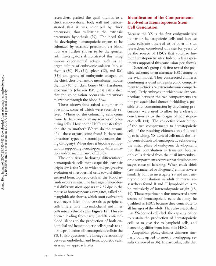
the generation of adult stem cells. In the caseof HSCs, the discussion was for a long timedominated by the direct observation of mam-malian and avian embryos, in which an ac-tive erythropoietic activity is first detected inthe yolk sac (YS). Within the mesodermallayer of the YS, homogeneous cellular aggre-gates appear in 7–7.5 days postcoitus (dpc) inthe mouse embryo and rapidly form a con-fined structure surrounded by cells that evolveto resemble endothelial cells morphologically,while the inner part of the aggregate com-prises erythrocytes. This cellular formationhas been called a blood island (Figure 1a).The erythrocytes in blood islands are large,nucleated cells that resemble erythrocyte pre-cursors in the BM and erythrocytes in lower(primitive) vertebrate groups, such as birds,fish, and amphibians. They never reach thefinal enucleated stage in situ, and for this rea-son they have been called primitive. Primitiveerythrocytes are the first detected hematopoi-etic cells of embryonic origin. It is logicalto conclude that, because the first identi-fiable hematopoietic cells in embryogenesisare present in the YS, HSCs would originatein the YS and lead to hematopoiesis at thissite.
The idea that HSCs originate in the YSfrom which (via the circulatory network thatforms thereafter) they could reach the fetalliver (FL), the major embryonic hematopoi-etic organ, and then the thymus and BM pre-vailed in the 1970s. Several experimental linesof evidence reinforced this hypothesis. Em-bryonic cells of YS origin isolated shortly afterblood island formation were injected in vivointo mouse embryos of related stages (8). His-tological analysis, done after birth, indicatedthat a variable, albeit sizable fraction of cells ofdonor origin, belonging to the hematopoieticlineage, was present in the thymus and BM ofthe recipient mice. Auerbach’s (9) group re-inforced these results by showing T cell dif-ferentiation potential in YS cells after fetalthymus organ culture. Interestingly, the peakof hematopoietic progenitors with lymphoidpotential occurred at 11 dpc, whereas the
www.annualreviews.org • Ontogeny of the Hematopoietic System 747
Ann
u. R
ev. I
mm
unol
. 200
7.25
:745
-785
. Dow
nloa
ded
from
arj
ourn
als.
annu
alre
view
s.or
gby
IN
SER
M-m
ulti-
site
acc
ount
on
11/0
1/07
. For
per
sona
l use
onl
y.

ANRV306-IY25-25 ARI 19 February 2007 16:31
Native mesoderm Hemangioblastic chord
Hemangioblast
Hemogenicendothelium
Committedendothelialprecursors
Committedhematopoietic
precursors
Differentiation of endothelialand hematopoietic lineages
Yolk sac endodermMesenchymal cells
Primitive erythroid cells
Primitive erythroid cell
Primitive erythroid cell
Endothelial cells
Endothelial cell
Primitive erythroid cell
Endothelial cell
Mesoderm
Mesoderm
a
b
c
21
Clonal growth
Specification toward hematopoieticand endothelial lineages
Differentiation
1
Clonal growth
Specification of hemogenicendothelial cells
Transdifferentiation intoprimitive erythroid precursor
Alternative model
Aggregation of specifiedendothelial and
hematopoietic precursors
Differentiation
2
Figure 1Mechanism of yolk sac (YS) blood island formation. (a) Schematic representation of the progressiveevolution of blood island mesodermal cells to a functional vascular network and primitive erythroid cells.Two alternative mechanism of YS blood island formation are shown. (b) Endothelial and hematopoieticcells may be generated directly from progenitors restricted to both differentiation pathways, thehemangioblast, or via the intermediate production of hemogenic endothelial cells. (c) Alternatively, thespecification into endothelial and hematopoietic cells might occur at earlier stages of differentiation (c).
748 Cumano · Godin
Ann
u. R
ev. I
mm
unol
. 200
7.25
:745
-785
. Dow
nloa
ded
from
arj
ourn
als.
annu
alre
view
s.or
gby
IN
SER
M-m
ulti-
site
acc
ount
on
11/0
1/07
. For
per
sona
l use
onl
y.

ANRV306-IY25-25 ARI 19 February 2007 16:31
intraembryonic compartment was consis-tently devoid of T cell differentiationpotential.
At the time, these observations were takenas evidence that HSCs are present in the YSbefore or around the time circulation is es-tablished. However, hematopoietic cells entercirculation rapidly between the four- and five-somite stage (10, 11), from which they reachthe hematopoietic organs. Therefore, isolat-ing embryonic hematopoietic tissues after thefive-somite stage does not allow the preciseidentification of the origin of HSCs.
Colony assays brought additional supportto the theory of the YS origin of HSCs. Moore& Metcalf (12) developed the first in vitroassays designed to reveal multipotency byanalysis of the progeny of single precursors.Limited cell types could be distinguished mor-phologically, but erythrocytes and polymor-phonuclear cells could be discriminated frommononuclear elements. The authors used cy-tokines present in conditioned media to stim-ulate these cultures as the main mitotic agentresponsible for colony formation in semisolidcultures. A careful time-course analysis ofsingle hematopoietic progenitors and theirprogeny indeed showed that the first multilin-eage colonies were obtained from YS popula-tions isolated shortly after the onset of bloodisland formation. These experiments showedthat progenitors with multilineage differenti-ation potential are of YS origin and are thefirst to be generated.
In the late 1970s, Herzenberg’s (13) lab-oratory used cells from different fractions ofthe embryo to reconstitute the B cell com-partment of adult irradiated recipient mice.The results of this experiment were puzzling.They obtained B cells of donor origin, asdistinguished by their immunoglobulin allo-type, with YS derivatives with similar if notlower efficiency than that of the cells iso-lated from the intraembryonic compartmentin a region confined to the subdiaphragma.This experiment, done with embryos afterthe establishment of circulation, raised thepossibility that lymphocyte precursors could
be present in the intraembryonic compart-ment before the colonization of the FL. Thisobservation remained largely forgotten untilexperiments done in the avian model, usingchicken/chicken or quail/chicken chimeras,showed that long-lasting contribution to thehematopoietic compartment was obtainedfrom progenitors present in the body of theembryo rather than in the YS (14). Theprevailing hypothesis in the late 1980s wasthus based on evidence from experiments onmouse; it stated that HSCs first originated inthe YS.
EXPERIMENTAL APPROACHOF DEVELOPMENTALHEMATOPOIESIS: STRENGTHSAND LIMITATIONS OF ANIMALMODELS
Recent progress in understanding the stepsinvolved in HSC development relies on datagathered in different animal models. Here,we briefly describe the major features of eachmodel and their respective contribution to theprogress and breakthroughs in developmentalhematopoiesis.
Amphibian and Avian Models
Amphibians and avians constitute the old-est vertebrate models in developmental biol-ogy, owing to the availability and large sizeof embryos. They are also amenable to thetransplantation of embryonic territories, thusallowing developmental fate analyses. Theconstruction of interspecific chimeras (first inthe avian model, then in amphibians) allowedthe definition of cell-fate analyses because thein vivo migration of small cell groups as wellas their contribution to the developing tis-sues can be followed through donor-recipientmarkers (15).
In the avian model, such chimeras resultfrom the combination of chick and quail tis-sues that can be discriminated from the mor-phology of nucleoli (condensed in quail anddispersed in chick) (16) and presently by using
www.annualreviews.org • Ontogeny of the Hematopoietic System 749
Ann
u. R
ev. I
mm
unol
. 200
7.25
:745
-785
. Dow
nloa
ded
from
arj
ourn
als.
annu
alre
view
s.or
gby
IN
SER
M-m
ulti-
site
acc
ount
on
11/0
1/07
. For
per
sona
l use
onl
y.
ANRV306-IY25-25 ARI 19 February 2007 16:31
species-specific antibodies (17). In some in-stances, investigators constructed chick-chickchimeras involving sex-mismatched (18) or al-logeneic (19) embryos to analyze late stagesof lymphoid development (including contri-bution to adulthood), as the initial synchronyof chick and quail embryonic development iseventually lost.
In the amphibian model, chimeras (firstconstructed in the frog Rana, then in the toadXenopus) combine tissues from chromosomal-ly distinct species (diploid versus triploid) (20,21). Besides its usefulness in tissue-fate trac-ing, the amphibian model is the oldest modelused to study the cascade of events leadingto embryonic patterning (22), and as such itproves highly suitable for investigating meso-derm determination toward a hematopoieticfate (23), in particular in the animal cap assayin which a dissected part of the gastrula nor-mally fated to produce ectodermal derivativesmay be turned into blood-forming mesodermon exposure to prospective inducing factors,such as BMP4 (see below).
Although antibodies against hematopoi-etic markers are available, ensuring the sort-ing and characterization of selected subsets,the limitations of these two models lie in thepoorly developed in vitro culture systems—although clonogenic assays that allow ery-thromyeloid precursor characterization areavailable for avian cells (24)— and routine/up-to-date in vivo assays of LTR, as well as inthe absence of tools to carry over genetic ap-proaches (homologous recombination).
Zebrafish
Interest in the zebrafish as a hematopoieticdevelopment model lies first in the poten-tial novel finding about the genetic controlof developmental hematopoiesis, thanks tothe large collection of chemically inducedmutants generated by two laboratories (25,26). The zebrafish constitutes a fast develop-ing and promising model because LTR as-says have been recently developed, as wellas phenotypic FACS analyses (27, 28), which
will complete the large panel of zebrafish as-sets, such as the availability of transgenic fishexpressing an increasing number of fluores-cent reporter proteins, the routine use of an-tisense technology, and lineage-tracing proto-cols. More importantly, the zebrafish providesthe possibility, owing to the transparency ofthe embryo, of following in situ cell migra-tion of selected groups or even single cells,without staining, at selected differentiationtimes and locations thanks to laser-drivenfluorochromes uncaging. Nevertheless, stud-ies of hematopoietic development in the ze-brafish are complicated owing to the absenceof hematopoiesis in the YS, with the first waveof hematopoietic cells appearing in a caudalsite named an intercellular cell mass.
THE FIRST CHALLENGESTO THE CLASSICAL MODELS
Successive Hematopoietic Sites
Historically, investigators approached the un-derstanding of hematopoietic developmentthrough observation of the sites at which dif-ferentiation occurs, an approach that provedsomehow misleading as to the origin of HSCsbecause it was based on the assumption thatthe HSC generation site could be moni-tored by their differentiation. The main con-clusion drawn from these analyses was that,in all species studied (Table 1), successivesites achieve the production of differentiatedhematopoietic cells until the definitive (adult)hematopoietic organs are fully developed andready to take over this function.
The first hematopoietic site, namely YSblood islands, is active from the earliest stageof organogenesis (soon after gastrulation). Be-fore the development of the hematopoieticorgans (the first being the thymus, then thespleen and, finally, the BM), hematopoiesissystematically occurs within an intermediatesite, the FL in the mouse and human, as wellas in the amphibian (23).
In the chick embryo, a function similar tothat of the FL seems to be carried out by a
750 Cumano · Godin
Ann
u. R
ev. I
mm
unol
. 200
7.25
:745
-785
. Dow
nloa
ded
from
arj
ourn
als.
annu
alre
view
s.or
gby
IN
SER
M-m
ulti-
site
acc
ount
on
11/0
1/07
. For
per
sona
l use
onl
y.

ANRV306-IY25-25 ARI 19 February 2007 16:31
Table 1 Successive hematopoietic sites in vertebrate embryos: location and functions
Initial generation site Second generation siteIntermediate
hematopoietic siteDefinitive
hematopoietic sitesFish (zebrafish) ICM: Generation +
differentiationAGM: Generation. Noovert differentiation
Unknown Thymus: T celldifferentiation
Kidney: HSCmaintenance +differentiation?
Amphibian (Xenopus) YS (VBI): Generation+ differentiation
AGM (DLP):Generation. No overtdifferentiation
FL: HSCmaintenance?HSC expansion?Differentiation
Thymus: T celldifferentiation
Spleen: differentiationKidney: HSCmaintenance +differentiation?
Avian (chick/quail) YS: Generation +differentiation
AGM: Generation. Noovert differentiation
Allantois: Generation?
Para-aortic foci:HSCmaintenance?HSC expansion?Differentiation
Thymus: T celldifferentiation
Bursa of Fabricius: Bcell differentiation
Spleen: differentiationBM: HSC maintenance+ differentiation
Mouse YS: Generation +differentiation
AGM: Generation. Nodifferentiation.Expansion?
Allantois/Placenta:Generation?Accumulation?Expansion?
FL: HSCmaintenance.HSC expansion.Differentiation
Thymus: T celldifferentiation
Spleen: B celldifferentiation
BM: HSC maintenance+ differentiation
Human YS: Generation +differentiation
AGM: Generation. Noovert differentiation.Expansion?
FL: HSCmaintenance.HSC expansion.Differentiation
Thymus: T celldifferentiation
Spleen: B celldifferentiation
BM: HSC maintenance+ differentiation
Abbreviations: AGM, aorta, gonads, and mesonephros; BM, bone marrow; DLP, dorsal lateral plate; FL, fetal liver; HSC, hematopoietic stem cell;ICM, intermediate cell mass; VBI, ventral blood island; YS, yolk sac.
structure called the para-aortic foci. In the fishembryo, no such intermediate hematopoieticsite has been identified.
Extrinsic Origin of HematopoieticPrecursors in the Various TissuesPerforming HematopoieticDifferentiation
The alternative hypotheses as to the originof blood cell precursors in the successive
organs sustaining hematopoietic cell produc-tion constituted a high controversy amongembryologists for approximately one century,namely (a) each site independently producesits own precursors; (b) blood cell productionin a given site depends on its colonization byprecursors born elsewhere; and (c) hypothesis(a) or (b) applies, depending on the consideredsite. The first conclusive experimental datawere obtained in the avian model. Using thechick-quail interspecific chimera setup,
www.annualreviews.org • Ontogeny of the Hematopoietic System 751
Ann
u. R
ev. I
mm
unol
. 200
7.25
:745
-785
. Dow
nloa
ded
from
arj
ourn
als.
annu
alre
view
s.or
gby
IN
SER
M-m
ulti-
site
acc
ount
on
11/0
1/07
. For
per
sona
l use
onl
y.
ANRV306-IY25-25 ARI 19 February 2007 16:31
researchers grafted the quail thymus to achick embryo dorsal body wall and demon-strated that it was colonized by chickprecursors, thus validating the extrinsicprecursors hypothesis (29). The need forthe developing hematopoietic organs to becolonized by extrinsic precursors via bloodflow was further shown to be the generalrule. Investigators demonstrated this usingvarious experimental setups, such as anorgan culture of embryonic anlagen [mousethymus (30), FL (31), spleen (32), and BM(33)] and grafts of embryonic anlagen onthe chick chorio-allantoic membrane [mousethymus (30), chicken bone (34)]. Parabiosisexperiments [chicken BM (35)] establishedthat the colonization occurs via precursorsmigrating through the blood flow.
These observations raised a number ofquestions, some of which were recently re-vived: Where do the colonizing cells comefrom? Is there one or many sources of colo-nizing cells? How do the HSCs transfer fromone site to another? Where do the stromaof all these organs come from? Is there oneor various types of stromal precursors dur-ing ontogeny? When does it become compe-tent in supporting hematopoietic differentia-tion and/or maintenance of HSCs?
The only tissue harboring differentiatedhematopoietic cells that escape this extrinsicorigin law is the YS, in which the progressiveevolution of mesodermal cells toward differ-entiated hematopoietic cells in the blood is-lands occurs in situ. The first sign of mesoder-mal differentiation appears at 7.25 dpc in themouse as homogeneous aggregates, called he-mangioblastic chords, which soon evolve intoerythrocyte-filled blood vessels as peripheralcells differentiate into endothelial and innercells into erythroid cells (Figure 1a). This se-quence leading from early (undifferentiated)blood islands to the production of both en-dothelial and hematopoietic cells signals to anin situ production of hematopoietic cells in theYS. It also questions the lineage relationshipbetween endothelial and hematopoietic cells,an issue we approach later.
Identification of the CompartmentsInvolved in Hematopoietic StemCell Generation
Because the YS is the first embryonic siteto harbor hematopoietic cells and becausethese cells are observed to be born in situ,researchers considered this site for years tobe the source of HSCs that colonize fur-ther hematopoietic sites. Indeed, a few exper-iments supported this conclusion (see above).
Dieterlen’s group (14) first tested the pos-sible existence of an alternate HSC source inthe avian model. They constructed chimerascombining a quail intraembryonic compart-ment to a chick YS (extraembryonic compart-ment). Early embryos, in which vascular con-nections between the two compartments arenot yet established (hence forbidding a pos-sible cross-contamination by circulating pre-cursors), were used to allow for a clear-cutconclusion as to the origin of hematopoi-etic cells (14). The respective contributionof the two compartments to hematopoieticcells of the resulting chimeras was followedup to hatching. YS-derived cells made the ma-jor contribution to hematopoietic cells duringthe initial phase of embryonic development,but this contribution is transient becauseonly cells derived from the quail intraembry-onic compartment are present at developmentstages close to hatching. When chick-chick(sex-mismatched or allogeneic) chimeras weresimilarly built to investigate YS and intraem-bryonic contribution in adult chimeras, re-searchers found B and T lymphoid cells tobe exclusively of intraembryonic origin (18,19). These experiments thus uncovered a newsource of hematopoietic cells that may bequalified as HSCs because they contribute toall lineages of the adult. They also establishedthat YS-derived cells lack the capacity eitherto sustain the production of hematopoieticcells or to give rise to lymphoid cells, andhence they differ from bona fide HSCs.
Amphibian ploidy-distinct chimeras sim-ilarly built up led to mostly overlapping re-sults (reviewed in 36). In particular, cells that
752 Cumano · Godin
Ann
u. R
ev. I
mm
unol
. 200
7.25
:745
-785
. Dow
nloa
ded
from
arj
ourn
als.
annu
alre
view
s.or
gby
IN
SER
M-m
ulti-
site
acc
ount
on
11/0
1/07
. For
per
sona
l use
onl
y.

ANRV306-IY25-25 ARI 19 February 2007 16:31
colonize the FL and produce an ery-thromyeloid as well as B lymphoid progenyderive from the dorsal lateral plate (DLP)(amphibian homologous to the intraembry-onic hemogenic site) (37). However, in thisgroup, the ventral blood islands (YS homol-ogous) also produce T lymphocytes of thelarva and adult (21), in addition to DLP-derived thymocytes. This discrepancy pointsto other standing questions in the field: Aremesodermal-derived hematopoietic precur-sors from the extra- and intraembryonic com-partment initially identical? Is there a YS pre-cursor limitation regarding the differentiationand self-renewal potential imposed by the en-vironment? Alternatively, is the determina-tion/generation process radically different forprecursors of extra- and intraembryonic ori-gin? The ability of amphibian YS precursorsto give rise to a lymphoid progeny would favorthe first hypothesis. More recently (38), fate-map tracing experiments showed that Xenopusventral blood islands and DLP derive fromdistinct blastomeres of the 32-cell embryo,thus establishing the independent generationof precursors in both sites.
In mammals, the first evidence of an in-traembryonic pool of hematopoietic precur-sors appeared in the early 1990s. Nishikawa’sgroup (39) and Cumano et al. (40) showedthat precursors endowed with a B lymphoidpotential could be recovered first from the in-traembryonic compartment and later in theYS. Godin et al. (41) established that the aorticregion of 9-dpc embryos was capable of givingrise to B cells on transfer to irradiated mice,whereas the YS precursors could not. Medvin-sky et al. (42) pointed to the same region—which encompasses the aorta, gonads, andmesonephros (AGM) at a latter stage (10.5dpc)—as containing CFU-S before this pre-cursor type can be recovered from the YS.
Investigators further characterized in-traembryonic hematopoietic precursors asmultipotent (32, 43) and capable of perform-ing long-term multilineage hematopoietic re-constitution (LTR activity) of adult irradi-ated mice, when taken after 10.5 dpc (44). At
Hemogenic:property of a sitethat generateshematopoieticprecursors byopposition to thehematopoietic sitewhere maintenanceand/ordifferentiationoccurs
AGM: aorta,gonads, andmesonephros
earlier developmental stages (9 dpc), this ac-tivity could only be evidenced on the directinjection of intraembryonic precursors intothe liver of myelo-ablated newborn recipients.Yoder et al. (45) concomitantly detected thisactivity (in larger amounts) in the correspond-ing YS that was similarly injected.
Studies combining phenotypic and cyto-logical analyses looked into the possible oc-currence of an intraembryonic hemogenic siteduring human ontogeny, which led to the con-clusion that intraembryonic precursors arealso present in the aorta region of human em-bryos (46, 47). However, a clear-cut homol-ogy to the two-generation events as foundin lower vertebrate chimeras could not bedrawn from these experiments: Intraembry-onic hematopoietic precursors first appear at8.5 dpc in the mouse (43), i.e., after the con-nection of YS and intraembryonic blood ves-sels, an event that occurs at 8 dpc (10, 48).Consequently, the recovery of intraembry-onic precursors from the aorta region couldresult either from an in situ generation orfrom a selective aggregation of precursorsborn elsewhere in a suitable intraembryonicniche.
Independent Generationof Hematopoietic Cells
Researchers undertook different approachesto answer the question of the independentgeneration of hematopoietic cells. An in-direct approach involved organ culture ofthe intraembryonic hemogenic site explantedat 10 dpc (i.e., at stages when LTR activ-ity on transfer to irradiated adult recipientsis not detectable) (49). After organ culture,the investigators recovered a greater num-ber of HSCs displaying LTR activity in adults(49), indicating either an increased de novoHSC generation or, alternatively, an amplifi-cation/maturation of the pool of precursorsinitially present in the explants (as the in-traembryonic hemogenic site already containsmultipotent precursors at the stage of AGMisolation) (43).
www.annualreviews.org • Ontogeny of the Hematopoietic System 753
Ann
u. R
ev. I
mm
unol
. 200
7.25
:745
-785
. Dow
nloa
ded
from
arj
ourn
als.
annu
alre
view
s.or
gby
IN
SER
M-m
ulti-
site
acc
ount
on
11/0
1/07
. For
per
sona
l use
onl
y.

ANRV306-IY25-25 ARI 19 February 2007 16:31
YS
Sp
AGM
SV
FL
AGM
Somatopleura
Splanchnopleura
YS
8 dpc:Before the establishment of
a functional vascular network
10.5 dpc:The hematogenic sites in the AGM
derives from the 8 dpc Sp
Sp
Ectoderm
Mesoderm
Endoderm
SAP
HIAC
Figure 2Hemogenic sites in the developing mouse embryo. (a) The respective localization and layer makeup ofthe extraembryonic yolk sac (YS) and presumptive intraembryonic splanchnopleura (Sp) are shown at thestage preceding the establishment of vacular connections between the two compartments. Both sitesassociate endoderm and mesoderm germ layers. (b) The Sp gives rise eventually to the para-aorticsplanchnopleura/aorta, gonads, and mesonephros (P-Sp/AGM), whose localization within the embryo isshown, as well as the tissue structure in the schematic cross-section of a 10.5-dpc embryo. The respectivelocalization of the hematopoietic intra-aortic clusters (HIACs) and subaortic patches (SAPs) is alsoshown in the enlarged aorta drawing.
Splanchnopleura(Sp): subdivision ofthe lateral platemesoderm by thecoelomic cavity thatassociates mesodermand endoderm
Another strategy involved organ cultureof the prospective territory that gives rise tothe aortic region, the splanchnopleura (Sp)(see Figure 2a), explanted at stages preced-ing both the establishment of blood vesselconnections between the YS and the embryoproper, and the appearance of intraembryonichematopoietic precursors (50, 51). In theseexperiments, the organ culture aimed to pre-serve the environment in which mesodermalcells may commit to a hematopoietic fate, such
as a proper relationship with the endoderm,that is required to ensure the generation ofhematopoietic cells from the nearby meso-derm, as discussed below. After the organ-culture step, hematopoietic cells could berecovered from the YS and from the intraem-bryonic hemogenic prospective territory, thusproving the existence of two independentwaves of hematopoietic cell generation in theearly embryo. Moreover, extra- and intraem-bryonic precursors differ strikingly in their
754 Cumano · Godin
Ann
u. R
ev. I
mm
unol
. 200
7.25
:745
-785
. Dow
nloa
ded
from
arj
ourn
als.
annu
alre
view
s.or
gby
IN
SER
M-m
ulti-
site
acc
ount
on
11/0
1/07
. For
per
sona
l use
onl
y.

ANRV306-IY25-25 ARI 19 February 2007 16:31
differentiation potential because, contrary toprecursors derived from the intraembryoniccompartment, YS-derived precursors provedunable to generate a lymphoid progeny. Be-fore the organ-culture step, the intraembry-onic, but not the extraembryonic, cells weredevoid of hematopoietic potential in vitro, anobservation that may account for the failureof Moore & Metcalf (12) to detect the in-traembryonic precursors in their multilineagecolony assays. Similar conclusions applied tohuman ontogeny because the early YS lacksboth B lymphoid (on in vitro culture) and Tlymphoid [on fetal-thymus organ culture inNOD-SCID (nonobese diabetic–severe com-bined immunodeficient)] potentials, whereasprecursors derived from the intraembry-onic hemogenic site display both lymphoidpotentials (52).
To assay the in vivo engraftment capac-ity, Cumano et al. (51) transplanted earlyextra- and intraembryonic hematopoietic pre-cursors, isolated before circulation, into theRag-2−/− x γc−/− mouse strain (53), whichis alymphoid and also lacks natural killercell activity. The authors chose these recip-ients because MHC class I molecules arehardly expressed in embryonic cells before10.5 dpc (54), and hematopoietic precursorscould therefore be eliminated by radiation-resistant host natural killer cells when trans-ferred into conventional recipients. YS pre-cursors were capable of engrafting these miceand transiently gave rise to a myeloid progeny.However, no lymphoid progeny was ever pro-duced, and myeloid cell production was onlytransient because no contribution was everdetected three months after engraftment. Incontrast, precursors derived from the organ-cultured intraembryonic hemogenic site gen-erated myeloid and lymphoid cells that couldbe detected in the periphery and in the BMup to eight months after transplantation.
Because precursors displaying the multi-lineage and LTR activity that characterizeHSCs could be derived from the prospec-tive intraembryonic hemogenic site explantedat precirculatory stages and not from the
corresponding YS, intraembryonic precursorsmay be responsible for the seeding of defini-tive hematopoietic organs. These conclusionspoint to an absence of lymphoid potential inthe precirculatory YS (55, 56).
THE INTRAEMBRYONICHEMOGENIC SITE
General Location: P-Sp/AGM
Both in vivo and in vitro functional ap-proaches and analyses performed in situ pointto the aorta and underlying mesenchyme asthe source of intraembryonic HSC.
Experimental analyses. In the avian model,cytological (57) (see below) and in vitro (24)analyses found that intraembryonic HSCs arelocated in the region neighboring the dor-sal aorta. Furthermore, on grafting a quailaorta into the dorsal mesentery of a chick em-bryo (58), investigators found that quail aorta–derived HSCs contributed to para-aortic foci(57), an intermediate differentiation site thatmay correspond to the FL of other vertebratespecies as it harbors cells from all hematopoi-etic lineages, as well as transplantable lym-phoid cells (in cycloheximide-treated chicken)(59). Interestingly, when the quail aorta wascleared of contaminating mesenchymal cells,no hematopoietic contribution was detected,an observation that led the authors to con-clude that hematopoietic precursors likelyarise from tissue surrounding the dorsal aorta(58).
As stated above, intraembryonic precur-sors are found in mammals in an area thatincludes the AGM (Figure 2b). To determineprecisely the site of intraembryonic HSC pro-duction, researchers subdivided the mouseAGM into its various components and assayedit for precursor enrichment in vitro. The iso-lated aorta and surrounding tissue harboredthe majority of multipotent precursors (32).A similar approach, undertaken by Dzierzak’s(60) group in an in vivo assay, established thatthe aorta region, rather than the urogenital
www.annualreviews.org • Ontogeny of the Hematopoietic System 755
Ann
u. R
ev. I
mm
unol
. 200
7.25
:745
-785
. Dow
nloa
ded
from
arj
ourn
als.
annu
alre
view
s.or
gby
IN
SER
M-m
ulti-
site
acc
ount
on
11/0
1/07
. For
per
sona
l use
onl
y.

ANRV306-IY25-25 ARI 19 February 2007 16:31
HIACs:hematopoieticintra-aortic clusters
SAPs: subaorticpatches
compartment, displays the highest frequencyof LTR cells.
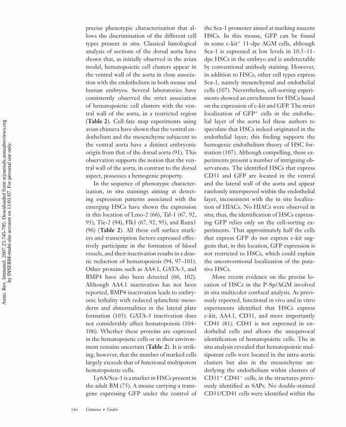
Cytological analyses. Cytological analysesuncovered two structures that appear linkedto the process of intraembryonic HSC gen-eration (Figure 2b). The first comprisedhematopoietic intra-aortic clusters (HIACs)inserted inside the ventral wall of the dorsalaorta, which have been observed in bird (57,61, 62), amphibian (38), mouse (63, 64), andhuman embryos (46). In situ phenotype anal-yses, in some instances linked to in vitro andin vivo potential assessment, indicate that theclusters comprise immature hematopoieticcells, as well as more differentiated cells, suchas macrophages. In the mouse embryo, intra-arterial clusters are also detected in the um-bilical and omphalo-mesenteric (also calledvitelline) arteries (60, 63, 65); however, theircharacterization for in vitro and in vivo po-tentials, as well as phenotype, is less advancedthan that of the aortic clusters. The only avail-able functional data indicate that these twoarteries harbor LTR HSCs. However, con-trary to the aorta in which the LTR-HSCnumber increases on organ culture (49, 60),organ culture of either umbilical or omphalo-mesenteric arteries does not lead to an in-creased recovery of LTR HSCs (60). The cellsthat belong to the HIACs are inserted be-tween endothelial cells, resulting in a discon-tinuity in the endothelial layer, but also in thebasement membrane, which is disrupted atthe HIAC level (46, 62, 63). The morphol-ogy of both HIAC cells and the underlyingvessel wall suggests a migratory phenotypefor these cells but gives no indication on thedirection of a possible migratory route. In-terestingly, HIAC are not detected from thebeginning of intraembryonic hematopoieticprecursor generation [8.5–9 dpc in the mouse(43)], but only when the number of generatedprecursors reaches a maximal level [10.5–11dpc (32)].
The tissue below the aortic floor also ap-pears potentially involved in the HSC gener-ation process. It comprises the second struc-
ture discovered through cytological analysis,called subaortic patches (SAPs). SAPs werefirst identified through the expression of theGATA-3 transcription factor (66) and arepresent below the aorta for the whole du-ration of intraembryonic hematopoietic pre-cursor generation, contrary to the HIACs,which are only detected at the peak of in-traembryonic HSC production. SAPs, whichare preferentially located below the HIACs atthe time when these are present, from 10.5to 11.5 dpc (63), disappear after 12.5 dpc,when intraembryonic precursors are nolonger generated (32). Although a similarstructure has also been identified in the humanAGM (67, 68), SAPs are not characterizedas such in other species, even if expression-pattern analyses report the expression in thearea underlying the HIACs of a large arrayof genes and proteins involved in hematopoi-etic cell development (Table 2). Interest-ingly, these SAPs do not seem to occur belowthe umbilical or omphalo-mesenteric arteries(I. Godin, personal observation), so the above-mentioned difference between these two ar-teries and the aorta regarding in vitro HSCamplification on organ culture may be relatedto the different type of environment. Theonly direct evidence for the involvement ofHIACs and the SAPs/subaortic area in thegeneration of intraembryonic HSCs comesfrom lineage-tracing experiments performedin Xenopus embryos, in which both intra-aorticclusters and the subaortic area are found toexpress hematopoietic markers, as well as theβ-galactosidase reporter, on injection of theblastomeres that gives rise to the DLP (38).
Phenotypic Analysis and PreciseLocation of HematopoieticStem Cells
The observation that HSCs appear in a de-fined location around the dorsal aorta be-tween 9–12 dpc led to an attempt to character-ize the surface-marker expression pattern thatcharacterizes them and to visualize emergingHSCs in the Sp/AGM. HSCs in the BM are
756 Cumano · Godin
Ann
u. R
ev. I
mm
unol
. 200
7.25
:745
-785
. Dow
nloa
ded
from
arj
ourn
als.
annu
alre
view
s.or
gby
IN
SER
M-m
ulti-
site
acc
ount
on
11/0
1/07
. For
per
sona
l use
onl
y.

ANRV306-IY25-25 ARI 19 February 2007 16:31
Table 2 In situ expression patterns in the various AGM compartment involved in HSC generationa
Localization in the intraembryonic hemogenic siteMarker CE HIACs HSCs in SAPs SAPs Reference(s)Adhesion moleculesCD31 + + + + H: 46; M: 65, 84CD34 + + +∗ +∗ H: 46, 95, 181; M: 64, 65; ∗I. Godin,
unpublishedAA4.1 + + + + M: 66, 79, 84Endomucin + + ? − M: 191VE-cadherin + + ? − H: 67V-CAM (CD106) + + ? − H: 192CD41 − + +∗ −∗ A: 193; M: ∗84CD43 − + ? − H: 194CD44 − + ? − H: 194CD164 − + − H: 194Tenascine C − to +∗ − to +∗ + + H: 67; A: ∗195ALCAM/CD166/BEN −∗ to + − ? +∗ H: ∗181; M: 196H-CAM − + ? − H: 192WASP − + ? − H: 192Tyrosine kinases receptorsc-Kit + + ? ? H: 67; M: 98, 197Flk-1/VEGF-R2/KDR + − to ± ? ? H: 67, 95; M: 98; A: 62; Zf: 149, 198Flt3/Flk2/STK-1 + + ? − H: 67Tie2/tek + + ? ? M: 97Growth factors/morphogensFlt3-ligand + + ? − H: 67BMP-4 − ± ? + H: 105; M: A. Manaia & I. Godin,
personal observationVEGF + + ? − H: 67TGFβ-1 ± + ? − H: 105Transcription factorsGATA-2 + + +∗ +∗ H: 67; M: ∗84; XL: 38; Zf: 149, 198GATA-3 − + +∗ +∗ H: ∗199; XL: 38; Zf: 200; M: ∗66, 84c-Myb − + ND − H: 95; A: 201; Zf: 198Lmo2 ± + + − M: 66; Zf: 198AML-1 ± + ? − M: 99; Zf: 38Tal-1/SCL + + +∗ −∗ H: 67, 95; M: 96 (∗I. Godin,
unpublished); XL: 38; Zf: 19, 149OthervWf + − ND + M: 65CD45 − ∗few cells to + − − H: 46, 67; A: 12, 62; M: ∗66, 84SMA − − ? + H: 47; M: 66
aWhenever contradictory expression patterns are found, ∗ indicates the corresponding publication.
www.annualreviews.org • Ontogeny of the Hematopoietic System 757
Ann
u. R
ev. I
mm
unol
. 200
7.25
:745
-785
. Dow
nloa
ded
from
arj
ourn
als.
annu
alre
view
s.or
gby
IN
SER
M-m
ulti-
site
acc
ount
on
11/0
1/07
. For
per
sona
l use
onl
y.

ANRV306-IY25-25 ARI 19 February 2007 16:31
P-Sp: para-aorticsplanchnopleura
P-Sp/AGM:intraembryonichemogenic site from8.5–11.5 dpc; theP-Sp 8.5–10 dpcevolves into theAGM at 10–11.5 dpc
characterized by the expression of c-kit andSca-1 within the Lin− fraction (75). Althoughit contains all HSCs, this fraction is far fromhomogeneous, and it was later fractionatedwith Flk2 and CD34 in Flk2−CD34− long-term and Flk2+ CD34+ short-term reconsti-tuting cells (76). HSCs can also be isolated bythe expression of CD150 and the absence ofCD48 (77). In the FL, HSCs express most ofthe above-mentioned markers, although theycoexpress low levels of CD11b (78).
Surface-marker analysis. One of the firstcharacterizations of hematopoietic cells in thepara-aortic splanchnopleura (P-Sp; intraem-bryonic hemogenic site at 8.5–10 dpc) pointedto the expression of AA4.1 (43), later char-acterized by Lemischka et al. (79), as allow-ing a substantial enrichment of multipotentcells that differentiate in vitro. In the samestudy, multipotent cells were not enriched inthe Sca-1+ fraction. Subsequently, Sanchezet al. (80) showed that long-term reconsti-tuting cells were all in the c-kit+ fractionof the AGM and that they coexpress CD34and low levels of Mac-1 (CD11b). The find-ing that some cells within this region alsoexpress CD31, VE-cadherin, and Tie-2 (ex-pressed in endothelial cells) and that a fractionlack CD45 (a pan-hematopoietic marker) ledsome authors to propose that an endothelialcompartment in this region contained HSCs(81–83, 170).
In an attempt to homogenize and extendthe information on cell surface markers withinthe P-Sp/AGM region, a recent study (81)showed that 10.5-dpc multipotent cells, whichalso show LTR activity, were all indeed c-kit+
but expressed low levels of CD45. They allalso coexpress CD31, which was subsequentlyshown to be present in hematopoietic cells inthe FL and BM and thus cannot be taken as astrict endothelial marker (82, 83). In this sameregion, Mac-1+ cells are also present, andthey correspond to monocyte/macrophage-committed precursors likely of YS origin (81).In situ labeling experiments have shown thatCD41 is a marker of early hematopoietic cells
but is absent in endothelial cells (81, 84–86).In adult BM, CD41 expression is restrictedto megakaryocytes (87). The coexpression ofCD41 (a strict hematopoietic marker) on allc-kit+ cells also shows that no endothelialcomponents are present in this fraction at thisstage of development (71). c-kit+ cells alsoexpress, as previously mentioned, a batteryof endothelial markers (CD34, CD31, VE-cadherin, Tie-2), and some are even Flk1+
(Figure 3). Whether this coincidence reflectsa common precursor or a functional require-ment remains to be determined. As devel-opment progresses, c-kit+ cells express in-creasing levels of CD45, which labels mosthematopoietic precursors in 11.5 dpc (88).
As circulation is established at the four-to-five-somite stage (8 dpc) (10, 48), no cleardifference in surface phenotype was ever ob-tained between YS and AGM hematopoieticprecursors. In YS, the first expressed mark-ers are again c-kit and CD41 (85, 86, 89). By10.5 dpc, the hematopoietic populations inboth sites appear similar. However, a quan-titative in vitro differentiation potential assayrevealed that YS c-kit+ cells exhibit a lowerfrequency of multipotent cells than AGM,while having a much higher frequency ofmyeloid progenitors (81). These myeloid pro-genitors originate in the YS, although somemight also be of intraembryonic origin (90).A careful examination of the expression pro-files shows that the frequency of AA.1, CD34,Tie-2 bright, and c-kit+ cells is considerablyhigher in AGM than in YS hematopoieticcells (Figure 3c). When isolated by flow cy-tometry, these brightly stained cells comprisemultipotent cells at frequencies close to 1:1(A. Cumano & I. Godin, unpublished obser-vations). In conclusion, no particular pheno-type distinguishes between the multipotentlong-term reconstituting cells and YS-derivederythromyeloid precursors.
Direct visualization of hematopoieticstem cells in the aorta, gonads, andmesonephros. The direct visualization ofthe emerging HSCs in the AGM requires a
758 Cumano · Godin
Ann
u. R
ev. I
mm
unol
. 200
7.25
:745
-785
. Dow
nloa
ded
from
arj
ourn
als.
annu
alre
view
s.or
gby
IN
SER
M-m
ulti-
site
acc
ount
on
11/0
1/07
. For
per
sona
l use
onl
y.

ANRV306-IY25-25 ARI 19 February 2007 16:31
Figure 3Flow-cytometric profiles of cell suspensions obtained from 10.5-dpc aorta, gonads, and mesonephros(AGM) and yolk sac (YS). (a) Profile of the staining of AGM cell suspensions with CD31, Tie-2, CD34,and AA4.1 gated on both c-kit+ hematopoietic cells and c-kit− cells. Because most hematopoieticmultipotent cells do not express CD45, these cells that comprise monocytes and macrophages at thisstage were gated out. (b) Flow-cytometry profiles showing the representation of Sca-1, Tie-2, Flk1, andc-kit in AGM cell suspensions. In this experiment macrophages were gated out using the Mac-1 (CD11b)antibody. (c) Flow-cytometry profiles comparing AGM and YS cell suspensions for the expression ofCD31, VE-cadherin, c-kit, and CD34.
www.annualreviews.org • Ontogeny of the Hematopoietic System 759
Ann
u. R
ev. I
mm
unol
. 200
7.25
:745
-785
. Dow
nloa
ded
from
arj
ourn
als.
annu
alre
view
s.or
gby
IN
SER
M-m
ulti-
site
acc
ount
on
11/0
1/07
. For
per
sona
l use
onl
y.
ANRV306-IY25-25 ARI 19 February 2007 16:31
precise phenotypic characterization that al-lows the discrimination of the different celltypes present in situ. Classical histologicalanalysis of sections of the dorsal aorta haveshown that, as initially observed in the avianmodel, hematopoietic cell clusters appear inthe ventral wall of the aorta in close associa-tion with the endothelium in both mouse andhuman embryos. Several laboratories haveconsistently observed the strict associationof hematopoietic cell clusters with the ven-tral wall of the aorta, in a restricted region(Table 2). Cell-fate map experiments usingavian chimera have shown that the ventral en-dothelium and the mesenchyme subjacent tothe ventral aorta have a distinct embryonicorigin from that of the dorsal aorta (91). Thisobservation supports the notion that the ven-tral wall of the aorta, in contrast to the dorsalaspect, possesses a hemogenic property.
In the sequence of phenotypic character-ization, in situ stainings aiming at detect-ing expression patterns associated with theemerging HSCs have shown the expressionin this location of Lmo-2 (66), Tal-1 (67, 92,93), Tie-2 (94), Flk1 (67, 92, 95), and Runx1(96) (Table 2). All these cell surface mark-ers and transcription factors expressed effec-tively participate in the formation of bloodvessels, and their inactivation results in a dras-tic reduction of hematopoiesis (94, 97–101).Other proteins such as AA4.1, GATA-3, andBMP4 have also been detected (66, 102).Although AA4.1 inactivation has not beenreported, BMP4 inactivation leads to embry-onic lethality with reduced splanchnic meso-derm and abnormalities in the lateral plateformation (103). GATA-3 inactivation doesnot considerably affect hematopoiesis (104–106). Whether these proteins are expressedin the hematopoietic cells or in their environ-ment remains uncertain (Table 2). It is strik-ing, however, that the number of marked cellslargely exceeds that of functional multipotenthematopoietic cells.
Ly6A/Sca-1 is a marker in HSCs present inthe adult BM (75). A mouse carrying a trans-gene expressing GFP under the control of
the Sca-1 promoter aimed at marking nascentHSCs. In this mouse, GFP can be foundin some c-kit+ 11-dpc AGM cells, althoughSca-1 is expressed at low levels in 10.5–11-dpc HSCs in the embryo and is undetectableby conventional antibody staining. However,in addition to HSCs, other cell types expressSca-1, namely mesenchymal and endothelialcells (107). Nevertheless, cell-sorting experi-ments showed an enrichment for HSCs basedon the expression of c-kit and GFP. The strictlocalization of GFP+ cells in the endothe-lial layer of the aorta led these authors tospeculate that HSCs indeed originated in theendothelial layer; this finding supports thehemogenic endothelium theory of HSC for-mation (107). Although compelling, these ex-periments present a number of intriguing ob-servations. The identified HSCs that expressCD31 and GFP are located in the ventraland the lateral wall of the aorta and appearrandomly interspersed within the endotheliallayer, inconsistent with the in situ localiza-tion of HIACs. No HIACs were observed insitu; thus, the identification of HSCs express-ing GFP relies only on the cell-sorting ex-periments. That approximately half the cellsthat express GFP do not express c-kit sug-gests that, in this location, GFP expression isnot restricted to HSCs, which could explainthe unconventional localization of the puta-tive HSCs.
More recent evidence on the precise lo-cation of HSCs in the P-Sp/AGM involvedin situ multicolor confocal analysis. As previ-ously reported, functional in vivo and in vitroexperiments identified that HSCs expressc-kit, AA4.1, CD31, and more importantlyCD41 (81). CD41 is not expressed in en-dothelial cells and allows the unequivocalidentification of hematopoietic cells. The insitu analysis revealed that hematopoietic mul-tipotent cells were located in the intra-aorticclusters but also in the mesenchyme un-derlying the endothelium within clusters ofCD31+ CD41− cells, in the structures previ-ously identified as SAPs. No double-stainedCD31/CD41 cells were identified within the
760 Cumano · Godin
Ann
u. R
ev. I
mm
unol
. 200
7.25
:745
-785
. Dow
nloa
ded
from
arj
ourn
als.
annu
alre
view
s.or
gby
IN
SER
M-m
ulti-
site
acc
ount
on
11/0
1/07
. For
per
sona
l use
onl
y.

ANRV306-IY25-25 ARI 19 February 2007 16:31
endothelial layer of the aorta, as in the studypreviously described. We are therefore leftwith some contradictory results that neverthe-less coincide to identify HSCs in close associ-ation with the ventral wall of the dorsal aortaboth in intra-aortic clusters and some within aparticular set of mesenchymal cells in the sub-jacent region (81). Still, all these experimentscombined have not provided conclusive evi-dence for either the generation process or theexact location of the differentiating cells.
OTHER EXTRA-EMBRYONICGENERATION SITES?
In the avian embryo, the allantois combinesboth mesoderm and endoderm and as suchmay potentially contribute to blood cells, asit harbors the same embryonic layer makeupas the YS and the early intraembryonichemogenic site. Caprioli et al. (69) experimen-tally tested this possibility by grafting quailprecirculatory allantoic buds into the coelomof chick recipients. The host embryo BM,analyzed at the onset of hematopoietic ac-tivity, harbored allantois-derived hematopoi-etic and endothelial cells, suggesting thatboth hematopoietic and endothelial precur-sors may emerge in situ in precirculatory al-lantoic buds and reach the BM via the bloodflow.
Two recent publications (107a,b) indicatethat such potential is present in the mouse al-lantois, although it is purely mesodermal: Pre-vious results showed that no hematopoieticcontribution could be obtained from precir-culatory allantoic explants in vitro (70, 71).However, by bringing in an organ culture,both authors reveal an erythromyeloid po-tential in the allantois. Moreover, Zeigleret al. (107a) obtained a similar hematopoieticprogeny from chorion maintained in organculture.
At later stages, two groups (72, 73) re-ported that the mouse placenta might serve asa stem cell reservoir starting at 11 dpc (i.e., atthe onset of FL colonization by AGM-derivedHSCs) because cells recovered from this lo-
cation display LTR activity when transferredinto adult irradiated recipients. Placenta LTRHSCs are comprised in a population express-ing c-Kit and CD34 (72, 73), as well as CD31,CD41, and CD45 (73), a phenotype typical ofFL or circulating HSCs at the developmentaltime analyzed (12 dpc).
Three mutually nonexclusive phenomenamay explain the formerly unnoticed presenceof HSCs in the mid-gestation placenta. First,placental HSCs may simply reflect the pres-ence of this cell type in systemic circulation,their recovery from this site being facilitatedby the abundance of blood in this physiolog-ical sponge. Second, the placenta may pro-vide an environment suitable for peripheralblood HSC accumulation/adhesion and ex-pansion, the functional significance of whichremains to be determined. Third, the placentamay perform de novo HSC generation, an is-sue difficult to assess owing to the presenceof HSCs in the vascular network during thestages when these cells are retrieved from theplacenta (11 to 15 dpc), from the AGM (up to12.5 dpc), and later from the FL.
Two groups attempted a comparativequantification of LTR HSCs to approachthese possibilities. The highest frequency ofplacenta HSCs occurs at 12.5 dpc (72), whenthe generation process in the AGM ceases(32), concomitantly with the beginning of FLactivity in HSC expansion and differentiation.The low frequency of HSCs—1 per 49,000cells; i.e., 12 HSCs per 12-dpc placenta (73)—argues in favor of a sequestration of circulat-ing cells, as does the lack of HSC expansionon placenta organ culture, although this latterfailure may also be attributed to inadequateculture conditions (73). This quantificationstrictly correlates with the number of mul-tipotent hematopoietic precursors previouslydetected in the 12-dpc placenta in an in vitroanalysis (74). In this later study, the number ofmultipotent precursors found in the periph-eral blood and FL at this stage exceeded thatof the placenta. In contrast, the comparisonof LTR-HSC content in the AGM, placenta,peripheral blood, and FL (72) suggests that
www.annualreviews.org • Ontogeny of the Hematopoietic System 761
Ann
u. R
ev. I
mm
unol
. 200
7.25
:745
-785
. Dow
nloa
ded
from
arj
ourn
als.
annu
alre
view
s.or
gby
IN
SER
M-m
ulti-
site
acc
ount
on
11/0
1/07
. For
per
sona
l use
onl
y.

ANRV306-IY25-25 ARI 19 February 2007 16:31
the highest number is found in the placenta,leading the authors to conclude that this or-gan is a suitable niche for HSC accumulationand/or in situ generation.
MODELS OF HEMATOPOIETICPRECURSOR GENERATION
Presently, only two sites of hematopoietic cellgeneration have been unambiguously identi-fied, namely the YS–blood island mesodermand the ventral aorta within the P-Sp/AGM.This identification was possible through ex-perimental approaches carried out before thepossible occurrence of cross-contaminationthrough systemic blood flow.
Extra- and intraembryonic hemogenicsites share a set of common features—thegerm layer components (both are Sp), thegenes involved in HSC generation (flk-1,Tal-1/SCL, and lmo2), and the precursorphenotype—but they also display a numberof differences (environment, gene expressed)that may be involved in the different poten-tials displayed by the generated precursors.
Does Mesoderm CommitmentToward a Hematopoietic LineageGives Rise to the Same Type ofHematopoietic Precursors in BothExtra- and IntraembryonicCompartments?
The striking opposing features of YS andintraembryonic hematopoietic precursors re-garding the differentiation potential andmaintenance capacity may reflect two al-ternative developmental pathways: (a) Thepath leading from extraembryonic mesodermto YS-hematopoietic precursors would com-pletely differ from that leading from intraem-bryonic mesoderm to HSCs; or (b) in bothcompartments, the sequence of events may beinitially identical, the mesoderm giving riseto the same type of hematopoietic precur-sor. Further limitation in YS-precursor self-renewal and differentiation potential wouldthus result from constraints imposed on
these YS-hematopoietic precursors by theenvironment.
A few experimental data suggest that extra-and intraembryonic native mesoderm have asimilar competence to giving rise to HSCs.The strongest evidence again comes from ex-periments performed in the amphibian model.In the course of reciprocal transplantation ex-periments in which the ventral blood island(extraembryonic hemogenic site) was trans-ferred to the DLP region (intraembryonichemogenic site) and vice versa, the contribu-tion of both precursor territories conformedto the site to which they were engrafted. Thiscapacity is lost at the neural stage (108). Theseresults, which imply that mesodermal precur-sors in both sites are equally competent, stressthe differential ability of the environmentsin both hemogenic sites to modulate the po-tential of the hematopoietic precursors. Thecapacity of amphibian YS precursors to giverise to T cells also favors a similar processof hematopoietic precursor generation in thetwo compartments (21).
A common mechanism of mesoderm de-termination toward hematopoietic fate is sug-gested by the fact that the genes so far identi-fied as absolutely required for hematopoieticcell generation (Flk-1, Tal-1/Scl, and lmo2; seebelow) similarly affect the extra- and intraem-bryonic compartments, whereas the genesonly required for full HSC development (suchas Gata-2 and Runx1) do not affect generationsensu stricto. In mammals, early YS-derivedprecursors gain multilineage long-term re-constituting activity on exposure to AGM-derived cell lines (109), suggesting that theintraembryonic environment positively regu-lates the potential of YS-derived precursors.However, this shift of activity has not beendescribed for other AGM-derived cell lines(110), nor have the mechanisms involved beenelucidated.
Supporting the concept of similar com-petence of the hematopoietic mesoderm isthe observation that features characterizingHSCs (LTR ability and multilineage differ-entiation potentials) are gained by YS-derived
762 Cumano · Godin
Ann
u. R
ev. I
mm
unol
. 200
7.25
:745
-785
. Dow
nloa
ded
from
arj
ourn
als.
annu
alre
view
s.or
gby
IN
SER
M-m
ulti-
site
acc
ount
on
11/0
1/07
. For
per
sona
l use
onl
y.

ANRV306-IY25-25 ARI 19 February 2007 16:31
precursors on HoxB4 forced expression (111).That native YS and AGM precursors share aninitial c-kit+CD31+CD41+CD45− pheno-type, despite their different outcomes, mightalso be relevant to this issue.
Together, these data give some weight tothe hypothesis of a permissive function ofenvironment on the differentiation and self-renewal potential of hematopoietic precur-sors. However, the amphibian transfer exper-iments seem to suggest that this competenceis transient.
Numerous data on hematopoietic cell gen-eration in the YS are available from experi-mental work performed in lower vertebratemodels, as well as phenotypic and lineage rela-tionship analyses in the murine model, mainlyin embryonic stem (ES) cells and to a lesserextent during normal ontogeny. In contrast,little is known about HSC generation in theAGM. We thus review the available informa-tion for the YS below and point to convergingand diverging mechanisms in the intraembry-onic compartment.
From Mesoderm to HematopoieticCells: Function of the Endoderm
A common feature during extra- and intraem-bryonic hematopoietic precursor ontogeny isthat hematopoietic cell generation occurs ina combination of mesoderm and endodermgerm layers called Sp, irrespective of the extra-or intraembryonic location (Figure 2a). Nev-ertheless, in the YS Sp, mesoderm is combinedto the visceral endoderm, which precedes gas-trulation. In contrast, in the intraembryonicSp (the presumptive AGM territory), meso-derm is tied to the definitive endoderm thatderives from the epiblast during gastrulation(112, 113).
The visceral endoderm exerts two distincteffects on endothelial and/or hematopoieticdevelopment. At the onset of gastrulation, thevisceral endoderm has an instructive effecton the whole epiblast to pattern the embryoand to specify the mesoderm and definitive
endoderm toward its various fates, includinghematopoietic and endothelial (114). Theseevents depend on endodermal Wnt signaling,combined with BMP4 signaling from the ex-traembryonic ectoderm, among others. (For areview on normal and ES cell epiblast pattern-ing, see Reference 115.) Once allocated to theYS, the extraembryonic mesoderm is then ca-pable of autonomously producing hematopoi-etic and endothelial cells. This was estab-lished first in the avian model (116) and thenin the murine model (114, 117) by experi-ments in which YS-mesoderm explants main-tained in vitro in the absence of contaminatingendodermal cells produced low levels of en-dothelial and hematopoietic cells. However,an adequate production of endothelial andhematopoietic cells requires the presence ofsoluble factors secreted by the endoderm be-cause such production is only reached whenthese explants are combined with visceralendoderm (114, 116).
Researchers first investigated the commit-ment of the mesoderm toward a hematopoi-etic fate in the lower vertebrate models [am-phibian (118, 119) and zebrafish (120, 121)]in which BMP4 signaling was identified as akey player. In the early murine embryo, thecompetence of the epiblast (the embryoniclayer that gives rise through gastrulation to allthe ectodermal and mesodermal derivatives,as well as the definitive endoderm) in produc-ing hematopoietic cells, which is revealed onculture on the OP9 stromal line (122), is pro-gressively restricted to the posterior fragmentof the epiblast. As a consequence, the anteriorepiblast, which is fated to produce neural tis-sue, is the first territory that loses the capacityto produce hematopoietic cells in such a con-dition. However, the addition of BMP4 to theculture allows this fragment to retain the abil-ity to produce hematopoietic cells (123). Ac-cordingly, BMP4 also promotes hematopoi-etic differentiation from murine (124) andhuman (125) ES cells. Park et al. (126) re-cently established in the murine ES modelthat BMP4 is required to induce mesodermal
www.annualreviews.org • Ontogeny of the Hematopoietic System 763
Ann
u. R
ev. I
mm
unol
. 200
7.25
:745
-785
. Dow
nloa
ded
from
arj
ourn
als.
annu
alre
view
s.or
gby
IN
SER
M-m
ulti-
site
acc
ount
on
11/0
1/07
. For
per
sona
l use
onl
y.

ANRV306-IY25-25 ARI 19 February 2007 16:31
precursors expressing the tyrosine kinase re-ceptor flk-1 and the bHLH transcriptionfactor Tal-1/SCL.
Gene invalidation in the mouse points toflk-1 and its ligand VEGF as essential for theinitiation of endothelial and hematopoieticdevelopment. Flk-1 is expressed in the earlymesoderm (127) and remains expressed after-ward in a large array of mesoderm-derivedcells types, including endothelial, hematopoi-etic, cardiac, and skeletal muscle cells (128).Flk-1 is strictly required for the generationof endothelial and hematopoietic cells in boththe extra- and intraembryonic compartment(100, 129), owing to a defect in the migratorybehavior of mesoderm cells (100), rather thanan inability of Flk-1−/− cells to produce en-dothelial and hematopoietic cells (130, 131).
Among the endoderm-derived factors thatmay influence blood cell development, VEGF,which is expressed by the visceral endoderm(132), stands out as a major factor for bloodisland hematopoietic and endothelial cell de-velopment (133, 134). In Drosophila, VEGFis involved in blood cell guidance (135). Inmammals, endoderm-derived VEGF may actas a cue to direct flk-1-positive mesodermalcells to the blood islands, but may also expandYS-erythroid cells (136). VEGF function inmesoderm-derived cell migration seems toapply in the intraembryonic compartment be-cause, in Xenopus embryos, VEGF secreted bythe hypochord (a transient structure of endo-dermal origin, present in amphibian and ze-brafish embryos) may guide angioblast migra-tion during the formation of the dorsal aorta(137). One may again hypothesize that suchan attraction may also target the intraembry-onic Sp, the mesodermal cells that give riseto intraembryonic HSCs, as well as the aortaangioblasts. Indeed, VEGF is expressed inthe intraembryonic Sp endoderm when thepaired aortae are already formed (i.e., at thetime when HSCs emerge from the precircula-tory Sp in organ culture) (138). Other factors(lmo2 and GATA-3), which display an endo-dermal expression restricted to the intraem-bryonic compartment (66), possibly play a
role in intraembryonic hematopoietic devel-opment. Some actors involved in the stepsleading from native mesoderm to hematopoi-etic cells in the extraembryonic compart-ment are thus present in the intraembryonichemogenic site.
Invalidation of BMP4 in the mouse (103)leads to a variable phenotype (owing to con-tamination with maternal protein) from nomesoderm to reduced blood island formation(in the YS) and reduction in lateral plate meso-derm formation (which gives rise to the in-traembryonic Sp from which the P-Sp/AGMdevelops). BMP4 is also expressed in the AGM(Table 2) in the area ventral to the aorticfloor that contains the SAPs, both in murine(I. Godin & A. Manaia, unpublished results)and human (102) embryos, and may also beinvolved in intraembryonic HSC production.
Initiation of Hematopoietic andEndothelial Cell Production
Gene invalidation in the mouse allowed theidentification of a set of genes (Tal-1/SCL andlmo2) that are equally required at the initialstage of hematopoietic cell production in bothextra- and intraembryonic hemogenic sites.The bHLH transcription factor Tal-1/SCLand the transcription regulator lmo2 are bothexpressed in the extraembryonic mesodermimmediately before YS blood islands are mor-phologically identifiable [Tal-1/SCL (93, 139,140), lmo2 (66, 141)] and thereafter are ex-pressed in both endothelial and hematopoieticcells. The disruption of these two genes pro-duces a similar phenotype, namely the absenceof YS blood cells and a reduction in endothe-lial cells [Tal-1/Scl (142, 143), lmo2 (144)].Moreover, in complementation chimera, nocontribution of invalidated ES cells to furtherstages of hematopoietic development (FL)was observed, meaning that these genes arealso required for HSC generation in the AGM[Tal-1/Scl (98, 99), lmo2 (97)].
In the zebrafish, disrupting the Cloche geneleads to a severe reduction in the amountof generated endothelial and hematopoietic
764 Cumano · Godin
Ann
u. R
ev. I
mm
unol
. 200
7.25
:745
-785
. Dow
nloa
ded
from
arj
ourn
als.
annu
alre
view
s.or
gby
IN
SER
M-m
ulti-
site
acc
ount
on
11/0
1/07
. For
per
sona
l use
onl
y.
ANRV306-IY25-25 ARI 19 February 2007 16:31
precursors, a defect partially rescued by aforced expression of Tal-1/Scl, thus placingthis gene (whose mammalian homolog is stillunknown) upstream of Tal-1/Scl (145). More-over, Tal-1/Scl overexpression causes an over-production of endothelial and hematopoieticprecursors (lateral plate mesoderm), at the ex-pense of the mesoderm normally fated to pro-duce the somites (paraxial) and pronephros(intermediate) (146).
Whereas all the genes necessary forhematopoietic cell generation (and differen-tiation) in the YS are also required for HSCproduction in the P-Sp/AGM, the reverse isnot true; the latter depends on a set of geneswhose disruption does not interfere with YS-derived hematopoiesis. Among these genes,some (Runx-1 and GATA-2) are first expressedin the extraembryonic mesoderm, then by YSendothelial and hematopoietic precursors (96,141), as well as in the AGM (81, 96, 147). Theinvalidation of these genes leads to embryoniclethality between 9.5 and 11.5 dpc (at the stageextending from the starting hemogenic activ-ity in the P-Sp/AGM and the onset of HSCexpansion and differentiation activity in theFL) (101, 148–150).
Type of Precursors Produced:Hemangioblast? HemogenicEndothelium? Other Alternatives?
The hierarchy tree leading from native meso-derm to hematopoietic precursors in theYS and AGM is currently unknown, al-though analysis of ES cell differentiationprovides increasing information on extraem-bryonic mesoderm differentiation toward theendothelial and hematopoietic lineages, espe-cially when they are compared with pheno-type and potential analyses gathered duringnormal ontogeny.
Because of the close developmental re-lationship between the endothelial andhematopoietic lineages, the current modelsfor hematopoietic precursor generation em-phasize a direct ontogenetic link betweenthe two lineages. (a) In the YS, the cur-
Hemangioblast:mesoderm-derivedbipotent precursorthat gives rise toendothelial andhematopoietic cells
rently accepted model is that hematopoieticcell generation involves an intermediate andtransient bipotent precursor endowed withthe ability to give rise to both endothelialand hematopoietic cells within the blood is-land (Figure 1b). (b) As for the intraembry-onic compartment, most authors stand for anHSC generation process involving the trans-differentiation of the endothelial cells fromthe aortic floor, qualified as hemogenic en-dothelium (see Figure 4, below).
Extraembryonic compartment. Ranvierfirst put forward the hemangioblast modelas early as 1874 (cited by Reference 151).He primarily considered the first step of thecytological evolution of YS blood islands,the hemangioblastic chord when cells inthe chord do not yet display morphologicaldifferences (step 1 in Figure 1a). The secondmodel considered by early embryologistswas that of the hemogenic endothelium,according to which already differentiatedendothelial cells are able to give rise tohematopoietic precursors. The holders ofthis model (reviewed in 151) focused onthe intermediate step of blood island evo-lution, when morphologically identifiableendothelial cells are still linked by a celljunction to the inner cells of the blood islandduring the lumen formation process (step 2in Figure 1a).
Two possible mechanisms may accountfor the simultaneous development of en-dothelial and hematopoietic cells from ag-gregates of seemingly identical cells. Ei-ther hemangioblastic aggregates result fromhemangioblast expansion and differentiationand would thus be clonal in origin, or theyresult from selective adherence of lineage-unrelated mesodermal cells (Figures 1b,c). Toaddress this question, Tam’s group (152) setout to allocate in the epiblast the cells thatwill ultimately give rise to YS endothelial andhematopoietic cells (152). The authors or-thotopically grafted epiblast fragments takenat various gastrulation time points fromLacZ- or GFP-expressing embryos to normal
www.annualreviews.org • Ontogeny of the Hematopoietic System 765
Ann
u. R
ev. I
mm
unol
. 200
7.25
:745
-785
. Dow
nloa
ded
from
arj
ourn
als.
annu
alre
view
s.or
gby
IN
SER
M-m
ulti-
site
acc
ount
on
11/0
1/07
. For
per
sona
l use
onl
y.

ANRV306-IY25-25 ARI 19 February 2007 16:31
1
2 3
1
2
3
Hemogenic endothelium model
Subaortic generation model
HIAC
SAP
1 Transdifferentiation of endothelial cells from the aortic floor
2 HSC generation leading to HIAC formation
3 HSC release in the blood flow (leading to fetal liver colonization)
1 HSC generation within the SAP
2 Migration towards the aortic floor and transendothelial migrationleading to HIAC formation
3 HSC release in the blood flow (leading to fetal liver colonization)
Expression pattern of HSCs
in the HIAC or within the SAP:
c-kit+ AA4.1+ CD31+
CD34+ CD41+ CD45+
GATA-3+ GATA-2+ Lmo2+
Expression pattern of the
nonhematopoietic SAP:
GATA-3+ GATA-2+ BMP4+
AA4.1+ CD31+ CD34+
CD45– CD41– Lmo2+
Figure 4Schematic representation of the different models of HSC generation in the AGM region. Thehemogenic endothelium and the subaortic generation models are shown. The surface markers andtranscription factors expressed by the newly formed hematopoietic cells in the HIACs (orange) and themesenchyme in the SAPs (blue) are shown.
766 Cumano · Godin
Ann
u. R
ev. I
mm
unol
. 200
7.25
:745
-785
. Dow
nloa
ded
from
arj
ourn
als.
annu
alre
view
s.or
gby
IN
SER
M-m
ulti-
site
acc
ount
on
11/0
1/07
. For
per
sona
l use
onl
y.

ANRV306-IY25-25 ARI 19 February 2007 16:31
embryos and allowed them to develop in vitrofor a few days. Examination of the distributionand nature of labeled cells indicated that a si-multaneous contribution to endothelial andhematopoietic cells constitutes a rare event.Cells aimed at giving rise to erythroid cells areproduced first, whereas epiblast contributionto the majority of endothelial cells takes placelater during gastrulation. Recently, Ueno et al.(152a) developed another approach to test theclonal origin of hemangioblastic aggregates,by following the contribution to YS bloodislands of the progeny of ES cells labeledin three different colors, after injection in awild-type blastocyst. Within individual bloodislands, endothelial and hematopoietic com-ponents were multicolored, showing the poly-clonal nature of hemangioblastic aggregates.These observations lead to the conclusion thatprecursors for the endothelial and hematopoi-etic lineages are independently recruited dur-ing the gastrulation stages analyzed but mightbe present at earlier stages. Thus, clonality forall components of a single blood island is thusinconsistent with these experimental data, andthere is no direct evidence of a lineage rela-tionship between the endothelial layer of theblood island and the primitive erythrocyteswithin.
Nevertheless, more relevant to develop-mental hematopoiesis than whether the he-mangioblast exists is the overall analysis ofmesodermal cell evolution in the ES modeland in the YS that has been developed in re-cent years. These studies allowed the charac-terization of intermediate stages from nativemesoderm to hemangioblastic mesoderm dur-ing ES development.
It is now accepted that the developmen-tal steps occurring during the differentiationof ES cells and embryoid bodies recapitulatethose taking place during normal ontogeny.During developmental hematopoiesis, this isparticularly well documented for the eventsoccurring during extraembryonic precursorgeneration and differentiation, based on thesequence of expression of various mark-ers (e.g., transcription factors, growth fac-
tor receptors, adhesion molecules) correlatedto in vitro analyses of precursors isolatedfrom ES, embryoid bodies, or YS at variousdevelopment stages.
Keller’s group (153) identified, within dif-ferentiating embryoid bodies, the sequenceof intermediate mesodermal precursors thatgenerate hematopoietic and endothelial cells.The first colonies to appear after the initia-tion of expression of the mesodermal markerbrachyury, called transitional (153), corre-spond to the developmental stage imme-diately preceding the determination of themesoderm toward the hemangioblastic lin-eage. These transitional colonies produce car-diomyocytes as well as hematopoietic and en-dothelial cells. The next step in the sequenceinvolves the specification among brachyury-expressing mesodermal cells into a flk− sub-population that leads to cardiomyocytes (154),whereas brachyury+/flk-1+, which is presentin the posterior part of the primitive streakof brachyuryGFP/+ embryos (155), containsblast colony–forming cells (156–158). Blastcolony–forming cells, which no longer ex-press brachyury, are considered the in vitroES-derivative equivalent to hemangioblasts.Their development depends on VEGF andgives rise to endothelial and hematopoieticcells (157, 158). In a complementary ap-proach, Nishikawa’s group (159) set out tocorrelate the phenotype and in vitro po-tential of ES cells at various differentiationstages, at both the population and single-celllevel. Flk-1+ single ES cells gave rise to en-dothelial and hematopoietic cells, as well asto smooth muscle cells (lateral plate meso-derm derivatives) (159), so their differenti-ation potential is larger than the bipotentfate attributed to the hemangioblast. Indeed,the same group recently showed that Flk-1+
ES cells also produce paraxial mesodermderivatives (myocytes, chondrocytes, osteo-cytes) in addition to lateral plate mesodermderivatives (160). Within the Flk-1+ sub-population, mesodermal precursors endowedwith hemangioblastic potential are mostlycomposed in the Flk1+VE-cadherin+CD45−
www.annualreviews.org • Ontogeny of the Hematopoietic System 767
Ann
u. R
ev. I
mm
unol
. 200
7.25
:745
-785
. Dow
nloa
ded
from
arj
ourn
als.
annu
alre
view
s.or
gby
IN
SER
M-m
ulti-
site
acc
ount
on
11/0
1/07
. For
per
sona
l use
onl
y.

ANRV306-IY25-25 ARI 19 February 2007 16:31
subset, sorted either from differentiating EScells or from the YS and embryo body of9.5-dpc embryos. However, some heman-gioblastic progeny is also obtained from theFlk1+VE-cadherin−CD45− subset (55, 161).In both subpopulations sorted from ES cellcultures or from the YS and caudal part of9.5-dpc embryos, the ability to produce anerythromyeloid progeny strictly correlateswith the expression of α4-integrin (162).
Although both Keller’s and Nishikawa’sgroup have shown in a rigorous single-cell ex-perimental setup the existence of a commonprecursor for hematopoietic and endothelialcells and established the conditions that al-low them to develop in vitro, other differen-tiation potentials have not been investigated,and the strict bilineage nature of these cellsawaits confirmation. The hypothesis of a he-mangioblastic origin of blood cells in the YSis thus widely accepted but is still waiting fora direct demonstration.
As mentioned above, the first differenti-ated hematopoietic cells appear in the YSblood islands at the neural plate stage (7.0 dpc)(141). The best-characterized population atthis stage is the first wave of primitive ery-throcytes. The other cell types present in theearly YS are primitive megakaryocytes proneto an accelerated production of platelets anda reduced ploidy compared with adult typemegakaryocytes (163) and macrophages (90,141). Interestingly, these various cell typesfirst derive from monopotent precursors, theproduction of pluripotent (erythromyeloid)precursors representing a secondary eventthat occurs at approximately 8 dpc (90, 141),so in the YS, the mature hematopoietic cellsappear before immature precursors in an in-verted pathway compared with later stages ofontogeny and adulthood.
Intraembryonic compartment. As statedabove, a major difference in extra- andintraembryonic mesoderm differentiation isthat the YS Spl mesoderm gives rise simul-taneously to endothelial and hematopoieticderivatives, whereas in the intraembryonic
compartment, vascular development occurswithout concomitant hematopoietic cell pro-duction. Indeed, recent analyses performedduring gastrulation indicate that precursorsaimed to give rise to the intraembryonic vas-culature, which do not display hematopoieticpotential, are recruited independently fromthose that will produce extraembryonic en-dothelial and hematopoietic cells (71). As aconsequence, the hypothesis of a common he-mangioblastic precursor in the intraembry-onic Sp does not appear likely. However, thelocalization of intraembryonic HSCs in theaortic region points again to possible lin-eage relationships between endothelial andhematopoietic cells.
In the early nineteenth century, re-searchers believed that the intra-aortic clus-ters arising in the aorta derived from theunderlying endothelium but differed fromthe hemogenic endothelium of the YS (step2 in Figure 1a) by the type of producedcells, “which would be more immature inthe aorta and would only secondarily giverise to red cells” (reviewed in 151, but seealso 164). The theory of an endothelial ori-gin of AGM HSCs was revived with the re-discovery of the aortic clusters, which fol-lowed the identification of intraembryonicHSCs. In support of this model were the con-cordant results obtained by various groupsaiming to characterize the phenotype ofAGM HSCs. Most of the used markers—VE-cadherin, the angiopoietin-1 receptor Tie-2/Tek, CD31, Runx (165–167)—except Sca-1,which is expressed late during ontogeny, areexpressed by native mesodermal cells and re-main expressed by mesodermal cells’ endothe-lial and hematopoietic derivatives, includingcells from the intra-aortic clusters. To dis-criminate endothelial and hematopoietic cells,the authors looked for the expression of thepan-leukocyte marker CD45 and the abil-ity to uptake acetylated low-density lipopro-teins (AcLDLs) as a marker for endothelialcells. In all instances, HSCs were identifiedin the fraction negative for CD45 expressionand, when tested, positive for AcLDL uptake,
768 Cumano · Godin
Ann
u. R
ev. I
mm
unol
. 200
7.25
:745
-785
. Dow
nloa
ded
from
arj
ourn
als.
annu
alre
view
s.or
gby
IN
SER
M-m
ulti-
site
acc
ount
on
11/0
1/07
. For
per
sona
l use
onl
y.

ANRV306-IY25-25 ARI 19 February 2007 16:31
leading the authors (107, 161, 168–171) log-ically to conclude that the endothelial cellswere able to give rise to intraembryonic HSCsthrough a transient transdifferentiation pro-cess (Figure 4).
An alternative to this hemogenic endothe-lium pathway is that native AGM HSCswould lack CD45 and that more cell typesthan initially thought would be capable ofAcLDL uptake during ontogeny. Indeed, arecent study aiming to clarify the phenotypeof native HSCs and gain some insight intotheir in situ localization demonstrated thatHSCs expressed, in addition to the CD45−
c-kit+ CD31+, the CD41 antigen, which wasexpressed only by hematopoietic cells (81).These AGM HSCs express a set of transcrip-tion factors shared by hematopoietic and en-dothelial cells (Lmo2 and GATA-2), but alsoGATA-3, which is not expressed by endothe-lial cells. Cells expressing these transcriptionfactors and cell surface markers were furtherallocated in situ both within HIACs and SAPs,indicating that they do not colocalize with en-dothelial cells. These HSCs display AcLDLuptake capacity, an ability shared by mostAGM cells at this stage (81). An alternativemodel that takes into account the extendedphenotype of native HSCs and their in situlocalization could be drawn from this model(Figure 4). According to this model, HSCswould be generated within SAPs, whose na-ture and function remain to be elucidated.HSCs would migrate toward the aortic floorand reach the blood flow to colonize the FL.This translocation across the ventral endothe-lium of the aorta would lead to the appear-ance of HIACs, which are detected only whenthe number of AGM HSCs reaches a peak at10.5 dpc (32).
FATE OF HEMATOPOIETICCELLS
Environment
As mentioned above, the particular location ofHSCs in the BM and their interactions with
the surrounding stroma are difficult to inves-tigate by conventional imaging techniques. Asa consequence, the nature of the cellular in-teractions in the BM that might determinewhether an HSC is resting or entering a cellcycle and whether, on division, differentia-tion or self-renewal occurs remains poorly un-derstood. Recently, direct visualization of thepresumptive HSCs in the BM and their inter-active cellular partners has been achieved.
Suda’s (172) group has concentrated on therole of the molecule angiopoietin-1 (Ang-1) inthis interaction. Although expressed by bothhematopoietic and what have been identifiedas osteoblastic cells, Ang-1 has been shown tobe responsible for the maintenance of HSCsin a resting multipotent stage of undifferen-tiation. In parallel studies, Scadden and col-laborators (173) showed that in a transgenicmouse, in which the number of osteoblastsis increased by the expression of a constitu-tively active parathyroid hormone receptor,not only is the bone mass increased, but sois the number of HSCs. The mediator forthis increase has been identified as the Notchligand Jagged1 (173, 174). Interestingly, al-though HSCs appear increased, stromal linesderived from these mice are unable to transferthe phenotype and appear grossly abnormal intheir capacity to induce BM formation in ec-topic explants (175).
Along the same line, mice with a condition-ally inactivated BMP receptor exhibit highernumbers of osteoblastic cells that expressN-cadherin. As a consequence, higher num-bers of HSCs are present in the BM. The Wntsignaling mediator β-catenin was shown tobe expressed by HSCs as the interaction oc-curs (176, 177). In this case, as in the above-mentioned studies, HSCs were located closeto the bone surface and mainly in trabecularbone.
Taken together, these three studies indi-cate that resting HSCs are located in BM inclose contact with osteoblastic cells, and thenumber of HSCs is limited by the number ofosteoblasts, so an increase in the number of os-teoblasts leads to an increase in the number of
www.annualreviews.org • Ontogeny of the Hematopoietic System 769
Ann
u. R
ev. I
mm
unol
. 200
7.25
:745
-785
. Dow
nloa
ded
from
arj
ourn
als.
annu
alre
view
s.or
gby
IN
SER
M-m
ulti-
site
acc
ount
on
11/0
1/07
. For
per
sona
l use
onl
y.
ANRV306-IY25-25 ARI 19 February 2007 16:31
HSCs. The molecular partners that are func-tionally determinant in this interaction arestill a matter of speculation. β-catenin condi-tional inactivated mice have normal numbersof HSCs in the BM, indicating that this sig-naling pathway is not essential for maintain-ing HSCs (178), but β-catenin overexpressionleads to a block in HSC differentiation, result-ing in the accumulation of HSCs (178a,b).
More recently, Morrison’s group describedan alternative combination of surface mark-ers that allow the purification of HSCs inBM. The expression pattern of SLAM fam-ily members allowed them to distinguish be-tween long- and short-term reconstitutingcells. Thus, HSCs express a unique combina-tion of CD150+, CD48−, and CD224− (77).BM cells isolated according to this protocolexhibit a frequency of LTR close to 1:3, sim-ilar to previously published enrichment pro-cedures. Using this simplified identificationscheme, the authors analyzed the anatomicallocation of HSCs in the BM and spleen andshowed that HSCs are in close contact to thebone surface (as previously mentioned) butalso that a large fraction of these cells werein contact with the endothelium of sinusoids.In summary, HSCs in the BM appear to be incontact with either mesenchymal cells in thebone surface or with endothelial cells in thesinusoids.
Little is known about the hematopoieticenvironmental properties in the FL. The largeexpansion of HSCs in the FL between 12 and15 dpc makes this environment particularlyinteresting in understanding HSC amplifica-tion. However, information on the precise lo-calization of HSCs in the FL is almost nonex-istent. In the FL, the hematopoietic nicheis obviously different from that in BM, andno differentiated osteoblasts are present inthis site. However, myelo-supportive stroma,able in vitro to differentiate into mesenchy-mal components (osteoblast, chondrocytes,and adipocytes), have been isolated fromthis organ (179, 180). Therefore, in the FLthe amplification of the HSC pool relies onthis myelo-supportive stroma, on endothe-
lial cells, or on an unidentified cell type (orpathway). The elucidation of the function ofeach of these cell types will be fundamentalto understanding the processes that lead tohematopoiesis.
Almost paradoxically, more studies areavailable in the expression of particular molec-ular patterns in the close vicinity of the emerg-ing HSCs in the AGM. Although many wereinitially done to recognize the HSCs them-selves, and from there to deduce the mech-anisms of HSC generation, these studies arevaluable in that they identified a number ofmolecules that are expressed by neighbor-ing cells. Manaia et al. (66) identified cellu-lar clusters that were localized in the ventralaspect of the dorsal aorta, subjacent to the en-dothelial layer. These structures, the SAPs,coincide in time with the presence of intra-aortic hematopoietic clusters and with the de-tection of hematopoietic multipotent cells inthis region. Cells within the SAPs expresshematopoietic markers. The first describedwere GATA-2, GATA-3, and AA4.1 (66, 81).The number of cells detected in the SAPsexceeds the number of hematopoietic cellsdetected in this region; thus, another uniden-tified cellular component coexists in theSp-AGM region in close association with theemerging HSCs. Later, cells within the SAPswere shown to express CD31 and CD34, bothcoexpressed by endothelial and hematopoi-etic cells (81; A. Cumano & I. Godin, un-published data). These observations were alsolater done in human embryos in which GATA-3, BMP4, and tenascin-C were detected inthis region (67, 68, 181) (Table 2). Althoughmost markers are coexpressed by endothelialand hematopoietic cells, GATA-3 and BMP4stand out as conspicuously absent from en-dothelial cells. Thus, although the presenceof endothelial cells in SAPs cannot be ruledout, another unidentified cell type coexists inthis region.
After the presence of stromal cells in thisregion was first suggested, it was further eval-uated (110, 180, 182). Although stromal celllines have been developed from total AGM
770 Cumano · Godin
Ann
u. R
ev. I
mm
unol
. 200
7.25
:745
-785
. Dow
nloa
ded
from
arj
ourn
als.
annu
alre
view
s.or
gby
IN
SER
M-m
ulti-
site
acc
ount
on
11/0
1/07
. For
per
sona
l use
onl
y.
ANRV306-IY25-25 ARI 19 February 2007 16:31
cell suspensions, their location in vivo in re-lation to the hematopoietic cells has not beendetermined. In addition, an ex vivo evaluationof the number of osteoblastic progenitors inthis region calculated an average of one to twoprogenitors per embryo (180). This reducedrepresentation of osteoblast precursors doesnot favor the hypothesis of a physiological roleof these cells in the generation and/or expan-sion of HSCs, as they are unlikely to contacta large number of these cells.
The Hematopoietic Environment
Stem cells, irrespective of where they arespecified, will likely be nonfunctional unlessthey home to the specific location where theycan expand and differentiate. The HSCs aremostly found in adult BM, where in close con-tact with the surrounding stroma they will ei-ther maintain their stem cell nature or differ-entiate into a mature cell type. The stromalcell compartment in the BM is ill defined andincludes endothelial cells that surround thesinusoids and mesenchymal cells that can beeither interspersed in the marrow, in closecontact the bone surface, or with the en-dothelial cells. These are two well-defined lin-eages with specific functions. Mesenchymalcells were defined in the 1970s as an indepen-dent lineage that comprises osteoblasts, chon-drocytes, adipocytes, and fibroblast-like cellsthat secrete cytokines important for the ex-pansion of differentiated hematopoietic pre-cursors, the myelo-supportive stroma (183).These different cell types coexist in the BMand can be generated in clonal in vivo assaysfrom a unique progenitor, the mesenchymalstem cell. Cells with such properties can befound in the BM of adult mice and humans.One of the main problems when analyzingthe stromal cell compartment in a sample ofBM is the difficulty in identifying these dif-ferent cell types by surface markers. In themouse, these cells are also not highly prolifer-ative in vitro. Researchers have therefore re-lied on the establishment of stable lines that
have been the product either of long-term cul-ture and selection or of active transformation(T SV40). We must keep in mind that thesestromal cell lines might or might not pre-serve the phenotypic characteristics and func-tion performed in vivo. They have neverthe-less been extremely important in improvingthe in vitro differentiation tests in that they areunique in supplying the differentiation and/orsurvival signals required for the analysis ofhematopoietic single-cell differentiation po-tential. However, in spite of some isolated re-ports, no maintenance of LTR activity of cul-tured HSCs has been consistently obtainedunder these conditions (184). Thus, the sig-nals delivered in such in vitro culture systemsdo not favor the self-renewal of HSCs.
The technical limitations in the analysisof the BM hematopoietic environment statedabove largely justify the slow progress in thecharacterization of the in vivo function ofthese cells. Only recently have major advancesin imaging technology allowed initial studiesthat attempt to characterize in situ the mi-croanatomy of the interactions between HSCsand surrounding cells (this complex of inter-actions has been generally named the niche).Another even less well-characterized cell typein close association with capillary vessels, thepericytes, might or might not contribute tohematopoiesis. Because, in the absence of spe-cific cell markers, their lineage assignmentis the position they occupy in close associa-tion with endothelial cells in capillaries, theymay or may not be a homogeneous popula-tion. However, within the general designationof pericyte, there is a cell type distinct fromthe endothelial lineages, which is important inproviding the integrity of the capillaries andwhose contribution to the niche is ill defined.
HSCs in the Sp/AGM, as they are gen-erated, likely do not proceed to in situ dif-ferentiation. Experiments aiming at detect-ing intermediate hematopoietic precursors,such as those identified in the YS and laterin FL and BM, revealed that the vast majorityof hematopoietic cells in this region remain
www.annualreviews.org • Ontogeny of the Hematopoietic System 771
Ann
u. R
ev. I
mm
unol
. 200
7.25
:745
-785
. Dow
nloa
ded
from
arj
ourn
als.
annu
alre
view
s.or
gby
IN
SER
M-m
ulti-
site
acc
ount
on
11/0
1/07
. For
per
sona
l use
onl
y.

ANRV306-IY25-25 ARI 19 February 2007 16:31
multipotent (32). The few erythroid- andmyeloid-restricted precursors found between9–12 dpc are also found in the circulatingblood and thus are assumed to be contami-nants of the cell preparation. Moreover, mul-tipotent hematopoietic progenitors are foundin the circulating blood as soon as 10 dpcwhen HSC generation has not attained a max-imum (74). This observation suggests thatHSCs rapidly enter circulation, from whichthey can reach the different hematopoietic or-gans. As mentioned above, compelling evi-dence supports the notion that the FL, thy-mus, or BM is colonized by exogenous HSCsrather than supporting in situ HSC genera-tion from some unidentified undifferentiatedmesodermal cell. Thus, the P-Sp/AGM canbe considered as a site of HSC generation butnot a hematopoietic site because the condi-tions for supporting hematopoietic differen-tiation are not active.
Recently, it was shown that interleukin-3 may play a role in the amplification/maintenance of HSCs in the AGM as a Runx-1 effector (185). This surprising observationhighlights that hematopoietic cytokines mayoperate at early stages of HSC development.It nevertheless raises questions about the tim-ing and cell types involved in their synthesisin the embryo.
The Fetal Liver
Hematopoietic cells colonize the FL, start-ing at 10 dpc, and in situ hematopoietic ac-tivity is evident thereafter. Considering thelimited number of HSCs generated in theP-Sp/AGM (500–1000 cells) (32) and the ex-tent of hematopoietic activity observed atearly stages in FL, it is likely that the majorityof cells that initially colonize the FL are pro-genitors of YS origin that rapidly differentiateto enucleated erythrocytes and myeloid cells.Thus, within 24 h, between 10 and 11 dpc,the frequency of primitive erythrocytes de-creases from 80% to 10% of the circulatingcells (A. Cumano & I. Godin, unpublished
observation). These enucleated erythrocytesprobably reach terminal differentiation in theFL by exposure to erythropoietin. Unlikethe BM, FL provides a unique set of envi-ronmental conditions that allow the expan-sion of HSCs. Until 15 dpc, HSCs expandin numbers and acquire the final character-istic surface markers that define them in theadult BM (78). In addition to the expansionof HSCs, the FL supports the differentiationof erythrocytes, myeloid cells, and lympho-cytes. Although some hierarchical lineage dif-ferentiation is already established in the FLand common lymphoid progenitors have beenidentified, the differentiation potential ofthese cells appears less strict than that in BM.Thus, the common lymphoid progenitorsshow some restricted monocyte/macrophagepotential; B/macrophage-restricted progeni-tors have been isolated; and growth require-ments, at least within the B lineage, seem tovary from those in BM (186, 187).
The bases for these differences have notbeen defined, but environmental conditionsthat are obviously different from those in BMmight play a role in this phenomenon. Thus,although stromal cells have been isolated fromthe FL and they exhibit similar properties tothose in BM, the complex cell interactions es-tablished in this location that constitute theniche for hematopoiesis might differ consid-erably. In the absence of markers that allowthe direct identification of the mesenchymalcompartment, the direct visualization of theinteraction between the hematopoietic andstromal compartment in the FL is virtuallyundocumented.
The origin of HSCs that colonize the FLand undergo expansion and differentiation hasalso been a matter of debate. The discovery ofHSCs in the AGM region raised the possibil-ity that they would migrate to the FL and fromthere to the BM. In the chicken chimeric sys-tem, investigators convincingly showed thatintraembryonic hematopoietic cells, in con-trast to their YS counterparts, contribute al-most exclusively to adult hematopoiesis. In
772 Cumano · Godin
Ann
u. R
ev. I
mm
unol
. 200
7.25
:745
-785
. Dow
nloa
ded
from
arj
ourn
als.
annu
alre
view
s.or
gby
IN
SER
M-m
ulti-
site
acc
ount
on
11/0
1/07
. For
per
sona
l use
onl
y.

ANRV306-IY25-25 ARI 19 February 2007 16:31
the mouse, lineage-tracing experiments havebeen more difficult to perform. A transgenicmouse that expresses Cre-recombinase un-der the control of the CD41 promoter wascrossed to the Rosa-26 loxp mouse. CD41was expressed in HSCs in the AGM re-gion. Surprisingly, although the contributionof recombined hematopoietic cells to fetalhematopoiesis was high, adult BM HSCs didnot show a significant percentage of rear-rangements. These results argue that subse-quent waves of HSCs, generated after the ex-tinguishment of AGM production, are themain contributors to adult hematopoiesis(84). A subsequent work used the SCL pro-moter to drive CreERT and to induce, on ta-moxifen administration, Cre-mediated dele-tion in Cre reporter mice. The results showthat induction around 10 dpc marked mosthematopoietic cells in mid-gestation, andthese cells effectively contributed to adultBM hematopoiesis. This experiment shows,in contrast to the previous one, that produc-tion of new HSCs after 10–11 dpc is reduced,and if it occurs, it does not significantly con-tribute to adult hematopoiesis (188). Both ex-periments are not conclusive; therefore, di-rect evidence for the lifelong contribution ofAGM HSCs to adult BM hematopoiesis is stillmissing. SCL is expressed in endothelial cellsand HSCs, and both experiments rely on thecorrect expression of the transgene. More-over, the SCL-mediated deletion in HSCsfrom the AGM was not directly assessed, andYS-derived hematopoietic cells also expressSCL. Therefore, although it is generally ac-cepted that HSCs are not generated in theFL or BM, direct evidence is still inconclu-sive, and, in addition to the P-Sp/AGM, laterwaves of YS HSCs could contribute to adulthematopoiesis, as suggested Medvinsky andcolleagues (189).
The Fetal Spleen
The fetal spleen is a transient hematopoieticorgan starting between 13–14 dpc until the
first weeks of postnatal life. HSCs circulatingfrom the FL home to the spleen at 13–14 dpc,and hematopoietic differentiation occursthere (32). HSCs do not expand significantlyin the fetal spleen in contrast to the FL (190).In the absence of a robust maintenance of anHSC pool in this site, hematopoietic differ-entiation relies on the presence of interme-diate precursors that either derive from dif-ferentiation of HSCs or home directly fromthe FL to the fetal spleen. Particular lin-eages of stromal cells might condition fe-tal spleen hematopoiesis and restrict or favorthe differentiation of particular hematopoieticcomponents (190).
CONCLUSION
Compelling evidence that HSCs originateduring embryonic development and succes-sively colonize the FL, spleen, and BM sug-gests that they are generated in a restrictedperiod of time and in precise locations. Here,we discuss experiments that point to theP-Sp/AGM as the initial site of HSC produc-tion, although the YS and maybe the placentacontribute at later stages to this cell pool.Although gene-inactivation experiments haverevealed a number of tyrosine kinase recep-tors and transcription factors required for ad-equate HSC generation, the exact progeni-tor cell in which they act and the functionalactivity they exert are still elusive. The pastfew years have seen an increasing interest inthe hematopoiesis field in dissecting the com-plex cellular interactions that take place inthe BM between HSCs and the surroundingmesenchymal compartment (i.e., the niche).The comparative analysis of the mesenchymalcompartment in the P-Sp/AGM, but also inthe YS and in FL, will indicate the major de-velopmental pathways required for HSC for-mation and expansion. The understanding ofthese pathways will allow, in the future, themanipulation of undifferentiated mesodermcells or even of ES cells for large-scale HSCproduction in vitro.
www.annualreviews.org • Ontogeny of the Hematopoietic System 773
Ann
u. R
ev. I
mm
unol
. 200
7.25
:745
-785
. Dow
nloa
ded
from
arj
ourn
als.
annu
alre
view
s.or
gby
IN
SER
M-m
ulti-
site
acc
ount
on
11/0
1/07
. For
per
sona
l use
onl
y.

ANRV306-IY25-25 ARI 19 February 2007 16:31
ACKNOWLEDGMENTS
We acknowledge P. Bianco for helpful discussion. A.C. is supported by grants from the Pasteur-Weizmann Scientific Council; Eurostemcell European Consortium; and a joint grant fromINSERM GDRF, AFM, and the French Research Ministry. I.G. is supported by grants fromthe “Association pour la Recherche sur le Cancer” (Grant No. 3552) and Institut GustaveRoussy (CRI grant X74487).
LITERATURE CITED
1. Metcalf D. 1970. Studies on colony formation in vitro by mouse bone marrow cells. II.Action of colony stimulating factor. J. Cell Physiol. 76:89–99
2. Till JE, McCulloch EA. 1961. A direct measurement of the radiation sensitivity of normalmouse bone marrow cells. Radiat. Res. 14:213–22
3. Presents the firstexperimentalevidence of HSCself-renewalin vivo.
3. Keller G, Snodgrass R. 1990. Life span of multipotential hematopoietic stem cellsin vivo. J. Exp. Med. 171:1407–18
4. Snodgrass R, Keller G. 1987. Clonal fluctuation within the haematopoietic system ofmice reconstituted with retrovirus-infected stem cells. EMBO J. 6:3955–60
5. Passegue E, Wagers AJ, Giuriato S, Anderson WC, Weissman IL. 2005. Global analysisof proliferation and cell cycle gene expression in the regulation of hematopoietic stemand progenitor cell fates. J. Exp. Med. 202:1599–611
6. Wagers AJ, Christensen JL, Weissman IL. 2002. Cell fate determination from stem cells.Gene Ther. 9:606–12
7. Orkin SH, Zon LI. 2002. Hematopoiesis and stem cells: plasticity versus developmentalheterogeneity. Nat. Immunol. 3:323–28
8. Weissman I, Papaioannou V, Gardner R. 1978. Fetal hematopoietic origins of the adulthemolymphoid system. In Differentiation of Normal and Neoplastic Cells, ed. B Clarkson,P Mark, J Till, pp. 33–47. New York: Cold Spring Harbor Lab. Press
9. Liu CP, Auerbach R. 1991. In vitro development of murine T cells from prethymicand preliver embryonic yolk sac hematopoietic stem cells. Development 113:1315–23
10. McGrath KE, Koniski AD, Malik J, Palis J. 2003. Circulation is established in a stepwisepattern in the mammalian embryo. Blood 101:1669–76
11. Bertrand JY, Giroux S, Cumano A, Godin I. 2005. Hematopoietic stem cell developmentduring mouse embryogenesis. Methods Mol. Med. 105:273–88
12. Moore MAS, Metcalf D. 1970. Ontogeny of the haemopoietic system: yolk sac originof in vivo and in vitro colony forming cells in the developing mouse embryo. Br. J.Haematol. 18:279–96
13. Tyan ML, Herzenberg LA. 1968. Studies on the ontogeny of the mouse immune system.II. Immunoglobulin producing cells. J. Immunol. 101:446–50
14. Identifies theintraembryoniccompartment asthe source ofdefinitivehematopoiesis.
14. Dieterlen-Lievre F. 1975. On the origin of haemopoietic stem cells in the avianembryo: an experimental approach. J. Embryol. Exp. Morphol. 33:607–19
15. Dieterlen-Lievre F. 1984. Blood in chimeras. In Chimera in Developmental Biology, ed.N Le Douarin, A McLaren, pp. 133–63. London: Academic
16. Le Douarin N. 1969. Particularites du noyau interphasique chez la Caille japonaise (Co-turnix coturnix japonica). Utilisation de ces particularites comme ‘marquage biologique’dans les recherches sur les interactions tissulaires et les migration cellulaires au cours del’ontogenese. Bull. Biol. Fr. Belg. 103:435–52
774 Cumano · Godin
Ann
u. R
ev. I
mm
unol
. 200
7.25
:745
-785
. Dow
nloa
ded
from
arj
ourn
als.
annu
alre
view
s.or
gby
IN
SER
M-m
ulti-
site
acc
ount
on
11/0
1/07
. For
per
sona
l use
onl
y.
ANRV306-IY25-25 ARI 19 February 2007 16:31
17. Peault BM, Thiery JP, Le Douarin NM. 1983. Surface marker for hemopoietic andendothelial cell lineages in quail that is defined by a monoclonal antibody. Proc. Natl.Acad. Sci. USA 80:2976–80
18. Lassila O, Eskola J, Toivanen P, Martin C, Dieterlen-Lievre F. 1978. The origin oflymphoid stem cells studied in chick yolk sac-embryo chimaeras. Nature 272:353–54
19. Lassila O, Martin C, Dieterlen-Lievre F, Gilmour DG, Eskola J, Toivanen P. 1982.Migration of prebursal stem cells from the early chicken embryo to the yolk sac. Scand.J. Immunol. 16:265–68
20. Kau CL, Turpen JB. 1983. Dual contribution of embryonic ventral blood island anddorsal lateral plate mesoderm during ontogeny of hemopoietic cells in Xenopus laevis.J. Immunol. 131:2262–66
21. Bechtold TE, Smith PB, Turpen JB. 1992. Differential stem cell contributions to thy-mocyte succession during development of Xenopus laevis. J. Immunol. 148:2975–82
22. Heasman J. 2006. Patterning the early Xenopus embryo. Development 133:1205–1723. Evans T. 2001. Blood formation during Xenopus embryogenesis. In Hematopoiesis: A
Developmental Approach, ed. LI Zon, pp. 154–61. Oxford: Oxford Univ. Press24. Cormier F, Dieterlen-Lievre F. 1988. The wall of the chick embryo aorta harbours
M-CFC, G-CFC, GM-CFC and BFU-E. Development 102:272–8525. Weinstein BM, Schier AF, Abdelilah S, Malicki J, Solnica-Krezel L, et al. 1996.
Hematopoietic mutations in the zebrafish. Development 123:303–926. Ransom DG, Haffter P, Odenthal J, Brownlie A, Vogelsang E, et al. 1996. Charac-
terization of zebrafish mutants with defects in embryonic hematopoiesis. Development123:311–19
27. Traver D, Paw BH, Poss KD, Penberthy WT, Lin S, Zon LI. 2003. Transplantationand in vivo imaging of multilineage engraftment in zebrafish bloodless mutants. Nat.Immunol. 4:1238–46
28. Traver D, Winzeler A, Stern HM, Mayhall EA, Langenau DM, et al. 2004. Effects oflethal irradiation in zebrafish and rescue by hematopoietic cell transplantation. Blood104:1298–305
29. Le Douarin N, Jotereau FV. 1973. Origin and renewal of lymphocytes in the avianembryo thymuses studied in interspecific combinations. Nat. New Biol. 246:25–27
30. Fontaine-Perrus JC, Calman FM, Kaplan C, Le Douarin NM. 1981. Seeding of the10-day mouse embryo thymic rudiment by lymphocyte precursors in vitro. J. Immunol.126:2310–16
31. Houssaint E. 1981. Differentiation of the mouse hepatic primordium. II. Extrinsic originof the haemopoitic cell line. Cell Differ. 10:243–52
32. Godin I, Garcia-Porrero JA, Dieterlen-Lievre F, Cumano A. 1999. Stem cell emergenceand hemopoietic activity are incompatible in mouse intraembryonic sites. J. Exp. Med.190:43–52
33. Petrakis NL. 1966. Some physiological and developmental considerations of thetemperature-gradient hypothesis of bone marrow distribution. Am. J. Phys. Anthropol.25:119–29
34. Le Douarin NM, Houssaint E, Jotereau FV, Belo M. 1975. Origin of hemopoietic stemcells in embryonic bursa of Fabricius and bone marrow studied through interspecificchimeras. Proc. Natl. Acad. Sci. USA 72:2701–5
35. Moore MA, Owen JJ. 1965. Chromosome marker studies on the development of thehaemopoietic system in the chick embryo. Nature 208:956–89
36. Turpen JB. 1998. Induction and early development of the hematopoietic and immunesystems in Xenopus. Dev. Comp. Immunol. 22:265–78
www.annualreviews.org • Ontogeny of the Hematopoietic System 775
Ann
u. R
ev. I
mm
unol
. 200
7.25
:745
-785
. Dow
nloa
ded
from
arj
ourn
als.
annu
alre
view
s.or
gby
IN
SER
M-m
ulti-
site
acc
ount
on
11/0
1/07
. For
per
sona
l use
onl
y.

ANRV306-IY25-25 ARI 19 February 2007 16:31
37. Chen XD, Turpen JB. 1995. Intraembryonic origin of hepatic hematopoiesis in Xenopuslaevis. J. Immunol. 154:2557–67
38. Presentsevidence for anindependent originof the extra- andintraembryonichematopoieticprecursors.
38. Ciau-Uitz A, Walmsley M, Patient R. 2000. Distinct origins of adult and embry-onic blood in Xenopus. Cell 102:787–96
39. Ogawa M, Nishikawa S, Ikuta K, Yamamura F, Naito M, Takahashi K. 1988. B cellontogeny in murine embryo studied by a culture system with the monolayer of a stromalcell clone, ST2: B cell progenitor develops first in the embryonal body rather than inthe yolk sac. EMBO J. 7:1337–43
40. Cumano A, Furlonger C, Paige C. 1993. Differentiation and characterization of B-cellprecursors detected in the yolk sac and embryo body of embryos beginning at the 10–12somite stage. Proc. Natl. Acad. Sci. USA 90:6429–33
41. Godin I, Garcia Porrero JA, Coutinho A, Dieterlen-Lievre F, Marcos MAR. 1993. Para-aortic splanchnopleura contains B1a lymphocyte precursors. Nature 364:67–69
42. Medvinsky AL, Samoylina NL, Muller AM, Dzierzak EA. 1993. An early preliver in-traembryonic source of CFU-S in the developing mouse. Nature 364:64–67
43. Godin I, Dieterlen-Lievre F, Cumano A. 1995. Emergence of multipotent hematopoieticcells in the yolk sac and paraaortic splanchnopleura in mouse embryo, beginning at 8.5days postcoitus. Proc. Natl. Acad. Sci. USA 92:773–77
44. Muller AM, Medvinsky A, Strouboulis J, Grosveld F, Dzierzak E. 1994. Developmentof hematopoietic stem cell activity in the mouse embryo. Immunity 1:291–301
45. Yoder MC, Hiatt K, Dutt P, Mukherjee P, Bodine DM, Orlic D. 1997. Characterizationof definitive lymphohematopoietic stem cells in the day 9 murine yolk sac. Immunity7:335–44
46. Tavian M, Coulombel L, Luton D, Clemente HS, Dieterlen-Lievre F, Peault B. 1996.Aorta-associated CD34+ hematopoietic cells in the early human embryo. Blood 87:67–72
47. Tavian M, Hallais MF, Peault B. 1999. Emergence of intraembryonic hematopoieticprecursors in the preliver human embryo. Development 126:793–803
48. Bertrand JY, Manaia A, Van Celst J, Cumano A, Godin I. 2006. Origin and fateof hematopoietic precursors in the early mouse embryo. In Hematopoietic StemCell Development, ed. I Godin, A Cumano, pp. 108–23. Georgetown, TX: LandesBiosci./Eurekah.com; New York: Kluwer/Plenum
49. Medvinsky A, Dzierzak E. 1996. Definitive hematopoiesis is autonomously initiated bythe AGM region. Cell 86:897–906
50. Provides thefirst evidence for anindependentgeneration of intra-andextraembryonichematopoieticprecursors in themouse.
50. Cumano A, Dieterlen-Lievre F, Godin I. 1996. Lymphoid potential, probed beforecirculation in mouse, is restricted to caudal intraembryonic splanchnopleura. Cell
86:907–1651. Cumano A, Ferraz JC, Klaine M, Di Santo JP, Godin I. 2001. Intraembryonic, but
not yolk sac hematopoietic precursors, isolated before circulation, provide long-termmultilineage reconstitution. Immunity 15:477–85
52. Providesexperimentalevidence thathumanhematopoieticdevelopmentfollows the samerule as defined inanimal models.
52. Tavian M, Robin C, Coulombel L, Peault B. 2001. The human embryo, but not itsyolk sac, generates lympho-myeloid stem cells: mapping multipotent hematopoi-etic cell fate in intraembryonic mesoderm. Immunity 15:487–95
53. Colucci F, Soudais C, Rosmaraki E, Vanes L, Tybulewicz VL, Di Santo JP. 1999. Dissect-ing NK cell development using a novel alymphoid mouse model: investigating the roleof the c-abl proto-oncogene in murine NK cell differentiation. J. Immunol. 162:2761–65
54. Jaffe L, Jeannotte L, Bikoff EK, Robertson EJ. 1990. Analysis of β 2-microglobulin geneexpression in the developing mouse embryo and placenta. J. Immunol. 145:3474–82
776 Cumano · Godin
Ann
u. R
ev. I
mm
unol
. 200
7.25
:745
-785
. Dow
nloa
ded
from
arj
ourn
als.
annu
alre
view
s.or
gby
IN
SER
M-m
ulti-
site
acc
ount
on
11/0
1/07
. For
per
sona
l use
onl
y.

ANRV306-IY25-25 ARI 19 February 2007 16:31
55. Nishikawa SI, Nishikawa S, Hirashima M, Matsuyoshi N, Kodama H. 1998. Progressivelineage analysis by cell sorting and culture identifies FLK1+VE-cadherin+ cells at adiverging point of endothelial and hemopoietic lineages. Development 125:1747–57
56. Yokota T, Huang J, Tavian M, Nagai Y, Hirose J, et al. 2006. Tracing the first waves oflymphopoiesis in mice. Development 133:2041–51
57. Dieterlen-Lievre F, Martin C. 1981. Diffuse intraembryonic hemopoiesis in normal andchimeric avian development. Dev. Biol. 88:180–91
58. Le Douarin NM, Dieterlen-Lievre F, Oliver PD. 1984. Ontogeny of primary lymphoidorgans and lymphoid stem cells. Am. J. Anat. 170:261–99
59. Lassila O, Eskola J, Toivanen P, Dieterlen-Lievre F. 1980. Lymphoid stem cells in theintraembryonic mesenchyme of the chicken. Scand. J. Immunol. 11:445–48
60. de Bruijn MF, Speck NA, Peeters MC, Dzierzak E. 2000. Definitive hematopoieticstem cells first develop within the major arterial regions of the mouse embryo. EMBOJ. 19:2465–74
61. Pardanaud L, Yassine F, Dieterlen-Lievre F. 1989. Relationship between vasculogenesis,angiogenesis and haemopoiesis during avian ontogeny. Development 105:473–85
62. Jaffredo T, Gautier R, Eichmann A, Dieterlen-Lievre F. 1998. Intraaortic hemopoieticcells are derived from endothelial cells during ontogeny. Development 125:4575–83
63. Garcia-Porrero JA, Godin IE, Dieterlen-Lievre F. 1995. Potential intraembryonichemogenic sites at preliver stages in the mouse. Anat. Embryol. 192:425–35
64. Wood HB, May G, Healy L, Enver T, Morriss-Kay GM. 1997. CD34 expression pat-terns during early mouse development are related to modes of blood vessel formationand reveal additional sites of hematopoiesis. Blood 90:2300–11
65. Garcia-Porrero JA, Manaia A, Jimeno J, Lasky LL, Dieterlen-Lievre F, Godin IE. 1998.Antigenic profiles of endothelial and hemopoietic lineages in murine intraembryonichemogenic sites. Dev. Comp. Immunol. 22:303–19
66. Manaia A, Lemarchandel V, Klaine M, Max-Audit I, Romeo P, et al. 2000. Lmo2 andGATA-3 associated expression in intraembryonic hemogenic sites. Development 127:643–53
67. Marshall CJ, Moore RL, Thorogood P, Brickell PM, Kinnon C, Thrasher AJ. 1999.Detailed characterization of the human aorta-gonad-mesonephros region reveals mor-phological polarity resembling a hematopoietic stromal layer. Dev. Dyn. 215:139–47
68. Marshall CJ, Thrasher AJ. 2001. The embryonic origins of human haematopoiesis. Br.J. Haematol. 112:838–50
69. Caprioli A, Jaffredo T, Gautier R, Dubourg C, Dieterlen-Lievre F. 1998. Blood-borneseeding by hematopoietic and endothelial precursors from the allantois. Proc. Natl. Acad.Sci. USA 95:1641–46
70. Downs KM, Gifford S, Blahnik M, Gardner RL. 1998. Vascularization in the murineallantois occurs by vasculogenesis without accompanying erythropoiesis. Development125:4507–20
71. Furuta C, Ema H, Takayanagi S, Ogaeri T, Okamura D, et al. 2006. Discordant de-velopmental waves of angioblasts and hemangioblasts in the early gastrulating mouseembryo. Development 133:2771–79
72. Gekas C, Dieterlen-Lievre F, Orkin SH, Mikkola HK. 2005. The placenta is a niche forhematopoietic stem cells. Dev. Cell 8:365–75
73. Ottersbach K, Dzierzak E. 2005. The murine placenta contains hematopoietic stem cellswithin the vascular labyrinth region. Dev. Cell 8:377–87
74. Delassus S, Cumano A. 1996. Circulation of hematopoietic progenitors in the mouseembryo. Immunity 4:97–106
www.annualreviews.org • Ontogeny of the Hematopoietic System 777
Ann
u. R
ev. I
mm
unol
. 200
7.25
:745
-785
. Dow
nloa
ded
from
arj
ourn
als.
annu
alre
view
s.or
gby
IN
SER
M-m
ulti-
site
acc
ount
on
11/0
1/07
. For
per
sona
l use
onl
y.

ANRV306-IY25-25 ARI 19 February 2007 16:31
75. Spangrude GJ, Heimfield DS, Weissman IL. 1988. Purification and characterization ofmouse hematopoietic stem cells. Science 241:58–62
76. Yang L, Bryder D, Adolfsson J, Nygren J, Mansson R, et al. 2005. Identification ofLin−Sca1+kit+CD34+Flt3− short-term hematopoietic stem cells capable of rapidly re-constituting and rescuing myeloablated transplant recipients. Blood 105:2717–23
77. Kiel MJ, Yilmaz OH, Iwashita T, Yilmaz OH, Terhorst C, Morrison SJ. 2005. SLAMfamily receptors distinguish hematopoietic stem and progenitor cells and reveal en-dothelial niches for stem cells. Cell 121:1109–21
78. Morrison SJ, Hemmati HD, Wandycz AM, Weissman IL. 1995. The purificationand characterization of fetal liver hematopoietic stem cell. Proc. Natl. Acad. Sci. USA92:10302–6
79. Petrenko O, Beavis A, Klaine M, Kittappa R, Godin I, Lemischka I. 1999. The molecularcharacterization of the fetal stem cell marker AA4. Immunity 10:691–700
80. Sanchez MJ, Holmes A, Miles C, Dzierzak E. 1996. Characterization of the first defini-tive hematopoietic stem cells in the AGM and liver of the mouse embryo. Immunity5:513–25
81. Demonstratesin situ localizationof embryonic HSCsbased on thephenotype definedin vitro.
81. Bertrand JY, Giroux S, Golub R, Klaine M, Jalil A, et al. 2005. Characterizationof purified intraembryonic hematopoietic stem cells as a tool to define their siteof origin. Proc. Natl. Acad. Sci. USA 102:134–39
82. Baumann CI, Bailey AS, Li W, Ferkowicz MJ, Yoder MC, Fleming WH. 2004. PECAM-1 is expressed on hematopoietic stem cells throughout ontogeny and identifies a popu-lation of erythroid progenitors. Blood 104:1010–16
83. Yokota T, Kouro T, Hirose J, Igarashi H, Garrett KP, et al. 2003. Unique propertiesof fetal lymphoid progenitors identified according to RAG1 gene expression. Immunity19:365–75
84. Emambokus NR, Frampton J. 2003. The glycoprotein IIb molecule is expressed on earlymurine hematopoietic progenitors and regulates their numbers in sites of hematopoiesis.Immunity 19:33–45
85. Ferkowicz MJ, Starr M, Xie X, Li W, Johnson SA, et al. 2003. CD41 expression definesthe onset of primitive and definitive hematopoiesis in the murine embryo. Development130:4393–403
86. Mikkola HK, Fujiwara Y, Schlaeger TM, Traver D, Orkin SH. 2003. Expression ofCD41 marks the initiation of definitive hematopoiesis in the mouse embryo. Blood101:508–16
87. Debili N, Wendling F, Cosman D, Titeux M, Florindo C, et al. 1995. The Mpl receptor isexpressed in the megakaryocytic lineage from late progenitors to platelets. Blood 85:391–401
88. Matsubara A, Iwama A, Yamazaki S, Furuta C, Hirasawa R, et al. 2005. Endomucin,a CD34-like sialomucin, marks hematopoietic stem cells throughout development.J. Exp. Med. 202:1483–92
89. Li W, Johnson SA, Shelley WC, Ferkowicz M, Morrison P, et al. 2003. Primary en-dothelial cells isolated from the yolk sac and para-aortic splanchnopleura (P-Sp) supportthe expansion of adult marrow stem cells in vitro. Blood 102:4345–53
90. Bertrand JY, Jalil A, Klaine M, Jung S, Cumano A, Godin I. 2005. Three pathways tomature macrophages in the early mouse yolk sac. Blood 106:3004–11
91. Pardanaud L, Luton D, Prigent M, Bourcheix LM, Catala M, Dieterlen-Lievre F. 1996.Two distinct endothelial lineages in ontogeny, one of them related to hemopoiesis.Development 122:1363–71
778 Cumano · Godin
Ann
u. R
ev. I
mm
unol
. 200
7.25
:745
-785
. Dow
nloa
ded
from
arj
ourn
als.
annu
alre
view
s.or
gby
IN
SER
M-m
ulti-
site
acc
ount
on
11/0
1/07
. For
per
sona
l use
onl
y.

ANRV306-IY25-25 ARI 19 February 2007 16:31
92. Labastie MC, Cortes F, Romeo PH, Dulac C, Peault B. 1998. Molecular identity ofhematopoietic precursor cells emerging in the human embryo. Blood 92:3624–35
93. Elefanty AG, Begley CG, Hartley L, Papaevangeliou B, Robb L. 1999. SCL expres-sion in the mouse embryo detected with a targeted lacZ reporter gene demonstrates itslocalization to hematopoietic, vascular, and neural tissues. Blood 94:3754–63
94. Takakura N, Huang XL, Naruse T, Hamaguchi I, Dumont DJ, et al. 1998. Critical roleof the TIE2 endothelial cell receptor in the development of definitive hematopoiesis.Immunity 9:677–86
95. Yoshida H, Takakura N, Hirashima M, Kataoka H, Tsuchida K, et al. 1998. Hematopoi-etic tissues, as a playground of receptor tyrosine kinases of the PDGF-receptor family.Dev. Comp. Immunol. 22:321–32
96. North T, Gu TL, Stacy T, Wang Q, Howard L, et al. 1999. Cbfa2 is required for theformation of intra-aortic hematopoietic clusters. Development 126:2563–75
97. Yamada Y, Warren AW, Dobson C, Forster A, Pannell R, Rabbitts TH. 1998. The Tcell leukemia LIM protein Lmo2 is necessary for adult mouse hematopoiesis. Proc. Natl.Acad. Sci. USA 95:3890–95
98. Porcher C, Swat W, Rockwell K, Fujiwara Y, Alt FW, Orkin SH. 1996. The T-cellleukemia oncoprotein SCL/tal-1 is essential for development of all hematopoietic lin-eages. Cell 86:47–57
99. Robb L, Elwood NJ, Elefanty AG, Kontgen F, Li R, et al. 1996. The scl gene product isrequired for the generation of all hematopoietic lineages in the adult mouse. EMBO J.15:4123–29
100. Shalaby F, Ho J, Stanford WL, Fischer KD, Schuh AC, et al. 1997. A requirement forFlk1 in primitive and definitive hematopoiesis and vasculogenesis. Cell 89:981–90
101. Wang Q, Stacy T, Binder M, Marin-Padilla M, Sharpe AH, Speck NA. 1996. Disruptionof the Cbfa2 gene causes necrosis and hemorrhaging in the central nervous system andblocks definitive hematopoiesis. Proc. Natl. Acad. Sci. USA 93:3444–49
102. Marshall CJ, Kinnon C, Thrasher AJ. 2000. Polarized expression of bone morphogeneticprotein-4 in the human aorta-gonad-mesonephros region. Blood 96:1591–93
103. Winnier G, Blessing M, Labosky PA, Hogan BL. 1995. Bone morphogenetic protein-4is required for mesoderm formation and patterning in the mouse. Genes Dev. 9:2105–16
104. Ting CN, Olson MC, Barton KP, Leiden JM. 1996. Transcription factor GATA-3 isrequired for development of T-cell lineage. Nature 384:474–78
105. Lim KC, Lakshmanan G, Crawford SE, Gu Y, Grosveld F, Engel JD. 2000. GATA-3loss leads to embryonic lethality due to noradrenaline deficiency of the sympatheticnervous system. Nat. Genet. 25:209–12
106. Samson SI, Richard O, Tavian M, Ranson T, Vosshenrich CA, et al. 2003. GATA-3 pro-motes maturation, IFN-γ production, and liver-specific homing of NK cells. Immunity19:701–11
107. de Bruijn MF, Ma X, Robin C, Ottersbach K, Sanchez MJ, Dzierzak E. 2002.Hematopoietic stem cells localize to the endothelial cell layer in the midgestation mouseaorta. Immunity 16:673–83
107a. Zeigler BM, Sujiyama D, Chen M, Guo Y, Downd KM, Speck NA. 2006. The al-lantois and chorion, when isolated before circulation or chorio-allantoic fusion, havehematopoietic potential. Development 133:4183–92
107b. Corbel C, Salaun J, Belo-Diabangouaya P, Dieterlen-Lievre F. 2007. Hematopoieticpotential of the pre-fusion allantois. Dev. Biol. 301:478–88
108. Turpen JB, Kelley CM, Mead PE, Zon LI. 1997. Bipotential primitive-definitivehematopoietic progenitors in the vertebrate embryo. Immunity 7:325–34
www.annualreviews.org • Ontogeny of the Hematopoietic System 779
Ann
u. R
ev. I
mm
unol
. 200
7.25
:745
-785
. Dow
nloa
ded
from
arj
ourn
als.
annu
alre
view
s.or
gby
IN
SER
M-m
ulti-
site
acc
ount
on
11/0
1/07
. For
per
sona
l use
onl
y.
ANRV306-IY25-25 ARI 19 February 2007 16:31
109. Matsuoka S, Tsuji K, Hisakawa H, Xu M, Ebihara Y, et al. 2001. Generation of definitivehematopoietic stem cells from murine early yolk sac and paraaortic splanchnopleures byaorta-gonad-mesonephros region-derived stromal cells. Blood 98:6–12
110. Oostendorp RA, Harvey KN, Kusadasi N, de Bruijn MF, Saris C, et al. 2002. Stromalcell lines from mouse aorta-gonads-mesonephros subregions are potent supporters ofhematopoietic stem cell activity. Blood 99:1183–89
111. Kyba M, Perlingeiro RC, Daley GQ. 2002. HoxB4 confers definitive lymphoid-myeloidengraftment potential on embryonic stem cell and yolk sac hematopoietic progenitors.Cell 109:29–37
112. Rossant J. 1995. Development of the extraembryonic lineages. Semin. Dev. Biol. 6:237–47113. Tam P, Behringer RR. 1997. Mouse gastrulation: the formation of a mammalian body
plan. Mech. Dev. 68:3–25114. Belaoussoff M, Farrington SM, Baron MH. 1998. Hematopoietic induction and respeci-
fication of A-P identity by visceral endoderm signaling in the mouse embryo. Development125:5009–18
115. Gadue P, Huber TL, Nostro MC, Kattman S, Keller GM. 2005. Germ layer inductionfrom embryonic stem cells. Exp. Hematol. 33:955–64
116. Kessel J, Fabian B. 1987. Inhibitory and stimulatory influences on mesodermal erythro-poiesis in the early chick blastoderm. Development 101:45–49
117. Palis J, McGrath KE, Kingsley PD. 1995. Initiation of hematopoiesis and vasculogenesisin murine yolk sac explants. Blood 86:156–63
118. Maeno M, Mead PE, Kelley C, Xu RH, Kung HF, et al. 1996. The role of BMP-4 andGATA-2 in the induction and differentiation of hematopoietic mesoderm in Xenopuslaevis. Blood 88:1965–72
119. Mead PE, Brivanlou IH, Kelley CM, Zon LI. 1996. BMP-4-responsive regulation ofdorsal-ventral patterning by the homeobox protein Mix.1. Nature 382:357–60
120. Neave B, Holder N, Patient R. 1997. A graded response to BMP-4 spatially coordinatespatterning of the mesoderm and ectoderm in the zebrafish. Mech. Dev. 62:183–95
121. Lieschke GJ, Oates AC, Paw BH, Thompson MA, Hall NE, et al. 2002. Zebrafish SPI-1(PU.1) marks a site of myeloid development independent of primitive erythropoiesis:implications for axial patterning. Dev. Biol. 246:274–95
122. Nakano T, Kodama H, Honjo T. 1996. In vitro development of primitive and definitiveerythrocytes from different precursors. Science 272:722–24
123. Kanatsu M, Nishikawa S. 1996. In vitro analysis of epiblast tissue potency for hematopoi-etic cell differentiation. Development 122:823–30
124. Johansson BM, Wiles MV. 1995. Evidence for involvement of activin A and bone mor-phogenetic protein 4 in mammalian mesoderm and hematopoietic development. Mol.Cell Biol. 15:141–51
125. Chadwick K, Wang L, Li L, Menendez P, Murdoch B, et al. 2003. Cytokines and BMP-4promote hematopoietic differentiation of human embryonic stem cells. Blood 102:906–15
126. Park C, Afrikanova I, Chung YS, Zhang WJ, Arentson E, et al. 2004. A hierarchicalorder of factors in the generation of FLK1- and SCL-expressing hematopoietic andendothelial progenitors from embryonic stem cells. Development 131:2749–62
127. Yamaguchi TP, Dumont DJ, Conlon RA, Breitman ML, Rossant J. 1993. flk-1, anflt-related receptor tyrosine kinase is an early marker for endothelial cell precursors.Development 118:489–98
128. Ema M, Takahashi S, Rossant J. 2006. Deletion of the selection cassette, but not cis-actingelements, in targeted Flk1-lacZ allele reveals Flk1 expression in multipotent mesodermalprogenitors. Blood 107:111–17
780 Cumano · Godin
Ann
u. R
ev. I
mm
unol
. 200
7.25
:745
-785
. Dow
nloa
ded
from
arj
ourn
als.
annu
alre
view
s.or
gby
IN
SER
M-m
ulti-
site
acc
ount
on
11/0
1/07
. For
per
sona
l use
onl
y.
ANRV306-IY25-25 ARI 19 February 2007 16:31
129. Shalaby F, Rossant J, Yamaguchi TP, Gertsenstein M, Wu XF, et al. 1995. Failure ofblood-island formation and vasculogenesis in Flk-1-deficient mice. Nature 376:62–66
130. Hidaka M, Stanford WL, Bernstein A. 1999. Conditional requirement for the Flk-1receptor in the in vitro generation of early hematopoietic cells. Proc. Natl. Acad. Sci.USA 96:7370–75
131. Schuh AC, Faloon P, Hu QL, Bhimani M, Choi K. 1999. In vitro hematopoietic andendothelial potential of flk-1−/− embryonic stem cells and embryos. Proc. Natl. Acad. Sci.USA 96:2159–64
132. Breier G, Clauss M, Risau W. 1995. Coordinate expression of vascular endothelialgrowth factor receptor-1 (flt-1) and its ligand suggests a paracrine regulation of murinevascular development. Dev. Dyn. 204:228–39
133. Bautch VL, Redick SD, Scalia A, Harmaty M, Carmeliet P, Rapoport R. 2000. Char-acterization of the vasculogenic block in the absence of vascular endothelial growthfactor-A. Blood 95:1979–87
134. Damert A, Miquerol L, Gertsenstein M, Risau W, Nagy A. 2002. Insufficient VEGFAactivity in yolk sac endoderm compromises haematopoietic and endothelial differentia-tion. Development 129:1881–92
135. Cho NK, Keyes L, Johnson E, Heller J, Ryner L, et al. 2002. Developmental control ofblood cell migration by the Drosophila VEGF pathway. Cell 108:865–76
136. Martin R, Lahlil R, Damert A, Miquerol L, Nagy A, et al. 2004. SCL interacts withVEGF to suppress apoptosis at the onset of hematopoiesis. Development 131:693–702
137. Cleaver O, Krieg PA. 1998. VEGF mediates angioblast migration during developmentof the dorsal aorta in Xenopus. Development 125:3905–14
138. Flamme I, Breier G, Risau W. 1995. Vascular endothelial growth factor (VEGF) andVEGF receptor 2 (flk-1) are expressed during vasculogenesis and vascular differentiationin the quail embryo. Dev. Biol. 169:699–712
139. Kallianpur AR, Jordan JE, Brandt SJ. 1994. The SCL/TAL gene is expressed in pro-genitors for both the hematopoietic and vascular systems during embryogenesis. Blood83:1200–8
140. Drake CJ, Brandt SJ, Trusk TC, Little CD. 1997. TAL1/SCL is expressed in endothelialprogenitor cells/angioblasts and defines a dorsal-to-ventral gradient of vasculogenesis.Dev. Biol. 192:17–30
141. Silver L, Palis J. 1997. Initiation of murine embryonic erythropoiesis: a spatial analysis.Blood 89:1154–64
142. Robb L, Lyons I, Li R, Hartley L, Kontgen F, et al. 1995. Absence of yolk sachematopoiesis from mice with a targeted disruption of the scl gene. Proc. Natl. Acad.Sci. USA 92:7075–79
143. Shivdasani RA, Mayer EL, Orkin SH. 1995. Absence of blood formation in mice lackingthe T-cell leukaemia oncoprotein tal-1/SCL. Nature 373:432–34
144. Warren AJ, Colledge WH, Carlton MBL, Evans MJ, Smith AJH, Rabbitts TH. 1994.The oncogenic cysteine-rich LIM domain protein Rbtn-2 is essential for erythroiddevelopment. Cell 78:45–57
145. Stainier DY, Weinstein BM, Detrich HW, Zon LI, Fishman MC. 1995. Cloche, an earlyacting zebrafish gene, is required by both the endothelial and hematopoietic lineages.Development 121:3141–50
146. Gering M, Rodaway AR, Gottgens B, Patient RK, Green AR. 1998. The SCL genespecifies haemangioblast development from early mesoderm. EMBO J. 17:4029–45
www.annualreviews.org • Ontogeny of the Hematopoietic System 781
Ann
u. R
ev. I
mm
unol
. 200
7.25
:745
-785
. Dow
nloa
ded
from
arj
ourn
als.
annu
alre
view
s.or
gby
IN
SER
M-m
ulti-
site
acc
ount
on
11/0
1/07
. For
per
sona
l use
onl
y.

ANRV306-IY25-25 ARI 19 February 2007 16:31
147. Minegishi N, Ohta J, Yamagiwa H, Suzuki N, Kawauchi S, et al. 1999. The mouseGATA-2 gene is expressed in the para-aortic splanchnopleura and aorta-gonads andmesonephros region. Blood 93:4196–207
148. North TE, Stacy T, Matheny CJ, Speck NA, de Bruijn MF. 2004. Runx1 is expressedin adult mouse hematopoietic stem cells and differentiating myeloid and lymphoid cells,but not in maturing erythroid cells. Stem Cells 22:158–68
149. Yokomizo T, Ogawa M, Osato M, Kanno T, Yoshida H, et al. 2001. Requirement ofRunx1/AML1/PEBP2αB for the generation of haematopoietic cells from endothelialcells. Genes Cells 6:13–23
150. Tsai FY, Orkin SH. 1997. Transcription factor GATA-2 is required for proliferation/survival of early hematopoietic cells and mast cell formation, but not for erythroid andmyeloid terminal differentiation. Blood 89:3636–43
151. Maximow AA. 1924. Relation of blood cells to connective tissues and endothelium.Physiol. Rev. 4:533–63
152. Kinder SJ, Tsang TE, Quinlan GA, Hadjantonakis AK, Nagy A, Tam PP. 1999. Theorderly allocation of mesodermal cells to the extraembryonic structures and the antero-posterior axis during gastrulation of the mouse embryo. Development 126:4691–701
152a. Ueno H, Weissman IL 2006. Clonal analysis of mouse development reveals a polyclonalorigin for yolk sac blood islands. Dev. Cell 11:519–33
153. Robertson SM, Kennedy M, Shannon JM, Keller G. 2000. A transitional stage in thecommitment of mesoderm to hematopoiesis requiring the transcription factor SCL/tal-1. Development 127:2447–59
154. Kouskoff V, Lacaud G, Schwantz S, Fehling HJ, Keller G. 2005. Sequential developmentof hematopoietic and cardiac mesoderm during embryonic stem cell differentiation. Proc.Natl. Acad. Sci. USA 102:13170–75
155. Huber TL, Kouskoff V, Fehling HJ, Palis J, Keller G. 2004. Haemangioblast commit-ment is initiated in the primitive streak of the mouse embryo. Nature 432:625–30
156. Fehling HJ, Lacaud G, Kubo A, Kennedy M, Robertson S, et al. 2003. Tracking meso-derm induction and its specification to the hemangioblast during embryonic stem celldifferentiation. Development 130:4217–27
157. Kennedy M, Firpo M, Choi K, Wall C, Robertson S, et al. 1997. A common precursorfor primitive erythropoiesis and definitive haematopoiesis. Nature 386:488–93
158. Demonstratesthe first significantcontribution todefine, indifferentiating EScells, the pathwayleading frommesoderm tohematopoietic andendothelial cells.
158. Choi K, Kennedy M, Kazarov A, Papadimitriou JC, Keller G. 1998. A commonprecursor for hematopoietic and endothelial cells. Development 125:725–32
159. Yamashita J, Itoh H, Hirashima M, Ogawa M, Nishikawa S, et al. 2000. Flk1-positivecells derived from embryonic stem cells serve as vascular progenitors. Nature 408:92–96
160. Sakurai H, Era T, Jakt LM, Okada M, Nakai S, et al. 2006. In vitro modeling of paraxialand lateral mesoderm differentiation reveals early reversibility. Stem Cells 24:575–86
161. Nishikawa SI, Nishikawa S, Kawamoto H, Yoshida H, Kizumoto M, et al. 1998. In vitrogeneration of lymphohematopoietic cells from endothelial cells purified from murineembryos. Immunity 8:761–69
162. Ogawa M, Kizumoto M, Nishikawa S, Fujimoto T, Kodama H, Nishikawa SI. 1999.Expression of α4-integrin defines the earliest precursor of hematopoietic cell lineagediverged from endothelial cells. Blood 93:1168–77
163. Xu MJ, Matsuoka S, Yang FC, Ebihara Y, Manabe A, et al. 2001. Evidence for thepresence of murine primitive megakaryocytopoiesis in the early yolk sac. Blood 97:2016–22
164. Smith RA, Glomski CA. 1982. “Hemogenic endothelium” of the embryonic aorta: Doesit exist? Dev. Comp. Immunol. 6:359–68
782 Cumano · Godin
Ann
u. R
ev. I
mm
unol
. 200
7.25
:745
-785
. Dow
nloa
ded
from
arj
ourn
als.
annu
alre
view
s.or
gby
IN
SER
M-m
ulti-
site
acc
ount
on
11/0
1/07
. For
per
sona
l use
onl
y.

ANRV306-IY25-25 ARI 19 February 2007 16:31
165. Breier G, Breviario F, Caveda L, Berthier R, Schnurch H, et al. 1996. Molecular cloningand expression of murine vascular endothelial-cadherin in early stage development ofcardiovascular system. Blood 87:630–41
166. Drake CJ, Fleming PA. 2000. Vasculogenesis in the day 6.5 to 9.5 mouse embryo. Blood95:1671–79
167. Dumont DJ, Fong GH, Puri MC, Gradwohl G, Alitalo K, Breitman ML. 1995. Vascu-larization of the mouse embryo: a study of flk-1, tek, tie, and vascular endothelial growthfactor expression during development. Dev. Dyn. 203:80–92
168. Hamaguchi I, Huang XL, Takakura N, Tada J, Yamaguchi Y, et al. 1999. In vitrohematopoietic and endothelial cell development from cells expressing TEK receptorin murine aorta-gonad-mesonephros region. Blood 93:1549–56
169. Oberlin E, Tavian M, Blazsek I, Peault B. 2002. Blood-forming potential of vascularendothelium in the human embryo. Development 129:4147–57
170. North TE, de Bruijn MF, Stacy T, Talebian L, Lind E, et al. 2002. Runx1 expres-sion marks long-term repopulating hematopoietic stem cells in the midgestation mouseembryo. Immunity 16:661–72
171. Taoudi S, Morrison AM, Inoue H, Gribi R, Ure J, Medvinsky A. 2005. Progressivedivergence of definitive haematopoietic stem cells from the endothelial compartmentdoes not depend on contact with the fetal liver. Development 132:4179–91
172. Arai F, Hirao A, Ohmura M, Sato H, Matsuoka S, et al. 2004. Tie2/angiopoietin-1signaling regulates hematopoietic stem cell quiescence in the bone marrow niche. Cell118:149–61
173. Calvi LM, Adams GB, Weibrecht KW, Weber JM, Olson DP, et al. 2003. Osteoblasticcells regulate the haematopoietic stem cell niche. Nature 425:841–46
174. Stier S, Ko Y, Forkert R, Lutz C, Neuhaus T, et al. 2005. Osteopontin is a hematopoieticstem cell niche component that negatively regulates stem cell pool size. J. Exp. Med.201:1781–91
175. Kuznetsov SA, Riminucci M, Ziran N, Tsutsui TW, Corsi A, et al. 2004. The interplayof osteogenesis and hematopoiesis: Expression of a constitutively active PTH/PTHrPreceptor in osteogenic cells perturbs the establishment of hematopoiesis in bone and ofskeletal stem cells in the bone marrow. J. Cell Biol. 167:1113–22
176. Zhang J, Niu C, Ye L, Huang H, He X, et al. 2003. Identification of the haematopoieticstem cell niche and control of the niche size. Nature 425:836–41
177. Reya T, Duncan AW, Ailles L, Domen J, Scherer DC, et al. 2003. A role for Wntsignaling in self-renewal of haematopoietic stem cells. Nature 423:409–14
178. Cobas M, Wilson A, Ernst B, Mancini SJ, MacDonald HR, et al. 2004. β-catenin isdispensable for hematopoiesis and lymphopoiesis. J. Exp. Med. 199:221–29
178a. Kirstetter P, Anderson K, Porse BT, Jacobsen SEW, Nerlov C. 2006. Activation ofthe canonical Wnt pathway leads to loss of hematopoietic stem cell repopulation andmultilineage differentiation block. Nat. Immunol. 10:1048–56
178b. Scheller M, Huelsken J, Rosenbauer F, Taketo MM, Birchmeier W, et al. 2006.Hematopoietic stem cell and multilineage defects generated by constitutive β-cateninactivation. Nat. Immunol. 10:1038–47
179. Hackney JA, Charbord P, Brunk BP, Stoeckert CJ, Lemischka IR, Moore KA. 2002. Amolecular profile of a hematopoietic stem cell niche. Proc. Natl. Acad. Sci. USA 99:13061–66
180. Mendes SC, Robin C, Dzierzak E. 2005. Mesenchymal progenitor cells localize withinhematopoietic sites throughout ontogeny. Development 132:1127–36
www.annualreviews.org • Ontogeny of the Hematopoietic System 783
Ann
u. R
ev. I
mm
unol
. 200
7.25
:745
-785
. Dow
nloa
ded
from
arj
ourn
als.
annu
alre
view
s.or
gby
IN
SER
M-m
ulti-
site
acc
ount
on
11/0
1/07
. For
per
sona
l use
onl
y.

ANRV306-IY25-25 ARI 19 February 2007 16:31
181. Cortes F, Debacker C, Peault B, Labastie MC. 1999. Differential expression ofKDR/VEGFR-2 and CD34 during mesoderm development of the early human em-bryo. Mech. Dev. 83:161–64
182. Oostendorp RA, Medvinsky AJ, Kusadasi N, Nakayama N, Harvey K, et al. 2002. Em-bryonal subregion-derived stromal cell lines from novel temperature-sensitive SV40 Tantigen transgenic mice support hematopoiesis. J. Cell Sci. 115:2099–108
183. Friedenstein AJ, Chailakhyan RK, Latsinik NV, Panasyuk AF, Keiliss-Borok IV. 1974.Stromal cells responsible for transferring the microenvironment of the hemopoietictissues. Cloning in vitro and retransplantation in vivo. Transplantation 17:331–40
184. Dexter TM. 1982. Stromal cell associated haemopoiesis. J. Cell Physiol. Suppl. 1:87–94185. Robin C, Ottersbach K, Durand C, Peeters M, Vanes L, et al. 2006. An unexpected role
for IL-3 in the embryonic development of hematopoietic stem cells. Dev. Cell 11:171–80186. Godin I, Cumano A. 2002. The hare and the tortoise: an embryonic haematopoietic
race. Nat. Rev. Immunol. 2:593–604187. Igarashi H, Kouro T, Yokota T, Comp PC, Kincade PW. 2001. Age and stage depen-
dency of estrogen receptor expression by lymphocyte precursors. Proc. Natl. Acad. Sci.USA 98:15131–36
188. Gothert JR, Gustin SE, Hall MA, Green AR, Gottgens B, et al. 2005. In vivo fate-tracing studies using the Scl stem cell enhancer: Embryonic hematopoietic stem cellssignificantly contribute to adult hematopoiesis. Blood 105:2724–32
189. Kumaravelu P, Hook L, Morrison AM, Ure J, Zhao S, et al. 2002. Quantitative devel-opmental anatomy of definitive haematopoietic stem cells/long-term repopulating units(HSC/RUs): role of the aorta-gonad-mesonephros (AGM) region and the yolk sac incolonization of the mouse embryonic liver. Development 129:4891–99
190. Bertrand JY, Desanti GE, Lo-Man R, Leclerc C, Cumano A, Golub R. 2006. Fetalspleen stroma drives macrophage commitment. Development 133:3619–28
191. Brachtendorf G, Kuhn A, Samulowitz U, Knorr R, Gustafsson E, et al. 2001. Early ex-pression of endomucin on endothelium of the mouse embryo and on putative hematopoi-etic clusters in the dorsal aorta. Dev. Dyn. 222:410–19
192. Marshall C. 2006. Intraembryonic development of hematopoietic stem cells during hu-man ontogeny: expression analysis. In Hematopoietic Stem Cell Development, ed. I Godin,A Cumano, pp. 142–53. Georgetown, TX: Landes Biosci./Eurekah.com; New York:Kluwer/Plenum
193. Ody C, Vaigot P, Quere P, Imhof BA, Corbel C. 1999. Glycoprotein IIb-IIIa is expressedon avian multilineage hematopoietic progenitor cells. Blood 93:2898–906
194. Watt SM, Butler LH, Tavian M, Buhring HJ, Rappold I, et al. 2000. Functionally de-fined CD164 epitopes are expressed on CD34+ cells throughout ontogeny but displaydistinct distribution patterns in adult hematopoietic and nonhematopoietic tissues. Blood95:3113–24
195. Anstrom KK, Tucker RP. 1996. Tenascin-C lines the migratory pathways of avian pri-mordial germ cells and hematopoietic progenitor cells. Dev. Dyn. 206:437–46
196. Ohneda O, Ohneda K, Arai F, Lee J, Miyamoto T, et al. 2001. ALCAM (CD166): itsrole in hematopoietic and endothelial development. Blood 98:2134–42
197. Bernex F, De Sepulveda P, Kress C, Elbaz C, Delouis C, Panthier JJ. 1996. Spatial andtemporal patterns of c-kit-expressing cells in WlacZ/+ and WlacZ/WlacZ mouse embryos.Development 122:3023–33
198. Thompson MA, Ransom DG, Pratt SJ, MacLennan H, Kieran MW, et al. 1998. Thecloche and spadetail genes differentially affect hematopoiesis and vasculogenesis. Dev. Biol.197:248–69
784 Cumano · Godin
Ann
u. R
ev. I
mm
unol
. 200
7.25
:745
-785
. Dow
nloa
ded
from
arj
ourn
als.
annu
alre
view
s.or
gby
IN
SER
M-m
ulti-
site
acc
ount
on
11/0
1/07
. For
per
sona
l use
onl
y.
ANRV306-IY25-25 ARI 19 February 2007 16:31
199. Debacker C, Catala M, Labastie MC. 1999. Embryonic expression of the humanGATA-3 gene. Mech. Dev. 85:183–87
200. Neave B, Rodaway A, Wilson SW, Patient R, Holder N. 1995. Expression of zebrafishGATA3 (gta3) during gastrulation and neurulation suggests a role in the specification ofcell fate. Mech. Dev. 51:169–82
201. Vandenbunder B, Pardanaud L, Jaffredo T, Mirabel MA, Stehelin D. 1989. Comple-mentary patterns of expression of c-ets 1, c-myb and c-myc in the blood-forming systemof the chick embryo. Development 107:265–74
www.annualreviews.org • Ontogeny of the Hematopoietic System 785
Ann
u. R
ev. I
mm
unol
. 200
7.25
:745
-785
. Dow
nloa
ded
from
arj
ourn
als.
annu
alre
view
s.or
gby
IN
SER
M-m
ulti-
site
acc
ount
on
11/0
1/07
. For
per
sona
l use
onl
y.
AR306-FM ARI 13 February 2007 11:22
Annual Review ofImmunology
Volume 25, 2007Contents
FrontispiecePeter C. Doherty � � � � � � � � � � � � � � � � � � � � � � � � � � � � � � � � � � � � � � � � � � � � � � � � � � � � � � � � � � � � � � � � � � � � � � � � � � � � � x
Challenged by Complexity: My Twentieth Century in ImmunologyPeter C. Doherty � � � � � � � � � � � � � � � � � � � � � � � � � � � � � � � � � � � � � � � � � � � � � � � � � � � � � � � � � � � � � � � � � � � � � � � � � � � � �1
The Impact of Glycosylation on the Biological Function and Structureof Human ImmunoglobulinsJames N. Arnold, Mark R. Wormald, Robert B. Sim, Pauline M. Rudd,
and Raymond A. Dwek � � � � � � � � � � � � � � � � � � � � � � � � � � � � � � � � � � � � � � � � � � � � � � � � � � � � � � � � � � � � � � � � � � 21
The Multiple Roles of Osteoclasts in Host Defense: Bone Remodelingand Hematopoietic Stem Cell MobilizationOrit Kollet, Ayelet Dar, and Tsvee Lapidot � � � � � � � � � � � � � � � � � � � � � � � � � � � � � � � � � � � � � � � � � � � � � � � 51
Flying Under the Radar: The Immunobiology of Hepatitis CLynn B. Dustin and Charles M. Rice � � � � � � � � � � � � � � � � � � � � � � � � � � � � � � � � � � � � � � � � � � � � � � � � � � � � � 71
Resolution Phase of Inflammation: Novel EndogenousAnti-Inflammatory and Proresolving Lipid Mediators and PathwaysCharles N. Serhan � � � � � � � � � � � � � � � � � � � � � � � � � � � � � � � � � � � � � � � � � � � � � � � � � � � � � � � � � � � � � � � � � � � � � � � �101
Immunobiology of Allogeneic Hematopoietic Stem CellTransplantationLisbeth A. Welniak, Bruce R. Blazar, and William J. Murphy � � � � � � � � � � � � � � � � � � � � � � �139
Effector and Memory CTL DifferentiationMatthew A. Williams and Michael J. Bevan � � � � � � � � � � � � � � � � � � � � � � � � � � � � � � � � � � � � � � � � � � �171
TSLP: An Epithelial Cell Cytokine that Regulates T CellDifferentiation by Conditioning Dendritic Cell MaturationYong-Jun Liu, Vasilli Soumelis, Norihiko Watanabe, Tomoki Ito,
Yui-Hsi Wang, Rene de Waal Malefyt, Miyuki Omori, Baohua Zhou,and Steven F. Ziegler � � � � � � � � � � � � � � � � � � � � � � � � � � � � � � � � � � � � � � � � � � � � � � � � � � � � � � � � � � � � � � � � � � �193
Discovery and Biology of IL-23 and IL-27: Related but FunctionallyDistinct Regulators of InflammationRobert A. Kastelein, Christopher A. Hunter, and Daniel J. Cua � � � � � � � � � � � � � � � � � � � � � �221
v
Ann
u. R
ev. I
mm
unol
. 200
7.25
:745
-785
. Dow
nloa
ded
from
arj
ourn
als.
annu
alre
view
s.or
gby
IN
SER
M-m
ulti-
site
acc
ount
on
11/0
1/07
. For
per
sona
l use
onl
y.

AR306-FM ARI 13 February 2007 11:22
Improving T Cell Therapy for CancerAnn M. Leen, Cliona M. Rooney, and Aaron E. Foster � � � � � � � � � � � � � � � � � � � � � � � � � � � � � � � �243
Immunosuppressive Strategies that are Mediated by Tumor CellsGabriel A. Rabinovich, Dmitry Gabrilovich, and Eduardo M. Sotomayor � � � � � � � � � � � �267
The Biology of NKT CellsAlbert Bendelac, Paul B. Savage, and Luc Teyton � � � � � � � � � � � � � � � � � � � � � � � � � � � � � � � � � � � � � �297
Regulation of Cellular and Humoral Immune Responses by the SLAMand SAP Families of MoleculesCindy S. Ma, Kim E. Nichols, and Stuart G. Tangye � � � � � � � � � � � � � � � � � � � � � � � � � � � � � � � � � �337
Mucosal Dendritic CellsAkiko Iwasaki � � � � � � � � � � � � � � � � � � � � � � � � � � � � � � � � � � � � � � � � � � � � � � � � � � � � � � � � � � � � � � � � � � � � � � � � � � � � �381
Immunologically Active Autoantigens: The Role of Toll-LikeReceptors in the Development of Chronic Inflammatory DiseaseAnn Marshak-Rothstein and Ian R. Rifkin � � � � � � � � � � � � � � � � � � � � � � � � � � � � � � � � � � � � � � � � � � � � �419
The Immunobiology of SARSJun Chen and Kanta Subbarao � � � � � � � � � � � � � � � � � � � � � � � � � � � � � � � � � � � � � � � � � � � � � � � � � � � � � � � � � �443
Nonreceptor Protein-Tyrosine Phosphatases in Immune Cell SignalingLily I. Pao, Karen Badour, Katherine A. Siminovitch, and Benjamin G. Neel � � � � � � �473
Fc Receptor-Like MoleculesRandall S. Davis � � � � � � � � � � � � � � � � � � � � � � � � � � � � � � � � � � � � � � � � � � � � � � � � � � � � � � � � � � � � � � � � � � � � � � � � � �525
The Death Domain Superfamily in Intracellular Signaling of Apoptosisand InflammationHyun Ho Park, Yu-Chih Lo, Su-Chang Lin, Liwei Wang, Jin Kuk Yang,
and Hao Wu � � � � � � � � � � � � � � � � � � � � � � � � � � � � � � � � � � � � � � � � � � � � � � � � � � � � � � � � � � � � � � � � � � � � � � � � � � � �561
Cellular Responses to Viral Infection in Humans: Lessons fromEpstein-Barr VirusAndrew D. Hislop, Graham S. Taylor, Delphine Sauce, and Alan B. Rickinson � � � � � �587
Structural Basis of Integrin Regulation and SignalingBing-Hao Luo, Christopher V. Carman, and Timothy A. Springer � � � � � � � � � � � � � � � � � � �619
Zoned Out: Functional Mapping of Stromal SignalingMicroenvironments in the ThymusHoward T. Petrie and Juan Carlos Zuniga-Pflucker � � � � � � � � � � � � � � � � � � � � � � � � � � � � � � � � � � �649
T Cells as a Self-Referential, Sensory OrganMark M. Davis, Michelle Krogsgaard, Morgan Huse, Johannes Huppa,
Bjoern F. Lillemeier, and Qi-jing Li � � � � � � � � � � � � � � � � � � � � � � � � � � � � � � � � � � � � � � � � � � � � � � � � � � �681
The Host Defense of Drosophila melanogasterBruno Lemaitre and Jules Hoffmann � � � � � � � � � � � � � � � � � � � � � � � � � � � � � � � � � � � � � � � � � � � � � � � � � � �697
vi Contents
Ann
u. R
ev. I
mm
unol
. 200
7.25
:745
-785
. Dow
nloa
ded
from
arj
ourn
als.
annu
alre
view
s.or
gby
IN
SER
M-m
ulti-
site
acc
ount
on
11/0
1/07
. For
per
sona
l use
onl
y.

AR306-FM ARI 13 February 2007 11:22
Ontogeny of the Hematopoietic SystemAna Cumano and Isabelle Godin � � � � � � � � � � � � � � � � � � � � � � � � � � � � � � � � � � � � � � � � � � � � � � � � � � � � � � � �745
Chemokine:Receptor Structure, Interactions, and AntagonismSamantha J. Allen, Susan E. Crown, and Tracy M. Handel � � � � � � � � � � � � � � � � � � � � � � � � � �787
IL-17 Family Cytokines and the Expanding Diversity of EffectorT Cell LineagesCasey T. Weaver, Robin D. Hatton, Paul R. Mangan, and Laurie E. Harrington � � �821
Indexes
Cumulative Index of Contributing Authors, Volumes 15–25 � � � � � � � � � � � � � � � � � � � � � � � �853
Cumulative Index of Chapter Titles, Volumes 15–25 � � � � � � � � � � � � � � � � � � � � � � � � � � � � � � � � �860
Errata
An online log of corrections to Annual Review of Immunology chapters (if any, 1997 tothe present) may be found at http://immunol.annualreviews.org/errata.shtml
Contents vii
Ann
u. R
ev. I
mm
unol
. 200
7.25
:745
-785
. Dow
nloa
ded
from
arj
ourn
als.
annu
alre
view
s.or
gby
IN
SER
M-m
ulti-
site
acc
ount
on
11/0
1/07
. For
per
sona
l use
onl
y.