One-step isolation of sappanol and brazilin from...
6
BMB Reports BMB Rep. 2015; 48(5): 289-294 www.bmbreports.org *Corresponding author. Tel: +82-33-650-3653; Fax: +82-33-650- 3679; E-mail: [email protected] http://dx.doi.org/10.5483/BMBRep.2015.48.5.189 Received 3 September 2014, Revised 19 September 2014, Accepted 19 September 2014 Keywords: Antioxidant, Caesalpinia sappan, Centrifugal partition chromatography, Homoisoflavonoids, N-retinylidene-N-retinyl-etha- nolamine (A2E) photooxidation ISSN: 1976-670X (electronic edition) Copyright ⓒ 2015 by the The Korean Society for Biochemistry and Molecular Biology This is an open-access article distributed under the terms of the Creative Commons Attribution Non-Commercial License (http://creativecommons.org/li- censes/by-nc/4.0) which permits unrestricted non-commercial use, distribution, and reproduction in any medium, provided the original work is properly cited. One-step isolation of sappanol and brazilin from Caesalpinia sappan and their effects on oxidative stress-induced retinal death Golam Mezbah Uddin 1,3 , Chul Young Kim 2 , Donghwa Chung 3 , Kyung-A Kim 1 & Sang Hoon Jung 1, * 1 Natural Products Research Center, Korea Institute of Science and Technology (KIST) Gangneung Institute, Gangneung 201-340, 2 College of Pharmacy, Hanyang University, Ansan 426-791, 3 Department of Marine Food Science and Technology, Gangneung-Wonju National University, Gangneung 210-702, Korea Caesalpinia sappan is a well-distributed plant that is cultivated in Southeast Asia, Africa, and the Americas. C. sappan has been used in Asian folk medicine and its extract has been shown to have pharmacological effects. Two homoisoflavo- noids, sappanol and brazilin, were isolated from C. sappan by using centrifugal partition chromatography (CPC), and tested for protective effects against retinal cell death. The isolated homoisoflavonoids produced approximately 20-fold inhibition of N-retinylidene-N-retinyl-ethanolamine (A2E) photooxidation in a dose-dependent manner. Of the 2 compounds, brazilin showed better inhibition (197.93 ± 1.59 M of IC50). Cell viability tests and PI/Hoechst 33342 double staining method indicated that compared to the negative control, sappanol significantly attenuated H2O2-induced retinal death. The com- pounds significantly blunted the up-regulation of intracellular reactive oxygen species (ROS), and sappanol inhibited lipid peroxidation in a concentration-dependent manner. Thus, both compounds represent potential antioxidant treatments for re- tinal diseases. [BMB Reports 2015; 48(5): 289-294] INTRODUCTION Retinal ganglion cells (RGC) are one of the types of cells that make up the retina. The gradual loss of RGC is involved in the pathophysiology of glaucoma (1). In glaucoma treatment, re- duction of elevated intraocular pressure (IOP) does not always diminish the progression of glaucoma. Proper understanding of the mechanisms behind RGC death and neuroprotection is crucial for the development of new effective treatments (2). Oxidative stress has been shown to lead to RGC cell death and can be a primary cause of glaucoma (3). The proposed connection between oxidative stress and neuroprotection in- dicates the potential for regulation of the antioxidant defense system to protect against RGC death, which is a major element in the pathogenesis of glaucoma (4, 5). Light is necessary for vision, but photons from the visible re- gion can be absorbed by cellular chromophores and lead to cell death owing to imbalanced ROS production (6, 7). N-reti- nylidene-N-retinyl-ethanolamine (A2E) is a major component of lipofuscin, which accumulates with age on the retinal pig- ment epithelium (RPE) layer, and is considered a blue light-ab- sorbing retinal chromophore that can mediate retinal damage. The phototoxic injurious effect of blue light on RPE can cause secondary changes in other retinal cells that can lead to retinal diseases (8). Natural sources with various properties, especially anti-oxi- dant effects, have been suggested to prevent and treat neuro- degenerative diseases such as glaucoma (9). Among the iso- lation techniques used to extract active ingredients from natu- ral sources, CPC is an effective choice because of its large scale elution, wide variety of possible solvents, ability to use small amounts of solvents, and relatively quick one-step proc- ess (10). Caesalpinia sappan (Leguminosae) is well distributed and cultivated in Southeast Asia, Africa, and the Americas. It is a hardwood with anti-bacterial, anti-inflammatory, emmenagogue, and analgesic properties that have led to its use in Asian folk medicine (11). The antioxidant properties of C. sappan heart- wood extract have been showed previously in both in vitro and in vivo models (12). Because of the numerous biological effects of C. sappan, especially its antioxidant effects, in this study we isolated and identified compounds from this medici- nal plant using centrifugal partition chromatography (CPC), and evaluated their ability to inhibit oxidative stress-induced retinal cell death. Another potential mechanism of retinal cell damage was evaluated by testing the capacity of the isolated
Transcript of One-step isolation of sappanol and brazilin from...

BMB Reports
BMB Rep. 2015; 48(5): 289-294www.bmbreports.org
*Corresponding author. Tel: +82-33-650-3653; Fax: +82-33-650- 3679; E-mail: [email protected]
http://dx.doi.org/10.5483/BMBRep.2015.48.5.189
Received 3 September 2014, Revised 19 September 2014, Accepted 19 September 2014
Keywords: Antioxidant, Caesalpinia sappan, Centrifugal partition chromatography, Homoisoflavonoids, N-retinylidene-N-retinyl-etha-nolamine (A2E) photooxidation
ISSN: 1976-670X (electronic edition)Copyright ⓒ 2015 by the The Korean Society for Biochemistry and Molecular Biology
This is an open-access article distributed under the terms of the Creative Commons Attribution Non-Commercial License (http://creativecommons.org/li-censes/by-nc/4.0) which permits unrestricted non-commercial use, distribution, and reproduction in any medium, provided the original work is properly cited.
One-step isolation of sappanol and brazilin from Caesalpinia sappan and their effects on oxidative stress-induced retinal deathGolam Mezbah Uddin1,3, Chul Young Kim2, Donghwa Chung3, Kyung-A Kim1 & Sang Hoon Jung1,*1Natural Products Research Center, Korea Institute of Science and Technology (KIST) Gangneung Institute, Gangneung 201-340, 2College of Pharmacy, Hanyang University, Ansan 426-791, 3Department of Marine Food Science and Technology, Gangneung-Wonju National University, Gangneung 210-702, Korea
Caesalpinia sappan is a well-distributed plant that is cultivated in Southeast Asia, Africa, and the Americas. C. sappan has been used in Asian folk medicine and its extract has been shown to have pharmacological effects. Two homoisoflavo-noids, sappanol and brazilin, were isolated from C. sappan by using centrifugal partition chromatography (CPC), and tested for protective effects against retinal cell death. The isolated homoisoflavonoids produced approximately 20-fold inhibition of N-retinylidene-N-retinyl-ethanolamine (A2E) photooxidation in a dose-dependent manner. Of the 2 compounds, brazilin showed better inhibition (197.93 ± 1.59 M of IC50). Cell viability tests and PI/Hoechst 33342 double staining method indicated that compared to the negative control, sappanol significantly attenuated H2O2-induced retinal death. The com-pounds significantly blunted the up-regulation of intracellular reactive oxygen species (ROS), and sappanol inhibited lipid peroxidation in a concentration-dependent manner. Thus, both compounds represent potential antioxidant treatments for re-tinal diseases. [BMB Reports 2015; 48(5): 289-294]
INTRODUCTION
Retinal ganglion cells (RGC) are one of the types of cells that make up the retina. The gradual loss of RGC is involved in the pathophysiology of glaucoma (1). In glaucoma treatment, re-duction of elevated intraocular pressure (IOP) does not always diminish the progression of glaucoma. Proper understanding of
the mechanisms behind RGC death and neuroprotection is crucial for the development of new effective treatments (2).
Oxidative stress has been shown to lead to RGC cell death and can be a primary cause of glaucoma (3). The proposed connection between oxidative stress and neuroprotection in-dicates the potential for regulation of the antioxidant defense system to protect against RGC death, which is a major element in the pathogenesis of glaucoma (4, 5).
Light is necessary for vision, but photons from the visible re-gion can be absorbed by cellular chromophores and lead to cell death owing to imbalanced ROS production (6, 7). N-reti-nylidene-N-retinyl-ethanolamine (A2E) is a major component of lipofuscin, which accumulates with age on the retinal pig-ment epithelium (RPE) layer, and is considered a blue light-ab-sorbing retinal chromophore that can mediate retinal damage. The phototoxic injurious effect of blue light on RPE can cause secondary changes in other retinal cells that can lead to retinal diseases (8).
Natural sources with various properties, especially anti-oxi-dant effects, have been suggested to prevent and treat neuro-degenerative diseases such as glaucoma (9). Among the iso-lation techniques used to extract active ingredients from natu-ral sources, CPC is an effective choice because of its large scale elution, wide variety of possible solvents, ability to use small amounts of solvents, and relatively quick one-step proc-ess (10).
Caesalpinia sappan (Leguminosae) is well distributed and cultivated in Southeast Asia, Africa, and the Americas. It is a hardwood with anti-bacterial, anti-inflammatory, emmenagogue, and analgesic properties that have led to its use in Asian folk medicine (11). The antioxidant properties of C. sappan heart-wood extract have been showed previously in both in vitro and in vivo models (12). Because of the numerous biological effects of C. sappan, especially its antioxidant effects, in this study we isolated and identified compounds from this medici-nal plant using centrifugal partition chromatography (CPC), and evaluated their ability to inhibit oxidative stress-induced retinal cell death. Another potential mechanism of retinal cell damage was evaluated by testing the capacity of the isolated

Neuroprotective effects of C. sappanGolam Mezbah Uddin, et al.
290 BMB Reports http://bmbreports.org
A B
C D
Fig. 1. CPC separation (A) of the crude extract from Caesalpinia sappan. Solvent system: Ethyl Acetate : Acetonitrile: Water (1:1:2, v/v); flow-rate of the mobile phase, 2.0 ml/min; revolution speed, 800 rpm; sample size, 350 mg; injection volume, 5 ml; detection wave-length, 280 nm; retention of the stationary phase, 60%; Peaks III and V in the CPC chromatogram correspond to sappanol (B) and brazil-in (C), respectively. Chemical structures of isolated compounds from C. sappan (D).
compounds to inhibit A2E photooxidation.
RESULTS AND DISCUSSION
Isolation of sappanol and brazilin using CPCSeparation of natural products using CPC is dependent on the partitioning behaviors of the target compounds between 2 im-miscible two-phase solvents that are used as a mobile phase and a stationary phase. In order to choose a suitable two-phase solvent system, several two-phase solvent systems were tested and their partition coefficients (K values) were calculated. Using the selected solvent system (ethyl acetate:acetonitrile: water, 1:1:2, v/v), the ethyl acetate-soluble material (350 mg) was subjected to CPC and separated. As shown in Fig. 1A, peaks were separated very well in the CPC elution chromato-gram, and 2 homoisoflavonoids were isolated, as shown in Fig. 1B and 1C. The isolated homoisoflavonoids were deter-mined to be sappanol and brazilin using NMR data (Fig. 1D).
Effects of sappanol and brazilin on A2E photooxidationSappanol and brazilin isolated from C. sappan were tested for their effects on A2E photooxidation, and results are shown in Table 1. The photooxidation of A2E is reflected by the reduced content of A2E in a sample after 450 nm illumination. Irradi-ation of A2E in the absence of the isolated compounds caused
a substantial decrease in the absorbance of the A2E peak, such that levels were approximately 5% of those of non-irradiated controls. The loss of A2E was diminished in the presence of sappanol and brazilin in a concentration-dependent manner.
Lutein, zeaxanthin, and meso-zeaxanthin accumulate in the retina as yellowish pigments. The presence of lutein is consid-ered to be responsible for retinal diseases such as age-related macular degeneration and cataract, but also protects the eye from light. The low dietary availability of lutein has led to its consumption as a supplement (13). To study the effects of ho-moisoflavonoids on A2E photooxidation, they were compared with lutein.
During aging, A2E accumulates over the retinal pigment epi-thelium, and has been implicated in the pathogenesis of retinal disorders. Increased sensitivity of the retinal cells to visible light due to photooxidation is a concern in age-related macular degeneration (14). In this study, homoisoflavonoids inhibited A2E photooxidation in a dose-dependent manner. Levels of A2E in irradiated samples in the presence of the isolated com-pounds were more than 20-fold higher than in the absence of these compounds. Compared to sappanol, brazilin showed better inhibition of A2E photooxidation (Table 1). Thus, brazil-in can be considered to be a candidate for further study as a prospective treatment for retinal diseases.

Neuroprotective effects of C. sappanGolam Mezbah Uddin, et al.
291http://bmbreports.org BMB Reports
Sample
Photochem*
Sample
Lipid Peroxidation**
Concentration(M)
Inhibition(%)
IC50
(M)Concentration
(g/ml)Inhibition
(%)IC50
(M)
Sappanol
Brazilin
Lutein
400200100 50400200100 50200
58.7838.1121.6210.9268.0150.4932.6820.0786.49
316.14 ± 3.38
197.93 ± 1.59
-
EGCG
Sappanol
1 0.5 0.25 0.125 0.0625 0.0312510 5 2.5 1.25 0.625 0.3125
82.91 ± 2.5062.22 ± 4.3539.14 ± 8.6224.86 ± 3.0318.99 ± 2.4112.74 ± 3.7356.49 ± 1.9133.09 ± 4.8622.85 ± 4.1915.44 ± 3.5412.09 ± 1.05 9.95 ± 3.85
0.50
8.59
*Sappanol and brazilin reduce A2E photooxidation. The photooxidation of A2E is reflcted in the reduced content of A2E in a sample after 450 nm illumination. The loss of A2E was diminished in the presence of sappanol and brazilin at the indicated concentrations. Mean ± S.E.M. of four experiments. **The effect of different concentrations of sappanol on inhibition of lipid peroxidation induced by sodium nitropruside (SNP) in rat forebrain homogenates. Results are means values ± S.E.M. of three independent experiments.
Table 1. The effect of different concentrations of compounds isolated from C. sappan on reduction of A2E photooxidation and inhibition of lipid per-oxidation
A B
C
Fig. 2. (A) Effect of C. sappan on the viability of RGC-5 cells exposed to 300 M H2O2 for 24 h as measured by the MTT assay. 0.2 mM EGCG was used as a positive control. The results are mean values with error bars in-dicating ± S.E.M. where n = 5 independent experiment (*P < 0.05, **P < 0.01, ***P < 0.001). (B) Representative fluorescence microscopy of PI (red) and Hoechst 33342 (blue) staining. a. Control RGC-5 cells in culture. b. 300 M H2O2 treated RGC-5 cells in culture. c-e. Pre-treatment with sap-panol (0.1 to 10 g/ml concentration) followed by H2O2. f. Pre-treatment with EGCG (0.2 mM concentration) followed by H2O2. Scale bar = 50 m. (C) PI positive cells were counted using a cell counter under a fluorescence microscope at 100 times magnification and four representative images were used to estimate the percent of PI positive cells of total cell numbers (Minimum 200 cells /well were counted). **P < 0.01, ***P < 0.001 versuscontrol.
Neuroprotective effects of C. sappanGolam Mezbah Uddin, et al.
292 BMB Reports http://bmbreports.org
A B
C D
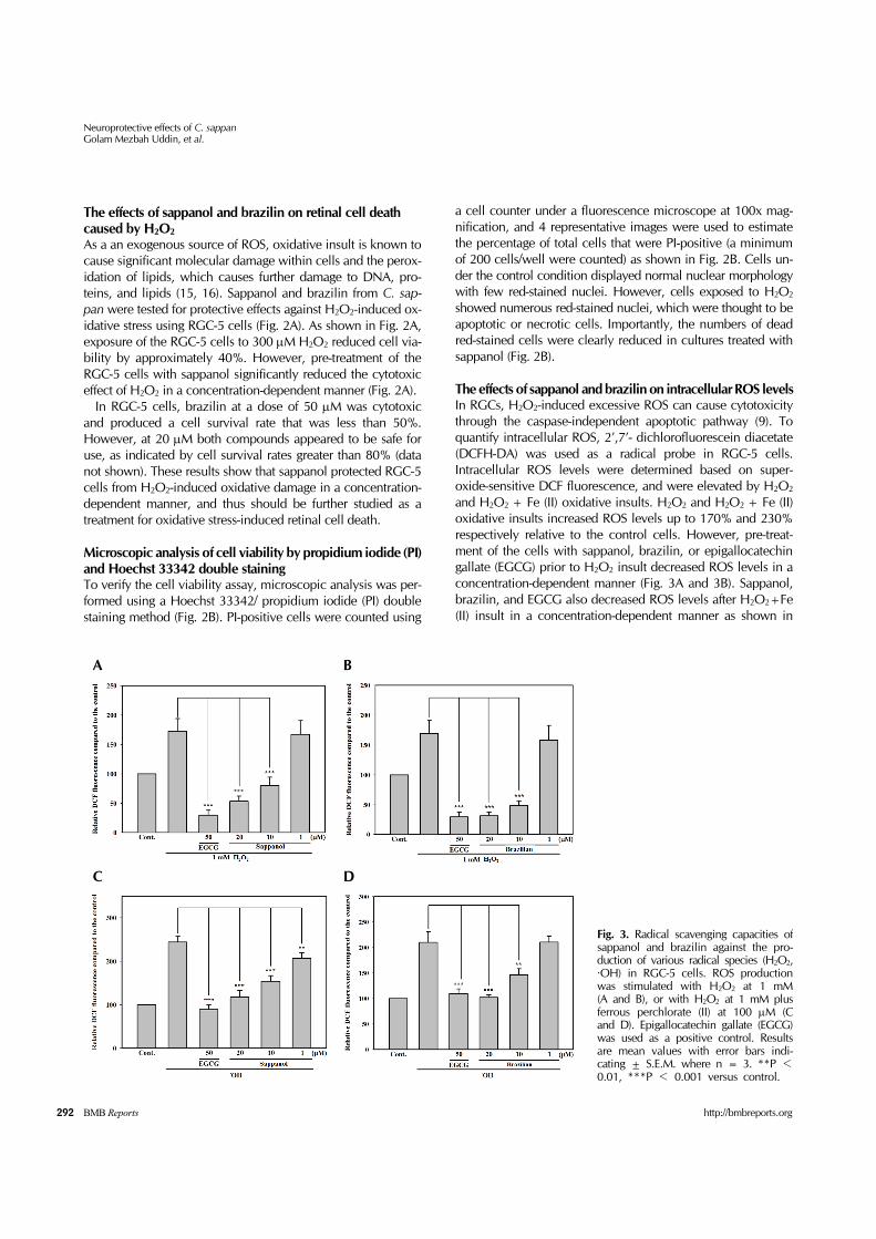
Fig. 3. Radical scavenging capacities of sappanol and brazilin against the pro-duction of various radical species (H2O2,·OH) in RGC-5 cells. ROS production was stimulated with H2O2 at 1 mM (A and B), or with H2O2 at 1 mM plusferrous perchlorate (II) at 100 M (C and D). Epigallocatechin gallate (EGCG)was used as a positive control. Results are mean values with error bars indi-cating ± S.E.M. where n = 3. **P <0.01, ***P < 0.001 versus control.
The effects of sappanol and brazilin on retinal cell death caused by H2O2
As a an exogenous source of ROS, oxidative insult is known to cause significant molecular damage within cells and the perox-idation of lipids, which causes further damage to DNA, pro-teins, and lipids (15, 16). Sappanol and brazilin from C. sap-pan were tested for protective effects against H2O2-induced ox-idative stress using RGC-5 cells (Fig. 2A). As shown in Fig. 2A, exposure of the RGC-5 cells to 300 M H2O2 reduced cell via-bility by approximately 40%. However, pre-treatment of the RGC-5 cells with sappanol significantly reduced the cytotoxic effect of H2O2 in a concentration-dependent manner (Fig. 2A).
In RGC-5 cells, brazilin at a dose of 50 M was cytotoxic and produced a cell survival rate that was less than 50%. However, at 20 M both compounds appeared to be safe for use, as indicated by cell survival rates greater than 80% (data not shown). These results show that sappanol protected RGC-5 cells from H2O2-induced oxidative damage in a concentration- dependent manner, and thus should be further studied as a treatment for oxidative stress-induced retinal cell death.
Microscopic analysis of cell viability by propidium iodide (PI) and Hoechst 33342 double stainingTo verify the cell viability assay, microscopic analysis was per-formed using a Hoechst 33342/ propidium iodide (PI) double staining method (Fig. 2B). PI-positive cells were counted using
a cell counter under a fluorescence microscope at 100x mag-nification, and 4 representative images were used to estimate the percentage of total cells that were PI-positive (a minimum of 200 cells/well were counted) as shown in Fig. 2B. Cells un-der the control condition displayed normal nuclear morphology with few red-stained nuclei. However, cells exposed to H2O2 showed numerous red-stained nuclei, which were thought to be apoptotic or necrotic cells. Importantly, the numbers of dead red-stained cells were clearly reduced in cultures treated with sappanol (Fig. 2B).
The effects of sappanol and brazilin on intracellular ROS levelsIn RGCs, H2O2-induced excessive ROS can cause cytotoxicity through the caspase-independent apoptotic pathway (9). To quantify intracellular ROS, 2’,7’- dichlorofluorescein diacetate (DCFH-DA) was used as a radical probe in RGC-5 cells. Intracellular ROS levels were determined based on super-oxide-sensitive DCF fluorescence, and were elevated by H2O2 and H2O2 + Fe (II) oxidative insults. H2O2 and H2O2 + Fe (II) oxidative insults increased ROS levels up to 170% and 230% respectively relative to the control cells. However, pre-treat-ment of the cells with sappanol, brazilin, or epigallocatechin gallate (EGCG) prior to H2O2 insult decreased ROS levels in a concentration-dependent manner (Fig. 3A and 3B). Sappanol, brazilin, and EGCG also decreased ROS levels after H2O2+Fe (II) insult in a concentration-dependent manner as shown in

Neuroprotective effects of C. sappanGolam Mezbah Uddin, et al.
293http://bmbreports.org BMB Reports
Fig. 3C and 3D. These results show that sappanol and brazilin isolated from C. sappan attenuated ROS production caused by two different radical species.
The effect of sappanol on MDA contentROS produce malondialdehyde (MDA), which is an end-prod-uct of lipid peroxidation, the hallmark of ROS-induced injury (17). To study the inhibitory effects of sappanol on lipid perox-idation, sodium nitropruside (SNP)-induced production of MDA was measured using a thiobarbituric acid reactive species (TBARS) assay in rat brain homogenates. MDA levels indicated that 20 M SNP increased lipid peroxidation compared to the untreated control group. However, sappanol inhibited the pro-duction of MDA in a concentration-dependent manner with an IC50 value of 8.59 M (Table 1). SNP-induced TBARS for-mation was dose-dependently attenuated by sappanol; how-ever, its effect was not greater than that of positive control EGCG.
The effects of sappanol and brazilin on apoptotic cell death caused by sodium azideBecause of its redox potential, ROS generation was also exam-ined in the context of the four-complex mitochondrial electron transport chain, of which complex IV is inhibited by sodium azide (SAZ). It has been demonstrated that antioxidants are not able to reverse this SAZ inhibition (18). Sappanol and brazilin were tested for protective effects against SAZ insult. In RGC-5 cells, exposure to 15 mM SAZ caused moderate reductions in cell viability. Interestingly, pretreatment with sappanol and brazilin did not inhibit cell death (data not shown). Therefore, in contrast to their effects on H2O2-induced oxidative stress in RGC-5 cells, compounds isolated from C. sappan did not pro-tect complex IV of the mitochondrial electron chain.
ConclusionsSappanol and Brazilin showed protective effects against oxida-tive stress-induced retinal cell death, and attenuated A2E pho-tooxidation. Thus, these natural compounds isolated from C. sappan represent potential treatments for oxidative stress-in-duced retinal diseases. Further study will illuminate the mech-anisms involved in these protective effects and the full neuro-protective abilities of these compounds.
MATERIALS AND METHODS
Materials and Methods are described in the online data supple-ment, available at http://www.bmbreports.org/.
ACKNOWLEDGEMENTS
The RGC-5 cells were kindly gifted by Alcon Research, Ltd. This work was financially supported by an intramural grant (2Z04381) from the Korea Institute of Science and Technology (KIST), Republic of Korea.
REFERENCES
1. Almasieh M, Wilson AM, Morquette B, Cueva Vargas JL and Di Polo A (2012) The molecular basis of retinal gan-glion cell death in glaucoma. Prog Retin Eye Res 31, 152-181
2. Wein FB and Levin LA (2002) Current understanding of neuroprotection in glaucoma. Curr Opin Ophthalmol 13, 61-67
3. Osborne NN, Melena J, Chidlow G and Wood JP (2001) A hypothesis to explain ganglion cell death caused by vas-cular insults at the optic nerve head: possible implication for the treatment of glaucoma. Br J Ophthalmol 85, 1252- 1259
4. Kim KA, Kang KD, Lee EH, Nho CW and Jung SH (2011) Edible wild vegetable, Gymnaster koraiensis protects reti-nal ganglion cells against oxidative stress. Food Chem Toxicol 49, 2131-2143
5. Levkovitch-Verbin H, Martin KR, Quigley HA, Baumrind LA, Pease ME and Valenta D (2002) Measurement of ami-no acid levels in the vitreous humor of rats after chronic intraocular pressure elevation or optic nerve transection. J Glaucoma 11, 396-405
6. Godley BF, Shamsi FA, Liang FQ, Jarrett SG, Davies S and Boulton M (2005) Blue light induces mitochondrial DNA damage and free radical production in epithelial cells. J Biol Chem 280, 21061-21066
7. Hockberger PE, Skimina TA, Centonze VE et al (1999) Activation of flavin-containing oxidases underlies light-in-duced production of H2O2 in mammalian cells. Proc Natl Acad Sci U S A 96, 6255-6260
8. Gaillard ER, Avalle LB, Keller LM, Wang Z, Reszka KJ and Dillon JP (2004) A mechanistic study of the photooxi-dation of A2E, a component of human retinal lipofuscin. Exp Eye Res 79, 313-319
9. Jung SH, Kim BJ, Lee EH and Osborne NN (2010) Isoquercitrin is the most effective antioxidant in the plant Thuja orientalis and able to counteract oxidative-induced damage to a transformed cell line (RGC-5 cells). Neurochem Int 57, 713-721
10. Ingkaninan K, Ijzerman AP, Taesotikult T and Verpoorte R (1999) Isolation of opioid-active compounds from Taber-naemontana pachysiphon leaves. J Pharm Pharmacol 51, 1441-1446
11. Xie YW, Ming DS, Xu HX, Dong H and But PP (2000) Vasorelaxing effects of Caesalpinia sappan involvement of endogenous nitric oxide. Life Sci 67, 1913-1918
12. Badami S, Moorkoth S, Rai SR, Kannan E and Bhojraj S (2003) Antioxidant activity of Caesalpinia sappan heart-wood. Biol Pharm Bull 26, 1534-1537
13. Kijlstra A, Tian Y, Kelly ER and Berendschot TT (2012) Lutein: more than just a filter for blue light. Prog Retin Eye Res 31, 303-315
14. Yoon KD, Yamamoto K, Ueda K, Zhou J and Sparrow JR (2012) A novel source of methylglyoxal and glyoxal in ret-ina: implications for age-related macular degeneration. PLoS One 7, e41309
15. Esterbauer H (1993) Cytotoxicity and genotoxicity of lip-id-oxidation products. Am J Clin Nutr 57, 779S-785S; dis-cussion 785S-786S

Neuroprotective effects of C. sappanGolam Mezbah Uddin, et al.
294 BMB Reports http://bmbreports.org
16. Veal EA, Day AM and Morgan BA (2007) Hydrogen per-oxide sensing and signaling. Mol Cell 26, 1-14
17. Dib M, Garrel C, Favier A, Robin V and Desnuelle C (2002) Can malondialdehyde be used as a biological marker of progression in neurodegenerative disease? J Neurol 249, 367-374
18. Ji D, Kamalden TA, del Olmo-Aguado S and Osborne NN (2011) Light- and sodium azide-induced death of RGC-5 cells in culture occurs via different mechanisms. Apoptosis 16, 425-437
19. Xu P, Guan S, Feng R, Tang R and Guo D (2012) Separa-tion of four homoisoflavonoids from Caesalpinia sappan by high-speed counter-current chromatography. Phytochem Anal 23, 228-231
20. Van Bergen NJ, Wood JP, Chidlow G et al (2009) Recharacterization of the RGC-5 retinal ganglion cell line.
Invest Ophthalmol Vis Sci 50, 4267-427221. Mosmann T (1983) Rapid colorimetric assay for cellular
growth and survival: application to proliferation and cyto-toxicity assays. J Immunol Methods 65, 55-63
22. Shimazawa M, Nakajima Y, Mashima Y and Hara H (2009) Docosahexaenoic acid (DHA) has neuroprotective effects against oxidative stress in retinal ganglion cells. Brain Res 1251, 269-275
23. Zhang B and Osborne NN (2006) Oxidative-induced reti-nal degeneration is attenuated by epigallocatechin gallate. Brain Res 1124, 176-187
24. Jung SH, Kang KD, Ji D et al (2008) The flavonoid baicalin counteracts ischemic and oxidative insults to retinal cells and lipid peroxidation to brain membranes. Neurochem Int 53, 325-337