One-Month Global Longitudinal Strain Identifies Patients ... the potential to identify patients at...
14
RESEARCH ARTICLE One-Month Global Longitudinal Strain Identifies Patients Who Will Develop Pacing- Induced Left Ventricular Dysfunction over Time: The Pacing and Ventricular Dysfunction (PAVD) Study Fozia Zahir Ahmed 1,2 *, Manish Motwani 2 , Colin Cunnington 2 , Chun Shing Kwok 3,4 , Catherine Fullwood 5,6 , Delvac Oceandy 1 , Alan Fitchet 7 , Grahame Kevin Goode 8 , Matthew Luckie 2 , Amir Masood Zaidi 2 , Rajdeep Khattar 9 , Mamas Andreas Mamas 3,4 1 Cardiovascular Institute, Faculty of Medical and Human Sciences, University of Manchester, Manchester, United Kingdom, 2 Department of Cardiology, Manchester Heart Centre, Manchester Royal Infirmary, Manchester, United Kingdom, 3 Cardiovascular Research Group, Institutes of Science and Technology in Medicine and Primary Care, University of Keele, Keele, United Kingdom, 4 University Hospital North Midlands, Stoke-on-Trent, United Kingdom, 5 Manchester Biomedical Research Centre, Central Manchester University Hospitals NHS Foundation Trust, Manchester Academic Health Sciences Centre, Manchester, United Kingdom, 6 Department of Biostatistics, Institute of Population Health, University of Manchester, Manchester, United Kingdom, 7 Department of Cardiology, Lancashire Cardiac Centre, Blackpool, United Kingdom, 8 Department of Cardiology, Salford Royal Foundation Trust, Stott Lane, Salford, United Kingdom, 9 Department of Cardiology, Royal Brompton Hospital and Cardiovascular Biomedical Research Unit, Imperial College, London, United Kingdom * [email protected] Abstract Background Predicting which individuals will have a decline in left ventricular (LV) function after pace- maker implantation remains an important challenge. We investigated whether LV global lon- gitudinal strain (GLS), measured by 2D speckle tracking strain echocardiography, can identify patients at risk of pacing-induced left ventricular dysfunction (PIVD) or pacing- induced cardiomyopathy (PICMP). Methods Fifty-five patients with atrioventricular block and preserved LV function underwent dual- chamber pacemaker implantation and were followed with serial transthoracic echocardiog- raphy for 12 months for the development of PIVD (defined as a reduction in LV ejection frac- tion (LVEF) !5 percentage points at 12 months) or PICMP (reduction in LVEF to <45%). Results At 12 months, 15 (27%) patients developed PIVD; of these, 4 patients developed PICMP. At one month, GLS was significantly lower in the 15 patients who subsequently developed PIVD, compared to those who did not (n = 40) (GLS -12.6 vs. -16.4 respectively; p = 0.022). PLOS ONE | DOI:10.1371/journal.pone.0162072 January 17, 2017 1 / 14 a1111111111 a1111111111 a1111111111 a1111111111 a1111111111 OPEN ACCESS Citation: Ahmed FZ, Motwani M, Cunnington C, Kwok CS, Fullwood C, Oceandy D, et al. (2017) One-Month Global Longitudinal Strain Identifies Patients Who Will Develop Pacing-Induced Left Ventricular Dysfunction over Time: The Pacing and Ventricular Dysfunction (PAVD) Study. PLoS ONE 12(1): e0162072. doi:10.1371/journal. pone.0162072 Editor: Alessandro Parolari, University of Milano, ITALY Received: April 28, 2016 Accepted: August 17, 2016 Published: January 17, 2017 Copyright: © 2017 Ahmed et al. This is an open access article distributed under the terms of the Creative Commons Attribution License, which permits unrestricted use, distribution, and reproduction in any medium, provided the original author and source are credited. Data Availability Statement: All relevant data are presented in the paper. Minimal dataset is uploaded as a supplementary file. Funding: FZA was supported by a research grant funded by Medtronic. The funders had no role in study design, data collection and analysis, decision to publish, or preparation of the manuscript. Competing Interests: FZA was supported by a research grant funded by Medtronic. This does not
Transcript of One-Month Global Longitudinal Strain Identifies Patients ... the potential to identify patients at...
RESEARCH ARTICLE
One-Month Global Longitudinal Strain
Identifies Patients Who Will Develop Pacing-
Induced Left Ventricular Dysfunction over
Time: The Pacing and Ventricular Dysfunction
(PAVD) Study
Fozia Zahir Ahmed1,2*, Manish Motwani2, Colin Cunnington2, Chun Shing Kwok3,4,
Catherine Fullwood5,6, Delvac Oceandy1, Alan Fitchet7, Grahame Kevin Goode8,
Matthew Luckie2, Amir Masood Zaidi2, Rajdeep Khattar9, Mamas Andreas Mamas3,4
1 Cardiovascular Institute, Faculty of Medical and Human Sciences, University of Manchester, Manchester,
United Kingdom, 2 Department of Cardiology, Manchester Heart Centre, Manchester Royal Infirmary,
Manchester, United Kingdom, 3 Cardiovascular Research Group, Institutes of Science and Technology in
Medicine and Primary Care, University of Keele, Keele, United Kingdom, 4 University Hospital North
Midlands, Stoke-on-Trent, United Kingdom, 5 Manchester Biomedical Research Centre, Central Manchester
University Hospitals NHS Foundation Trust, Manchester Academic Health Sciences Centre, Manchester,
United Kingdom, 6 Department of Biostatistics, Institute of Population Health, University of Manchester,
Manchester, United Kingdom, 7 Department of Cardiology, Lancashire Cardiac Centre, Blackpool, United
Kingdom, 8 Department of Cardiology, Salford Royal Foundation Trust, Stott Lane, Salford, United Kingdom,
9 Department of Cardiology, Royal Brompton Hospital and Cardiovascular Biomedical Research Unit,
Imperial College, London, United Kingdom
Abstract
Background
Predicting which individuals will have a decline in left ventricular (LV) function after pace-
maker implantation remains an important challenge. We investigated whether LV global lon-
gitudinal strain (GLS), measured by 2D speckle tracking strain echocardiography, can
identify patients at risk of pacing-induced left ventricular dysfunction (PIVD) or pacing-
induced cardiomyopathy (PICMP).
Methods
Fifty-five patients with atrioventricular block and preserved LV function underwent dual-
chamber pacemaker implantation and were followed with serial transthoracic echocardiog-
raphy for 12 months for the development of PIVD (defined as a reduction in LV ejection frac-
tion (LVEF)�5 percentage points at 12 months) or PICMP (reduction in LVEF to <45%).
Results
At 12 months, 15 (27%) patients developed PIVD; of these, 4 patients developed PICMP. At
one month, GLS was significantly lower in the 15 patients who subsequently developed
PIVD, compared to those who did not (n = 40) (GLS -12.6 vs. -16.4 respectively; p = 0.022).
PLOS ONE | DOI:10.1371/journal.pone.0162072 January 17, 2017 1 / 14
a1111111111
a1111111111
a1111111111
a1111111111
a1111111111
OPENACCESS
Citation: Ahmed FZ, Motwani M, Cunnington C,
Kwok CS, Fullwood C, Oceandy D, et al. (2017)
One-Month Global Longitudinal Strain Identifies
Patients Who Will Develop Pacing-Induced Left
Ventricular Dysfunction over Time: The Pacing and
Ventricular Dysfunction (PAVD) Study. PLoS ONE
12(1): e0162072. doi:10.1371/journal.
pone.0162072
Editor: Alessandro Parolari, University of Milano,
ITALY
Received: April 28, 2016
Accepted: August 17, 2016
Published: January 17, 2017
Copyright: © 2017 Ahmed et al. This is an open
access article distributed under the terms of the
Creative Commons Attribution License, which
permits unrestricted use, distribution, and
reproduction in any medium, provided the original
author and source are credited.
Data Availability Statement: All relevant data are
presented in the paper. Minimal dataset is
uploaded as a supplementary file.
Funding: FZA was supported by a research grant
funded by Medtronic. The funders had no role in
study design, data collection and analysis, decision
to publish, or preparation of the manuscript.
Competing Interests: FZA was supported by a
research grant funded by Medtronic. This does not
When patients with PICMP were excluded, one month GLS was significantly reduced com-
pared to baseline whereas LVEF was not. One-month GLS had high predictive accuracy for
determining subsequent development of PIVD or PICMP (AUC = 0.80, optimal GLS thresh-
old: <−14.5, sensitivity 82%, specificity 75%); and particularly PICMP (AUC = 0.86, optimal
GLS threshold: <−13.5, sensitivity 100%, specificity 71%).
Conclusions
GLS is a novel predictor of decline in LV systolic function following pacemaker implantation,
with the potential to identify patients at risk of PIVD before measurable changes in LVEF are
apparent. GLS measured one month after implantation has high predictive accuracy for
identifying patients who later develop PIVD or PICMP.
Introduction
Right ventricular (RV) pacing is associated with a reduction in left ventricular (LV) systolic
function, [1] thought to be mediated by pacing-induced ventricular dyssynchrony. [2, 3] The
prevalence of heart failure after RV pacing is reported to range from 3–31%, the variation
largely depending on the definition and methods used to define heart failure as well as the pop-
ulation examined. [4, 5, 6, 7, 8, 9] Moreover, randomized trials in RV paced individuals have
shown a near 3-fold increased risk of hospitalization for heart failure when the cumulative per-
centage of ventricular pacing (Cum%VP) exceeds 40%. [5, 10] The term pacing-induced car-
diomyopathy (PICMP) has been used to describe clinically significant left ventricular systolic
dysfunction (ejection fraction <45%) attributable to RV pacing, occurring in the absence of
other causes of cardiomyopathy. [4, 7] However, lesser degrees of pacing-induced LV dysfunc-
tion (PIVD) have also been observed in up to two-thirds of patients with normal baseline LV
function. [3, 11, 12] Despite this, clinical guidelines do not currently recommend routine car-
diac imaging after pacemaker implantation, and PICMP/PIVD may therefore go undetected
until the onset of heart failure symptoms. [8]
In view of this consideration, a priori identification of patients at risk of developing heart
failure after RV pacing would be of considerable clinical value as these patients may benefit
from heightened clinical surveillance and possible upgrade to biventricular pacing, which has
been shown to reverse PIVD and LV remodeling. [13, 14, 15] A non-invasive test able to iden-
tify such individuals is, therefore, highly desirable. Two-dimensional (2D) speckle tracking
strain echocardiography (STE) has been shown to detect early signs of LV systolic dysfunction
in a range of cardiomyopathies before a measurable reduction in LVEF. [16, 17, 18, 19, 20]
Objectives
We investigated whether STE can be used to predict the development of PIVD and PICMP
after pacemaker implantation.
Methods
Subjects
The Pacing And Ventricular Dysfunction (PAVD) Study is an investigator-initiated prospec-
tive observational cohort study designed to investigate predictors of PIVD. Sixty subjects with
Longitudinal Strain and Pacing-Induced Heart Failure
PLOS ONE | DOI:10.1371/journal.pone.0162072 January 17, 2017 2 / 14
alter our adherence to PLOS ONE policies on
sharing of materials.

high-grade atrioventricular block scheduled to undergo dual chamber pacemaker implanta-
tion were recruited from three UK centres. Eligible patients were aged 18 years or over, had
preserved LVEF (�55%) and second-degree or third-degree atrioventricular block. All patients
were required to provide written informed consent for inclusion. Exclusion criteria were: preg-
nancy, myocardial infarction or coronary revascularization within prior 3 months, atrial fibril-
lation (AF), haemodynamically significant valvular heart disease (�moderate in severity),
structural heart abnormality including LV dilatation (according to British Society of Echocar-
diography reference ranges), LVEF<55%, New York Heart Association (NYHA) functional
class III or IV, significant respiratory disease, history of carcinoma within 5 years, autoim-
mune disorders, rheumatoid arthritis or treatment with disease modifying drugs including
mineralocorticoid receptor antagonists.
Study protocol
The Central Manchester research ethics committee and institutional review board reviewed
and approved the current clinical study in 2012. All clinical investigations were conducted
according to the principles expressed in the Declaration of Helsinki. Written informed consent
was obtained from all participants. Baseline evaluation consisted of (i) assessment of NYHA
functional class and age-adjusted Charlson score [21] (ii) 12-lead electrocardiogram (ECG)
(iii) standard transthoracic echocardiography. The protocol required that patients undergo
standardized implantation of a dual chamber permanent pacemaker with positioning of the
ventricular lead at the RV apex. Fluoroscopy and x-ray were used to record the anatomical
location of the RV lead in each case. Pacemakers were programmed to DDDR mode with the
manufacturer’s algorithms to minimize ventricular pacing enabled. Measurements (i)-(iii)
were repeated at 1 and 12 months after pacemaker implantation, as well as measurement of
the cumulative percentage of ventricular pacing (Cum%VP) from stored pacemaker diagnos-
tics. At follow-up, where the patient was not paced at the time of undertaking pacemaker inter-
rogation, pacing at 60 pulses per minute was enabled for the duration of the
echocardiographic examination.
Echocardiography
Image acquisition. Standard two-dimensional echocardiography was performed before
and after pacemaker implantation (baseline, 1 and 12 months) using an iE-33 ultrasound sys-
tem with a 1–5MHz phased-array probe (Philips Medical System, Andover, USA). Examina-
tions were performed in the left lateral decubitus position by an experienced operator.
Acquisitions from standard parasternal long-axis, apical and LV short axis views were
obtained at end-expiration with sector width and depth optimized to allow for complete myo-
cardial visualization while maximizing frame rate (60–110 frames per second). Cine loops
were acquired for 3 consecutive cycles. Two-dimensional LVEF were measured using Simp-
son’s biplane method. [22] PIVD was defined as an absolute decline in LVEF by�5 percentage
points. PICMP was defined as a reduction in LVEF to<45%. A single experienced operator
who was blinded to the pacing interrogation results performed all echocardiographic analyses.
Speckle-tracking strain analysis. Strain was evaluated off-line from digitally stored
images using Qlab 9 (cardiac motion quantification (CMQ); Phillips Medical Systems) soft-
ware package. Longitudinal strain for individual myocardial segments was measured from the
apical four-chamber, two-chamber and long axis views (16 segment AHA/ASE model). [23] In
end-diastole, automated border tracking was enabled, before manual adjustment using a point
and click approach to ensure that the endocardial and epicardial borders were included in the
region of interest. In cases of poor tracking, fine-tuning was performed manually after
Longitudinal Strain and Pacing-Induced Heart Failure
PLOS ONE | DOI:10.1371/journal.pone.0162072 January 17, 2017 3 / 14

cineloop playback and tracing was repeated and adjusted until tracking was considered opti-
mal by visual analysis. Individual segments that returned positive strain values, and those with
persistently poor tracking despite manual optimisation, were excluded from analysis. Peak
strain for the segment was defined as the peak negative value on the time strain curve for the
entire cardiac cycle. Peak regional longitudinal strain was measured in 16 myocardial regions
and a weighted mean was used to derive global longitudinal strain (GLS).
Longitudinal LV dyssynchrony was evaluated using the standard deviation of time to peak
strain (TPS-SD), as previously described. [24] Briefly, time to peak strain was measured from
the interval from onset of the Q wave to peak negative strain throughout the cardiac cycle.
Time-strain curves for the 12 basal and mid LV segments were generated and the TPS-SD for
these segments was calculated. [24] Dyssynchrony was defined as TPS-SD >60ms.
Reproducibility of data
Echocardiographic images for ten patients were independently analyzed twice, at intervals of
greater than 2 weeks, by two investigators (FZA and ML). Both operators were blinded to the
results of the first measurement and from each other. Intraobserver variability for GLS mea-
surements was small (mean arithmetic difference in GLS 0.41, (3.3%)). Intraobserver variabil-
ity for LVEF was 4.2%. Interobserver variability for GLS and LVEF was 5.4% and 5.1%
respectively.
Statistical analysis
Continuous data are presented as mean ± SD. Categorical data are presented as frequencies
and percentages. Normally distributed variables were compared using an unpaired t-test with
Welch’s correction. Categorical data were compared using the Fisher’s exact test. Non-nor-
mally distributed data were compared using the Mann-Whitney U-test or Kruskall-Wallis test
as appropriate. Statistical analyses were performed using GraphPad Prism (version 6.0e for
Mac, GraphPad Software, San Diego, California, USA) and Stata (StataCorp. 2015. Stata Statis-
tical Software: Release 14. College Station, TX: StataCorp LP. Receiver operating characteristic
(ROC) analysis was performed to determine the accuracy of one month GLS to predict (i)
PIVD including PICMP and (ii) specifically PICMP. The optimal GLS thresholds from the
ROC curve were determined using the maximum Youden index. [25] Multivariate logistic
regression analysis was performed to identify predictors of PIVD. (S1 File).
Results
Study population
Between October 2012 and May 2014, 172 patients were screened for the study. Sixty patients
with second or third-degree atrioventricular block and preserved LV systolic function fulfilled
criteria for inclusion. After pacemaker implantation, five patients were excluded due to subop-
timal echocardiographic images for GLS measurement (n = 3) or non-apical RV pacing leads
(n = 2). Therefore, the study population consisted of 55 patients (72.2 ±14.4 years; 35 (64%)
men) of whom 2 were undergoing temporary transvenous pacing prior to permanent pace-
maker implantation. Baseline clinical characteristics for the study participants are given in
Table 1. Fig 1 outlines the patient distribution for the study.
Incidence of ventricular dysfunction
PIVD occurred in 8/55 (15%) patients at 1 month, but there were no cases of PICMP at this
timepoint. At 12 months, PIVD was observed in 15/55 (27%) patients, including 4 patients
Longitudinal Strain and Pacing-Induced Heart Failure
PLOS ONE | DOI:10.1371/journal.pone.0162072 January 17, 2017 4 / 14
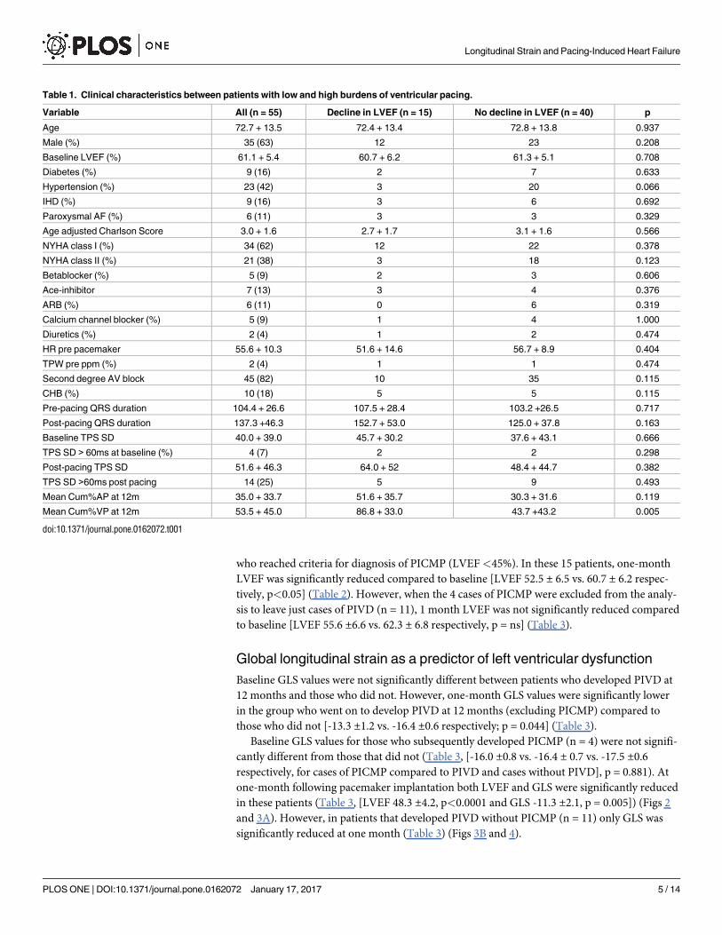
who reached criteria for diagnosis of PICMP (LVEF<45%). In these 15 patients, one-month
LVEF was significantly reduced compared to baseline [LVEF 52.5 ± 6.5 vs. 60.7 ± 6.2 respec-
tively, p<0.05] (Table 2). However, when the 4 cases of PICMP were excluded from the analy-
sis to leave just cases of PIVD (n = 11), 1 month LVEF was not significantly reduced compared
to baseline [LVEF 55.6 ±6.6 vs. 62.3 ± 6.8 respectively, p = ns] (Table 3).
Global longitudinal strain as a predictor of left ventricular dysfunction
Baseline GLS values were not significantly different between patients who developed PIVD at
12 months and those who did not. However, one-month GLS values were significantly lower
in the group who went on to develop PIVD at 12 months (excluding PICMP) compared to
those who did not [-13.3 ±1.2 vs. -16.4 ±0.6 respectively; p = 0.044] (Table 3).
Baseline GLS values for those who subsequently developed PICMP (n = 4) were not signifi-
cantly different from those that did not (Table 3, [-16.0 ±0.8 vs. -16.4 ± 0.7 vs. -17.5 ±0.6
respectively, for cases of PICMP compared to PIVD and cases without PIVD], p = 0.881). At
one-month following pacemaker implantation both LVEF and GLS were significantly reduced
in these patients (Table 3, [LVEF 48.3 ±4.2, p<0.0001 and GLS -11.3 ±2.1, p = 0.005]) (Figs 2
and 3A). However, in patients that developed PIVD without PICMP (n = 11) only GLS was
significantly reduced at one month (Table 3) (Figs 3B and 4).
Table 1. Clinical characteristics between patients with low and high burdens of ventricular pacing.
Variable All (n = 55) Decline in LVEF (n = 15) No decline in LVEF (n = 40) p
Age 72.7 + 13.5 72.4 + 13.4 72.8 + 13.8 0.937
Male (%) 35 (63) 12 23 0.208
Baseline LVEF (%) 61.1 + 5.4 60.7 + 6.2 61.3 + 5.1 0.708
Diabetes (%) 9 (16) 2 7 0.633
Hypertension (%) 23 (42) 3 20 0.066
IHD (%) 9 (16) 3 6 0.692
Paroxysmal AF (%) 6 (11) 3 3 0.329
Age adjusted Charlson Score 3.0 + 1.6 2.7 + 1.7 3.1 + 1.6 0.566
NYHA class I (%) 34 (62) 12 22 0.378
NYHA class II (%) 21 (38) 3 18 0.123
Betablocker (%) 5 (9) 2 3 0.606
Ace-inhibitor 7 (13) 3 4 0.376
ARB (%) 6 (11) 0 6 0.319
Calcium channel blocker (%) 5 (9) 1 4 1.000
Diuretics (%) 2 (4) 1 2 0.474
HR pre pacemaker 55.6 + 10.3 51.6 + 14.6 56.7 + 8.9 0.404
TPW pre ppm (%) 2 (4) 1 1 0.474
Second degree AV block 45 (82) 10 35 0.115
CHB (%) 10 (18) 5 5 0.115
Pre-pacing QRS duration 104.4 + 26.6 107.5 + 28.4 103.2 +26.5 0.717
Post-pacing QRS duration 137.3 +46.3 152.7 + 53.0 125.0 + 37.8 0.163
Baseline TPS SD 40.0 + 39.0 45.7 + 30.2 37.6 + 43.1 0.666
TPS SD > 60ms at baseline (%) 4 (7) 2 2 0.298
Post-pacing TPS SD 51.6 + 46.3 64.0 + 52 48.4 + 44.7 0.382
TPS SD >60ms post pacing 14 (25) 5 9 0.493
Mean Cum%AP at 12m 35.0 + 33.7 51.6 + 35.7 30.3 + 31.6 0.119
Mean Cum%VP at 12m 53.5 + 45.0 86.8 + 33.0 43.7 +43.2 0.005
doi:10.1371/journal.pone.0162072.t001
Longitudinal Strain and Pacing-Induced Heart Failure
PLOS ONE | DOI:10.1371/journal.pone.0162072 January 17, 2017 5 / 14

Fig 1. Patient distribution through the study at 12 months. Algorithm showing total number of cases considered for recruitment and the
reasons for exclusion, leading to selection of the final 55 patients.
doi:10.1371/journal.pone.0162072.g001
Table 2. Differences in LVEF and GLS values between patients with and without pacing-induced LV
dysfunction.
Decline in LVEF PIVD and PICMP cases
(n = 15)
No decline in LVEF
(n = 40)
p
LVEF
Baseline 60.7 + 6.2 61.3 + 5.1 0.780
1 month 52.5 + 6.5* 60.4 + 4.5 0.002
12 months 46.7 + 8.9* 58.7 4.5 0.010
Global longitudinal
strain
Baseline -16.3 + 0.5 -17.5 + 0.6 0.515
1 month -12.6 + 0.9* -16.4 + 0.6 0.022
12 months -11.9 + 2.5* -15.8 + 3.9 0.008
* denotes significantly reduced compared to baseline measurement (p<0.05).
doi:10.1371/journal.pone.0162072.t002
Longitudinal Strain and Pacing-Induced Heart Failure
PLOS ONE | DOI:10.1371/journal.pone.0162072 January 17, 2017 6 / 14
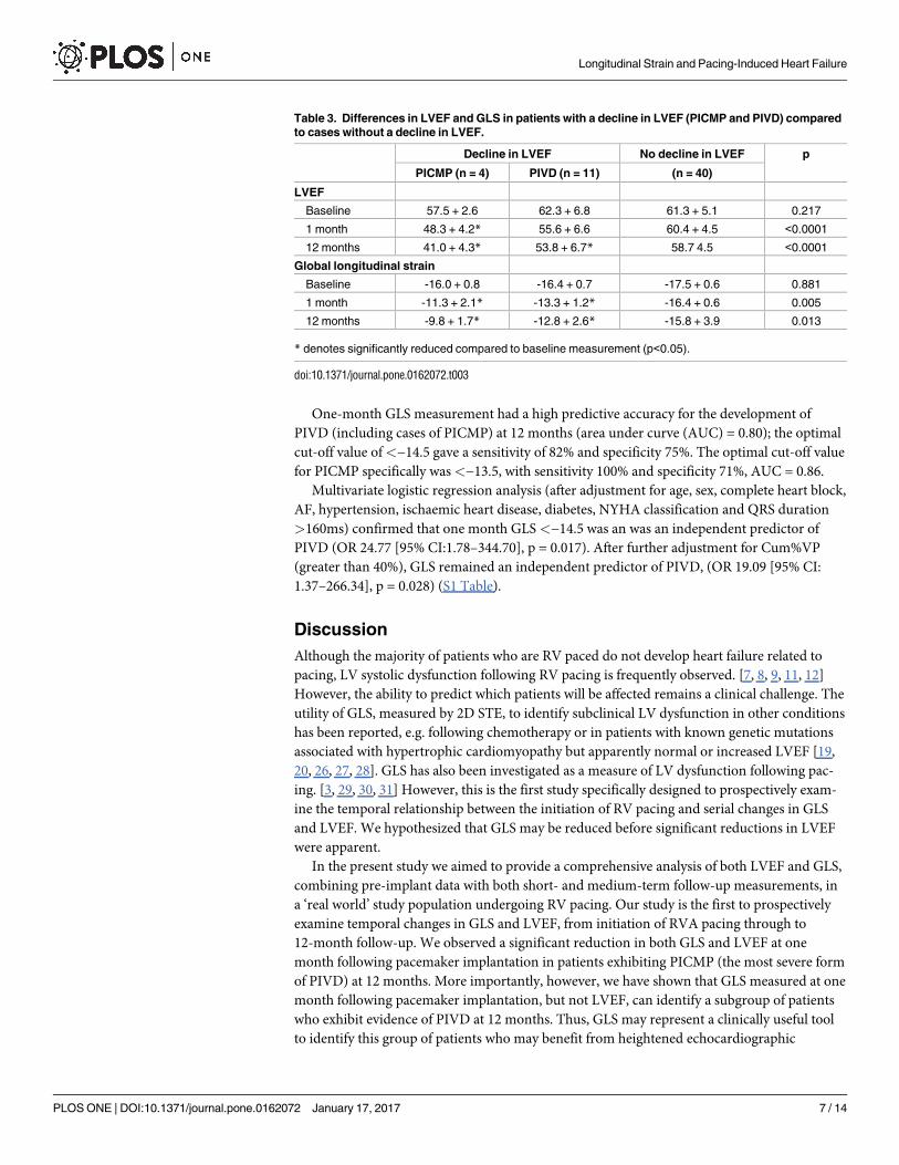
One-month GLS measurement had a high predictive accuracy for the development of
PIVD (including cases of PICMP) at 12 months (area under curve (AUC) = 0.80); the optimal
cut-off value of<−14.5 gave a sensitivity of 82% and specificity 75%. The optimal cut-off value
for PICMP specifically was <−13.5, with sensitivity 100% and specificity 71%, AUC = 0.86.
Multivariate logistic regression analysis (after adjustment for age, sex, complete heart block,
AF, hypertension, ischaemic heart disease, diabetes, NYHA classification and QRS duration
>160ms) confirmed that one month GLS <−14.5 was an was an independent predictor of
PIVD (OR 24.77 [95% CI:1.78–344.70], p = 0.017). After further adjustment for Cum%VP
(greater than 40%), GLS remained an independent predictor of PIVD, (OR 19.09 [95% CI:
1.37–266.34], p = 0.028) (S1 Table).
Discussion
Although the majority of patients who are RV paced do not develop heart failure related to
pacing, LV systolic dysfunction following RV pacing is frequently observed. [7, 8, 9, 11, 12]
However, the ability to predict which patients will be affected remains a clinical challenge. The
utility of GLS, measured by 2D STE, to identify subclinical LV dysfunction in other conditions
has been reported, e.g. following chemotherapy or in patients with known genetic mutations
associated with hypertrophic cardiomyopathy but apparently normal or increased LVEF [19,
20, 26, 27, 28]. GLS has also been investigated as a measure of LV dysfunction following pac-
ing. [3, 29, 30, 31] However, this is the first study specifically designed to prospectively exam-
ine the temporal relationship between the initiation of RV pacing and serial changes in GLS
and LVEF. We hypothesized that GLS may be reduced before significant reductions in LVEF
were apparent.
In the present study we aimed to provide a comprehensive analysis of both LVEF and GLS,
combining pre-implant data with both short- and medium-term follow-up measurements, in
a ‘real world’ study population undergoing RV pacing. Our study is the first to prospectively
examine temporal changes in GLS and LVEF, from initiation of RVA pacing through to
12-month follow-up. We observed a significant reduction in both GLS and LVEF at one
month following pacemaker implantation in patients exhibiting PICMP (the most severe form
of PIVD) at 12 months. More importantly, however, we have shown that GLS measured at one
month following pacemaker implantation, but not LVEF, can identify a subgroup of patients
who exhibit evidence of PIVD at 12 months. Thus, GLS may represent a clinically useful tool
to identify this group of patients who may benefit from heightened echocardiographic
Table 3. Differences in LVEF and GLS in patients with a decline in LVEF (PICMP and PIVD) compared
to cases without a decline in LVEF.
Decline in LVEF No decline in LVEF p
PICMP (n = 4) PIVD (n = 11) (n = 40)
LVEF
Baseline 57.5 + 2.6 62.3 + 6.8 61.3 + 5.1 0.217
1 month 48.3 + 4.2* 55.6 + 6.6 60.4 + 4.5 <0.0001
12 months 41.0 + 4.3* 53.8 + 6.7* 58.7 4.5 <0.0001
Global longitudinal strain
Baseline -16.0 + 0.8 -16.4 + 0.7 -17.5 + 0.6 0.881
1 month -11.3 + 2.1* -13.3 + 1.2* -16.4 + 0.6 0.005
12 months -9.8 + 1.7* -12.8 + 2.6* -15.8 + 3.9 0.013
* denotes significantly reduced compared to baseline measurement (p<0.05).
doi:10.1371/journal.pone.0162072.t003
Longitudinal Strain and Pacing-Induced Heart Failure
PLOS ONE | DOI:10.1371/journal.pone.0162072 January 17, 2017 7 / 14
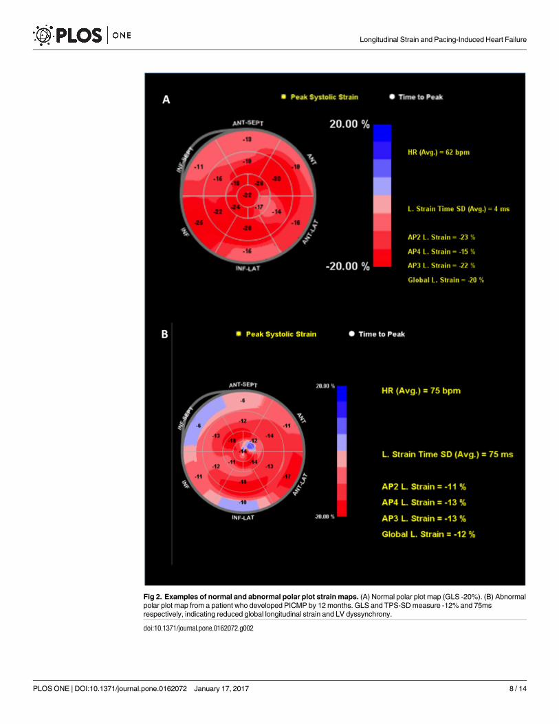
Fig 2. Examples of normal and abnormal polar plot strain maps. (A) Normal polar plot map (GLS -20%). (B) Abnormal
polar plot map from a patient who developed PICMP by 12 months. GLS and TPS-SD measure -12% and 75ms
respectively, indicating reduced global longitudinal strain and LV dyssynchrony.
doi:10.1371/journal.pone.0162072.g002
Longitudinal Strain and Pacing-Induced Heart Failure
PLOS ONE | DOI:10.1371/journal.pone.0162072 January 17, 2017 8 / 14

surveillance of LV dysfunction following pacemaker implantation. Furthermore, a GLS of
<−14.5 at one month had high sensitivity for predicting the development of PIVD (including
PICMP) at 12-months, with a value below this threshold being associated with a 19-fold
increased risk of developing PIVD. Importantly, this finding was independent of high pacing
burden (Cum%VP >40%) in multivariate logistic regression analysis. The wide confidence
intervals observed in the multivariate analysis are notable, and in part due to the small sample
size. We observed that adjustments made the confidence intervals wider, indicating that the
estimates are sensitive to multiple variables (supplementary file), further supporting the need
for better powered studies in the future. Although this study identified a one-month GLS
threshold, beyond which PIVD was more likely, we did not specifically identify a critical
Fig 3. (A) Global longitudinal strain for cases of all cases of PIVD (PICMP included). Global longitudinal strain was significantly lower in
patients with a decline in LVEF� 5% compared to cases without (one month GLS -12.6 ± 0.9 vs. -16.4 ±0.6 respectively; p = 0.022). One
and 12 month GLS were reduced compared to baseline for cases of with a decline in LVEF at 12 months (PIVD and PICMP), but not for
cases without a decline in LVEF (PIVD and PICMP: baseline GLS, -16.3 ±0.5 vs. -12.6 ±0.9 and -11.9 ±2.5; p = 0.012. No decline in LVEF:
baseline GLS -17.5 ±0.6 vs. -16.4 ±0.6 and -15.8 ±3.9; p = 0.311). (B) Global longitudinal strain for cases with PIVD (PICMP excluded). One
and 12 month GLS were significantly reduced for cases of PIVD compared to baseline (Baseline GLS -16.4 ±0.7 vs -13.3 ±.2 and -12.8 ±2.6
respectively; p = 0.024).
doi:10.1371/journal.pone.0162072.g003
Longitudinal Strain and Pacing-Induced Heart Failure
PLOS ONE | DOI:10.1371/journal.pone.0162072 January 17, 2017 9 / 14
burden of Cum%VP beyond which changes in GLS would be observed, though acknowledge
that it would be desirable to explore this in future studies.
Our finding of reduced GLS after RV apical (RVA) pacing is consistent with previous stud-
ies. [3, 29, 30, 31] However, previous studies have considered either acute or chronic effects of
RVA pacing in isolation; serial assessment of GLS compared to LVEF has not been systemati-
cally evaluated. Furthermore, these studies have often been limited by focusing on individual
measurements (e.g. GLS or LVEF, rather than evaluating both parameters in the same sub-
jects), single time-point follow-up, or highly selected populations making no allowance for var-
iations in pacing burden (e.g. pacing-dependent patients only, or following AV nodal
ablation). [3, 29, 30, 31] For example, Delgado et al. examined the acute effects of RVA pacing
in patients with preserved LV function and reported a significant decline in both LVEF and
GLS, but the long term impact and risk of PIVD progression or PICMP was not described.
[31] In a sub-study of the Protection of Left Ventricular Function During Right Ventricular
Pacing (Protect-PACE) trial, there was no significant difference in baseline GLS between the
RVA pacing group and controls (RV high-septal pacing). [30] Notably, however, GLS was not
evaluated prior to pacing, with the baseline measurement being obtained after pacemaker
implantation. After 2-years, GLS was reported to be significantly reduced in the RVA group
compared to controls, but this study does not yield information regarding the tempo of
changes from baseline to 2 years, nor if there was an earlier timepoint for possible therapeutic
intervention. [30] Similarly, Ahmed et al. evaluated the effects of RV apical pacing on LVEF at
2 years. In a retrospective study, predictors of a decline of LVEF >5% were examined among
patients undergoing AV node ablation for atrial fibrillation and pacemaker implantation. GLS
performed a median of 4-months after initiation of pacing was significantly reduced in
patients who had a decline in LVEF >5% at 2-years compared to those who did not. [3]
The Pacing and Cardiac Enlargement (PACE) study was a prospective study that compared
measured LVEF in patients randomized to RVA or biventricular pacing. [4] In RVA patients
with PIVD at 1 year, [4] further significant reductions in LVEF were observed when follow-up
was extended to 2 years (7% vs. 9.9% reduction in LVEF at 1 and 2 years respectively). [11]
Thus, relatively small reductions in LVEF at 12 months, such as those observed in the subjects
in our study, may progress to result in more clinically significant reductions in LVEF with
extended follow-up, emphasizing the importance of GLS in identifying this patient group at an
early stage of the disease process. Moreover, in the present study, a significant decline in both
one-month GLS and LVEF was observed in cases that subsequently developed PICMP at 12
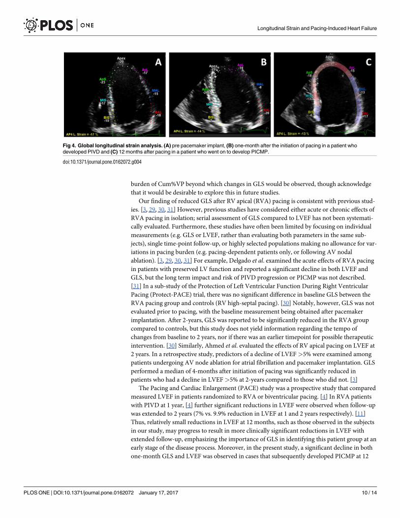
Fig 4. Global longitudinal strain analysis. (A) pre pacemaker implant, (B) one-month after the initiation of pacing in a patient who
developed PIVD and (C) 12 months after pacing in a patient who went on to develop PICMP.
doi:10.1371/journal.pone.0162072.g004
Longitudinal Strain and Pacing-Induced Heart Failure
PLOS ONE | DOI:10.1371/journal.pone.0162072 January 17, 2017 10 / 14
months, but a decline in GLS alone at one month in those that developed less severe PIVD.
Therefore, we demonstrate the utility of one-month GLS to not only risk stratify patients fol-
lowing pacemaker implantation, but also to predict the magnitude of the ensuing decline in
LVEF when the presence of absence of a decline in LVEF of>5% was also considered.
A reduction in LVEF to<50% has important clinical implications. Although the aetiology
of LV dysfunction differs and direct comparisons cannot be made, in the Framingham study,
LVEF between 40% and 50%, even if asymptomatic, was associated with a nearly four-fold
increase in the risk of heart failure and a 1.9-fold increase in the risk of mortality compared to
patients with an LVEF >50%. [32] In the current study, by 12 months, 15 patients (80% of
whom had a high burden of ventricular pacing) had a significant decline in LVEF� 5 percent-
age points, of whom 4 had a more severe decline in LVEF to<45% i.e. reaching the threshold
for diagnosis of PICMP. Absolute decline in LVEF at 12 months for cases of PICMP (n = 4)
was 16.5 percentage points compared to 8.4 percentage points for the remaining less severe
PIVD (n = 11) cases (p = 0.029). Though PICMP may be considered a more clinically impor-
tant situation that requires expedient consideration of upgrade to biventricular pacing, the
clinical relevance of asymptomatic “smaller” reductions in LVEF has been widely studied and
should not be overlooked as progressive declines in LVEF have been reported with extended
follow-up. [3, 4, 11, 32] Although the latter subgroup of patients with PICMP may seem small,
the current study postulates that simple screening using echocardiography (inexpensive and
widely available) should be considered in order to capture these high-risk patients at an earlier
time-point, especially considering the potential long-term health economic burden that is
associated with heart failure.
Serial assessment of LVEF has become widely accepted as a research tool for measuring the
deleterious effects of RV pacing on LV systolic function. However, the main disadvantage of
this practice is that a significant reduction in LVEF may represent the final phenotype of a
pathophysiological process. [33] In contrast, abnormal GLS may represent an earlier stage in
the disease process before a significant reduction in LVEF occurs, as was observed in cases of
PIVD in this study.[19, 20]
Limitations
Only 35 patients had non-contrast images suitable for three-dimensional (3D) analysis. There-
fore, in this study LVEF was calculated using semi-automated non-contrast 2D methods, a
practice that more closely reflects real world clinical practice. In this study the cut-off GLS to
detect PIVD was calculated to be -14.5 and therefore reported to one decimal place. Although
the values of GLS obtained using different vendor software is considered interchangeable,
Qlab software itself reports GLS values as a whole integer. This limitation has been considered
by previous studies also.
Alternative pacing sites were not examined in the current study. However, as septal pacing
is routinely performed in real world practice, it would be desirable for future studies of RV
pacing to establish whether one-month GLS significantly differs between RVA and septal pac-
ing sites. Four patients developed PICMP; due to the small sample size, and because all patients
who had PICMP had a 1 month GLS<14.5, multivariate logistic regression analysis could not
be performed for this cohort. Because of the lower incidence of PICMP compared to lesser
degrees of PIVD, such a study would have required significantly more patients and longer fol-
low-up. In addition, as outlined in the discussion, the observation that the confidence interval
widens with multiple adjustments indicates that the model is sensitive to multiple adjustments
(supplementary file). In view of these considerations, a larger study is needed to validate the
feasibility and clinical utility of GLS to predict PIVD.
Longitudinal Strain and Pacing-Induced Heart Failure
PLOS ONE | DOI:10.1371/journal.pone.0162072 January 17, 2017 11 / 14
Finally, although both clinical and animal models have previously explored the temporal
relationship between RV pacing, LV dimensions and LVEF, few have expanded this analysis to
also take into account GLS. It would be desirable for future clinical studies to examine a more
extensive set of echocardiographic variables in order to establish whether those individuals
with reduced GLS had subtle abnormalities in LV dimensions and diastolic function at
baseline.
Conclusion
GLS is easily performed in the clinical setting, and when measured at one month following
pacemaker implantation shows potential for identifying patients at high risk of subsequent
development of PIVD or PICMP, at a time when standard echocardiographic measurements
such as LVEF may be unchanged from baseline. Patients with abnormal GLS at one month
may benefit from more intensive clinical follow-up and echocardiographic surveillance, with a
view to upgrade to biventricular pacing. The utility of GLS in this setting warrants further
research.
Supporting Information
S1 File. The minimal dataset used to perform the multivariate analysis is presented herein.
(PDF)
S1 Table. Effect of global longitudinal strain <14.5 compared to >14.5 on risk of decline
in LVEF�5% at 12 months according to level of adjustments.
(DOCX)
Acknowledgments
We are grateful to Dr M Lutaaya and Mr S Allen for their assistance in data collection for this
study.
Author Contributions
Conceptualization: FZA M. Mamas RK.
Formal analysis: CSK M. Motwani CF.
Funding acquisition: FZA AZ M. Mamas RK.
Investigation: AF GG FZA DO.
Methodology: M. Mamas CC AZ FZA.
Supervision: DO M. Mamas AZ.
Validation: FZA ML CSK.
Writing – original draft: FZA CC M. Mamas.
Writing – review & editing: RK CSK CF M. Motwani GG AF DO.
References1. Tantengco MV, Thomas RL, Karpawich PP. Left ventricular dysfunction after long-term right ventricular
apical pacing in the young. J Am Coll Cardiol 2001; 37:2093–100. PMID: 11419893
Longitudinal Strain and Pacing-Induced Heart Failure
PLOS ONE | DOI:10.1371/journal.pone.0162072 January 17, 2017 12 / 14
2. Tops LF, Suffoletto MS, Bleeker GB, Boersma E, van der Wall EE, Gorcsan J et al. Speckle-tracking
radial strain reveals left ventricular dyssynchrony in patients with permanent right ventricular pacing. J
Am Coll Cardiol 2007; 50:1180–8. doi: 10.1016/j.jacc.2007.06.011 PMID: 17868811
3. Ahmed M, Gorcsan J, Marek J, Ryo K, Haugaa K, Ludwig D et al. Right ventricular apical pacing-
induced left ventricular dyssynchrony is associated with a subsequent decline in ejection fraction. Heart
Rhythm 2014; 11:602–8. doi: 10.1016/j.hrthm.2013.12.020 PMID: 24333287
4. Yu CM, Chan JY, Zhang Q, Omar R, Yip GW, Hussin A et al. Biventricular pacing in patients with brady-
cardia and normal ejection fraction. N Engl J Med 2009; 361:2123–34. doi: 10.1056/NEJMoa0907555
PMID: 19915220
5. Sweeney MO, Hellkamp AS, Ellenbogen KA, Greenspon AJ, Freedman RA, Lee KL, et al. Adverse
effect of ventricular pacing on heart failure and atrial fibrillation among patients with normal baseline
QRS duration in a clinical trial of pacemaker therapy for sinus node dysfunction. Circulation 2003;
107:2932–7. doi: 10.1161/01.CIR.0000072769.17295.B1 PMID: 12782566
6. Nahlawi M, Waligora M, Spies SM, Bonow RO, Kadish AH, Goldberger JJ. Left ventricular function dur-
ing and after right ventricular pacing. Journal of the American College of Cardiology 2004; 44:1883–8.
doi: 10.1016/j.jacc.2004.06.074 PMID: 15519023
7. Dreger H, Maethner K, Bondke H, Baumann G, Melzer C. Pacing-induced cardiomyopathy in patients
with right ventricular stimulation for >15 years. Europace 2012; 14:238–42. doi: 10.1093/europace/
eur258 PMID: 21846642
8. Thackray SD, Witte KK, Nikitin NP, Clark AL, Kaye GC, Cleland JG. The prevalence of heart failure and
asymptomatic left ventricular systolic dysfunction in a typical regional pacemaker population. Eur Heart
J 2003; 24:1143–52. PMID: 12804929
9. Zhang XH, Chen H, Siu CW, Yiu KH, Chan WS, Lee KL, et al. New-onset heart failure after permanent
right ventricular apical pacing in patients with acquired high-grade atrioventricular block and normal left
ventricular function. J Cardiovasc Electrophysiol 2008; 19:136–41. doi: 10.1111/j.1540-8167.2007.
01014.x PMID: 18005026
10. Wilkoff BL, Cook JR, Epstein AE, Hallstrom AP, Hsia H, Kutalek SP, et al. Dual-chamber pacing or ven-
tricular backup pacing in patients with an implantable defibrillator: the Dual Chamber and VVI Implant-
able Defibrillator (DAVID) Trial. JAMA 2002; 288:3115–23. PMID: 12495391
11. Chan JY, Fang F, Zhang Q, Fung JW, Razali O, Azlan H, et al. Biventricular pacing is superior to right
ventricular pacing in bradycardia patients with preserved systolic function: 2-year results of the PACE
trial. Eur Heart J 2011; 32:2533–40. doi: 10.1093/eurheartj/ehr336 PMID: 21875860
12. Ahmed FZ, Khattar RS, Zaidi AM, Neyses L, Oceandy D, Mamas M. Pacing-induced cardiomyopathy:
pathophysiological insights through matrix metalloproteinases. Heart Fail Rev 2013.
13. Horwich T, Foster E, De Marco T, Tseng Z, Saxon L. Effects of resynchronization therapy on cardiac
function in pacemaker patients "upgraded" to biventricular devices. J Cardiovasc Electrophysiol 2004;
15:1284–9. doi: 10.1046/j.1540-8167.2004.04279.x PMID: 15574179
14. Vatankulu MA, Goktekin O, Kaya MG, Ayhan S, Kucukdurmaz Z, Sutton R, et al. Effect of long-term
resynchronization therapy on left ventricular remodeling in pacemaker patients upgraded to biventricu-
lar devices. Am J Cardiol 2009; 103:1280–4. doi: 10.1016/j.amjcard.2009.01.023 PMID: 19406272
15. Dilaveris P, Pantazis A, Giannopoulos G, Synetos A, Gialafos J, Stefanadis C. Upgrade to biventricular
pacing in patients with pacing-induced heart failure: can resynchronization do the trick? Europace 2006;
8:352–7. doi: 10.1093/europace/eul015 PMID: 16635995
16. Prinzen FW, Hunter WC, Wyman BT, McVeigh ER. Mapping of regional myocardial strain and work dur-
ing ventricular pacing: experimental study using magnetic resonance imaging tagging. J Am Coll Cardiol
1999; 33:1735–42. PMID: 10334450
17. Amundsen BH, Crosby J, Steen PA, Torp H, Slordahl SA, Stoylen A. Regional myocardial long-axis
strain and strain rate measured by different tissue Doppler and speckle tracking echocardiography
methods: a comparison with tagged magnetic resonance imaging. Eur J Echocardiogr 2009; 10:229–
37. doi: 10.1093/ejechocard/jen201 PMID: 18650220
18. Amundsen BH, Helle-Valle T, Edvardsen T, Torp H, Crosby J, Lyseggen E, et al. Noninvasive myocar-
dial strain measurement by speckle tracking echocardiography: validation against sonomicrometry and
tagged magnetic resonance imaging. J Am Coll Cardiol 2006; 47:789–93. doi: 10.1016/j.jacc.2005.10.
040 PMID: 16487846
19. Nakai H, Takeuchi M, Nishikage T, Lang RM, Otsuji Y. Subclinical left ventricular dysfunction in asymp-
tomatic diabetic patients assessed by two-dimensional speckle tracking echocardiography: correlation
with diabetic duration. Eur J Echocardiogr 2009; 10:926–32. doi: 10.1093/ejechocard/jep097 PMID:
19622532
20. Fallah-Rad N, Walker JR, Wassef A, Lytwyn M, Bohonis S, Fang T, et al. The utility of cardiac biomark-
ers, tissue velocity and strain imaging, and cardiac magnetic resonance imaging in predicting early left
Longitudinal Strain and Pacing-Induced Heart Failure
PLOS ONE | DOI:10.1371/journal.pone.0162072 January 17, 2017 13 / 14

ventricular dysfunction in patients with human epidermal growth factor receptor II-positive breast cancer
treated with adjuvant trastuzumab therapy. J Am Coll Cardiol 2011; 57:2263–70. doi: 10.1016/j.jacc.
2010.11.063 PMID: 21616287
21. Charlson ME, Pompei P, Ales KL, MacKenzie CR. A new method of classifying prognostic comorbidity
in longitudinal studies: development and validation. J Chronic Dis 1987; 40:373–83. PMID: 3558716
22. Lang RM, Bierig M, Devereux RB, Flachskampf FA, Foster E, Pellikka PA, et al. Recommendations for
chamber quantification: a report from the American Society of Echocardiography’s Guidelines and Stan-
dards Committee and the Chamber Quantification Writing Group, developed in conjunction with the
European Association of Echocardiography, a branch of the European Society of Cardiology. J Am Soc
Echocardiogr 2005; 18:1440–63. doi: 10.1016/j.echo.2005.10.005 PMID: 16376782
23. Mor-Avi V, Lang RM, Badano LP, Belohlavek M, Cardim NM, Derumeaux G, et al. Current and evolving
echocardiographic techniques for the quantitative evaluation of cardiac mechanics: ASE/EAE consen-
sus statement on methodology and indications endorsed by the Japanese Society of Echocardiogra-
phy. J Am Soc Echocardiogr. United States 2011:277–313.
24. Mele D, Pasanisi G, Capasso F, De Simone A, Morales MA, Poggio D, et al. Left intraventricular myo-
cardial deformation dyssynchrony identifies responders to cardiac resynchronization therapy in patients
with heart failure. Eur Heart J 2006; 27:1070–8. doi: 10.1093/eurheartj/ehi814 PMID: 16574689
25. Youden WJ. Index for rating diagnostic tests. Cancer 1950; 3:32–5. PMID: 15405679
26. Serri K, Reant P, Lafitte M, Berhouet M, Le Bouffos V, Roudaut R, et al. Global and regional myocardial
function quantification by two-dimensional strain: application in hypertrophic cardiomyopathy. J Am Coll
Cardiol 2006; 47:1175–81. doi: 10.1016/j.jacc.2005.10.061 PMID: 16545649
27. Ho CY, Carlsen C, Thune JJ, Havndrup O, Bundgaard H, Farrohi F, et al. Echocardiographic strain
imaging to assess early and late consequences of sarcomere mutations in hypertrophic cardiomyopa-
thy. Circ Cardiovasc Genet 2009; 2:314–21. doi: 10.1161/CIRCGENETICS.109.862128 PMID:
20031602
28. Saito M, Okayama H, Yoshii T, Higashi H, Morioka H, Hiasa G, et al. Clinical significance of global two-
dimensional strain as a surrogate parameter of myocardial fibrosis and cardiac events in patients with
hypertrophic cardiomyopathy. Eur Heart J Cardiovasc Imaging 2012; 13:617–23. doi: 10.1093/
ejechocard/jer318 PMID: 22271116
29. Inoue K, Okayama H, Nishimura K, Saito M, Yoshii T, Hiasa G, et al. Right ventricular septal pacing pre-
serves global left ventricular longitudinal function in comparison with apical pacing: analysis of speckle
tracking echocardiography. Circ J 2011; 75:1609–15. PMID: 21597204
30. Saito M, Kaye G, Negishi K, Linker N, Gammage M, Kosmala W, et al. Dyssynchrony, contraction effi-
ciency and regional function with apical and non-apical RV pacing. Heart 2015; 101:600–8. doi: 10.
1136/heartjnl-2014-306990 PMID: 25666325
31. Delgado V, Tops LF, Trines SA, Zeppenfeld K, Marsan NA, Bertini M, et al. Acute effects of right ventric-
ular apical pacing on left ventricular synchrony and mechanics. Circ Arrhythm Electrophysiol 2009;
2:135–45. doi: 10.1161/CIRCEP.108.814608 PMID: 19808458
32. Wang TJ, Evans JC, Benjamin EJ, Levy D, LeRoy EC, Vasan RS, et al. Natural history of asymptomatic
left ventricular systolic dysfunction in the community. Circulation 2003; 108:977–82. doi: 10.1161/01.
CIR.0000085166.44904.79 PMID: 12912813
33. Kerkhove D, Fontaine C, Droogmans S, De Greve J, Tanaka K, Van De Veire N, et al. How to monitor
cardiac toxicity of chemotherapy: time is muscle! Heart. England 2014:1208–17.
Longitudinal Strain and Pacing-Induced Heart Failure
PLOS ONE | DOI:10.1371/journal.pone.0162072 January 17, 2017 14 / 14