One-electron oxidation of an oxoiron(IV) complex oxidation of an oxoiron(IV) complex to form an ½O...
6
One-electron oxidation of an oxoiron(IV) complex to form an ½O═ Fe V ═NR þ center Katherine M. Van Heuvelen a,b,1 , Adam T. Fiedler a,b,1,2 , Xiaopeng Shan a,b , Raymond F. De Hont c , Katlyn K. Meier c , Emile L. Bominaar c,3 , Eckard Münck c,3 , and Lawrence Que, Jr. a,b,3 a Department of Chemistry, University of Minnesota, Minneapolis, MN 55455; b Center for Metals in Biocatalysis, University of Minnesota, Minneapolis, MN 55455; and c Department of Chemistry, Carnegie Mellon University, Pittsburgh, PA 15213 Edited by Edward I. Solomon, Stanford University, Stanford, CA, and approved June 6, 2012 (received for review April 20, 2012) Oxoiron(V) species are postulated to be involved in the mechan- isms of the arene cis-dihydroxylating Rieske dioxygenases and of bioinspired nonheme iron catalysts for alkane hydroxylation, olefin cis-dihydroxylation, and water oxidation. In an effort to obtain a synthetic oxoiron(V) complex, we report herein the one- electron oxidation of the S ¼ 1 complex ½Fe IV ðOÞðTMCÞðNCCH 3 Þ 2þ (1, where TMC is tetramethylcyclam) by treatment with tert -butyl hydroperoxide and strong base in acetonitrile to generate a meta- stable S ¼ 1 2 complex 2 at −44 °C, which has been characterized by UV-visible, resonance Raman, Mössbauer, and EPR methods. The defining spectroscopic characteristic of 2 is the unusual x∕y aniso- tropy observed for the 57 Fe and 17 O A tensors associated with the high-valent Fe═O unit and for the 14 N A tensor of a ligand derived from acetonitrile. As shown by detailed density functional theory (DFT) calculations, the unusual x∕y anisotropy observed can only arise from an iron center with substantially different spin popula- tions in the d xz and d yz orbitals, which cannot correspond to an Fe IV ═O unit but is fully consistent with an S ¼ 1 2 Fe V center, like that found for ½Fe V ðOÞðTAMLÞ − (where TAML is tetraamido macrocyclic ligand), the only well-characterized oxoiron(V) complex reported. Mass spectral analysis shows that the generation of 2 entails the addition of an oxygen atom to 1 and the loss of one positive charge. Taken together, the spectroscopic data and DFTcalculations support the formulation of 2 as an iron(V) complex having axial oxo and acetylimido ligands, namely ½Fe V ðOÞðTMCÞðNCðOÞCH 3 Þ þ . high-valent iron-oxo ∣ bioinorganic chemistry F ormally oxoiron(V) oxidants are postulated in the catalytic cy- cles of several iron enzymes that carry out difficult oxidations. Most prominent of these are cytochromes P450, which can hydro- xylate strong C─H bonds (1–3), even those of methane (1, 2). Recent evidence has demonstrated that the active oxidant can be best described as having an oxoiron(IV) unit supported by a porphyrin cation radical (4). On the other hand, the Rieske di- oxygenases activate O 2 at an active site with a 2-His-1-carboxylate facial triad motif to effect the cis-dihydroxylation of C═C bonds in the biodegradation of aromatic complexes (5). For these non- heme iron enzymes, the proposed Fe V ═O oxidant is as yet un- observed. However, unlike the porphyrin ligand in cytochrome P450, none of the ligands in the nonheme iron active site appears likely to undergo one-electron oxidation to stabilize the high-va- lent state. Fe V ═O oxidants are also implicated in alkane hydro- xylation (6, 7), olefin epoxidation and cis-dihydroxylation (8–10), and water oxidation (11) by bioinspired nonheme iron catalysts supported by neutral tetradentate ligands, and direct evidence for the formation of oxoiron(V) oxidants has been obtained in two cases by mass spectrometry (10, 12). Despite the wealth of synthetic oxoiron(IV) complexes identified during the last decade (13), to date there is only one spectroscopically well-character- ized example of an oxoiron(V) complex, ½Fe V ðOÞðTAMLÞ − (where TAML is tetraamido macrocyclic ligand), which is stabi- lized by the tetraanionic nature of the TAML ligand (14). Given the ligand environments of the Rieske dioxygenases and the syn- thetic iron catalysts, we have been investigating the feasibility of generating an oxoiron(V) complex supported by a polydentate ligand of lower charge. Toward this end, we have focused on the one-electron oxidation of the oxoiron(IV) complex ½Fe IV ðOÞ ðTMCÞðNCCH 3 Þ 2þ (1, see Fig. 1), where the oxoiron(IV) moiety is supported by the neutral macrocyclic tetramethylcyclam (TMC) ligand (15). We chose complex 1 as a starting point for this effort because it is structurally well-characterized and exhi- bits good thermal stability. One-electron oxidation of 1 could be effected by the addition of tert-butyl hydroperoxide and strong base, and our observations are reported herein. Results and Discussion The reaction of 1 in CH 3 CN at −44 °C with at least three equivalents t BuOOH followed by the addition of 1–2 equivalents strong base (KO t Bu or NBu 4 OH) generates an orange complex (2) that exhibits distinct UV-visible features at 410 nm (ε ¼ 4; 000 M −1 cm −1 ) and 780 nm (ε ¼ 430 M −1 cm −1 ) (Fig. 2A). Subsequent addition of five equivalents strong acid, e.g., HBF 4 or HClO 4 , produces a green species (2-H þ ) associated with spec- tral features at 425 nm (ε ¼ 4; 100 M −1 cm −1 ), 600 nm (ε ¼ 680 M −1 cm −1 ), and 750 nm (ε ¼ 530 M −1 cm −1 ) (Fig. 2A). (The molar extinction coefficients shown are based on samples for which Mössbauer quantification of the relevant chromo- phores has been carried out.) Reaction of 2-H þ with an excess of strong base readily regenerates 2, indicating that 2 and 2-H þ comprise a conjugate acid/base pair. These complexes are gener- ated in approximately 50% yield relative to 1, as determined by Mössbauer spectroscopy (vide infra). Unlike 1, which is stable in MeCN at −44 °C, 2 and 2-H þ decay with t 1∕2 ¼ 60 and 30 min, respectively. Notably, both 2 and 2-H þ are S ¼ 1 2 species that ex- hibit very similar rhombic EPR spectra (Fig. 2A, Inset) with g ave ∼ 2.0, suggesting that the S ¼ 1 species 1 has undergone one- electron oxidation to form 2. The resonance Raman (rR) spectra of 2 and 2-H þ obtained with 413-nm excitation show features associated with an Fe═O unit. Complex 2 exhibits an intense vibrational feature at 798 cm −1 with an associated overtone at 1; 587 cm −1 (Fig. 2B); corresponding features for 2-H þ are observed at 811 and 1; 613 cm −1 . All four features downshift when 18 O-labeled 1 is used as the precursor; the observed downshifts are in accord with values calculated for an Fe═O diatomic oscillator using Hooke’s law (e.g., Δν ¼ −35 and −36 cm −1 , respectively). The Author contributions: E.L.B., E.M., and L.Q. designed research; K.M.V.H., A.T.F., X.S., R.F.D.H., K.K.M., E.L.B., and E.M. performed research; K.M.V.H., A.T.F., X.S., R.F.D.H., K.K.M., E.L.B., E.M., and L.Q. analyzed data; and K.M.V.H., A.T.F., E.L.B., E.M., and L.Q. wrote the paper. The authors declare no conflict of interest. This article is a PNAS Direct Submission. 1 K.M.V.H. and A.T.F. contributed equally to this work. 2 Present address: Department of Chemistry, Marquette University, Milwaukee, WI 53233. 3 To whom correspondence may be addressed. E-mail: [email protected] or [email protected] or [email protected]. This article contains supporting information online at www.pnas.org/lookup/suppl/ doi:10.1073/pnas.1206457109/-/DCSupplemental. www.pnas.org/cgi/doi/10.1073/pnas.1206457109 PNAS ∣ July 24, 2012 ∣ vol. 109 ∣ no. 30 ∣ 11933–11938 CHEMISTRY
Transcript of One-electron oxidation of an oxoiron(IV) complex oxidation of an oxoiron(IV) complex to form an ½O...
One-electron oxidation of an oxoiron(IV) complexto form an ½O═FeV
═NR�þ centerKatherine M. Van Heuvelena,b,1, Adam T. Fiedlera,b,1,2, Xiaopeng Shana,b, Raymond F. De Hontc, Katlyn K. Meierc,Emile L. Bominaarc,3, Eckard Münckc,3, and Lawrence Que, Jr.a,b,3
aDepartment of Chemistry, University of Minnesota, Minneapolis, MN 55455; bCenter for Metals in Biocatalysis, University of Minnesota, Minneapolis,MN 55455; and cDepartment of Chemistry, Carnegie Mellon University, Pittsburgh, PA 15213
Edited by Edward I. Solomon, Stanford University, Stanford, CA, and approved June 6, 2012 (received for review April 20, 2012)
Oxoiron(V) species are postulated to be involved in the mechan-isms of the arene cis-dihydroxylating Rieske dioxygenases andof bioinspired nonheme iron catalysts for alkane hydroxylation,olefin cis-dihydroxylation, and water oxidation. In an effort toobtain a synthetic oxoiron(V) complex, we report herein the one-electron oxidation of the S ¼ 1 complex ½FeIVðOÞðTMCÞðNCCH3Þ�2þ(1, where TMC is tetramethylcyclam) by treatment with tert -butylhydroperoxide and strong base in acetonitrile to generate a meta-stable S ¼ 1
2 complex 2 at −44 °C, which has been characterized byUV-visible, resonance Raman, Mössbauer, and EPR methods. Thedefining spectroscopic characteristic of 2 is the unusual x∕y aniso-tropy observed for the 57Fe and 17O A tensors associated with thehigh-valent Fe═O unit and for the 14N A tensor of a ligand derivedfrom acetonitrile. As shown by detailed density functional theory(DFT) calculations, the unusual x∕y anisotropy observed can onlyarise from an iron center with substantially different spin popula-tions in the dxz and dyz orbitals, which cannot correspond to anFeIV
═O unit but is fully consistent with an S ¼ 12 Fe
V center, like thatfound for ½FeVðOÞðTAMLÞ�− (where TAML is tetraamido macrocyclicligand), the only well-characterized oxoiron(V) complex reported.Mass spectral analysis shows that the generation of 2 entails theaddition of an oxygen atom to 1 and the loss of one positivecharge. Taken together, the spectroscopic data and DFTcalculationssupport the formulation of 2 as an iron(V) complex having axial oxoand acetylimido ligands, namely ½FeVðOÞðTMCÞðNCðOÞCH3Þ�þ.
high-valent iron-oxo ∣ bioinorganic chemistry
Formally oxoiron(V) oxidants are postulated in the catalytic cy-cles of several iron enzymes that carry out difficult oxidations.
Most prominent of these are cytochromes P450, which can hydro-xylate strong C─H bonds (1–3), even those of methane (1, 2).Recent evidence has demonstrated that the active oxidant canbe best described as having an oxoiron(IV) unit supported bya porphyrin cation radical (4). On the other hand, the Rieske di-oxygenases activate O2 at an active site with a 2-His-1-carboxylatefacial triad motif to effect the cis-dihydroxylation of C═C bondsin the biodegradation of aromatic complexes (5). For these non-heme iron enzymes, the proposed FeV
═O oxidant is as yet un-observed. However, unlike the porphyrin ligand in cytochromeP450, none of the ligands in the nonheme iron active site appearslikely to undergo one-electron oxidation to stabilize the high-va-lent state. FeV
═O oxidants are also implicated in alkane hydro-xylation (6, 7), olefin epoxidation and cis-dihydroxylation (8–10),and water oxidation (11) by bioinspired nonheme iron catalystssupported by neutral tetradentate ligands, and direct evidencefor the formation of oxoiron(V) oxidants has been obtained intwo cases by mass spectrometry (10, 12). Despite the wealth ofsynthetic oxoiron(IV) complexes identified during the last decade(13), to date there is only one spectroscopically well-character-ized example of an oxoiron(V) complex, ½FeVðOÞðTAMLÞ�−(where TAML is tetraamido macrocyclic ligand), which is stabi-lized by the tetraanionic nature of the TAML ligand (14). Giventhe ligand environments of the Rieske dioxygenases and the syn-thetic iron catalysts, we have been investigating the feasibility of
generating an oxoiron(V) complex supported by a polydentateligand of lower charge. Toward this end, we have focused on theone-electron oxidation of the oxoiron(IV) complex ½FeIVðOÞðTMCÞðNCCH3Þ�2þ (1, see Fig. 1), where the oxoiron(IV) moietyis supported by the neutral macrocyclic tetramethylcyclam(TMC) ligand (15). We chose complex 1 as a starting point forthis effort because it is structurally well-characterized and exhi-bits good thermal stability. One-electron oxidation of 1 could beeffected by the addition of tert-butyl hydroperoxide and strongbase, and our observations are reported herein.
Results and DiscussionThe reaction of 1 in CH3CN at −44 °C with at least threeequivalents tBuOOH followed by the addition of 1–2 equivalentsstrong base (KO tBu or NBu4OH) generates an orange complex(2) that exhibits distinct UV-visible features at 410 nm (ε ¼4;000 M−1 cm−1) and 780 nm (ε ¼ 430 M−1 cm−1) (Fig. 2A).Subsequent addition of five equivalents strong acid, e.g., HBF4
or HClO4, produces a green species (2-Hþ) associated with spec-tral features at 425 nm (ε ¼ 4;100 M−1 cm−1), 600 nm (ε ¼680 M−1 cm−1), and 750 nm (ε ¼ 530 M−1 cm−1) (Fig. 2A).(The molar extinction coefficients shown are based on samplesfor which Mössbauer quantification of the relevant chromo-phores has been carried out.) Reaction of 2-Hþ with an excessof strong base readily regenerates 2, indicating that 2 and 2-Hþcomprise a conjugate acid/base pair. These complexes are gener-ated in approximately 50% yield relative to 1, as determined byMössbauer spectroscopy (vide infra). Unlike 1, which is stable inMeCN at −44 °C, 2 and 2-Hþ decay with t1∕2 ¼ 60 and 30 min,respectively. Notably, both 2 and 2-Hþ are S ¼ 1
2species that ex-
hibit very similar rhombic EPR spectra (Fig. 2A, Inset) withgave ∼ 2.0, suggesting that the S ¼ 1 species 1 has undergone one-electron oxidation to form 2.
The resonance Raman (rR) spectra of 2 and 2-Hþ obtainedwith 413-nm excitation show features associated with an Fe═Ounit. Complex 2 exhibits an intense vibrational feature at798 cm−1 with an associated overtone at 1;587 cm−1 (Fig. 2B);corresponding features for 2-Hþ are observed at 811 and1;613 cm−1. All four features downshift when 18O-labeled 1 isused as the precursor; the observed downshifts are in accordwith values calculated for an Fe═O diatomic oscillator usingHooke’s law (e.g., Δν ¼ −35 and −36 cm−1, respectively). The
Author contributions: E.L.B., E.M., and L.Q. designed research; K.M.V.H., A.T.F., X.S.,R.F.D.H., K.K.M., E.L.B., and E.M. performed research; K.M.V.H., A.T.F., X.S., R.F.D.H.,K.K.M., E.L.B., E.M., and L.Q. analyzed data; and K.M.V.H., A.T.F., E.L.B., E.M., and L.Q.wrote the paper.
The authors declare no conflict of interest.
This article is a PNAS Direct Submission.1K.M.V.H. and A.T.F. contributed equally to this work.2Present address: Department of Chemistry, Marquette University, Milwaukee, WI 53233.3To whom correspondence may be addressed. E-mail: [email protected] [email protected] or [email protected].
This article contains supporting information online at www.pnas.org/lookup/suppl/doi:10.1073/pnas.1206457109/-/DCSupplemental.
www.pnas.org/cgi/doi/10.1073/pnas.1206457109 PNAS ∣ July 24, 2012 ∣ vol. 109 ∣ no. 30 ∣ 11933–11938
CHEM
ISTR
Y

observed vibrational frequencies of 798 and 811 cm−1 fall at thelow end of the range of νFe═O values previously observed for 1(839 cm−1) and its axial-ligand-substituted derivatives ½FeIVðOÞðTMCÞðN3Þ�þ (814 cm−1) and ½FeIVðOÞðTMCÞðCF3CO2Þ�þ(854 cm−1) (16). Thus the lower νFe═O’s of 2 and 2-Hþ suggestthe presence of a highly basic trans anionic ligand that weakensthe Fe═O bond.
To obtain further insight into the nature of the S ¼ 12species,
we have studied samples of 2 and 2-Hþ with Mössbauer spectro-scopy. In a typical sample, 2 and 2-Hþ represented 40–55% of thetotal Fe in a sample. Fig. 3A shows a 4.2 K Mössbauer spectrumof 2 recorded in a 50 mT field applied parallel to the observed γrays. In addition to 2, which accounts for approximately 55% ofFe, the sample contained several S ¼ 1 FeIV species yieldingquadrupole doublets for 1 (green, ΔEQ ¼ 1.23 mm∕s, δ ¼0.17 mm∕s, approximately 8%), ½FeIVðOÞðTMCÞðOHÞ�þ (1-OH:blue, ΔEQ ¼ 0.16 mm∕s, δ ¼ 0.15 mm∕s, 16%), and an uniden-tified FeIV species (ΔEQ ∼ 0.6 mm∕s, δ ∼ 0.15 mm∕s, ≈10%).There is also a high-spin FeIII species (g ∼ 6.9, 5.1, ≈10%), which,
fortunately, absorbs mostly outside the spectral region of 2. Simi-larly, the sample of 2-Hþ studied here contained the followingspecies: S ¼ 1
2complex 2-Hþ (41%), two S ¼ 1 complexes 1
(27%) and 1-OH (2%), and S ¼ 5∕2 FeIII (30%, see SIAppendix).
The presence of multiple contaminants in the sample for 2complicates the analysis of the S ¼ 1
2species of interest. However,
twoMössbauer attributes allow us to uniquely associate a spectralcomponent with the S ¼ 1
2species, and to eliminate the S ¼ 1 and
S ¼ 52contaminants. First, we can record spectra by applying a
weak (e.g., 50 mT) magnetic field either parallel or perpendicularto the observed γ rays. If the intensities of the absorption lines aredifferent between parallel and perpendicular field, the speciesmust be associated with an EPR active species; in thisway a unique association between an S ¼ 1
2EPR signal and a
Mössbauer spectral component is established. Second, for a dif-ference spectrum thus taken, all contributions from species yield-ing quadrupole doublets cancel, leaving us with the analysis of apure S ¼ 1
2paramagnetic species. This procedure yielded the dif-
ference spectrum of Fig. 3B, which represents 2.To simulate the Mössbauer and EPR spectra, we used the
S ¼ 12spin Hamiltonian, as follows:
H ¼ gβB • Sþ S • Að57FeÞ • I − gnβnB • I
þHQ þ ΣnS • AðnÞ • IðnÞ; [1]
Fig. 1. Proposed steps in the one-electron oxidation of 1.
Fig. 2. Spectroscopic data for 2 and 2-Hþ. (A) UV-visible spectra of 1 (blackdashed line), 2 (thick red line), and 2-Hþ (blue solid line) collected at −44 °C.(Inset) EPR spectra of 2 in CD3CN and 2-Hþ in 1∶9 CH3CN∶CH2Cl2. (B) Reso-nance Raman spectra of 2 (Left) and 2-Hþ (Right) collected at 77 K using 413-nm laser excitation for frozen solution samples prepared with 16O (black line)and 18O (red line). Acetonitrile solvent peaks are marked as “s”.
Fig. 3. A 4.2 K Mössbauer spectra of 2 and 2-Hþ. Mössbauer spectra ofsamples containing 2 (A) and 2-Hþ (C) recorded in a 50 mT field applied par-allel to the observed γ rays. (B) Difference spectrum parallel minus perpen-dicular, representing 2, obtained with 50 mT applied fields. Red lines aresimulations for 2 and 2-Hþ based on Eq. 1, using the parameters listed belowand in Table 1. The major FeIV
═O contaminants are shown by the green (1)and blue (1-OH) lines. The black solid line in A is a spectral simulation for thesum of 2 (55% of Fe), 1 (8%), and 1-OH (16%). The doublets in C represent27% of 1 and 2% of 1-OH. The arrows in C point to absorption due to ahigh-spin FeIII contaminant (30%). The δ, ΔEQ, and η values used in the simu-lations are þ0.10ð4Þ mm∕s, −0.5 mm∕s, and −3, respectively, for 2 andþ0.10ð4Þ mm∕s, −0.2 mm∕s, and −3, respectively, for 2-Hþ.
11934 ∣ www.pnas.org/cgi/doi/10.1073/pnas.1206457109 Van Heuvelen et al.
where Að57FeÞ is the 57Fe magnetic hyperfine tensor, I is the57Fe nuclear spin operator, and HQ describes the 57Fe quadru-pole interaction. As needed, the last term will be used to describeligand hyperfine interactions observed by EPR; n ¼ 14N, 15N, or17O. From extensive simulations of the spectra of 2 and 2-Hþ,we obtained the parameters listed in Table 1 and SI Appendix,Fig. S6. Both 2 and 2-Hþ exhibit the same isomer shifts withinthe uncertainties, namely δ ¼ þ0.10ð4Þ mm∕s. The δ values aresmaller than those reported for S ¼ 1 FeIVðOÞðTMCÞ complexes(range 0.15–0.19 mm∕s) (16), suggesting that the oxidation stateof the iron site in 2 and 2-Hþ is above FeIV. More importantly, 2and 2-Hþ exhibit unusual 57Fe A tensors with large x∕y anisotro-py, suggesting an electronic structure that differs significantlyfrom that of 1.
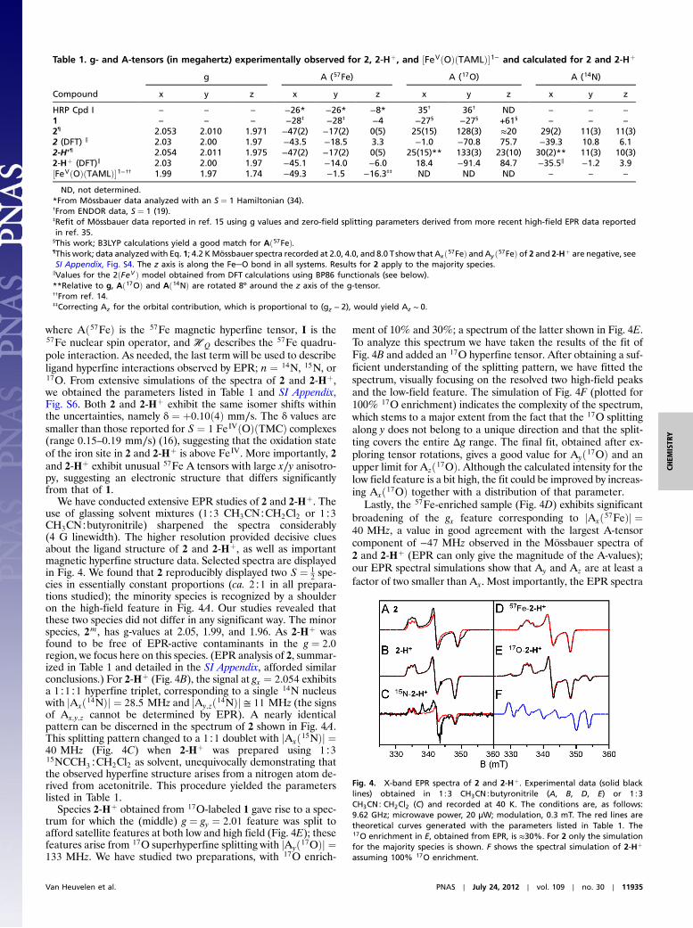
We have conducted extensive EPR studies of 2 and 2-Hþ. Theuse of glassing solvent mixtures (1∶3 CH3CN∶CH2Cl2 or 1∶3CH3CN∶butyronitrile) sharpened the spectra considerably(4 G linewidth). The higher resolution provided decisive cluesabout the ligand structure of 2 and 2-Hþ, as well as importantmagnetic hyperfine structure data. Selected spectra are displayedin Fig. 4. We found that 2 reproducibly displayed two S ¼ 1
2spe-
cies in essentially constant proportions (ca. 2∶1 in all prepara-tions studied); the minority species is recognized by a shoulderon the high-field feature in Fig. 4A. Our studies revealed thatthese two species did not differ in any significant way. The minorspecies, 2m, has g-values at 2.05, 1.99, and 1.96. As 2-Hþ wasfound to be free of EPR-active contaminants in the g ¼ 2.0region, we focus here on this species. (EPR analysis of 2, summar-ized in Table 1 and detailed in the SI Appendix, afforded similarconclusions.) For 2-Hþ (Fig. 4B), the signal at gx ¼ 2.054 exhibitsa 1∶1∶1 hyperfine triplet, corresponding to a single 14N nucleuswith jAxð14NÞj ¼ 28.5 MHz and jAy;zð14NÞj ≅ 11 MHz (the signsof Ax;y;z cannot be determined by EPR). A nearly identicalpattern can be discerned in the spectrum of 2 shown in Fig. 4A.This splitting pattern changed to a 1∶1 doublet with jAxð15NÞj ¼40 MHz (Fig. 4C) when 2-Hþ was prepared using 1∶315NCCH3∶CH2Cl2 as solvent, unequivocally demonstrating thatthe observed hyperfine structure arises from a nitrogen atom de-rived from acetonitrile. This procedure yielded the parameterslisted in Table 1.
Species 2-Hþ obtained from 17O-labeled 1 gave rise to a spec-trum for which the (middle) g ¼ gy ¼ 2.01 feature was split toafford satellite features at both low and high field (Fig. 4E); thesefeatures arise from 17O superhyperfine splitting with jAyð17OÞj ¼133 MHz. We have studied two preparations, with 17O enrich-
ment of 10% and 30%; a spectrum of the latter shown in Fig. 4E.To analyze this spectrum we have taken the results of the fit ofFig. 4B and added an 17O hyperfine tensor. After obtaining a suf-ficient understanding of the splitting pattern, we have fitted thespectrum, visually focusing on the resolved two high-field peaksand the low-field feature. The simulation of Fig. 4F (plotted for100% 17O enrichment) indicates the complexity of the spectrum,which stems to a major extent from the fact that the 17O splittingalong y does not belong to a unique direction and that the split-ting covers the entire Δg range. The final fit, obtained after ex-ploring tensor rotations, gives a good value for Ayð17OÞ and anupper limit for Azð17OÞ. Although the calculated intensity for thelow field feature is a bit high, the fit could be improved by increas-ing Axð17OÞ together with a distribution of that parameter.
Lastly, the 57Fe-enriched sample (Fig. 4D) exhibits significantbroadening of the gx feature corresponding to jAxð57FeÞj ¼40 MHz, a value in good agreement with the largest A-tensorcomponent of −47 MHz observed in the Mössbauer spectra of2 and 2-Hþ (EPR can only give the magnitude of the A-values);our EPR spectral simulations show that Ay and Az are at least afactor of two smaller than Ax. Most importantly, the EPR spectra
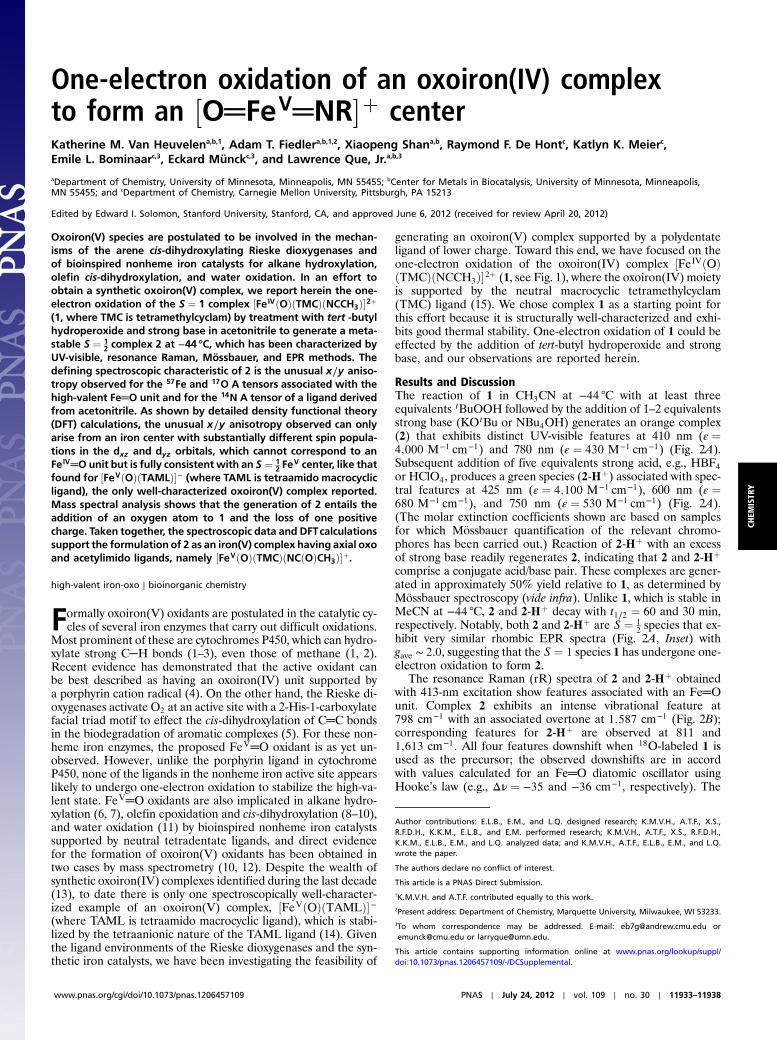
Table 1. g- and A-tensors (in megahertz) experimentally observed for 2, 2-Hþ, and ½FeVðOÞðTAMLÞ�1− and calculated for 2 and 2-Hþ
Compound
g A (57Fe) A (17O) A (14N)
x y z x y z x y z x y z
HRP Cpd I – – – −26* −26* −8* 35† 36† ND – – –1 – – – −28‡ −28‡ −4 −27§ −27§ +61§ – – –2¶ 2.053 2.010 1.971 −47(2) −17(2) 0(5) 25(15) 128(3) ≈20 29(2) 11(3) 11(3)2 (DFT) ∥ 2.03 2.00 1.97 −43.5 −18.5 3.3 −1.0 −70.8 75.7 −39.3 10.8 6.12-H+¶ 2.054 2.011 1.975 −47(2) −17(2) 0(5) 25(15)** 133(3) 23(10) 30(2)** 11(3) 10(3)2-Hþ (DFT)∥ 2.03 2.00 1.97 −45.1 −14.0 −6.0 18.4 −91.4 84.7 −35.5∥ −1.2 3.9½FeVðOÞðTAMLÞ�1−†† 1.99 1.97 1.74 −49.3 −1.5 −16.3‡‡ ND ND ND – – –
ND, not determined.*From Mössbauer data analyzed with an S ¼ 1 Hamiltonian (34).†From ENDOR data, S ¼ 1 (19).‡Refit of Mössbauer data reported in ref. 15 using g values and zero-field splitting parameters derived from more recent high-field EPR data reportedin ref. 35.
§This work; B3LYP calculations yield a good match for Að57FeÞ.¶This work; data analyzedwith Eq. 1; 4.2 KMössbauer spectra recorded at 2.0, 4.0, and 8.0 Tshow that Axð57FeÞ and Ay ð57FeÞ of 2 and 2-Hþ are negative, seeSI Appendix, Fig. S4. The z axis is along the Fe─O bond in all systems. Results for 2 apply to the majority species.
∥Values for the 2ðFeV Þ model obtained from DFT calculations using BP86 functionals (see below).**Relative to g, Að17OÞ and Að14NÞ are rotated 8º around the z axis of the g-tensor.††From ref. 14.‡‡Correcting Az for the orbital contribution, which is proportional to (gz − 2), would yield Az ∼ 0.
Fig. 4. X-band EPR spectra of 2 and 2-Hþ. Experimental data (solid blacklines) obtained in 1∶3 CH3CN∶butyronitrile (A, B, D, E) or 1∶3CH3CN∶CH2Cl2 (C) and recorded at 40 K. The conditions are, as follows:9.62 GHz; microwave power, 20 μW; modulation, 0.3 mT. The red lines aretheoretical curves generated with the parameters listed in Table 1. The17O enrichment in E, obtained from EPR, is ≈30%. For 2 only the simulationfor the majority species is shown. F shows the spectral simulation of 2-Hþ
assuming 100% 17O enrichment.
Van Heuvelen et al. PNAS ∣ July 24, 2012 ∣ vol. 109 ∣ no. 30 ∣ 11935
CHEM
ISTR
Y

of the 57Fe-enriched sample establish that the largest 57Fe A-ten-sor component is along x, the same direction for which the largest14N splitting is observed. It should be noted that the EPR spectraestablish a spatial correlation between the dominant componentsof the 57Fe, 17O, and 14N hyperfine tensors that provides insightinto the electronic structure of 2 and 2-Hþ.
Our accumulated spectroscopic data suggest that 2 and 2-Hþare one-electron oxidized derivatives of 1 and consist of aðTMCÞFe═O unit with an axial ligand derived from acetonitrile.In Fig. 1, we propose that 2 is formed by the attack of the t-bu-tylperoxide anion on the bound acetonitrile to form a peroxyimi-dic ester intermediate (17) that then undergoes homolyticcleavage of the O─O bond. To verify that homolysis occurs in thisinstance, we replaced tBuOOH with 2-methyl-1-phenylpropan-2-yl hydroperoxide (MPPH), a useful mechanistic probe to distin-guish between homolytic and heterolytic O─O bond cleavage(18). Although substitution of MPPH for tBuOOH did not dimin-ish the yield of 2, product analysis by GC showed quantitativeconversion of MPPH to benzaldehyde. This result demonstratesthat O─O bond homolysis occurs to generate 2. The formulationof 2 as shown in Fig. 1 is supported by low temperature electro-spray ionization (ESI)-MS studies of 2 that reveal a feature atm∕z 385.2, consistent with the formulation of 2 as ½FeðOÞðTMCÞðNCCH3Þ þO�þ (SI Appendix, Figs. S8 and S9). This fea-ture shifts to m∕z 388.2 upon preparation of 2 in CD3CN anddisappears after the sample is allowed to decay. We postulatein Fig. 1 two possible limiting electronic descriptions for 2: anoxoiron(IV) unit with an axial acetaminyl radical ligand(2ðFeIVN•Þ) at one end, and an iron(V) center with trans oxoand acetylimido ligands (2ðFeV Þ) at the other end. Our effortsto refine electronic descriptions for 2 and 2-Hþ more preciselyare discussed below.
The unusual A-tensor anisotropies in the xy plane listed inTable 1 for 2 and 2-Hþ indicate that their electronic structuresmust be significantly different from that of 1. The 57Fe and17O tensors of 1 as well as those of horseradish peroxidase com-pound I, an S ¼ 1 FeIV═O species antiferromagnetically coupledto a porphyrin radical (19), are axial with respect to the Fe═Obond, the z axis in our coordinate frames. The axial anisotropyobserved reflects the ðdxzÞ1ðdyzÞ1 electronic configuration asso-ciated with the S ¼ 1 FeIV═O unit. In contrast, 2 and 2-Hþ havedrastically different A-tensor components along x and y, indicat-ing substantially different unpaired spin densities on the dxz anddyz orbitals. This distribution can be achieved by transferring theelectron from dxz to the px orbital of the aminyl ligand to createan FeV═O center with a trans imido ligand, a notion supported bythe similar 57Fe A-tensor anisotropy observed for the bona fideFeV═O complex ½FeVðOÞðTAMLÞ�− (Table 1) (14).
Density functional theory (DFT) studies of 2 and 2-Hþ lendsubstantial support to the above conclusion (see SI Appendix,Tables S1–S7 for full computational details). For comparison,DFT calculations were also carried out for the experimentallycharacterized complexes 1 and ½FeVðOÞðTAMLÞ�− as well asthe hypothetical iron(V) species 1ox (a computational model ob-tained by removing one electron from 1; italics denote purelycomputational models). The use of the hybrid functional B3LYPled to the 2ðFeIVN•Þ description, which is similar to that re-ported for HRP Cpd I, but with the radical trans rather thancis to the oxo group. This FeIV-aminyl radical description withan iron ðdxzÞ1ðdyzÞ1 electron configuration predicts 2 and 2-Hþto have essentially axial 57Fe and 17OA-tensors, inconsistent withthe experimental data. In stark contrast, the use of the pure func-tional BP86 generated the 2ðFeV Þ limiting description with aniron ðdyzÞ1ðdxzÞ0 electron configuration and yielded solutions thatreproduce the observed anisotropies of the 57Fe A-tensor andrationalize the essential features of the entire dataset. In thismodel, the acetaminyl radical becomes an acetylimido ligand,and 2 and 2-Hþ are best formulated as complexes having
½O═FeV═NR�þ units (see Fig. 1). The dependence of the calcu-
lated ground state on whether a hybrid (B3LYP) or pure (BP86)functional is used may be related to the propensity of Hartree-Fock exchange to stabilize high spin states in the case of the for-mer functional. A similar dependence on the type of functionalsused has been reported for the description of electronic struc-tures of fFeNOg7 and fFeO2g8 complexes, systems involvinga noninnocent ligand radical (see for example refs. 20 and 21).In all cases, the functional chosen for further investigation mostclosely reproduced the experimentally observed spectroscopicparameters to generate an experimentally validated computa-tional model.
Table 2 compares the unpaired spin populations calculated forkey orbitals of several complexes. Interestingly, although the dyzspin population remains more or less constant at about 0.58across the series, the dxz spin population is equal to the dyz spinpopulation for 1 and 2ðFeIVN•Þ but decreases progressively for2ðFeV Þ, 1ox, and ½FeVðOÞðTAMLÞ�−. As viewed from theprogression of dxz spin density listed in Table 2, the electronicconfiguration of 2 more closely resembles that of 1ox and½FeVðOÞðTAMLÞ�− than of 1 and 2ðFeIVN•Þ. Because the trans-fer of electron density from the metal dxz orbital to the singly oc-cupied px orbital of the acetaminyl radical is not complete, theaxial nitrogen retains a net β spin population in px (n ¼ −0.30),which is much smaller than the value (n ¼ −0.85) calculated for2ðFeIVN•Þ. Consistent with these ideas, the Mulliken Fe spinpopulations for 2ðFeV Þ and 2ðFeV Þ-Hþ are calculated to be0.86 and 0.77, respectively, which is lower than the 1.30 value cal-culated for 1. The expectation value of the operator S2 providesquantitative insight into the oxidation state predicted by DFT.The 2ðFeV Þ model was found to have hΨBP86jS2jΨBP86i ¼ 0.94,which is close to the ideal value of SðSþ 1Þ ¼ 0.75 for S ¼ 1
2FeV
but considerably smaller than the value of 1.75 anticipatedfor the broken symmetry state (BS) of 2ðFeIVN•Þ. Using the ex-pansion jΨBP86 > ¼ cjFeV , S ¼ 1
2> þð1-c2Þ1∕2jFeIVN•, BS >
and the expectation values hS ¼ 1∕2jS2jS ¼ 1∕2i ¼ 0.75 andhBSjS2jBSi ¼ 1.75, we obtain c2 ¼ 0.81 from hΨBP86jS2jΨBP86i ¼0.75 c2 þ 1.75ð1-c2Þ ¼ 0.94. As spin unrestricted calculationsshow always some degree of spin contamination, the FeV char-acter of 2 (and 2-Hþ) is greater than 80%.
If we view 2 and 2-Hþ as FeV complexes, the spin densities atthe axial ligand atoms must originate from covalent spin polar-ization (22). Interaction of the oxo OðpyÞ electron pair withthe FeðdyzÞ orbital results in partial transfer of a β oxo electronto pair off some of the α spin density associated with the unpairedFeðdyzÞ electron, leaving a net α spin population in OðpyÞ (blue inFig. 5). The imido NðpxÞ electron pair transfers net α spin densityto the empty FeðdxzÞ orbital, guided by Hund’s rule, leaving a netβ spin population in NðpxÞ (red in Fig. 5). The electron pair in theout-of-plane NðpyÞ orbital of the imido ligand is calculated byDFT to be 10;000 cm−1 lower in energy than the redox-activein-plane NðpxÞ orbital and thus is much less involved in covalentdelocalization to the metal.
Table 2. Calculated unpaired spin populations n ¼ nα-nβ of Fe-dxz,Fe-dyz, and Naxial-px orbitals
FeVðOÞðTAMLÞ* 1oxðFeV Þ† 2ðFeV Þ‡ 1 2 (FeIVN•)§
Fe-dxz 0.07 0.15 0.23 0.58 0.58Fe-dyz 0.57 0.63 0.53 0.58 0.58Naxial-px – ∼0 −0.30 0 −0.85
*Tiago de Oliveira et al. (14).†Computational model obtained by removing one electron from 1.‡BP86 solution.§B3LYP solution, S ¼ 1 FeIV
═O coupled to S ¼ 1∕2 radical on N of axial ligand.
11936 ∣ www.pnas.org/cgi/doi/10.1073/pnas.1206457109 Van Heuvelen et al.

The resulting spin densities shown in Fig. 5 rationalize thespatial pattern observed in the A-tensors for 17O, 57Fe, and 14N.Thus, the net β spin population of the NðpxÞ gives a magnetichyperfine tensor with a large component along x and two smallercomponents along y and z for 2 (and 2-Hþ), with calculated Ax;y;zvalues of (−39, 11, 6) MHz for 14N in good agreement with thedata. The net α spin population of FeðdyzÞ produces a spin-dipo-lar hyperfine contribution with a large negative component alongx, which together with the negative Fermi contact term yields amagnetic hyperfine tensor of which the dominant componentis along x; the calculated Ax;y;z values of (−44, −19, 3) MHz(Table 1) are also in excellent agreement with the results obtainedfor 2 and 2-Hþ. Finally, the net α spin population of OðpyÞ givesan 17O A-tensor that has a large component along y, with Ax;y;zvalues of (−1, −71, þ76) MHz. The agreement for 17O betweenexperiment and calculations is not as good as that for 57Fe and14N; Ay is still too small and Az comes out too large but, impor-tantly, the calculated tensor lacks axial symmetry around z andexhibits the x∕y anisotropy observed experimentally. Finally,the g-values calculated for 2ðFeV Þ and reported in Table 1 matchboth in magnitude and direction the experimental data for 2and 2-Hþ. Thus the congruence of the experimental EPR resultswith the 2ðFeV Þ model provides a strong rationale for the de-scription of 2 and 2-Hþ as FeV complexes.
In contrast, the νFe═O frequencies hint at an FeIV oxidationstate for 2 and 2-Hþ and therefore may at first glance favor2ðFeIVN•Þ over 2ðFeV Þ. It should be noted, however, thatνFe═O is a spectroscopic parameter that reflects the length ofthe Fe-oxo bond (23). The Fe─O bond in the DFT model2ðFeV Þ (1.70 Å) is longer than the oxoiron(V) bond in 1ox(1.60 Å) and the oxoiron(IV) bond in 1 (1.66 Å) due to deloca-lization of electron density over the oxoironimido moiety* (seeFig. 6) and is therefore predicted to exhibit a lower νFe═O than1ox. The 2ðFeV Þ model predicts νFe═O values of 785 cm−1 for2ðFeV Þ (νexp ¼ 798 cm−1) and of 829 cm−1 for 2ðFeV Þ-Hþ(νexp ¼ 811 cm−1) in excellent agreement with the trend ob-served in our experimental data, indicating that the imido ligandexerts a sizable trans influence on the Fe─O unit.
Similarly, the weaker Fe═O bonds of 2 and 2-Hþ are expectedto give rise to higher Mössbauer isomer shifts, as this spectro-scopic parameter reflects the 3 s electron density at the 57Fe nu-cleus and the extent to which the 3 s electrons are shielded by 3delectrons. Thus, we expect the δ value of 2 to be more positivethan that of an FeV complex with an O═Fe─NCCH3 unit. As afurther test of the trans influence of the O═Fe═N─CðOÞ─R uniton the value of δ, we calculated the properties for the hypothe-tical 1-electron reduced 2 (denoted 2red) and obtained an Fe─Obond length of 1.767 Å, which is 0.10 Å longer than the Fe─Odistance 1.664 Å calculated for 1. The increase in bond lengthis indeed accompanied by an increase of the δ value by0.09 mm∕s and a 164 cm−1 decrease in νFe═O. The significanttrans influence of the acetylimido ligand is also manifested inthe comparison of the calculated values for 2ðFeV Þ (rFe─O ¼1.701 Å, δ ¼ 0.03 mm∕s, νFe═O ¼ 785 cm−1) and 1ox (rFe─O ¼1.603 Å, δ ¼ −0.08 mm∕s, νFe═O ¼ 944 cm−1). These calcula-tions support our view that νFe═O and δ are poor indicatorsfor the oxidation state of 2 and 2-Hþ, without properly account-ing for the trans influence of the axial ligand. Judged from the fulldata set, 2 and 2-Hþ are best described as FeV complexes.
The characterization of 2 and 2-Hþ reported in this paper in-creases to three the number of synthetic oxoiron(V) complexesfor which detailed spectroscopic and electronic structure infor-mation is available. Such complexes demonstrate that it is possi-ble to stabilize such a high-valent state in an iron coordinationcomplex. For ½FeVðOÞðTAMLÞ�−, the iron(V) oxidation statewas achieved by the use of a tetraanionic tetraazamacrocyclicligand that mitigates the high charge of the metal center (14).In contrast, 2 and 2-Hþ are supported by a neutral tetraazama-crocycle, so the stabilization of the iron(V) state, it would seem,must derive from the presence of the charged axial oxo and imidoligands. The structure we favor for 2 is closely related to that of½MnVðOÞ2ðTPFPPÞ�− [where TPFPP ismeso-tetrakis(pentafluor-ophenyl)porphinate dianion], which is the only known example ofa first row transition metal containing an ½MVðOÞ2� moiety (24).Examples of nitridoiron(V) and nitridoiron(VI) complexes havealso been reported, demonstrating the ability of the N3− ligand toallow access to high-valent iron complexes, even with supportingpolydentate ligands that are neutral or monoanionic (25–29).Lastly, the characterization of 2 and 2-Hþ demonstrates the pos-sibility of a neutral nonheme ligand to support an Fe(V) centerand lends credence to mechanistic proposals for FeVðOÞðOHÞoxidants in arene cis-dihydroxylation by Rieske dioxygenases(5) and in alkane, olefin, and water oxidation by a series of bioin-spired synthetic iron catalysts (6–9, 11). For the latter, evidencefor the fleeting iron(V) intermediates has been obtained from low
Fig. 5. Spin density plots of geometry optimized BP86 solutions for 2ðFeV Þ,1ox , and ½FeðOÞðTAMLÞ�−. The plot for 2ðFeV Þ, shown in two views, reveals thecontours of the orbitals carrying spin density. From top to bottom, px (Nam),dyz (Fe), and py (17O). For 1ox, the TMC ligand has been rotated by approxi-mately 90° around the Fe═O bond relative to the orientation shown for2ðFeV Þ. Majority spin α in blue; minority spin β in red.
Fig. 6. Calculated geometric and spectral parameters for the energy-minimized BP86 models 1ox, 2, and 2-H+. Bond lengths are reported in ang-stroms “L” denotes the TMC ligand.
*Unfortunately, our Raman experiments have not been able to identify vibrational modesthat can be associated with the Fe═NR unit. This result may not be surprising, asvibrational data are also not available for the only previously reported six-coordinateFe═NR complex (32), although Raman data for four-coordinate Fe═NR complexes havebeen obtained (33).
Van Heuvelen et al. PNAS ∣ July 24, 2012 ∣ vol. 109 ∣ no. 30 ∣ 11937
CHEM
ISTR
Y

temperature spectroscopic analysis of catalytic reactions by EPR(30, 31) and by mass spectrometry (10, 12).
Experimental ProceduresPreparation of 1, 2, and 2-Hþ. All reagents and solvents were pur-chased from commercial sources and used as received unlessotherwise noted. As previously reported (15), a solution of½FeIIðTMCÞðO3SCF3Þ� in acetonitrile was reacted with 1.2equivalents of PhIO solubilized in methanol to obtain 1 in quan-titative yield, as monitored by the appearance of its distinctivechromophore at 820 nm. Compound 2 was prepared at −44 °Cin acetonitrile by the addition of at least three equivalents oftert-butyl hydroperoxide followed by the addition of 1–2 equiva-lents of NBu4OH or KO tBu. Subsequent addition of at least fiveequivalents of strong acid, HBF4 or HClO4, yielded 2-Hþ. Fortypercent 17O-enriched H2O was used to prepare the 17O EPRsamples.
Physical Methods. A Hewlett Packard 8453A diode-array spectro-meter equipped with a Unisoku Scientific Instruments cryostatto maintain a constant temperature was employed to collect elec-tronic absorption spectrum. Resonance Raman spectra were gen-erated via excitation with Krþ and Arþ lasers (Spectra PhysicsBeamLok 2060-RM) with a power of <10 mW at the samples,and the spectra were collected using an ACTON AM-506M3
monochromator and a Princeton Instruments ACTON PyLoNLN/CCD-1340 × 400 detector. X band (9.28 GHz) EPR spectrawere recorded on a Bruker ESR 300 spectrometer equipped withan Oxford ESR 910 liquid Helium cryostat and an Oxford tem-perature controller. Mössbauer spectra were recorded using JanisResearch Super-Varitemp dewars that allowed studies in appliedmagnetic fields up to 8.0 T. Mössbauer spectral simulations wereperformed using the WMOSS software package (SEE Co), andEPR spectra were simulated with SpinCount, a program devel-oped by Prof. M. P. Hendrich of Carnegie Mellon University.Isomer shifts are quoted relative to Fe metal at 298 K. ESI-MSexperiments were conducted using a Bruker BioTOF II massspectrometer. The spray chamber voltage was set to 4,000 Vandthe gas carrier temperature was set at 60 °C.
ACKNOWLEDGMENTS.We thank Professor Thomas C. Brunold at the Universityof Wisconsin-Madison for kindly providing access to his computer cluster.We are particularly grateful to two of the reviewers for suggesting a keyimprovement in our proposed mechanism in Figure 1. This work wassupported by National Institutes of Health Grants GM33162 and GM38767(to L.Q.) and EB001475 (to E.M.) and postdoctoral fellowships GM-093479(to K.M.V.H.) and GM-079839 (to A.T.F.) and National Science FoundationGrant CHE1058248 (to L.Q.) and Grant CHE070073 (to E.L.B.) throughTeraGrid resources provided by the National Center for SupercomputingApplications and Pittsburgh Supercomputing Center.
1. Denisov IG, Makris TM, Sligar SG, Schlichting I (2005) Structure and chemistry ofcytochrome P450. Chem Rev 105:2253–2278.
2. Zilly FE, et al. (2011) Tuning a P450 enzyme formethane oxidation.Angew Chem Int Ed50:2720–2724.
3. Kawakami N, Shoji O, Watanabe Y (2011) Use of perfluorocarboxylic acids to trickcytochrome P450BM3 into initiating the hydroxylation of gaseous alkanes. AngewChem Int Ed 50:5315–5318.
4. Rittle J, Green MT (2010) Cytochrome P450 compound I: Capture, characterization,and C─H bond activation kinetics. Science 330:933–937.
5. Kovaleva EG, Lipscomb JD (2008) Versatility of biological non-heme Fe(II) centers inoxygen activation reactions. Nat Chem Biol 4:186–193.
6. Chen K, Que L, Jr (2001) Stereospecific alkane hydroxylation by nonheme ironcatalysts: Mechanistic evidence for an FeV
═O active species. J Am Chem Soc123:6327–6337.
7. Company A, et al. (2007) Alkane hydroxylation by a nonheme iron catalyst that chal-lenges the heme paradigm for oxygenase action. J Am Chem Soc 129:15766–15767.
8. Chen K, Costas M, Kim J, Tipton AK, Que L, Jr (2002) Olefin cis-dihydroxylation versusepoxidation by nonheme iron catalysts: Two faces of an FeIII
─OOH coin. J Am ChemSoc 124:3026–3035.
9. Company A, et al. (2009) Olefin-dependent discrimination between two nonhemeHO-FeV
═ tautomeric species in catalytic H2O2 epoxidations. Chem Eur J 15:3359–3362.10. Chow TW-S, et al. (2010) Cis-dihydroxylation of alkenes with oxone catalyzed by iron
complexes of amacrocyclic tetraaza ligand and reactionmechanism by ESI-MS spectro-metry and DFT calculations. J Am Chem Soc 132:13229–13239.
11. Fillol JL, et al. (2011) Efficient water oxidation catalysts based on readily available ironcoordination complexes. Nat Chem 3:807–813.
12. Prat I, et al. (2011) Observation of FeðVÞ═O using variable temperature mass spectro-metry and its enzyme-like C─H and C═C oxidation reactions. Nat Chem 3:788–793.
13. Que L, Jr (2007) The road to non-heme oxoferryls and beyond. Acc Chem Res40:493–500.
14. Tiago de Oliveira F, et al. (2007) Chemical and spectroscopic evidence for an FeV─oxo
complex. Science 315:835–838.15. Rohde J-U, et al. (2003) Crystallographic and spectroscopic evidence for a nonheme
FeIV═O complex. Science 299:1037–1039.
16. Jackson TA, et al. (2008) Axial ligand effects on the geometric and electronic structuresof nonheme oxoiron(IV) complexes. J Am Chem Soc 130:12394–12407.
17. Payne GB, Deming PH, Williams PH (1961) Reactions of hydrogen peroxide. VII. Alkali-catalyzed epoxidation and oxidation using a nitrile as co-reactant. J Org Chem26:659–663.
18. MacFaul PA, Arends IWCE, Ingold KI, Wayner DDM (1997) Oxygen activation by metalcomplexes and alkyl hydroperoxides. Applications of mechanistic probes to explorethe role of alkoxyl radicals in alkane functionalization. J Chem Soc Perkin Trans2:135–146.
19. Roberts JE, Hoffman BM, Rutter R, Hager LP (1981) 17O ENDOR of horseradish perox-idase compound I. J Am Chem Soc 103:7654–7656.
20. Schenk G, Pau MYM, Solomon EI (2004) Comparison between the geometric andelectronic structures and reactivities of fFeNOg7 and fFeO2g8 complexes: A densityfunctional theory study. J Am Chem Soc 126:505–515.
21. Hopmann KH, Conradie J, Ghosh A (2009) Broken-symmetry DFT spin densities of ironnitrosyls, including Roussin’s red and black salts: Striking differences between pureand hybrid functionals. J Phys Chem B 113:10540–10547.
22. Drago RS (1992) Physical Methods for Chemists (Saunders, Philadelphia), 2nd Ed.23. Green MT (2006) Application of Badger’s rule to heme and non-heme iron–oxygen
bonds: An examination of ferryl protonation states. J Am Chem Soc 128:1902–1906.24. Jin N, Ibrahim M, Spiro TG, Groves JT (2007) Trans-dioxo manganese(V) porphyrins.
J Am Chem Soc 129:12416–12417.25. Meyer K, Bill E, Mienert B, Weyhermüller T, Wieghardt K (1999) Photolysis of cis- and
trans-½FeIIIðcyclamÞðN3Þ2 �þ complexes: Spectroscopic characterization of a nitridoiron(V) species. J Am Chem Soc 121:4859–4876.
26. Grapperhaus CA, Mienert B, Bill E, Weyhermüller T, Wieghardt K (2000) Mononuclear(nitrido)iron(V) and (oxo)iron(IV) complexes via photolysis of ½ðcyclam-acetatoÞFeIII
ðN3Þ�þ and ozonolysis of ½ðcyclam-acetatoÞFeIIIðO3SCF3Þ�þ in water/acetone mixtures.Inorg Chem 39:5306–5317.
27. Aliaga-Alcalde N, et al. (2005) The geometric and electronic structure of½ðcyclam-acetatoÞFeðNÞ�þ : A genuine iron(V) species with a ground-state spin S ¼ 1
2.Angew Chem Int Ed 44:2908–2912.
28. Berry JF, et al. (2006) An octahedral coordination complex of iron(VI). Science312:1937–1941.
29. Scepaniak JJ, et al. (2011) Synthesis, structure, and reactivity of an iron(V) nitride.Science 331:1049–1052.
30. Lyakin OY, Bryliakov KP, Britovsek GJP, Talsi EP (2009) EPR spectroscopic trapping of theactive species of nonheme iron-catalyzed oxidation. J Am Chem Soc 131:10798–10799.
31. Lyakin OY, Bryliakov KP, Talsi EP (2011) EPR, 1H and 2H NMR, and reactivity studiesof the iron-oxygen intermediates in bioinspired catalyst systems. Inorg Chem50:5526–5538.
32. Klinker EJ, et al. (2006) A tosylimido analogue of a nonheme oxoiron(IV) complex.Angew Chem Int Ed 45:7394–7397.
33. Mehn MP, Brown SD, Jenkins DM, Peters JC, Que L, Jr (2006) Vibrational spectroscopyand analysis of pseudo-tetrahedral complexes with metal imido bonds. Inorg Chem45:7417–7427.
34. Schulz CE, Rutter R, Sage JT, Debrunner PG, Hager LP (1984) Mössbauer and electronparamagnetic resonance studies of horseradish peroxidase and its catalytic intermedi-ates. Biochemistry 23:4743–4754.
35. Krzystek J, et al. (2008) Determination by high-frequency and -field EPR of zero-fieldsplitting in iron(IV) oxo complexes: Implications for intermediates in nonheme ironenzymes. Inorg Chem 47:3483–3485.
11938 ∣ www.pnas.org/cgi/doi/10.1073/pnas.1206457109 Van Heuvelen et al.


![Silver(I) Dicyanonitrosomethanide, Ag[ONC(CN)2] Complex as ... · pharmaceuticals, resin additives and in organic synthesis [1-3]. Oxidation of benzyl alcohol, direct oxidation of](https://static.fdocuments.us/doc/165x107/5f7eb9bfcca1b15c6c2b5c81/silveri-dicyanonitrosomethanide-agonccn2-complex-as-pharmaceuticals.jpg)