oncologists secondary - PNAS · duced, in addition to a primary crown-gall tumor, secondary...
6
BOTANY: BRA UN tumor sectors of a single individual and the physiological comparison of these cell lines in tissue cultures. While this is theoretically possible in animal and human tumors, it is fraught with such technical difficulties that it has not yet been accom- plished over more than very brief periods, whereas in these spruce tumors it can be done with relative ease. We believe that these tumors represent a promising addition to our roster of materials in which the processes of tumefaction can be profitably studied. 1 P. R. White, Quart. Rev. Biol., 26, 1-16, 1951. 2 P. R. White and W. F. Millington, Cancer Research, 14, 128-134, 1954; Am. J. Bot., 41, 353- 361, 1954. 3J. Reinert, Science, 123, 457-458, 1956. 4J. Reinert and P. R. White, Physiol. Plantarum, 9, 177-189, 1956. A PHYSIOLOGICAL BASIS FOR AUTONOMOUS GROWTH OF THE CROWN-GALL TUMOR CELL* BY ARMIN C. BRAUN ROCKEFELLER INSTITUTE FOR MEDICAL RESEARCH, NEW YORK We are commemorating on this occasion the description of a rather remarkable micro-organism. This bacterium, which is now known as Agrobacterium tume- faciens (Smith and Town.) Conn, is responsible for the initiation of the non-self- limiting neoplastic disease of plants known as crown gall. The isolation and characterization of a tumor-inducing bacterium shortly after the turn of the century attracted considerable interest among pathologists generally because at the time of that discovery no animal tumor had yet been produced experimentally. For a period of about twenty years Erwin F. Smith made detailed comparative studies of crown-gall and malignant animal tumors and found that these two types of growth had much in common. There appeared, however, to be one fundamental dif- ference. Smith believed that the continued unregulated proliferation of the crown-gall tumor cell was dependent upon continued stimulation by the inciting bacterium. Crown gall was, therefore, not generally accepted by oncologists as being comparable to true animal tumors because, as described by Smith, this plant disease appeared to be simply a bacterial-stimulated hyperplasia and not a truly independent growth' as are most animal cancers. In certain plant species such as the sunflower and Paris daisy there may be pro- duced, in addition to a primary crown-gall tumor, secondary tumors that arise at points distant from the seat of the primary growth. These secondary tumors are interesting because they are frequently free of the bacteria that initiate the pri- mary tumor. The finding that many of the secondary tumors are bacteria-free per- mitted the unequivocal demonstration of the truly independent nature of the crown- gall tumor cell.1 Sterile tissue isolated from the secondary tumors grew profusely and indefinitely on a culture medium that did not support the continued growth of normal cells of the type from which the tumor cells were derived. Small frag- ments of such tumor tissue implanted into a healthy host developed again into 344 PROC. N. A. S.
Transcript of oncologists secondary - PNAS · duced, in addition to a primary crown-gall tumor, secondary...
BOTANY: BRA UN
tumor sectors of a single individual and the physiological comparison of these celllines in tissue cultures. While this is theoretically possible in animal and humantumors, it is fraught with such technical difficulties that it has not yet been accom-plished over more than very brief periods, whereas in these spruce tumors it can bedone with relative ease.We believe that these tumors represent a promising addition to our roster of
materials in which the processes of tumefaction can be profitably studied.1 P. R. White, Quart. Rev. Biol., 26, 1-16, 1951.2 P. R. White and W. F. Millington, Cancer Research, 14, 128-134, 1954; Am. J. Bot., 41, 353-
361, 1954.3J. Reinert, Science, 123, 457-458, 1956.4J. Reinert and P. R. White, Physiol. Plantarum, 9, 177-189, 1956.
A PHYSIOLOGICAL BASIS FOR AUTONOMOUSGROWTH OF THE
CROWN-GALL TUMOR CELL*
BY ARMIN C. BRAUN
ROCKEFELLER INSTITUTE FOR MEDICAL RESEARCH, NEW YORK
We are commemorating on this occasion the description of a rather remarkablemicro-organism. This bacterium, which is now known as Agrobacterium tume-faciens (Smith and Town.) Conn, is responsible for the initiation of the non-self-limiting neoplastic disease of plants known as crown gall. The isolation andcharacterization of a tumor-inducing bacterium shortly after the turn of the centuryattracted considerable interest among pathologists generally because at the timeof that discovery no animal tumor had yet been produced experimentally. For aperiod of about twenty years Erwin F. Smith made detailed comparative studies ofcrown-gall and malignant animal tumors and found that these two types of growthhad much in common. There appeared, however, to be one fundamental dif-ference. Smith believed that the continued unregulated proliferation of thecrown-gall tumor cell was dependent upon continued stimulation by the incitingbacterium. Crown gall was, therefore, not generally accepted by oncologists asbeing comparable to true animal tumors because, as described by Smith, thisplant disease appeared to be simply a bacterial-stimulated hyperplasia and not atruly independent growth' as are most animal cancers.
In certain plant species such as the sunflower and Paris daisy there may be pro-duced, in addition to a primary crown-gall tumor, secondary tumors that arise atpoints distant from the seat of the primary growth. These secondary tumors areinteresting because they are frequently free of the bacteria that initiate the pri-mary tumor. The finding that many of the secondary tumors are bacteria-free per-mitted the unequivocal demonstration of the truly independent nature of the crown-gall tumor cell.1 Sterile tissue isolated from the secondary tumors grew profuselyand indefinitely on a culture medium that did not support the continued growth ofnormal cells of the type from which the tumor cells were derived. Small frag-ments of such tumor tissue implanted into a healthy host developed again into
344 PROC. N. A. S.
BOTANY: BRAUN
typical crown-gall tumors. Since such sterile cells isolated not only from secondarytumors but subsequently also from primary tumors of many plant species2 have not,in the more than ten years that they have been kept under observation, shown theslightest tendency to become less autonomous, they have generally been regardedas being permanently altered cells that reproduce true to type and against thegrowth of which there is no control mechanism in the host. These are the charac-teristics by which malignant animal cells are distinguished from healthy or merelyinflammatory cells. Thus the one basic difference thought by Smith to distinguishcrown-gall from malignant animal tumors has been removed.
In any analysis of a complex series of events such as those that occur during tumorformation, it is often convenient to subdivide, in so far as that is possible, the totalresult into a series of contributing events, each of which is essential for the con-summation of the completed process. In studying these events in the crown-galldisease, two distinct phases have now been recognized.3 In the first phase normalcells are altered to tumor cells which do not as yet develop into a neoplastic growth.Two known requirements must be satisfied to complete the inception phase.These have been termed "conditioning" and "induction."4,5 By "conditioning"is meant that only those plant cells that have been rendered susceptible as a resultof irritation accompanying a wound can be altered to tumor cells. An excellentcorrelation has been found to exist, moreover, between the stage in the normalwound-healing cycle in which normal cells are converted to tumor cells and the sizeand rate of growth of the resulting tumors.5 6 Induction, on the other hand, refersto the actual conversion of a conditioned host cell into a crown-gall tumor cell by anas yet essentially uncharacterized tumor-inducing principle elaborated by the incit-ing bacteria. Induction completes the first phase of tumor formation. The secondphase of tumor formation, according to this concept, is concerned with the con-tinued unregulated and autonomous growth of the tumor cell, once the cellular al-teration has been accomplished. The development of a satisfactory explanationfor the continued abnormal growth of a tumor cell in the absence of any recogniz-able infective agent represents a very real challenge not only to students of crowngall but also to those interested in autonomous growth generally. Perhaps themost important single concept that has arisen from the plant work is that con-cerned with the nature of the autonomous growth of a tumor cell. It is with thataspect of the problem that we should like to concern ourselves at this time.
In order to gain insight into the nature of autonomous growth, it is necessary tounderstand something of the processes involved in normal growth and develop-ment. Growth in all higher animals and plants is the result either of an enlarge-ment of the constituent cells of such organisms or of the combined processes of cellenlargement and cell division. These fundamental growth processes appear to bedependent for their development in plant cells upon specific substances that may besynthesized by plant cells themselves.7'8 It is now possible, moreover, to delimitunder fully controlled experimental conditions, with the use as a test object of cer-tain plant-cell types, these two growth processes. When, for example, tobaccopith parenchymal cells are treated with synthetic growth substances of the auxintype such as naphthalene acetic acid, the pith cells enlarge greatly in size but do notdivide. It is only when a second growth factor such as 6-furfurylaminopurine,9or the naturally occurring equivalent of that substance,8"10 is supplied to the pith
VOL. 44, 1958345
BOTANY: BRAUN
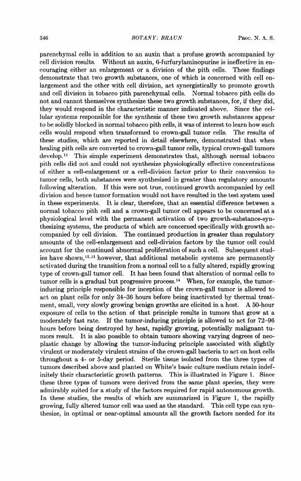
parenchymal cells in addition to an auxin that a profuse growth accompanied bycell division results. Without an auxin, 6-furfurylaminopurine is ineffective in en-couraging either an enlargement or a division of the pith cells. These findingsdemonstrate that two growth substances, one of which is concerned with cell en-largement and the other with cell division, act synergistically to promote growthand cell division in tobacco pith parenchymal cells. Normal tobacco pith cells donot and cannot themselves synthesize these two growth substances, for, if they did,they would respond in the characteristic manner indicated above. Since the cel-lular systems responsible for the synthesis of these two growth substances appearto be solidly blocked in normal tobacco pith cells, it was of interest to learn how suchcells would respond when transformed to crown-gall tumor cells. The results ofthese studies, which are reported in detail elsewhere, demonstrated that whenhealing pith cells are converted to crown-gall tumor cells, typical crown-gall tumorsdevelop." This simple experiment demonstrates that, although normal tobaccopith cells did not and could not synthesize physiologically effective concentrationsof either a cell-enlargement or a cell-division factor prior to their conversion totumor cells, both substances were synthesized in greater than regulatory amountsfollowing alteration. If this were not true, continued growth accompanied by celldivision and hence tumor formation would not have resulted in the test system usedin these experiments. It is clear, therefore, that an essential difference between anormal tobacco pith cell and a crown-gall tumor cell appears to be concerned at aphysiological level with the permanent activation of two growth-substance-syn-thesizing systems, the products of which are concerned specifically with growth ac-companied by cell division. The continued production in greater than regulatoryamounts of the cell-enlargement and cell-division factors by the tumor cell couldaccount for the continued abnormal proliferation of such a cell. Subsequent stud-ies have shown,'2 3 however, that additional metabolic systems are permanentlyactivated during the transition from a normal cell to a fully altered, rapidly growingtype of crown-gall tumor cell. It has been found that alteration of normal cells totumor cells is a gradual but progressive process.'4 When, for example, the tumor-inducing principle responsible for inception of the crown-gall tumor is allowed toact on plant cells for only 34-36 hours before being inactivated by thermal treat-ment, small, very slowly growing benign growths are elicited in a host. A 50-hourexposure of cells to the action of that principle results in tumors that grow at amoderately fast rate. If the tumor-inducing principle is allowed to act for 72-96hours before being destroyed by heat, rapidly growing, potentially malignant tu-mors result. It is also possible to obtain tumors showing varying degrees of neo-plastic change by allowing the tumor-inducing principle associated with slightlyvirulent or moderately virulent strains of the crown-gall bacteria to act on host cellsthroughout a 4- or 5-day period. Sterile tissue isolated from the three types oftumors described above and planted on White's basic culture medium retain indef-initely their characteristic growth patterns. This is illustrated in Figure 1. Sincethese three types of tumors were derived from the same plant species, they wereadmirably suited for a study of the factors required for rapid autonomous growth.In these studies, the results of which are summarized in Figure 1, the rapidlygrowing, fully altered tumor cell was used as the standard. This cell type can syn-thesize, in optimal or near-optimal amounts all the growth factors needed for its
346 PROC. N. A. S.

BOTANY: BRAUN
continued rapid abnormal growth from mineral salts and sucrose present in White'sbasic culture medium. The moderately fast-growing tumor cell required that thebasic medium be supplemented with glutamine, meso inositol, and a cell-enlargementfactor (naphthalene acetic acid) to achieve a growth rate comparable to that of thefully altered, rapidly growing type of tumor cell. The very slowly growing benigntumor cells altered in a 34-hour period required, in addition to the three com-pounds described above, asparagine as well as cytidylic and guanylic acids toachieve full growth. Asparagine did not, however, represent an absolute require-
Ri;a.\ia a,< s 9gIw i '1 .1' II "vt4ar!
i. .i .1 :^ is a] 't (f-II1 '4
B;a 'ni T(M nTy
91 V( " i :,Ig I;%,I ,)act
iX(U;i ff, Oll's3
H3 ia INo4 n..r,
'1 ,,i A l; sis.'h1t; -tsd s.;
gUses;4-n i3 :,o
Ba - M1 -ted ;,o
.i X:'.
ci
a -------- ... ..
FIG. 1-Relative rates of growth of three clones of crown-gall tumor tissue that show differentdegrees of neoplastic change, planted on White's basic medium. (Left), Fully altered rapidlygrowing tumor cells. (Upper left), Moderately fast-growing tumor cells. (Upper center), Veryslowly growing tumor cells. (Upper right), Normal cells of the type from which the tumor cellswere derived. While the three clones of the tumor cells grow continuously although at differentrates on the basic culture medium, normal cells of this type do not grow on that medium.Lower pictures and legends show minimal nutritional supplements needed by the three types oftissues to achieve a growth rate comparable to that of the fully altered tumor cell. (Photographsby J. A. Carlile.)
ment for rapid growth but served to stimulate somewhat the growth of tumor tissuefragments planted on an otherwise suitable culture medium. Certain amino acids,particularly proline and histidine, also appeared to have a stimulatory effect on thegrowth of this tissue, but this requirement seemed to be of a transient nature, sincethe amino acids did not appear to be required in the second transfer of the tissue tothe supplemented medium. It is clear from these experiments that, as the crown-gall tumor cell becomes more autonomous, its requirements in terms of externallysupplied growth factors become less exacting. More importantly, however, these
VOL. 44, 1958 347
N. ...) . IT' :.
;, "! '),%-i%,.,."!,%g"..
i I rl A ("I " '.
Ij l'-w - -d ("

BOTANY: BRAUN
studies clearly demonstrate that a series of well-defined growth-substance-synthe-sizing systems become gradually activated during the transition from the normalcell to the fully altered tumor cell, and the degree of activation of these systems de-termines the rate of growth of the tumor cell.Normal cells of the type from which the tumor cells were derived do not grow on
the basic medium. Thus, although the difference between the three types of tumorcells is quantitative because all grow continuously but at different rates on thebasic medium, the difference between the tumor cells and the normal cell is quali-tative. One qualitative difference found to exist in these studies was the absoluterequirement of the normal cell for 6-furfurylaminopurine or the naturally occurringequivalent of that substance. The addition of that compound to the basic me-dium or to the supplemented culture media did not stimulate growth of any of thetumor tissues. The normal cells also possess, in contrast with the tumor cells, anabsolute requirement as a supplement for a cell-enlargement factor such as naph-thalene acetic acid. The addition of 6-furfurylaminopurine and naphthalene aceticacid to the basic medium permits the very slow but limited growth of normal cells.However, only if the basic medium is supplemented with glutamine, asparagine,inositol, and guanylic and cytidylic acids in addition to the auxin and 6-furfuryl-aminopurine, do the normal cells achieve a growth rate comparable to that of thefully altered, rapidly growing type of tumor cell. These same substances, with theexception of 6-furfurylaminopurine, are required as supplements to the basic mediumfor the continued rapid growth of the normally slow-growing tumor cells. Themoderately fast-growing tumor cells require the addition of only three of thesecompounds to the basic medium to achieve rapid growth, while the fully alteredtumor cells can synthesize in optimal amounts all their requirements from themineral salts and sucrose present in the basic culture medium. It thus appearsthat, as a result of the transition from a normal cell to a fully altered, rapidly grow-ing crown-gall tumor cell, a series of quite distinct, but well-defined, growth-sub-stance-synthesizing systems becomes progressively activated. This leads to theproduction by the affected cell of greater than regulatory amounts of these growth-promoting substances. The continued production in greater than regulatoryamounts of these substances by the tumor cell could and most probably does ac-count for the continued unregulated proliferation of such a cell. Precisely howthe tumor-inducing principle associated with this disease accomplishes the simul-taneous unblocking of several apparently distinct and quite unrelated metabolicsystems remains a moot question. These results are understandable if it is as-sumed that some as yet uncharacterized master reaction within the cell is specificallybut gradually unblocked by the tumor-inducing principle and which, once activated,not only accomplishes the unblocking of several other growth-substance-synthe-sizing systems but also determines the rate at which the entire series of metabolicevents concerned with growth and cell division proceeds.The concept of growth autonomy presented above finds additional support in
other directions. It has been possible to reproduce under precisely defined experi-mental conditions and with the use of certain normal cell types as a test objectnot only the morphological growth patterns' 12 (slow and rapid disorganizedgrowths, teratoma-like structures) but also the histological (hypertrophy and hy-perplasia leading to disorganization and loss of function) as well as the cytological
PROC. N. A. S.348
BOTANY: BRAUN
(multinucleate giant cells, etc.) 15 abnormalities that characterize the tumorous statein crown gall. This was accomplished by varying the proportions of the cell-enlargement factor and the factor limiting for cell division in an otherwise suitableculture medium on which the normal cells were planted. These artificially stimu-lated normal cells, in contrast to crown-gall tumor cells, are self-limiting growths,and, when the externally supplied stimuli are removed, their growth promptlystops. The fact that such stimulated normal cells commonly show histological andcytological characteristics of true tumor cells but are themselves self-limitinggrowths indicates that the observed cellular abnormalities are the result rather thanthe cause of the tumorous state.The results of all of these studies strongly suggest that it is possible for a cell to
acquire the capacity for autonomous growth as a result of the permanent activationof a series of growth-substance-synthesizing systems, the products of which areconcerned specifically with growth accompanied by cell division. These systemsare precisely regulated in all normal plant cells.
* This investigation was supported in part by a research grant (PSH, C-2944 M & G) from theNational Cancer Institute, Public Health Service. This paper was presented at the NationalAcademy of Sciences, Symposium on Plant Tumors, autumn meetings, November 19, 1957.
1 A. C. Braun, Phytopathology, 31, 135, 1941; P. R. White and A. C. Braun, Cancer Research, 2,597, 1942; A. C. Braun and P. R. White, Phytopathology, 33, 85, 1943.
2 P. R. White, Am. J. Botany, 32, 237, 1945; R. S. deRopp, Phytopathology, 37, 201, 1947;A. C. Hildebrandt and A. J. Riker, Am. J. Botany, 36, 74, 1949; G. Morel, Ann. epiphyt., 14 (Ser.Path. veg. M6m. 5), 123, 1948; R. Gautheret, Comp. rend. Acad. sci. (Paris), 224, 1728, 1947.
3 A. C. Braun and T. Laskaris, these PROCEEDINGS, 28, 468, 1942.4 A. C. Braun, Ann. Rev. Plant Physiol., 5, 133, 1954.5 A. C. Braun, Growth, 16, 65, 1952.6 A. C. Braun, Brookhaven Symposia Biol., No. 6: Abnormal and Pathological Plant Growth,
115, 1954.7 F. C. Steward and S. M. Caplin, Science, 113, 518, 1951.8 J. R. Jablonski and F. Skoog, Physiol. Plantarum, 7, 16, 1954.9 C. 0. Miller, F. Skoog, F. S. Okumura, M. H. Von Saltza, and F. M. Strong, J. Am. Chem.
Soc., 77, 2662, 1955.10 A. C. Braun and U. NAf, Proc. Soc. Exp. Biol. Med., 86, 212, 1954.11 A. C. Braun, Cancer Research, 16, 53, 1956.12 A. C. Braun, Symposia Soc. Exptl. Biol., No. XI: The Biological Action of Growth Substances
132, 1957.13 A. C. Braun, J. Natl. Cancer Inst., 19, 753, 1957.14 A. C. Braun, Am. J. Botany, 30, 674, 1943; ibid., 34, 234, 1947; and Phytopathology, 41, 963,
1951.15 F. Skoog, Brookhaven Symposia Biol., No. 6: Abnormal and Pathological Plant Growth, 1,
1954; A. C. Braun and T. Stonier, Protoplasmatologia, 10, H. 5b, 1958.
VOL. 44) 1958 349