On sepsis - epidemiology, prediction and diagnostics ...
81
On sepsis - epidemiology, prediction and diagnostics Mellhammar, Lisa 2020 Document Version: Publisher's PDF, also known as Version of record Link to publication Citation for published version (APA): Mellhammar, L. (2020). On sepsis - epidemiology, prediction and diagnostics. Lund University, Faculty of Medicine. Total number of authors: 1 General rights Unless other specific re-use rights are stated the following general rights apply: Copyright and moral rights for the publications made accessible in the public portal are retained by the authors and/or other copyright owners and it is a condition of accessing publications that users recognise and abide by the legal requirements associated with these rights. • Users may download and print one copy of any publication from the public portal for the purpose of private study or research. • You may not further distribute the material or use it for any profit-making activity or commercial gain • You may freely distribute the URL identifying the publication in the public portal Read more about Creative commons licenses: https://creativecommons.org/licenses/ Take down policy If you believe that this document breaches copyright please contact us providing details, and we will remove access to the work immediately and investigate your claim.
Transcript of On sepsis - epidemiology, prediction and diagnostics ...

LUND UNIVERSITY
PO Box 117221 00 Lund+46 46-222 00 00
On sepsis - epidemiology, prediction and diagnostics
Mellhammar, Lisa
2020
Document Version:Publisher's PDF, also known as Version of record
Link to publication
Citation for published version (APA):Mellhammar, L. (2020). On sepsis - epidemiology, prediction and diagnostics. Lund University, Faculty ofMedicine.
Total number of authors:1
General rightsUnless other specific re-use rights are stated the following general rights apply:Copyright and moral rights for the publications made accessible in the public portal are retained by the authorsand/or other copyright owners and it is a condition of accessing publications that users recognise and abide by thelegal requirements associated with these rights. • Users may download and print one copy of any publication from the public portal for the purpose of private studyor research. • You may not further distribute the material or use it for any profit-making activity or commercial gain • You may freely distribute the URL identifying the publication in the public portal
Read more about Creative commons licenses: https://creativecommons.org/licenses/Take down policyIf you believe that this document breaches copyright please contact us providing details, and we will removeaccess to the work immediately and investigate your claim.

LISA M
ELLHA
MM
AR
O
n sepsis 2020:4
3
Department of Clinical Sciences, LundDivision of Infection Medicine
Lund University, Faculty of Medicine Doctoral Dissertation Series 2020:43
ISBN 978-91-7619-904-6 ISSN 1652-8220
On sepsisEpidemiology, prediction and diagnosticsLISA MELLHAMMAR
INFECTION MEDICINE | FACULTY OF MEDICINE | LUND UNIVERSITY
9789176
199046


On sepsis
Epidemiology, prediction and diagnostics

On sepsis Epidemiology, prediction and diagnostics
Lisa Mellhammar
DOCTORAL DISSERTATION
by due permission of the Faculty of Medicine, Lund University, Sweden. To be defended at Belfragesalen, BMC, Lund. 24th of April 2020 at 9:00
Faculty opponent Kristoffer Strålin, MD PhD
Karolinska Institutet

Organization LUND UNIVERSITY
Document name Doctoral Dissertation
Department of Clinical Sciences, Lund Division of Infection Medicine
Date of issue 24th of April 2020
Author(s) Lisa Mellhammar
Sponsoring organization
Title and subtitle On sepsis – epidemiology, prediction and diagnostics Abstract
The overall aims of this thesis were to improve prediction, diagnostics and knowledge on epidemiology of sepsis.
In paper I, we developed and evaluated an integrated platform for rapid analysis of sepsis-causing organisms directly from blood samples. Testing with blood samples spiked with bacteria and samples from septic patients indicate that the detection limit of the system is in the upper part of clinically relevant bacteria concentration range. The paper describes proof-of-principle for the integrated system for faster sepsis diagnostics.
In paper II, we assessed the incidence of hospital-treated sepsis in an entire population based on clinical findings. The annual incidence for severe sepsis (sepsis-2) was 687/100 000 person years (95% CI 549-824) and the annual incidence for sepsis-3 was 780/100 000 person years (95% CI 633-926). These estimates are closer to the true incidence of sepsis compared to estimates based on ICD-codes.
In paper III & IV, we evaluated different early warning scores for sepsis prediction and detection. We also developed and evaluated a candidate warning score for sepsis based on vital signs and heparin-binding protein. NEWS2 was superior to qSOFA and RETTS for screening for sepsis. Even with a statistical approach, we could not construct better warning scores for sepsis than NEWS2.
In paper V, patients with sepsis admitted to an ICU were retrospectively studied in a clinical chart review. We found a high proportion of bacteremic patients, probably due to that clinical chart review minimizes the misdiagnosis of other conditions. We also demonstrated higher mortality among bacteremic patients, than in non-bacteremic patients.
Key words Sepsis; Epidemiology; Early Warning Score; Bacteremia Classification system and/or index terms (if any)
Supplementary bibliographical information Language English
ISSN and key title 1652-8220
ISBN 978-91-7619-904-6
Recipient’s notes Number of pages 76 Price
Security classification
I, the undersigned, being the copyright owner of the abstract of the above-mentioned dissertation, hereby grant to all reference sources permission to publish and disseminate the abstract of the above-mentioned dissertation.
Signature Date 2020-03-13

On sepsis Epidemiology, prediction and diagnostics
Lisa Mellhammar
Coverphoto by Hady Yazdi Aznaveh
Copyright Lisa Mellhammar 2020
Paper 1 © Analytical Chemistry 2016
Paper 2 © Open Forum Infectious Diseases 2016
Paper 3 © Journal of Clinical Medicine 2019
Paper 4 © PLOS ONE 2020
Paper 5 © Lisa Mellhammar (Manuscript unpublished) 2020
Lund University, Faculty of Medicine Doctoral Dissertation Series 2020:43
ISBN 978-91-7619-904-6 ISSN 1652-8220
Printed in Sweden by Media-Tryck, Lund University Lund 2020

Table of Contents
Abstract .......................................................................................................... 9 Populärvetenskaplig sammanfattning på svenska ........................................ 10 List of papers ................................................................................................ 11 Abbreviations ............................................................................................... 12
The history ............................................................................................................ 13 The definitions ...................................................................................................... 15 The PIRO model ................................................................................................... 19 The epidemiology .................................................................................................. 21 The long-term effects ............................................................................................ 29 The pathophysiology ............................................................................................ 31 The therapy ........................................................................................................... 33 Bacteremia ............................................................................................................ 35 The acoustofluidics ............................................................................................... 37 Prediction and risk stratification scores ............................................................. 39 Aims ....................................................................................................................... 43 Paper I ................................................................................................................... 45
Materials and methods ................................................................................. 45 Results .......................................................................................................... 46 Discussion .................................................................................................... 46
Paper II .................................................................................................................. 49 Materials and methods ................................................................................. 49 Results .......................................................................................................... 49 Discussion .................................................................................................... 50
Paper III & IV ...................................................................................................... 51 Materials and methods ................................................................................. 51

Results .......................................................................................................... 52 Discussion .................................................................................................... 53
Paper V .................................................................................................................. 55 Materials & Methods .................................................................................... 55 Results .......................................................................................................... 55 Discussion .................................................................................................... 56
Future perspectives .............................................................................................. 57 Acknowledgements ............................................................................................... 61 References ............................................................................................................. 63

9
Abstract The overall aims of this thesis were to improve prediction, diagnostics and knowledge on epidemiology of sepsis.
In paper I, we developed and evaluated an integrated platform for rapid analysis of sepsis-causing organisms directly from blood samples. Testing with blood samples spiked with bacteria and samples from septic patients indicate that the detection limit of the system is in the upper part of clinically relevant bacteria concentration range. The paper describes proof-of-principle for the integrated system for faster sepsis diagnostics.
In paper II, we assessed the incidence of hospital-treated sepsis in an entire population based on clinical findings. The annual incidence for severe sepsis (sepsis-2) was 687/100 000 person years (95% CI 549-824) and the annual incidence for sepsis-3 was 780/100 000 person years (95% CI 633-926). These estimates are closer to the true incidence of sepsis compared to estimates based on ICD-codes.
In paper III & IV, we evaluated different early warning scores for sepsis prediction and detection. We also developed and evaluated a candidate warning score for sepsis based on vital signs and heparin-binding protein. NEWS2 was superior to qSOFA and RETTS for screening for sepsis. Even with a statistical approach, we could not construct better warning scores for sepsis than NEWS2.
In paper V, patients with sepsis admitted to an ICU were retrospectively studied in a clinical chart review. We found a high proportion of bacteremic patients, probably due to that clinical chart review minimizes the misdiagnosis of other conditions. We also demonstrated higher mortality among bacteremic patients, than in non-bacteremic patients.

10
Populärvetenskaplig sammanfattning på svenska Sepsis är ett livshotande tillstånd som uppstår när kroppens immunförsvar överreagerar på en infektion. Vid sepsis är det viktigt med snabb upptäckt och diagnos för att kunna påbörja behandling. Det övergripande syftet med denna avhandling var att förbättra upptäckt och diagnostik av sepsis samt öka kunskapen om dess epidemiologi. I vår första studie utvecklade och utvärderade vi ett integrerat system för snabb analys av sepsis-orsakande organismer direkt från blodprover. Det testade systemet fungerade såtillvida att det kunde påvisa och art-bestämma bakterier på mindre än 2 timmar. Systemet behöver vidareutvecklas för att öka känsligheten samt för att kunna identifiera fler bakteriearter. I vårt andra arbete beräknade vi förekomsten av sjukhusbehandlad sepsis. Den vanliga metoden för att beräkna hur många som drabbas av sepsis är att använda hälso- och sjukvårdsregistret. Dit skickas data var gång en patient skrivs ut från sjukhus. Datan i hälso- och sjukvårdsregistret innehåller information om vad patienten har vårdats för och alla sjukdomar är klassificerade med unika koder (International Classification of Disease, ICD). Patienter med sepsis har dessvärre sällan erhållit koder för sepsis, varför vi istället mätte och beräknade förekomsten av sepsis genom att granska journaler. Den årliga förekomsten av svår sepsis var cirka 700/100 000 person och år. Denna uppskattning är mer nära den verkliga förekomsten av sepsis jämfört med uppskattningar baserade på ICD-koder. I arbete III & IV utvärderade vi olika verktyg för att hitta patienter som har eller riskerar att utveckla sepsis. Av de utvärderade verktygen visade sig NEWS2 vara överlägset över qSOFA och RETTS för att förutspå och upptäcka sepsis. Vi försökte även utveckla ett nytt verktyg för att förutspå och upptäcka sepsis baserat på vitala parametrar och laboratorieprovet heparinbindande protein. Även med ett statistiskt tillvägagångssätt för att utveckla ett nytt verktyg kunde vi inte konstruera ett bättre verktyg för att hitta sepsis än NEWS2. Arbete V är en granskning av vilka mikroorganismer som orsakat sepsis bland patienter med sepsis som behövt intensivvård. Vi hittade en hög andel (54%) patienter med bakterier i blodbanan, s.k. bakteremi. Hos ytterligare en andel av patienterna kunde mikroorganismer hittas i andra prover men hos 30% av patienterna med sepsis hittades aldrig den utlösande mikroorganismen. Vi visade också att dödligheten i sepsis är högre bland patienter med bakteremi än bland icke-bakteremiska patienter.

11
List of papers
The thesis is based on the following studies, referred to in the text by their Roman numerals.
I. Integrated Acoustic Separation, Enrichment, and Microchip Polymerase ChainReaction Detection of Bacteria from Blood for Rapid Sepsis DiagnosticsOhlsson P, Evander M, Petersson K, Mellhammar L, Lehmusvuori A, KarhunenU, Soikkeli M, Seppä T, Tuunainen E, Spangar A, von Lode P, Rantakokko-Jalava K, Otto G, Scheding S, Soukka T, Wittfooth S, Laurell T. AnalyticalChemistry, 88: 9403-9411, 2016.
II. Sepsis Incidence: A Population-Based StudyMellhammar L, Wullt S, Lindberg Å, Lanbeck P, Christensson B, and Linder A.Open Forum Infectious Diseases, 3: 207, 2016.
III. NEWS2 Is Superior to qSOFA in Detecting Sepsis with Organ Dysfunction inthe Emergency DepartmentMellhammar L, Linder A, Tverring J, Christensson B, Boyd JH, Sendi P,Åkesson P, and Kahn F. Journal of Clinical Medicine, 8:1128, 2019.
IV. Scores for sepsis detection and risk stratification – construction of a novel scoreusing a statistical approach and validation of RETTSMellhammar L, Linder A, Tverring J, Christensson B, Boyd JH, Åkesson P, andKahn F. PLOS ONE, 15: 0229210, 2019.
V. Higher mortality in bacteremic sepsis - A propensity score matched studyMellhammar L, Whitlow C, Kander T, Christensson B, Kahn F, Linder A. Inmanuscript.

12
Abbreviations ACCP/ SCCM American College of Chest Physician and the Society of
Critical Care Medicine ARDS Acute respiratory distress syndrome AUC Area under receiver operating characteristic curve CI Confidence intervals CFU Colony forming unit DAMP Damage-associated molecular patterns ED Emergency department EHR Electronic health registries ESP EHR-based sepsis phenotyping HBP Heparin-binding protein ICD International classification of disease ICU Intensive care unit IV Intravenous LASSO Least absolute shrinkage and selector operator LCA Latent class analysis LOC Lab-on-a-chip MALDI-TOF MS Matrix-assisted laser desorption/ionization time-of-flight
mass spectrometry MAP Mean arterial pressure MEDS Mortality in the emergency department MTS Manchester Triage System NET Neutrophil extracellular traps NNE Number needed to evaluate PAMP Pathogen-associated molecular patterns PCR Polymerase chain reaction PIRO Predisposition, insult, response, organ dysfunction PNA-FISH Peptid nucleic acid fluorescent in situ hybridization PRR Pattern recognition receptors SBP Systolic blood pressure SSC Surviving sepsis campaign SHEWS Sepsis heparin-binding protein-based early warning score SIR Swedish intensive care registry SIRS Systemic inflammatory response syndrome SOFA Sequential organ failure assessment TNF Tumor necrosis factor TREWS Targeted real-time early warning score WHO World health organization

13
The history
The word sepsis is derived from Greek where it was first encountered in a Homer’s poem as a form of the word sepo, , meaning “I rot” (1). The syndrome of sepsis was, however, described even long before in ancient Egypt when previously curable wounds became incurable, once accompanied by fever, flush, perspiration, pus and odour. The term sepsis was used by Hippocrates, Aristotle and Plutarch, even without knowledge about the process of infection, as an accurate clinical description of systemic inflammation; “A local lesion, heated by humor afflux, makes the whole body become feverish. One can die because of this, especially on odd numbered days”. Sepsis has been recognized as a clinical entity through western civilization, even though the predominant concept of disease was to be caused by miasma. Its use declined in the medieval ages until the renaissance, but the word sepsis has persisted for more than 2 500 years with more or less unchanged meaning. In the 19th century, scientists like Semmelweis, Klebs, Pasteur, Koch and Lister revealed the germ theory of infectious diseases and sepsis. In 1904 Osler declared “The patient appears to die from the body’s response to infection rather than from it”. Still, the germ theory was predominant until the late 20th century when antibiotics were developed and it was evident that patients with sepsis died even after the pathogen was eradicated (2, 3).

14

15
The definitions
In the 20th century, multiple terms describing sepsis were used such as blood poisoning, bacteremia, sepsis, septicemia and septic syndrome. In 1992 the American College of Chest Physician and the Society of Critical Care Medicine (ACCP/ SCCM) international consensus conference published the first consensus definition of sepsis (sepsis-1). It aimed to improve bedside detection and allow standardization of research protocols. The ACCP/SCCM specified clinical criteria for systemic inflammatory response syndrome (SIRS) and defined sepsis as SIRS in the presence of infection. Furthermore, the ACCP/SCCM defined different degrees of severity of sepsis; severe sepsis when accompanied by organ dysfunction, hypoperfusion or hypotension and septic shock when complicated by persisting hypotension despite adequate fluid resuscitation (4). Since then, limitations of these definitions have been recognized. In an update in 2001 the list of diagnostic criteria were expanded, but due to lack of supporting evidence the sepsis definitions were only slightly altered (sepsis-2) (5). The sepsis definition remained largely unchanged until an improved understanding of the pathobiology of sepsis and the need to re-examine the current definitions of sepsis was recognized. Since sepsis has been proven to evolve from both immunologic and non-immunologic and both pro- and anti-inflammatory processes and not as a continuum, the distinction between sepsis and severe sepsis no longer had any pathobiological rational and was abandoned. Also, the two terms sepsis and severe sepsis were used interchangeably, which complicated clinical work and interpretations of research. New definitions improved the limitations of earlier versions and parsimony was prioritized. The new sepsis definition, designated sepsis-3, was established; Sepsis is a life-threatening organ dysfunction caused by a dysregulated host response to infection. Different scoring systems were evaluated for clinical criteria for sepsis. A pre-existing scoring-system; Sequential Organ Failure Assessment (SOFA) was found to be the most suitable for detection of organ dysfunction due to its predictive validity. An acute increase by 2 or more SOFA points represents organ dysfunction and along with infection are defined as sepsis. Septic shock is defined as sepsis with persisting hypotension requiring vasopressors to maintain mean arterial pressure (MAP) 65 mmHg and having serum lactate >2 mmol/L despite adequate volume resuscitation (6). Table 1 summarizes the sepsis definitions 1-3.

16
As long as there is no gold standard for sepsis against which the diagnostic criteria can be calibrated, the definitions will be arbitrary, imperfect and in need of revisions. Table 1. Summary of the sepsis definitions 1-3
Sepsis -1 Sepsis-2 Sepsis-3 Infection = microbial phenomenon characterized by an inflammatory response to the presence of microorganisms or the invasion of normally sterile host tissue by those organisms
Infection = pathologic process caused by the invasion of normally sterile tissue or fluid or body cavity by pathogenic or potentially pathogenic microorganisms
The definitions of infection were not addressed.
Sepsis = the systemic response to infection manifested by two or more of SIRS conditions: 1. Temperature >38°C or <36°C 2. Heart rate >90 beats/ minute 3. Respiratory rate >20 breaths per minute or PaCO2 <32 mmHg 4. WBC count >12,000/mm3, <4,000/mm3, or >10% immature forms
Sepsis = infection documented or suspected, and some of the following: General variables Fever (>38.3°C) Hypothermia ( 36°C) Heart rate >90 beats/ minute or >2 SD above the normal value for age Tachypnea >30 breaths/ minute Altered mental status Significant oedema or positive fluid balance (>20 mL/kg over 24 h) P-glucose >120 mg/dL or 7.7 mmol/L in the absence of diabetes Inflammatory variables WBC count >12,000/ L, <4,000/ L or >10% immature forms Plasma C-reactive protein >2 SD above the normal value Plasma procalcitonin >2 SD above the normal value Hemodynamic variables SBP 90 mmHg, MAP 70, or an SBP decrease >40 mmHg in adults or >2 SD below normal for age Mixed venous oxygen saturation >70% Cardiac index >3.5 L/min/m2 Organ dysfunction variables PaO2/FIO2 300 urine output 0.5 mL/kg/hr or 45 mmol/L for at least 2 hrs Creatinine increase >0.5 mg/dL INR >1.5 or aPTT >60 seconds Ileus (absent bowel sounds) Platelet count 100,000/ L Plasma total bilirubin >4 mg/dL or 70 mmol/L Tissue perfusion variables Hyperlactatemia (>1 mmol/L) Decreased capillary refill or mottling
Sepsis is defined as life-threatening organ dysfunction caused by a dysregulated host response to infection. Organ dysfunction can be identified as an acute change in total SOFA score 2 points consequent to the infection.
Severe sepsis = sepsis associated with organ dysfunction, hypoperfusion (may include, but are not limited to lactic acidosis, oliguria, or an acute alteration in mental status) or hypotension (SBP 90 mmHg or an SBP decrease >40 mmHg from baseline in the absence of other causes for hypotension)
Severe sepsis = sepsis complicated by organ dysfunction Organ dysfunction can be defined using the definitions developed by Marshall et al or by SOFA
The term severe sepsis is considered redundant

17
Table 1 continued
Sepsis -1 Sepsis-2 Sepsis-3 Septic shock = sepsis-induced hypotension despite adequate fluid resuscitation along with the presence of perfusion abnormalities
Septic shock = acute circulatory failure characterized by persistent arterial hypotension (a systolic arterial pressure <90 mm Hg or MAP<60 mm Hg or a reduction of ~40 mm Hg from baseline) in the absence of other causes for hypotension
Septic shock is a subset of sepsis in which underlying circulatory and cellular/metabolic abnormalities are profound enough to substantially increase mortality. Patients with septic shock can be identified with sepsis with persisting hypotension requiring vasopressors to maintain MAP 65 mm Hg and having a serum
lactate level >2 mmol/L (18mg/dL) despite adequate volume resuscitation
Table 2. The SOFA score
Organ System Score 0 1 2 3 4
Respiration PaO2/FiO2, mmHg
400 < 400 < 300 < 200 with respiratory support
< 100 with respiratory support
Coagulation Platelets x103/μL
150 149 - 100 99 - 50 49 -20 < 20
Liver Bilirubin, μmol/L
< 20 20-32 33-101 102-204 >204
Cardiovascular MAP 70 mmHg
MAP <70 mmHg
Catechol-aminea
Catechol-amineb
Catechol-aminec
Central nervous system, GCSd
15 13-14 10-12 6-9 <6
Renal Creatinine, μmol/L Urine output, mL/d
<110 110-170 171-299 300-440 <500
> 440 < 200
a Dopamine <5 μg/kg/min for at least 1 hour or dobutamine b Dopamine 5.1-15 or epinephrine 0.1 or norepinephrine 0.1μg/kg/min for at least 1 hour c Dopamine >15 or epinephrine or > 0.1 or norepinephrine >0.1μg/kg/min for at least 1 hour d Glascow Coma Scale

18

19
The PIRO model
PIRO is a concept for grading and classification of sepsis. The PIRO model was developed as an effort to classify the heterogenous sepsis patients similar to how oncologists for a long time have classified cancer patients. Cancer has many aetiologies with different clinical courses and responses to treatment. The TNM model in oncology divides patients with solid tumours according to tumour characteristics (T), presence of regional lymph node metastasis (N) and presence of distant metastasis (M). PIRO is based on the four elements of predisposition, insult, response and organ dysfunction to identify subgroups for prognosis, clinical management and inclusion in clinical therapeutic interventions. Predisposition factors are for example genetic profile, comorbid conditions, age and gender. Insult factors are for example site of infection, extension of infection, pathogen, and whether the infection is hospital-acquired or community-acquired. Response, represents the host response to infection and might be assessed by biomarker patterns. Organ dysfunction is the severity of disease. PIRO is not yet a fully developed staging system, rather a template that promotes the four domains to be considered for classification (7).

20

21
The epidemiology
As a heterogenous entity, sepsis’ contribution to history and to morbidity and mortality of today remains elusive. Its causes and pathogen-based recognition are well-known as the Black death, the small pox epidemic in the New World conquest, the Spanish flu, HIV and coronavirus. Data on sepsis epidemiology are often retrieved from administrative hospital discharge data by identifying patients with International Classification of Disease Ninth and Tenth revision codes (ICD-9 and ICD-10) for sepsis (8, 9). ICD was endorsed by the World Health Organization (WHO) in 1948. Introduction of new revisions has varied between countries and several countries have developed own modifications of the ICD which hamper comparisons (10). Also, factors such as quality of documentation and reimbursement incentives could influence the ICD-coding. Studies have demonstrated that the incidence of ICD-coded sepsis increases at a greater rate than infection (11). A significant proportion of an increase in sepsis codes was temporally related to policy changes affecting sepsis coding and reimbursement (12). Only a minority of the sepsis patients receive an explicit ICD code for sepsis, for example 18% in Sweden and one third in the U.S. (13, 14). Consequently, sepsis may be underestimated in administrative data irrespective of external incentives. In order to address this problem, various ICD combinations emerged that link infection and organ dysfunction codes to capture clinical sepsis (implicit coding). Depending on the code abstraction strategy used for case identification, estimates of sepsis incidence may vary by more than three-fold within the same cohort; e.g. from 13 to 43 per 100 000 person years in Sweden and from 300 to 1031 per 100 000 person years in the U.S. (15, 16). Comparing ICD-based studies of sepsis epidemiology with a gold standard of clinical chart review, an implicit coding strategy was found to have a sensitivity of 59% and a positive predictive value of 22% for the identification of sepsis cases in administrative data (17).
Clinical epidemiological data on sepsis are often limited to certain wards or populations, most often intensive care units (ICU). The incidence of intensive care-treated sepsis was for example estimated to 31 per 100 000 person years in Spain (18). Given that the majority of the sepsis patients are not treated in the ICU and that the number of ICU beds in the population is highly variable (e.g. 29 per 100 000 persons in Germany, and 6 per 100 000 persons in Sweden), these estimates fail to incorporate a substantial proportion of sepsis cases and are thus not comparable (19).

22
Population-based incidence estimates exist from just a few countries and they vary considerably, more likely due to methodological differences or different definitions of sepsis, than natural variation between countries. Henriksen et al reviewed all patients ≥15 years at one medical emergency department (ED) in Denmark and estimated anincidence of community-acquired severe sepsis of 457 per 100 000 person years (20).A large Chinese study of all hospitalizations among patients ≥18 years in one studycentre observed an incidence of sepsis of 236 per 100 000 person years (21).
Only a few population-level prospective studies exist. In the Faroe Islands, all community-acquired severe sepsis in patients ≥16 years were prospectively registered, equating in an incidence of 644 per 100 000 person years (22). Donnelly et al. found an incidence of sepsis-3 of 580 per 100 000 person years in data from the REGARDS cohort, which consists of longitudinal data from adults ≥45 years in the U.S. (23).
The increasing use of electronic health registries (EHR) allows estimations of sepsis epidemiology in large data sets (20, 24). For example, in the U.S. Rhee et al analyzed 10% of adult hospitalizations between 2009 and 2014 and found an incidence of sepsis of 530 per 100 000 person years. They validated their EHR-based method against manual clinical chart review as gold standard and presented a sensitivity of 70% (95% CI 53-92%) and a positive predictive value of 70% (95% CI 64-77%). Furthermore, the incidence and mortality of sepsis remained relatively stable over time when using EHR data, while the sepsis incidence based on ICD-codes increased and mortality decreased during the same time frame. This demonstrates again that ICD-based estimates may be vulnerable to clinical awareness, quality of documentation and coding practices whereas validation against clinical chart review or EHR data can help to improve the accuracy of estimates (13).
Recently, the first global report on epidemiology of sepsis was published in the Lancet. The global incidence and mortality of sepsis were estimated to 678 per 100 000 person years and 148 per 100 000 person years, respectively. There was a high variability in these estimates between countries, related to sociodemographic index. They also demonstrated decreases in sepsis incidence by 37% and in sepsis mortality by 53% from 1990 to 2017. Estimates were based on death certificate data and included input of cause-of-death and hospital care, allowing to model estimated global sepsis cases and deaths. However, modelling assumptions and imputation steps can introduce bias, as the model inputs were derived from the multiple cause of death data from four countries and hospital data from ten countries. These countries were high- and middle-income countries and data were subsequently extrapolated to low-income countries (25).
Table 3 and figure 1 summarize the highly diverse sepsis incidences (2–1336 per 100 000 person years) and mortalities (17-71%) from population-based studies on sepsis incidence in adults 2015-2019.

23
Tabl
e 3.
Sum
mar
y of
pop
ulat
ion-
base
d st
udie
s on
sep
sis
inci
denc
e in
adu
lts fr
om 2
015.
Aut
hor,
Year
, C
ount
ry
Popu
latio
nTo
tal n
umbe
r of
sep
sis
case
s In
cide
nce
(per
10
0 00
0 py
) In
-hos
pita
l m
orta
lity
(%)
Met
hod
& D
efin
ition
Cow
an, 2
015,
UK
(26)
ED
-trea
ted
seps
is &
sev
ere
seps
is,
sing
le c
ente
r 38
75
51
1 10
08
- R
etro
spec
tive,
clin
ical
cha
rt re
view
Se
psis
-2 d
efin
ition
Vakk
alan
ka, 2
019,
U
S (2
7)
ED-tr
eate
d, s
ever
e se
psis
, sta
te-w
ide
154
019
707
- R
etro
spec
tive,
hos
pita
l adm
inis
trativ
e da
ta b
ase
Im
plic
it se
psis
ICD
-9 c
odes
Yu, 2
018,
Tai
wan
(2
8)
ED-tr
eate
d, s
ever
e se
psis
, nat
ion-
wid
e 49
3 39
7 23
7 (2
002)
37
0 (2
012)
21
R
etro
spec
tive,
hea
lth in
sura
nce
data
bas
e Im
plic
it se
psis
ICD
-9 c
odes
Hen
rikse
n, 2
015,
D
enm
ark
(20)
C
omm
unity
-acq
uire
d se
psis
& s
ever
e se
psis
, sin
gle
cent
er
621
1071
26
5 45
7 Pr
ospe
ctiv
e, o
bser
vatio
nal s
tudy
Se
psis
-2 d
efin
ition
Don
nelly
, 201
7, U
S (2
3)
Com
mun
ity-a
cqui
red
seps
is,
long
itudi
nal c
ohor
t 10
80
580
Ret
rosp
ectiv
e an
alys
is o
f a lo
ngitu
dina
l coh
ort
Seps
is-3
def
initi
on
Todo
rovi
c, 2
019,
D
enm
ark
(22)
C
omm
unity
-acq
uire
d, h
ospi
tal-t
reat
ed
seps
is &
sev
ere
seps
is, s
ingl
e ce
ntre
58
314
1464
4 14
Pros
pect
ive,
obs
erva
tiona
l stu
dy
Seps
is-1
def
initi
on
Sim
ioni
, 201
5, It
aly
(29)
H
ospi
tal-t
reat
ed s
epsi
s si
ngle
cen
tre
104
19
0
110
(201
0)
200
(201
3)
- R
etro
spec
tive,
adm
inis
trativ
e da
ta b
ase
Ex
plic
it se
psis
ICD
-9 c
odes
Flei
schm
ann-
Stru
zek,
201
8,
Ger
man
y (1
7)
Hos
pita
l-tre
ated
sep
sis,
nat
ion-
wid
e
229
214
320
198
280
(201
0)
370
(201
5)
27
24
Ret
rosp
ectiv
e, a
dmin
istra
tive
data
bas
e Ex
plic
it se
psis
ICD
-10
code
s
Flei
schm
ann-
Stru
zek,
201
8,
Ger
man
y (3
0)
Hos
pita
l-tre
ated
, sev
ere
seps
is, n
atio
n-w
ide
87 9
73
136
542
108
(201
0)
158
(201
5)
48
42
Ret
rosp
ectiv
e, a
dmin
istra
tive
data
bas
e
Expl
icit
seps
is IC
D-1
0 co
des
Flei
schm
ann-
Stru
zek,
201
8,
Ger
man
y (3
0)
Hos
pita
l-tre
ated
, sev
ere
seps
is, n
atio
n-w
ide
770
258
1 16
6 06
1 94
2 (2
010)
1,
336
(201
5)
19
17
Ret
rosp
ectiv
e, a
dmin
istra
tive
data
bas
e Im
plic
it se
psis
ICD
-10
code
s
Zhou
, 201
7,
Chi
na (3
1)
Hos
pita
l-tre
ated
sep
sis
& se
vere
se
psis
, one
sub
dist
rict
1 71
6 49
8 66
7 19
4 21
26
R
etro
spec
tive,
clin
ical
cha
rt re
view
, Sep
sis-
1 de
finiti
on
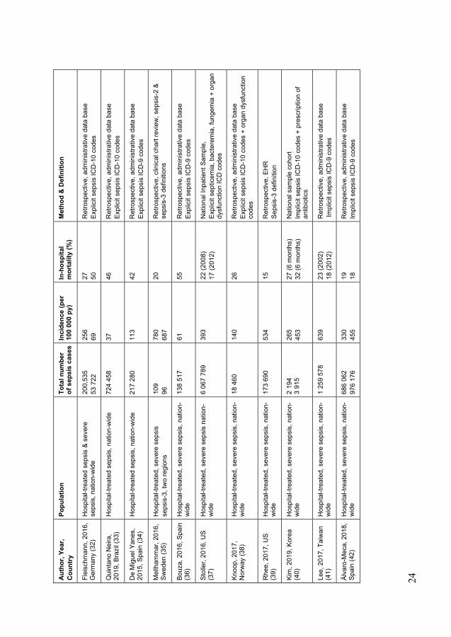
24
Aut
hor,
Year
, C
ount
ry
Popu
latio
nTo
tal n
umbe
r of
sep
sis
case
s In
cide
nce
(per
10
0 00
0 py
) In
-hos
pita
l m
orta
lity
(%)
Met
hod
& D
efin
ition
Flei
schm
ann,
201
6,
Ger
man
y (3
2)
Hos
pita
l-tre
ated
sep
sis
& se
vere
se
psis
, nat
ion-
wid
e 20
0,53
5 53
722
25
6 69
27
50
R
etro
spec
tive,
adm
inis
trativ
e da
ta b
ase
Expl
icit
seps
is IC
D-1
0 co
des
Qui
ntan
o N
eira
, 20
19, B
razi
l (33
) H
ospi
tal-t
reat
ed s
epsi
s, n
atio
n-w
ide
72
4 45
8 37
46
R
etro
spec
tive,
adm
inis
trativ
e da
ta b
ase
Expl
icit
seps
is IC
D-1
0 co
des
De
Mig
uel Y
anes
, 20
15, S
pain
(34)
H
ospi
tal-t
reat
ed s
epsi
s, n
atio
n-w
ide
21
7 28
0 11
3 42
R
etro
spec
tive,
adm
inis
trativ
e da
ta b
ase
Expl
icit
seps
is IC
D-9
cod
es
Mel
lham
mar
, 201
6,
Swed
en (3
5)
Hos
pita
l-tre
ated
, sev
ere
seps
is
seps
is-3
, tw
o re
gion
s
109
96
780
687
20
Ret
rosp
ectiv
e, c
linic
al c
hart
revi
ew, s
epsi
s-2
& se
psis
-3 d
efin
ition
s
Bouz
a, 2
016,
Spa
in
(36)
H
ospi
tal-t
reat
ed, s
ever
e se
psis
, nat
ion-
wid
e
138
517
61
55
Ret
rosp
ectiv
e, a
dmin
istra
tive
data
bas
e
Expl
icit
seps
is IC
D-9
cod
es
Stol
ler,
2016
, US
(37)
H
ospi
tal-t
reat
ed, s
ever
e se
psis
nat
ion-
wid
e 6
067
789
393
22 (2
008)
17
(201
2)
Nat
iona
l Inp
atie
nt S
ampl
e,
Expl
icit
sept
icem
ia, b
acte
rem
ia, f
unge
mia
+ o
rgan
dy
sfun
ctio
n IC
D c
odes
Knoo
p, 2
017,
N
orw
ay (3
8)
Hos
pita
l-tre
ated
, sev
ere
seps
is, n
atio
n-w
ide
18 4
60
140
26
Ret
rosp
ectiv
e, a
dmin
istra
tive
data
bas
e
Expl
icit
seps
is IC
D-1
0 co
des
+ or
gan
dysf
unct
ion
code
s
Rhe
e, 2
017,
US
(39)
H
ospi
tal-t
reat
ed, s
ever
e se
psis
, nat
ion-
wid
e 17
3 69
0 53
4 15
R
etro
spec
tive,
EH
R
Seps
is-3
def
initi
on
Kim
, 201
9, K
orea
(4
0)
Hos
pita
l-tre
ated
, sev
ere
seps
is, n
atio
n-w
ide
2 19
4 3
915
265
453
27 (6
mon
ths)
32
(6 m
onth
s)
Nat
iona
l sam
ple
coho
rt Im
plic
it se
psis
ICD
-10
code
s +
pres
crip
tion
of
antib
iotic
s
Lee,
201
7, T
aiw
an
(41)
H
ospi
tal-t
reat
ed, s
ever
e se
psis
, nat
ion-
wid
e 1
259
578
639
23 (2
002)
18
(201
2)
Ret
rosp
ectiv
e, a
dmin
istra
tive
data
bas
e Im
plic
it se
psis
ICD
-9 c
odes
Álva
ro-M
eca,
201
8,
Spai
n (4
2)
Hos
pita
l-tre
ated
, sev
ere
seps
is, n
atio
n-w
ide
686
062
976
176
330
455
19
18
Ret
rosp
ectiv
e, a
dmin
istra
tive
data
bas
e
Impl
icit
seps
is IC
D-9
cod
es

25
Hug
gan,
201
9, N
ew
Zeal
and
(43)
H
ospi
tal-t
reat
ed, s
ever
e se
psis
, one
re
gion
1
643
82
19
Ret
rosp
ectiv
e, a
dmin
istra
tive
data
bas
e
Impl
icit
seps
is IC
D-1
0 co
des
Goo
dwin
, 201
6, U
S (4
4)
Hos
pita
l-tre
ated
, sev
ere
seps
is, s
tate
-w
ide
24 3
95
717
18
Ret
rosp
ectiv
e, a
dmin
istra
tive
data
bas
e
Expl
icit
seps
is IC
D-9
cod
es
Dup
uis
2017
, Fr
ance
(45)
H
ospi
tal-t
reat
ed, s
eptic
sho
ck, n
atio
n-w
ide
421
699
(sep
tic
shoc
k on
ly)
136
(sep
tic s
hock
on
ly)
40 (s
eptic
sho
ck
only
) R
etro
spec
tive,
adm
inis
trativ
e da
ta b
ase
ICD
-10
sept
ic s
hock
cod
es o
r vas
opre
ssor
use
+
infe
ctio
n co
des
De
Mig
uel Y
anes
, 20
15, S
pain
(34)
H
ospi
tal-t
reat
ed, s
eptic
sho
ck, n
atio
n-w
ide
88 0
92 (s
eptic
sh
ock
only
) 46
(sep
tic s
hock
on
ly)
52 (s
eptic
sho
ck
only
) R
etro
spec
tive,
adm
inis
trativ
e da
ta b
ase
Expl
icit
sept
ic s
hock
ICD
-9 c
odes
Lore
ncio
, 201
8,
Spai
n (4
6)
Hos
pita
l-tre
ated
, sev
ere
seps
is, o
ne
regi
on
224
396
(all
year
s)
160
(200
5)
390
(200
16)
26
17
Ret
rosp
ectiv
e, a
dmin
istra
tive
data
bas
e
Impl
icit
seps
is IC
D-9
cod
es
Qui
ntan
o N
eira
, 20
19, B
razi
l (33
) IC
U-tr
eate
d se
psis
, nat
ion-
wid
e 21
0 81
7 11
65
R
etro
spec
tive,
adm
inis
trativ
e da
ta b
ase.
Ex
plic
it se
psis
ICD
-10
code
s
Nza
rora
, 201
6,
Rw
anda
(47)
IC
U-tr
eate
d se
psis
& s
ever
e se
psis
, tw
o st
udy
cent
res
220
396
2 3 71
65
Pr
ospe
ctiv
e co
hort
stud
y Se
psis
-1 d
efin
ition
Her
ran-
Mon
ge,
2017
, Spa
in (1
8)
ICU
-trea
ted,
sev
ere
seps
is, m
ulti
cent
re
231
31
37
Pros
pect
ive,
obs
erva
tiona
l stu
dy
Seps
is-2
def
initi
on
Kübl
er, 2
015,
Po
land
(48)
IC
U-tr
eate
d, s
ever
e se
psis
, mul
ti ce
ntre
36
4 19
1 69
(201
2)
60 (2
013)
-
Pros
pect
ive
surv
ey, s
urvi
ving
sep
sis
cam
paig
n gu
idel
ines
(Del
linge
r 200
8)
Mac
hado
, 201
7,
Braz
il (4
9)
ICU
-trea
ted,
sev
ere
seps
is, m
ulti
cent
re
794
290
56
Pros
pect
ive
obse
rvat
iona
l stu
dy
Seps
is-1
def
initi
on
Bertu
llo, 2
016,
U
rugu
ay (5
0)
ICU
-trea
ted,
sev
ere
seps
is, m
ulti
cent
re
153
19
55
Pros
pect
ive
obse
rvat
iona
l stu
dy
Seps
is-1
def
initi
on
Azká
rate
, 201
5,
Spai
n (5
1)
ICU
-trea
ted,
sev
ere
seps
is, s
ingl
e ce
ntre
1
136
2718
Pros
pect
ive
obse
rvat
iona
l stu
dy
Seps
is-2
def
initi
on
Alm
irall,
201
6,
Spai
n (5
2)
ICU
-trea
ted,
com
mun
ity-a
cqui
red,
se
vere
sep
sis,
sin
gle-
cent
er
917
52
19
.7
Pros
pect
ive
obse
rvat
iona
l stu
dy
Seps
is d
efin
ition
not
spe
cifie
d

26
Aut
hor,
Year
, C
ount
ry
Popu
latio
nTo
tal n
umbe
r of
sep
sis
case
s In
cide
nce
(per
10
0 00
0 py
) In
-hos
pita
l m
orta
lity
(%)
Met
hod
& D
efin
ition
Rhe
e, 2
017,
US
(39)
IC
U-tr
eate
d, s
ever
e se
psis
, nat
ion-
wid
e 94
956
29
2-
Ret
rosp
ectiv
e, E
HR
Seps
i s-3
def
initi
on
Flei
schm
ann-
Stru
zek,
201
8,
Ger
man
y (3
0)
ICU
-trea
ted
seps
is &
sev
ere
seps
is
natio
n-w
ide
76 5
57
104
705
49 5
84
73 4
19
94 (2
010)
12
7 (2
015)
61
(201
0)
86 (2
015)
- - 49
45
Ret
rosp
ectiv
e, a
dmin
istra
tive
data
bas
e Ex
plic
it se
psis
ICD
-10
code
s
Flei
schm
ann-
Stru
zek,
201
8,
Ger
man
y (3
0)
ICU
-trea
ted,
sev
ere
seps
is n
atio
n-w
ide
19
7 95
6 28
9 18
3 24
2 (2
010)
35
2 (2
015)
- -
Ret
rosp
ectiv
e, a
dmin
istra
tive
data
bas
e Im
plic
it se
psis
ICD
-10
code
s
Shan
kar-H
ari,
2017
, U
K (5
3)
ICU
-trea
ted
seps
is &
sev
ere
seps
is
natio
n-w
ide
197
724
197
142
102
102
31
32
Ret
rosp
ectiv
e, IC
U d
ata
base
Se
psis
-2 &
sep
sis-
3 de
finiti
ons
Zhou
, 201
7, C
hina
(5
4)
ICU
-trea
ted
seps
is &
sev
ere
seps
is
inci
denc
e, o
ne s
ubdi
stric
t 23
7 19
1 92
74
-
Ret
rosp
ectiv
e, c
linic
al c
hart
revi
ew
Seps
is-1
def
initi
on
Kim
, 201
9, K
orea
(4
0)
ICU
-trea
ted,
sev
ere
seps
is, n
atio
n-w
ide
747
1 20
8 91
(200
5)
140
(201
2)
- R
etro
spec
tive,
nat
iona
l dat
a ba
se. I
mpl
icit
seps
is
ICD
-10
code
s +
pres
crip
tion
of a
ntib
iotic
s
Yebe
nes,
201
7,
Spai
n (5
5)
ICU
-trea
ted,
sev
ere
seps
is, o
ne re
gion
23
236
61
R
etro
spec
tive,
adm
inis
trativ
e da
ta b
ase
Im
plic
it se
psis
ICD
-10
code
s
Hug
gan,
201
9, N
ew
Zeal
and
(43)
IC
U-tr
eate
d, s
ever
e se
psis
, one
regi
on
278
14
34
Ret
rosp
ectiv
e, a
dmin
istra
tive
data
bas
e
Impl
icit
seps
is IC
D-1
0 co
des
py =
per
son
year
s

27
Figure 1. Summary of sepsis mortality, from population-based studies on sepsis epidemiology in adults 2015-2019.

28

29
The long-term effects
Irrespective of chosen method for incidence and mortality estimate, there is a large number of sepsis survivors. Rudd et al assessed the number to 38 million persons each year (25).
From a large cohort of nationally sampled persons in the U.S. Iwashyna et al pulled out the subset which had been hospitalized for sepsis and demonstrated that new and persistent disability was common. Half of those reported with disability had not been treated in an ICU for the sepsis episode. Among disabilities demonstrated was a 3.5-fold increase in moderate to severe cognitive impairment. On average the persons had 1.5 new limitations in functional disability, such as inabilities to cook, manage toilet visits, dressing or bathing (56). A common approach for assessing long-term effects has been point-prevalence studies within a certain time interval following critical illness. These studies have shown high prevalence of mental illness including anxiety (30-40%), depression (28-34%) and post-traumatic stress disorder (36-42%) (57-59). Even though these studies do not have a baseline, prevalence is evidently higher than population norms. In a longitudinal cohort, Davydow et al found exactly the same prevalence of mental illness before and after sepsis (60). It is possible that mental illness is more common among people vulnerable to sepsis. Either way, mental illness is common and needs to be addressed following sepsis in order to promote recovery (61).
Forty percent of sepsis patients are re-hospitalized in the next 90 days. The most common reasons are recurrent sepsis followed by aspiration pneumonia, acute renal failure and cardiovascular events (62, 63). In a propensity score matched cohort, Prescott et al found 20% of sepsis patients to suffer from an attributable death the next two years as a result of sepsis (64).
The theoretical model for long-term effects of sepsis is that the patients do not return to homeostasis after sepsis but have an ongoing inflammation or immune suppression (65, 66).
Animal studies have demonstrated post-sepsis mice to be at increased risk of infection, to have an accelerated atherosclerotic disorder resulting in cardiovascular disease and an accelerated tumour growth causing cancer (67-69). However, after a systematic review and meta-analysis of sepsis and long-term mortality Shankar-Hari et al stated that epidemiological criteria for a causal relationship between sepsis and post-acute mortality is not consistently observed (70).

30
The long-term effects are complex interactions of risk factors for sepsis, sepsis treatment and, critical illness. One can conclude that there is a need to disentangle the effects of individual aspects of disease and treatment and to characterize and predict trajectories of recovery.

31
The pathophysiology
When a microorganism enters the body, pathogen-associated molecular patterns (PAMP) are recognized by pattern recognition receptors (PRR) and both are involved in activating the immune system (71). PAMPs are evolutionary conserved molecular structures expressed by different pathogens, recognized by the different PRRs that contribute to the induction of the profound dysregulated host response in sepsis. The PRRs also recognize damage-associated molecular patterns (DAMP), which are host molecules released from injured cells (72). The interaction between PRRs and their ligands leads to signalling cascades and release of various cytokines. This release will induce further cytokine production similar to a cytokine storm of pro- and anti-inflammatory cytokines of different categories such as tumour necrosis factor (TNF), interferons, growth factors and interleukins (73). In sepsis the host response fails to return to homeostasis in a complex way with components like excessive inflammation and immune suppression. Figure 2 (65, 74). The individual host response in sepsis is for example related to pathogen, focus of infection, immune status, epigenetic control of gene transcription and polymorphism in sepsis-associated genes (75, 76).
Figure 2. Schematic picture of pro- and antiinflammation in sepsis. Red = Inflammatory response, blue= antiinflammatory response
32
Sepsis is manifested as organ dysfunction. The multiple signal ways involved in transmitting the dysregulated host response through extraordinarily complex trajectories are not fully understood. Neutrophils activated in response to infection release neutrophil extracellular traps (NETs). NETs are web-like formations of extracellular DNA with histones, myeloperoxidase and elastase, involved in pathogen clearance, coagulation and vascular inflammation (77). Heparin-binding protein (HBP), also known as azurocidin or CAP37, is stored in neutrophils and is released from secretory vesicles and azurophilic granules of neutrophils after contact with bacterial products or neutrophil adhesion and can be augmented by the presence of specific antibodies towards bacterial structures (78-80). HBP binds to endothelial cells and is involved in the increase of vascular permeability, one of the hallmarks in sepsis (81). There are profound alterations of the endothelium in sepsis, including disruption of the endothelial barrier by the loss of glycocalyx, intercellular adhesions and other supportive molecules. The endothelial dysfunction contributes to vasodilation and tissue oedema which jointly with vasopressin deficiency, paradoxical downregulation of vasoconstrictive receptors, increase in nitric oxide and smooth muscle cell relaxation, contribute to arterial and further venous vasodilation (82). This reduces the pressure gradient, decreased venous return and cardiac output. Also, mitochondrial dysfunction, inflammation-induced cardiac dysfunction and impaired chronotropic response aggravate the cardiovascular dysfunction (83). Micro- and macrocirculation impairment contribute to global hypoperfusion and subsequent organ dysfunction. This is further exacerbated by occlusion of tissue beds due to platelet aggregation and thrombus formation. For example, hypoperfusion, inflammatory signals and oxidative stress can cause acute tubular damage in the kidneys. In the liver hypoxia during sepsis contributes to dysfunction. Hypoperfusion in the central nervous system causes dysfunction together with oxidative stress of mediators that diffuse through an intact blood-brain barrier. Furthermore, the inflammatory response can disrupt the blood-brain barrier and concomitant hepatic and renal dysfunction increase the level of toxins affecting the brain (84). Alveolar injury and increased pulmonary vascular permeability lead to an increased pulmonary dead space, impaired gas exchange, hypoxemia and hypercapnia, eventually causing acute respiratory distress syndrome (ARDS) (85). Epithelial barrier function is involved in the altered function of the gut. The increased permeability allows bacterial translocation and further gut injury due to effect of activated pancreatic enzymes (86, 87). Inflammation and coagulation are strongly linked and various coagulation pathways can be activated. A strong activation of the coagulation system can eventually lead to disseminated intravascular coagulation (DIC) and subsequent depletion of circulating platelets resulting in thrombocytopenia (88).

33
The therapy
Ever since Kumar et al published a study, showing that delay in commencing antimicrobials after the onset of hypotension during sepsis increased mortality by 7.6% by each hour for the first 6 hours, the effect of early antibiotics has been under debate (89). Increased survival with early antibiotic administration at least in patients with sepsis and hypotension has been demonstrated in several observational studies, yet not in others (90-92). A large meta-analysis concluded that there may be increased odds of death among patients reaching treatment after >3 hours, but this did not reach significance (93). There are no in-hospital randomized controlled trials, although in a Dutch study patients were randomized to receive antibiotics prehospitally without improved survival (94). Time-to-intervention is a complex variable confounded by many factors. For example, treatment is often delayed in more complicated patients and often given sooner in patients with more severe disease. The timing of an intervention often correlates with the timing of other interventions and also the onset of the disease is difficult to discriminate. Furthermore, studies are confounded by antibiotic therapy being inappropriate (95). Several studies have confirmed the connection between inappropriate antibiotic therapy and higher mortality and organ dysfunction (96, 97).
Clinical practice guidelines often rely on the Surviving Sepsis Campaign (SSC) which at present recommends that administration of intravenous (IV) empiric broad-spectrum antimicrobials are initiated as soon as possible after recognition and within 1 h for both sepsis and septic shock (98, 99). Source control, (the drainage of abscesses and removal of infected tissue or device) is prompted when required (100, 101). Oxygen is delivered, although higher arterial oxygen saturation, seems to be associated with higher mortality compared to a lower target (98, 102). For patients with hypotension, SSC recommends 30 mL/kg crystalloid fluid promptly initiated within the first hour after presentation. Neither this recommendation is uncontroversial, as there are risks possibly associated with under- and over-resuscitation (98, 99). Numerous studies have compared crystalloid fluids to colloids for sepsis resuscitation, without finding advantages for colloid fluids (103). If colloids are required, albumin is at present the drug of choice (104, 105). In order to maintain adequate organ perfusion, vasopressors are applied for patients with hypotension during or after fluid resuscitation (106). Norepinephrine is preferred since it has higher potency and less severe side effects (107). However, septic patients can be non-responders to some vasoactive drugs. Therefore, Chawla et al

34
suggested to start with multiple vasopressors of different mechanisms of action and then de-escalate after response - an approach similar to the use of broad-spectrum antibiotics in sepsis treatment (108).
In 2001, Rivers et al managed to reduce hospital mortality in sepsis by 16% with early goal-directed therapy (109). The algorithm used by Rivers was widely adopted into guidelines and practice. In more recent trials, these advantages have not been possible to demonstrate (110). Studies have compared early goal-directed therapy to usual care, with the possibility that advances in usual care blur the distinctions found by Rivers (110).
Steroids, partly as an anti-inflammatory strategy, have not been convincingly demonstrated to reduce mortality but have important secondary effects such as more rapid resolution of shock and earlier time to ICU discharge. Because of the ambiguous results and possible risk of harmful effects on patient outcome, guidelines only recommended steroids for patients who do not respond to fluid resuscitation and vasopressors (98, 111).
Evidently, there is no specific treatment for the sepsis reaction. Most pathophysiological pathways have been tested for modulation - thrombomodulin, activated protein C, interferon- , anti-TNF antibodies, TNF receptors, inhibitors of Toll-like receptor 4, platelet activating factor antagonists, complement inhibitors, ibuprofen, heparin, immunoglobulins and endotoxin removal - all with negative results in clinical studies (112). Part of all these negative results might be due to the heterogeneity in sepsis and targeted therapy can perhaps be effective when applied with personalized medicine, possibly with multiple action therapies. One example of how to accomplish this is with theranostics, i.e. biomarker directed therapy. Other unresolved issues are the treatment window and how to restore immune homeostasis. An endotype or subphenotype is a subset of a patient population defined by observable characteristics, distinguished from the population as a whole by natural history, disease manifestation and/or response to treatment (113). The identification of subphenotypes is well-established in asthma and cancer treatment. In sepsis, one has to establish subphenotypes without an exact definition. Ways to handle this problem are by using supervised and unsupervised clustering and machine learning.

35
Bacteremia
Bacteremia, i.e. bacterial presence in the blood stream, is often a result of pathogens seeding the blood from a focal source of infection, although sometimes the infectious focus is not identifiable. Bacteremia and its persistence is also an effect of clearance and asymtomatic, transient bacteremia is common in daily activities such as tooth brushing (114, 115). Population-based incidence for bacteremia is approximately 200 per 100 000 person years, with the most common isolates being E.coli and S.aureus (116-118). Bacteremia is detected in 15-30% of the sepsis patients but the opposite relationship is less reported (13, 22, 35, 119, 120). In a cohort of bacteremic patients in an ED 23-39% had septic shock (sepsis-2) with higher proportions of septic shock among older adults (121). There is a low bacterial density in bacteremia, presumably often as low as one colony forming unit (CFU)/mL. Consequently, the likelihood to sample at least one colony forming bacterium depends on the total sample volume, which is often insufficient (119, 122-125). It is further hampered by antimicrobial therapy given prior to sampling (126).
In clinical practice, bacteremia is diagnosed via blood culture. Blood culture was developed in the beginning of the 20th century and even though it has been elaborated with for example, continuous microbial detection the general method remains largely the same. Typically, sampling is followed by the time duration for transport and preparation, upon which incubation starts. Incubation takes 6-120 h followed by a conventional workflow of up to 48 more hours. This process can be shortened by molecular workflow like matrix-assisted laser desorption/ionization time-of-flight mass spectrometry (MALDI-TOF MS), peptid nucleic acid fluorescent in situ hybridization (PNA-FISH), microarray and polymerase chain reaction (PCR). However, most molecular methods are used on positive blood cultures due to the combination of low bacterial load in whole blood and a lack of sensitivity. Therefore, most molecular methods depend on the incubation of blood cultures, although shorter incubation is possible for MALDI-TOF MS, and need to be followed by sample preparation (127). A few methods can be applied directly on whole blood for instance, magnetic resonance-based diagnostics and PCR-methods that are dealing with the challenges of human DNA in blood and other components that interfere with PCR. These methods have a turnaround time of 3-6 hours but have had difficulties in achieving satisfying sensitivities (128, 129). There are approaches sequencing all bacteria in a sample out of the 16S-rDNA gene, still DNA

36
from a diversified microbiome exists in healthy blood donors and needs to be distinguished (130). Diagnosing bacteremia is important for several reasons; to facilitate diagnosing infection, for pathogen and resistance detection aiding tailored therapy, for rapid de-escalation of the generous use of broad-spectrum antimicrobials in sepsis and for its association with morbidity and mortality (131, 132). There is an unmet clinical need for a shorter time to positivity in diagnosing bacteremia since the preliminary and definite microbial results tend to arrive too late to influence clinical decisions in acute sepsis care.

37
The acoustofluidics
Microfluidics is the technology of systems that can handle small volumes of fluids in channels, which changes the flow properties from macrofluidics. In medical diagnostics microfluidics are often applied in a microchip. When used for sample preparation, it can be integrated with a detection into a miniaturized platform referred to as Lab-on-a-chip (LOC). Microfluidic techniques could be a mean of preparation for isolating bacteria from whole blood for rapid sample preparation. Microfluidic techniques in LOC platforms has the ability to provide automated, rapid diagnostics. Different microfluidic methods for separation of bacteria have been reported without achieving both a reasonable process time and satisfying bacteria recovery (133, 134).
Acoustophoresis means migration with sound. With ultrasound over microfluidic channels, acoustofluidics, one can move components which can be applied for cell sorting. Blood cells have been successfully removed from plasma by acoustofluidic devices using acoustic, size-based sorting (135, 136). Acoustofluidic sample preparation might be possible for bacterial enrichment and DNA purification, which includes removal of human DNA and PCR inhibitors.

38

39
Prediction and risk stratification scores
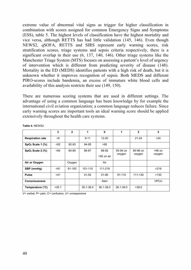
Patients with acute deterioration have clinical signs several hours before, while many ED patients develop sepsis within the first days after admittance but do not have organ dysfunction at presentation (137, 138). Various scores and systems have been developed to improve detection of patients with or at risk of clinical deterioration and to facilitate timely and effective clinical response. Early warning scores (EWS) was published in the mid 1990-ies, followed by modified version (MEWS) and numerous other EWS. They all aimed to help identifying patients at risk of deterioration, including sepsis as parts of “track-and-trigger” systems (139). A uniform system was developed out of expert review of different EWS and National Early Warning Score (NEWS) was applied in the U.K. Since then it has been reviewed and at present NEWS2 is used in the wards as well as in acute and ambulance settings for assessment of acute illness severity, detection of clinical deterioration and initiation of a timely and competent response, table 4 (140). Its predecessors have been validated for sepsis but not yet NEWS2 (141).
Although SIRS was a clinical criterion for sepsis, it has been helpful in early recognition of sepsis, table 1 (4). As a clinical criterion it has proven a lack of sensitivity as well as specificity but as an early warning score the sensitivity has been superior to other warning scores but on behalf of the specificity (137, 142).
QuickSOFA (qSOFA) was derived out of multiple logistic regression of candidate variables for in-hospital mortality, with the Bayesian criterion selection. The Bayesian criterion selection only retains the variables with the strongest improvement for the model i.e. parsimony was prioritized. qSOFA was launched along with the sepsis-3 definitions as a severity score among patients with a suspected infection. It assigns one point for each of three variables: altered mental status, systolic blood pressure (SBP) 100 mmHg and respiratory rate ≥22 breaths per minute. Patients with a score of 2 points or higher have a higher risk of in-hospital mortality (143). Validation studies have consistently shown high specificity but low sensitivity (137, 144, 145).
Rapid Emergency Triage and Treatment System (RETTS) was designed to find critically ill patients and those at risk of deterioration at admission and during ED stay. It is widely used for triage at EDs in Sweden (146, 147). RETTS uses one
40
extreme value of abnormal vital signs as trigger for higher classification in combination with scores assigned for common Emergency Signs and Symptoms (ESS), table 5. The highest levels of classification have the highest mortality and vice versa, although RETTS has had little validation (145, 146). Even though NEWS2, qSOFA, RETTS and SIRS represent early warning scores, risk stratification scores, triage systems and sepsis criteria respectively, there is a significant overlap in their use (6, 137, 140, 146). Other triage systems like the Manchester Triage System (MTS) focuses on assessing a patient’s level of urgency of intervention which is different from predicting severity of disease (148). Mortality in the ED (MEDS) identifies patients with a high risk of death, but it is unknown whether it improves recognition of sepsis. Both MEDS and different PIRO-scores include bandemia, an excess of immature white blood cells and availability of this analysis restricts their use (149, 150).
There are numerous scoring systems that are used in different settings. The advantage of using a common language has been knowledge by for example the international civil aviation organization; a common language reduces failure. Since early warning scores are important tools an ideal warning score should be applied extensively throughout the health care systems.
Table 4. NEWS2.
3 2 1 0 1 2 3
Respiration rate <9 9-11 12-20 21-24 >24
SpO2 Scale 1 (%) <92 92-93 94-95 >95
SpO2 Scale 2 (%) <84 84-85 86-87 88-92
>92 on air
93-94 on oxygen
95-96 on oxygen
>96 on oxygen
Air or Oxygen Oxygen Air
SBP (mmHg) <91 91-100 101-110 111-219 >219
Pulse <41 41-50 51-90 91-110 111-130 >130
Consciousness Alert VPCU
Temperature (°C) <35.1 35.1-36.0 36.1-38.0 38.1-39.0 >39.0
V= verbal, P= pain, C= confusion, U= unresponsive

41
Table 5. RETTS.
Red Orange Yellow Green
A Blocked airway or stridor
B Respiratory rate >30 or <8
SaO2<90 with oxygen (O2)
Respiratory rate >25
SaO2<90 without O2
SaO2 95 SaO2>95 without O2
C Heart rate >130 if sinus rythm, else >150
SBP <90
Heart rate >120 or <40 Heart rate >110 or <50 Heart rate 50-110
D Unconscious or cramps Somnolence Acute disorientation Alert
E Temperature >41 Temperature >38.5

42

43
Aims
The overall aim of the work presented in this thesis was to improve prediction, diagnostics and knowledge on epidemiology of sepsis.
Put into specific terms the aims were:
• To develop an integrated, dry-reagent based, disposable ready-to-usecartridge enabling rapid analysis of sepsis-causing organisms directly fromsuspected blood samples.
• To evaluate the performance of the integrated cartridge with clinical bloodsamples.
• To assess the incidence of hospital-treated sepsis in an entire populationbased on clinical findings.
• To compare the diagnostic accuracy of qSOFA, NEWS2 and RETTS forsepsis.
• To investigate whether plasma levels of HBP or lactate can improve theaccuracies for qSOFA or NEWS2 .
• To develop and evaluate a risk stratification score based on the mostpredictive, minimal set of vital signs, HBP and lactate plasma levels.
• To describe characteristics and outcome for patients with sepsis-3 admittedto the ICU with bacteremia, pathogen-detected but non-bacteremia, and for“sterile” sepsis.
• To identify subphenotypes of sepsis with a novel statistic approach.

44

45
Paper I
Materials and methods
The integrated system for pathogen detection consisted of an acoustophoretic separation chip which removed blood cells, followed by an acoustic trap for bacteria enrichment and a PCR chip for detection. Figure 3. The acoustophoretic separation chip consisted of a channel with a trifurcation at the end and a piezoelectrically induced, ultrasonic wave across. The frequency of the ultrasound was tuned so that the thickness of the capillary corresponds to half a wavelength. This causes a standing wave, which results in an acoustic force that attracts larger particles to the wave node. Blood cells were focused at the centre of the channel and removed from the centre of the trifurcation. Bacteria were suspended in the plasma and channelled to the acoustic trap. Bacteria are too small in itself to be captured by the acoustic forces, but can be trapped by secondary forces if larger particles are placed in the trap. We used polystyrene beads. The secondary forces are the result of scattering of sound between particles. Trapped bacteria were then washed and released into a PCR microchip which was placed in a PCR thermocycling. Four different PCR microchips were used for detection of Pseudomonas spp, S.aureus, E.coli and S.pneumoniae.
The different parts of the integrated system were tested for acoustic separation, bacteria enrichment, the detection limits for the PCR chips and sterilization. The integrated system was tested for blood samples spiked with bacteria and finally, for clinical samples from patients with suspected sepsis or febrile neutropenia. Patients were included prospectively in an observational study for comparison of the integrated system for bacteria detection to blood culture. Study samples were drawn at the same time as blood culture and analyzed in a blinded fashion.

46
Figure 3. The integrated microfluidic system. Modified and reprinted with permission from reference (151). Copyright © 2018, Springer Nature
Results Red blood cells were efficiently removed by the acoustic separation, but the integrated system enriched bacteria at a somewhat too low efficacy. The detection limits for the PCR chips were 3, 2, 400 and 100 CFU, respectively for S.aureus, S.pneumoniae, E.coli and Pseudomonas spp. In the spiked samples, the integrated system could detect 1000 P.putida /mL blood. The integrated process was completed within 2 hours.
In clinical samples, the integrated system detected E.coli in 2 out of 57 processed samples of which E.coli was retrieved in 4 blood cultures. S.aureus was retrieved in 3 blood cultures but none were detected by the integrated system. No S.pneumoniae positive cultures were found. The median incubation time to the first positive blood culture signal was 8.2 h. The acoustofluidic system detected 2 of the E.coli positive blood samples within 2 h. The samples where E.coli were detected, were the ones with the shortest incubation of blood cultures before positivity, which may be suggestive of high bacterial density.
Discussion This study provided a proof-of-principle for the novel, integrated, acoustophoresis-based bacteria detection system in blood samples from patients with suspected sepsis.
The integrated system can detect bacteria but not at sufficient rates at present execution and needs to be further optimized. The sensitivity of the integrated system depends on separation, trapping and detection. There were additional problems

47
processing the clinical samples; with platelets tending to attach to the capillary walls and higher sedimentation rates of red blood cells. Furthermore, there were difficulties to separate platelets from bacteria due to platelets being in the ranges of bacterial size and density. Possibly, bacteria were lost at the separation step because of bacterial clusters acting like larger particles, phagocytized bacteria and bacteria adhered to blood cells. One approach might be selective lysis of blood cells followed by trapping, other means could be to process larger sample volumes as well as detection of lower number of bacteria, which also should include broader range of pathogen detection.
The novel, integrated acoustophoresis-based bacteria detection in blood samples from patients with suspected sepsis will have to be improved, but a more rapid pathogen detection in sepsis with this new approach might be possible.

48

49
Paper II
Materials and methods The study was a clinical chart review of all patients 18 years in the regions of Skåne and Halland, who were started on IV antibiotic treatment on 4 dates evenly distributed over the year of 2015. Patients were included if they had at least one SIRS criteria or had experienced fever, chills, had a blood pressure of 110 mmHg or less or an altered mental status within ±12 h from the initiation of antibiotic therapy. Data were collected regarding demography, comorbid conditions, health care, vital signs and diagnostic results. Sepsis was defined as modifications of the sepsis-2 and sepsis-3 definitions and infection as modifications of the definitions presented by Calandra (4-6, 152). Population data were retrieved from Statistics Sweden. Descriptive statistics were used for characteristics and demographics of the patients included in the study.
Results 563 patients were started on IV antibiotic treatment on the four days of the survey. 482 patients were included in the study, of those 339 had a diagnosed infection. 74 patients had sepsis according to both definitions (severe sepsis and sepsis-3), additionally 35 patients had only sepsis-3 and 22 patients had only severe sepsis. The mean adult population in the regions for 2015 was 1 275 753. Thus, the annual incidence for patients treated with IV antibiotics for a diagnosed infection was 2425/100 000 person years (95% CI 2167-2683), the annual incidence for severe sepsis was 687/100 000 person years (95% CI 549-824) and the annual incidence for sepsis-3 was 780/100 000 person years (95% CI 633-926). The incidences increased with 10-year age strata but the numbers were too small to calculate age-adjusted estimates. The most common focus of infection was the respiratory tract followed by the urinary tract, the most common pathogens retrieved in blood cultures were Enterobacterales followed by S.aureus.

50
Discussion The incidence of sepsis was higher compared to most previous estimates and thus responsible for a larger burden of morbidity in society and in health care systems. There may be an increase in sepsis due to for example, increased awareness and sensitivity of diagnosis, an increasing number of immunocompromised patients, the use of invasive procedures, the emerging antimicrobial resistant microorganisms and the growth of the elderly population. More probable is that the high incidence is due to the methodology. Instead of using ICD-codes widely used in epidemiological sepsis studies, we used a labour-intensive method examining every clinical chart for admitted, adult patients started on IV antibiotic therapy on four dates in an entire population. We believe that the study design has contributed to the estimates being closer to the true burden of sepsis in Sweden. Furthermore, Sweden is a high-income country with free healthcare services accessible to everyone, which also contributes to a more accurate estimate of the true incidence of sepsis in similar countries. Still, the study population might not be a close representation of the national population since it was only conducted in two regions and had a small sample size. To rely on syndromic, clinical case definitions will likely introduce some misclassification errors.
At the time the study was performed, population-based sepsis incidence estimates from clinical data were rare. Since then, several studies on sepsis incidence based on clinical data have been published, which found similar incidence rates of sepsis in the Nordic countries (20, 22). A large Chinese study found annual incidences of 103/100 000 person years and 236/100 000 person years of severe sepsis and sepsis-3, respectively (21, 31). A prospective epidemiological study is the ideal method and should be encouraged since it might come even closer to the true sepsis burden. Nevertheless, prospective, comprehensive, population-based epidemiological studies are difficult to carry out. In order to obtain reliable, comparable population-level estimates clinical chart review is a feasible method and lately increasingly referred to as “gold standard”(13, 17, 153). Our study design can be used as a study template for studies examining the epidemiology of sepsis.
The introduction of new sepsis definitions necessitates comparison of epidemiological estimates in order to be able to use knowledge from previous studies. Clinicians need local epidemiological data for clinical decisions, for diagnostics and therapeutics when handling medical patients with acute infectious diseases. Apart from improving the care, knowledge on epidemiology is of importance for effective use of health resources.

51
Paper III & IV
Materials and methods We performed a retrospective analysis of data from two prospective, observational, multicentre, convenience trials of sepsis biomarkers at EDs. One of the cohorts consisted of patients diagnosed with an infection, cohort A. The second cohort consisted of mixed ED patients (with and without infection), cohort B.
We used a composite outcome for sepsis consisting of sepsis with organ dysfunction (sepsis-2), infection-related mortality within <72 h, or intensive care due to an infection.
Accuracy of risk stratification scores was evaluated for sepsis using area under receiver operating characteristic curve (AUC), sensitivity, specificity and their 95% CIs. AUCs were compared with DeLong’s test and proportions with Chi2-test. For 30-day mortality, odds ratio (OR) was calculated.
A locally weighted scatterplot smoothing (LOWESS) regression for eligible parameters with reference to sepsis generated smooth curves for selection of intervals for parameters. Selected parameters were dichotomized within the selected intervals and included in a least absolute shrinkage and selector operator (LASSO) model for construction of a risk stratification score. The LASSO method avoids correlating covariates from being included in a prediction model (154, 155). The LASSO included a 10-fold cross-validation with AUC optimization and was iterated 50 times. All values optimizing AUC in more than 50% of the LASSO analyses and with a coefficient ≥0.05 were then entered into a second set of LASSO regressions, unless the values were adjacent. If adjacent values, the one with the higher coefficient was chosen. Values included in more than 50% of the second set of LASSO regressions and with a coefficient of ≥0.05 within 1 standard error (SE) from max AUC were selected. These values were given a score proportional to their coefficients generated by the second set of LASSO regression and rounded to the closest integer. The cut-offs for these scores were set to also require scores from more than one parameter.

52
Results Cohort A consisted of 526 patients with a diagnosed infection, 288 with the composite outcome. Cohort B consisted of 645 patients, of whom 269 had a diagnosed infection and 191 experienced the composite outcome. In both cohorts, NEWS2 had significantly higher AUC, 0.80 (95% CI 0.75–0.83) and 0.70 (95% CI 0.65–0.74), than qSOFA, AUC 0.70 (95% CI 0.66–0.75) and 0.62 (95% CI 0.57–0.67) p < 0.01 and, p = 0.02, respectively, for the composite outcome. In a subgroup with lactate and HBP available at presentation (n=485 in cohort A, n=358 in cohort B) the addition of 1 point for HBP >30 ng/mL or lactate >2.0 mmol/L to NEWS2 and qSOFA were tested without improving the scores except for HBP increasing the AUC for qSOFA in cohort A to 0.78 (95% CI 0.74 to 0.82), p = 0.01.
506 patients, with a diagnosed infection from cohort A were evaluated for comparison of NEWS2 and RETTS as well, and also served as a derivation cohort for construction of a risk stratification score. The risk stratification score based on vital signs and HBP is called Sepsis Heparin binding protein-based Early Warning Score (SHEWS), table 6.
In 435 patients, of whom 184 had a diagnosed infection and 129 with the composite outcome in cohort B, RETTS was compared to NEWS2 and SHEWS was evaluated.
In both cohorts (A and B), AUC for the composite outcome was higher for NEWS2, 0.80 (95% CI 0.76-0.84) and 0.69 (95% CI 0.63-0.74), than RETTS, 0.74 (95% CI 0.70-0.79) and 0.55 (95% CI 0.49-0.60), p=0.05 and p <0.01, respectively. As derived from cohort A, SHEWS could only be evaluated in cohort B, where it had the highest AUC, 0.73 (95% CI 0.68 to 0.79), although it did not reach significance.
Table 6. SHEWS, Sepsis Heparin binding protein-based Early Warning Score.
1 2 3 4 5 6 7 8 Age >45 >60 >80 Mental Status Confused or
drowsy
Respiratory Frequency
>24
SBP (mmHg) <106 <100 DBP (mmHg) <78 <56 Heart Rate >110 HBP (ng/mL) >26 >30 >48 >54

53
Discussion NEWS2 was superior to qSOFA and RETTS for screening for sepsis both among infected patients and among undifferentiated patients at EDs. The addition of HBP, but not lactate, to vital signs improved the performance of qSOFA. SHEWS had the highest accuracy for sepsis detection, but even with a statistical approach we could not construct significantly better risk stratification scores for sepsis than NEWS2.
A major strength of this study is the validation of the scores both among infected patients and among unselected patients at the ED. A sepsis risk stratification score performs most likely better among infected patients. However, the initial assessment of whether the patient is infected or not has often proved to be wrong. A strength as well as a weakness is the composite outcome. A study physician has reviewed whether an infection and organ dysfunction was present which provides a more global view of patient outcome than the in-hospital mortality (143, 156-158). The weakness of the composite outcome was that several parameters in the risk stratification scores also define organ dysfunction and thus sepsis. We tried to minimize this bias by excluding organ dysfunction of the central nervous system from the outcome and when evaluating the effect of adding lactate to the risk stratification scores, hyperlactaemia was excluded from the sepsis definition (144, 159, 160). In a sensitivity analysis, we repeated the analyses with acute neurological dysfunction as well as hyperlactaemia included as organ dysfunction.
A severity score is not the same as a decision-making tool. A decision-making tool needs to be sensitive for a severe disease like sepsis, which qSOFA is not (137, 143, 145, 158, 159, 161). qSOFA was launched as a severity score but it has been widely applied as a decision-making tool. It was also recommended to use qSOFA to prompt clinicians to consider infection if not already recognized, investigate organ dysfunction and intensify care i.e. the role of an early warning score and decision-making tool (6). Thus, qSOFA needs to be validated compared to early warning scores as well. Even for prediction of mortality or ICU admission, qSOFA is inferior to NEWS and SIRS (137, 157, 161). qSOFA can focus attention to a patient with a high risk of deteroriation, but patients become qSOFA positive late before deteroriation (137).
NEWS2 is an aggregate weighted, multi-parameter early warning system which previously has been proven superior to systems like RETTS, that use one extreme value as trigger (162).
For prediction of sepsis, perhaps not the AUC but the sensitivity and the positive predictive value or in this context, number needed to evaluate (NNE), are more important metrics. Satisfying PPV are lower than for classic diagnostic tests, because in prediction tools, this only means having to perform further clinical workup. Suspected sepsis can be regarded in analogy with chest pain, because of the similarities in prevalence and severity (163). Patients with chest pain constitute

54
5% of all ED visits, and of those 5% have a myocardial infarction and 50% are commonly admitted for further clinical evaluation (164, 165). NEWS2 5 had a NNE below three in our study which should be acceptable. Furthermore, the continuous nature of NEWS2 gives the possibility to find the “sweet spot” between alarm fatigue and unfamiliarity with clinical response workflow.

55
Paper V
Materials & Methods Patients were identified from the Swedish Intensive Care Registry (SIR), a national register for intensive care. We performed a retrospective, clinical chart review of patients who received a sepsis diagnosis in a general mixed ICU in 2007-2014. Patients were included if they had sepsis (sepsis-3) and at least one blood culture sampled ± 48 h from ICU admission.
In a propensity score analysis bacteremic and non-bacteremic patients were matched 1:1 with regard to age, comorbidities, site of infection and antimicrobial therapy prior to blood cultures.
A Latent Class Analysis (LCA) was performed to identify unmeasured class membership. LCA is a statistical method for identifying unmeasured class membership among subjects i.e. subphenotypes. The goal of LCA is to derive subphenotypes, for which each subject has a high probability to belong to one of and a low probability to belong to all others.
Results 784 patients were identified as treated in the ICU with a sepsis diagnosis. From 140 patients, no blood cultures were drawn within the time interval for the study and additionally 95 patients did not fulfil a sepsis diagnosis and were excluded. 549 patients were included, 295 (54%) with bacteremia, 90 (16%) were non-bacteremic but had relevant pathogens detected from another body location and in 164 (30%) no relevant pathogen was detected in microbial samples. In the propensity score analysis (n=344) 90-day mortality was higher among bacteremic patients, 47%, than in non-bacteremic patients, 36%, p=0.04.
The LCA identified 8 classes, with different mortality rates, where pathogen detection in microbial samples were important factors for class distinction and outcome, yet not in itself but in combinations.

56
Discussion The main findings in this study are that bacteremia is associated with poor outcome and that a higher percentage of ICU patients with sepsis had positive blood cultures and other microbiological samples when analyzed with clinical chart reviewed sepsis diagnosis.
Sepsis is a highly heterogenous condition and different foci of infection have both different mortalities as well as different diagnostic yield of cultures and other microbial samples. We addressed this problem by a propensity matched analysis and demonstrated a higher mortality in bacteremic patients.
The major weakness of the study is the retrospective design. As microbiological samples were ordered as part of clinical workup, insufficient culture sampling might contribute to the microbiology negative cohorts. The major strength of this study is the clinical review. All infection diagnoses and all data from microbial analysis have been reviewed by an infectious disease specialist.
The high proportion of bacteremic patients, 54% in this study compared to 7-37% in other studies, can partially result from the clinical chart review (13, 24, 119, 166-169). The clinical chart review minimizes the misdiagnosis of other conditions. 95 (15%) patients not fulfilling infection or sepsis-3 definitions could have been included if we would have relied on administrative data like ICD-codes or EHR-algorithms based on blood cultures drawn. ICD-based strategies would even risk to include the 140 (18%) patients without blood culture drawn (166-168, 170).
Since bacteremic patients had even higher severity of illness and higher mortality than their non-bacteremic counterparts the high morbidity in this cohort can have contributed to the high proportion of bacteremic sepsis. The sterile sepsis proportion is similar to what has been found when analyzing patients with septic shock (171).
Still, 30% of the patients had sterile sepsis, with a mortality of 44%. With the high incidence of sepsis and the emerging antimicrobial resistance, sterile sepsis is a substantial cause of morbidity which needs to be further examined.
Even though sepsis is caused by a dysregulated host response, the severity might correspond to the bacterial load (172, 173).
When defining subphenotypes in sepsis, pathogen detection in microbial samples seems to have a high impact on probability of belonging to a class (5, 174, 175). LCA has shown potential for identification of treatment responsive subsets within ARDS and further LCA’s incorporating infection and other readouts of sepsis biology are needed (174).

57
Future perspectives
The poor accuracy of ICD-coding for sepsis has been well documented, why clinical chart reviews should be considered the “gold standard” in sepsis epidemiology studies (14-16, 176). Recently, EHR has become generally available in high income countries. More than 95% of acute-care hospitals in Sweden, Norway, the Netherlands, U.K. and New Zealand use EHR (126). By using EHR data and automated methods for sepsis diagnosis in epidemiology, it may be possible to implement a highly accurate system that outperforms prior approaches, reduces measurement burden and enables surveillance. Still, results from EHR-based studies need validation (177).
As long as the definition of a syndrome is used, clinical chart review risks to mis-classify sepsis as well. A perfect definition requires a perfect understanding of sepsis, which is not yet achieved. A little way down the road the definition might be based on pathology or biomarkers. In the meantime, the sepsis definitions are based on physiological patterns. A definition should ideally be bound by a “zone of rarity” in order to easily assign patients to have or not to have sepsis (178). Unfortunately, the frequency distribution of individuals with uncomplicated infection, organ dysfunction without infection or sepsis is not separated by zones of rarity and constitutes discrete peaks.
Angus et al rewrote the current sepsis definition as a statistical, logic statement where one can divide sepsis into a function of variables linked in a causal pathway (179):
Sepsis = ƒ (threat to life | organ dysfunction | dysregulated host response | infection) ( | = conditional on)
Each variable can be examined separately:
Threat to life can be easily assessed by mortality. Present and preceding sepsis definitions have advanced recognition, timely treatments and care. However, patients who are given and respond to treatment represent a group that still can have a threat to life. Therefore, other outcomes than mortality need to be considered in sepsis trials.

58
Organ dysfunction. Although most agree that organ dysfunction exists in sepsis, the functions of an organ can be multiple and all might not be known and/or properly measured. Whether a deviation from normal function is due to dysregulation or an appropriate stress response is not without controversy and needs to be established.
Dysregulated host response. The pro-inflammatory and anti-inflammatory processes are not completely understood. Yet, techniques such as mass spectrometry and bioinformatics are promising, increasingly available, can identify abnormal expression of biomarkers and can be developed for use in a time-critical framework.
Infection. The current and previous sepsis definitions have not defined details for infection. Even among critical care physicians, experienced in caring for patients with sepsis, there is still significant variability in diagnosing sepsis. According to a survey performed by Rhee et al in 2016, =0.29. In a subgroup of respondents who were very confident in their ability to apply sepsis definitions, the agreement was not better =0.28 (180). Since then, the organ dysfunction criteria have been clarified when launching the new definition of sepsis, but the definitions of infection were not addressed (6). The choice of criteria to define infected patients have an impact on for example incidence and mortality. Clear definitions of infection would facilitate comparable sepsis research. For instance, it would facilitate surveillance and estimates of the effects of vaccine coverage, antimicrobial resistance and sepsis care on sepsis epidemiology.
Guidelines for clinical management for sepsis are often packaged in bundles, under the concept of ‘one size fits all’, while almost all disciplines are hunting for personalization. Modulating the host’s response to infection has been proposed as treatment strategy for decades without efficacy. Hopes are that a personalized approach, with tailored therapy out of for example immune response profile, is effective. As hormone receptor status is used for treatment decisions in breast cancer, it is possible that stratified sepsis patients may benefit from, for example, immune modulation. Thus, the understanding of sepsis might gain from discriminating between different subphenotypes. Classifications of sepsis are traditionally based on common assumptions and methodologies. For example, SOFA was developed by experts based on a literature review and launched as criteria for sepsis definition due to its impact on prognoses. Subphenotypes may as well differ in treatment responses. At present a lot of research in the fields of proteomics, metabolomics, transcriptomics and epigenetics aim to improve definitions of patient populations to allow for personalized medicine. By the availability of EHR-data and automated methods, it may be possible to perform EHR-based sepsis phenotyping (ESP) as well (177).
EHR also enables to see trends in patient status for measured parameters. This provides another picture of the development, applied in for example targeted real-time early warning score (TREWS) (181). A large proportion of the sepsis patients come via ED with sepsis present on admission, where the observation time needs to

59
be shorter before detection of sepsis and hence, a slightly different approach might be needed (182, 183). Still, a large proportion of sepsis patients has utilized care within the week prior to sepsis and might be possible to detect (184).
Finally; sepsis is a global problem with the highest impact in countries with low socioeconomic index. Sepsis research is however almost exclusively from high- and middle income countries. In the future, researchers - and hopefully caregivers and politicians - should address this discrepancy. A first step has been undertaken by Rudd et al, by demonstrating the impact of the problem (25, 185).

60

61
Acknowledgements
Stort och innerligt tack till alla som hjälpt mig eller på olika sätt bidragit till min avhandling och tillvaron under arbetets gång. Särskilt stort tack till:
Adam, för att du har skapat och inkluderat mig i Lunds coolaste sepsisgrupp, där inget är för stort eller för smått för att ge sig in i och för allt du lärt mig, alltifrån ursprunget till pudelrockens mode till förhandlingstekniker.
Bertil, min förträffliga, ständige (bi-)handledare.
Fredrik, du har handlett med imponerande expertis och ihärdighet samtidigt som du trollat med schemat.
Alla kollegor på infektionskliniken, det är ni som gör det till en härlig arbetsplats med Malin vid rodret.
Mor, Far och mina syskon, Sara och Nina, för stöttning och samtidigt sunt ifrågasättande av akademiska meriter.
Alla goda vänner, särskilt tack Anna som varit med sedan första dagen på läkarutbildningen, genom barnuppfostran och som läst långa haranger.
Anders, ingenting hade varit vad det är utan dig - du har varit med varenda steg.
Ingrid, Inez och Uno, för ALLT, jag älskar er.

62

63
References
1. Geroulanos S, Douka ET. Historical perspective of the word "sepsis". Intensive care medicine. 2006;32(12):2077.
2. Hernández Botero JSnFnPr, María Cristina The History of Sepsis from Ancient Egypt to the XIX Century. IntechOpen. 2012;Sepsis - An Ongoing and Significant Challenge.
3. Yipp BG, Winston BW. Sepsis without SIRS is still sepsis. Annals of translational medicine. 2015;3(19):294.
4. Bone RC, Balk RA, Cerra FB, Dellinger RP, Fein AM, Knaus WA, et al. Definitions for sepsis and organ failure and guidelines for the use of innovative therapies in sepsis. The ACCP/SCCM Consensus Conference Committee. American College of Chest Physicians/Society of Critical Care Medicine. Chest. 1992;101(6):1644-55.
5. Levy MM, Fink MP, Marshall JC, Abraham E, Angus D, Cook D, et al. 2001 SCCM/ESICM/ACCP/ATS/SIS International Sepsis Definitions Conference. Intensive care medicine. 2003;29(4):530-8.
6. Singer M, Deutschman CS, Seymour CW, Shankar-Hari M, Annane D, Bauer M, et al. The Third International Consensus Definitions for Sepsis and Septic Shock (Sepsis-3). Jama. 2016;315(8):801-10.
7. Marshall JC. The PIRO (predisposition, insult, response, organ dysfunction) model: toward a staging system for acute illness. Virulence. 2014;5(1):27-35.
8. Angus DC, Linde-Zwirble WT, Lidicker J, Clermont G, Carcillo J, Pinsky MR. Epidemiology of severe sepsis in the United States: analysis of incidence, outcome, and associated costs of care. Crit Care Med. 2001;29(7):1303-10.
9. Martin GS, Mannino DM, Eaton S, Moss M. The epidemiology of sepsis in the United States from 1979 through 2000. The New England journal of medicine. 2003;348(16):1546-54.
10. Jette N, Quan H, Hemmelgarn B, Drosler S, Maass C, Moskal L, et al. The development, evolution, and modifications of ICD-10: challenges to the international comparability of morbidity data. Medical care. 2010;48(12):1105-10.
11. Rhee C, Murphy MV, Li L, Platt R, Klompas M. Comparison of trends in sepsis incidence and coding using administrative claims versus objective clinical data. Clinical infectious diseases : an official publication of the Infectious Diseases Society of America. 2015;60(1):88-95.
12. Gohil SK, Cao C, Phelan M, Tjoa T, Rhee C, Platt R, et al. Impact of Policies on the Rise in Sepsis Incidence, 2000-2010. Clinical infectious diseases : an official publication of the Infectious Diseases Society of America. 2016;62(6):695-703.

64
13. Rhee C, Dantes R, Epstein L, Murphy DJ, Seymour CW, Iwashyna TJ, et al. Incidence and Trends of Sepsis in US Hospitals Using Clinical vs Claims Data, 2009-2014. Jama. 2017;318(13):1241-9.
14. Wilhelms SB, Walther SM, Huss F, Sjoberg F. Severe sepsis in the ICU is often missing in hospital discharge codes. Acta anaesthesiologica Scandinavica. 2017;61(2):186-93.
15. Gaieski DF, Edwards JM, Kallan MJ, Carr BG. Benchmarking the incidence and mortality of severe sepsis in the United States. Crit Care Med. 2013;41(5):1167-74.
16. Wilhelms SB, Huss FR, Granath G, Sjoberg F. Assessment of incidence of severe sepsis in Sweden using different ways of abstracting International Classification of Diseases codes: difficulties with methods and interpretation of results. Crit Care Med. 2010;38(6):1442-9.
17. Fleischmann-Struzek C, Thomas-Ruddel DO, Schettler A, Schwarzkopf D, Stacke A, Seymour CW, et al. Comparing the validity of different ICD coding abstraction strategies for sepsis case identification in German claims data. PloS one. 2018;13(7):e0198847.
18. Herran-Monge R, Muriel-Bombin A, Garcia-Garcia MM, Merino-Garcia PA, Martinez-Barrios M, Andaluz D, et al. Epidemiology and Changes in Mortality of Sepsis After the Implementation of Surviving Sepsis Campaign Guidelines. Journal of intensive care medicine. 2019;34(9):740-50.
19. Rhodes A, Ferdinande P, Flaatten H, Guidet B, Metnitz PG, Moreno RP. The variability of critical care bed numbers in Europe. Intensive care medicine. 2012;38(10):1647-53.
20. Henriksen DP, Laursen CB, Jensen TG, Hallas J, Pedersen C, Lassen AT. Incidence rate of community-acquired sepsis among hospitalized acute medical patients-a population-based survey. Crit Care Med. 2015;43(1):13-21.
21. Tian HC, Zhou JF, Weng L, Hu XY, Peng JM, Wang CY, et al. Epidemiology of Sepsis-3 in a sub-district of Beijing: secondary analysis of a population-based database. Chinese medical journal. 2019.
22. Todorovic Markovic M, Pedersen C, Gottfredsson M, Todorovic Mitic M, Gaini S. Epidemiology of community-acquired sepsis in the Faroe Islands - a prospective observational study. Infectious diseases (London, England). 2019;51(1):38-49.
23. Donnelly JP, Safford MM, Shapiro NI, Baddley JW, Wang HE. Application of the Third International Consensus Definitions for Sepsis (Sepsis-3) Classification: a retrospective population-based cohort study. The Lancet Infectious diseases. 2017;17(6):661-70.
24. Vincent JL, Sakr Y, Sprung CL, Ranieri VM, Reinhart K, Gerlach H, et al. Sepsis in European intensive care units: results of the SOAP study. Crit Care Med. 2006;34(2):344-53.
25. Rudd KE, Johnson SC, Agesa KM, Shackelford KA, Tsoi D, Kievlan DR, et al. Global, regional, and national sepsis incidence and mortality, 1990-2017: analysis for the Global Burden of Disease Study. Lancet (London, England). 2020;395(10219):200-11.

65
26. Cowan SL, Holland JA, Kane AD, Frost I, Boyle AA. The burden of sepsis in the Emergency Department: an observational snapshot. European journal of emergency medicine : official journal of the European Society for Emergency Medicine. 2015;22(5):363-5.
27. Vakkalanka JP, Harland KK, Swanson MB, Mohr NM. Clinical and epidemiological variability in severe sepsis: an ecological study. Journal of epidemiology and community health. 2018;72(8):741-5.
28. Yu CW, Chang SS, Lai CC, Wu JY, Yen DW, Lee MG, et al. Epidemiology of Emergency Department Sepsis: A National Cohort Study Between 2001 and 2012. Shock (Augusta, Ga). 2019;51(5):619-24.
29. Simioni L VA, Lugarà M, Modena F, Restelli J, Rollo V, Del Monte D, Pittoni GM, Tognon L, Grezzana M. Sepsis: Epidemiological data on 1003 cases from 2004 to 2013 (ICD-IX codes) Greater sensitivity in diagnosis did not alter mortality. Italian Journal of Medicine. 2015.
30. Fleischmann-Struzek C, Mikolajetz A, Schwarzkopf D, Cohen J, Hartog C, Pletz M, et al. Challenges in Assessing the Burden of Sepsis and Understanding the Inequalities of Sepsis Outcomes between National Health Systems - Secular Trends in Sepsis and Infection Incidence and Mortality in Germany Intensive Care Med 2018;44(11):1826-35.
31. Zhou J, Tian H, Du X, Xi X, An Y, Duan M, et al. Population-Based Epidemiology of Sepsis in a Subdistrict of Beijing. Crit Care Med. 2017;45(7):1168-76.
32. Fleischmann C, Thomas-Rueddel DO, Hartmann M, Hartog CS, Welte T, Heublein S, et al. Hospital Incidence and Mortality Rates of Sepsis. Deutsches Arzteblatt international. 2016;113(10):159-66.
33. Quintano Neira RA, Hamacher S, Japiassu AM. Epidemiology of sepsis in Brazil: Incidence, lethality, costs, and other indicators for Brazilian Unified Health System hospitalizations from 2006 to 2015. PloS one. 2018;13(4):e0195873.
34. de Miguel-Yanes JM, Mendez-Bailon M, Jimenez-Garcia R, Hernandez-Barrera V, Perez-Farinos N, Lopez-de-Andres A. Trends in sepsis incidence and outcomes among people with or without type 2 diabetes mellitus in Spain (2008-2012). Diabetes Res Clin Pract. 2015;110(3):266-75.
35. Mellhammar L, Wullt S, Lindberg A, Lanbeck P, Christensson B, Linder A. Sepsis Incidence: A Population-Based Study. Open forum infectious diseases. 2016;3(4):ofw207.
36. Bouza C, Lopez-Cuadrado T, Amate-Blanco JM. Use of explicit ICD9-CM codes to identify adult severe sepsis: impacts on epidemiological estimates. Crit Care. 2016;20(1):313.
37. Stoller J, Halpin L, Weis M, Aplin B, Qu W, Georgescu C, et al. Epidemiology of severe sepsis: 2008-2012. Journal of critical care. 2016;31(1):58-62.
38. Knoop ST, Skrede S, Langeland N, Flaatten HK. Epidemiology and impact on all-cause mortality of sepsis in Norwegian hospitals: A national retrospective study. PloS one. 2017;12(11):e0187990.

66
39. Rhee C, Dantes R, Epstein L, Murphy DJ, Seymour CW, Iwashyna TJ, et al. Incidence and Trends of Sepsis in US Hospitals Using Clinical vs Claims Data, 2009-2014. JAMA : the journal of the American Medical Association. 2017;318(13):1241-9.
40. Kim J, Kim K, Lee H, Ahn S. Epidemiology of sepsis in Korea: a population-based study of incidence, mortality, cost and risk factors for death in sepsis. Clin Exp Emerg Med. 2019;6(1):49-63.
41. Lee CC, Yo CH, Lee MG, Tsai KC, Lee SH, Chen YS, et al. Adult sepsis - A nationwide study of trends and outcomes in a population of 23 million people. The Journal of infection. 2017;75(5):409-19.
42. Alvaro-Meca A, Jimenez-Sousa MA, Micheloud D, Sanchez-Lopez A, Heredia-Rodriguez M, Tamayo E, et al. Epidemiological trends of sepsis in the twenty-first century (2000-2013): an analysis of incidence, mortality, and associated costs in Spain. Popul Health Metr. 2018;16(1):4.
43. Huggan PJ, Bell A, Waetford J, Obertova Z, Lawrenson R. Evidence of High Mortality and Increasing Burden of Sepsis in a Regional Sample of the New Zealand Population. Open forum infectious diseases. 2017;4(3):ofx106.
44. Goodwin AJ, Nadig NR, McElligott JT, Simpson KN, Ford DW. Where You Live Matters: The Impact of Place of Residence on Severe Sepsis Incidence and Mortality. Chest. 2016;150(4):829-36.
45. Dupuis C, Bouadma L, Ruckly S, Perozziello A, Mourvillier B, Bailly S, et al. Septic shock in France from 2009 to 2014: Incidence, outcome, and associated costs of care. Annals of Intensive Care 2017;7((1)):32-3.
46. Lorencio C, Yébenes JC, Gonzalez Londoño J, Cleriès M, Vela E, Espinosa L, et al. Incidence and mortality of multiple organ failure (MOF) in septic patients. An 11 year review in Catalonia. Intensive Care Medicine Experimental. 2018;6.
47. Nzarora J, Beach ML, Riviello ED, Twagirumugabe T. Epidemiology And Outcomes Of Sepsis In Two Intensive Care Units In Rwanda. American journal of respiratory and critical care medicine. 2016;193.
48. Kubler A, Adamik B, Ciszewicz-Adamiczka B, Ostrowska E. Severe sepsis in intensive care units in Poland--a point prevalence study in 2012 and 2013. Anaesthesiol Intensive Ther. 2015;47(4):315-9.
49. Machado FR, Cavalcanti AB, Bozza FA, Ferreira EM, Angotti Carrara FS, Sousa JL, et al. The epidemiology of sepsis in Brazilian intensive care units (the Sepsis PREvalence Assessment Database, SPREAD): an observational study. The Lancet infectious diseases. 2017;17(11):1180-9.
50. Bertullo M, Carbone N, Brandes M, Silva M, Meiss H, Tejera D, et al. Epidemiología, diagnóstico y tratamiento de la sepsis severa en Uruguay: un estudio multicéntrico prospectivo. Rev méd Urug 2016;32(3):178-89.
51. Azkarate I, Choperena G, Salas E, Sebastian R, Lara G, Elosegui I, et al. Epidemiology and prognostic factors in severe sepsis/septic shock. Evolution over six years. Medicina intensiva / Sociedad Espanola de Medicina Intensiva y Unidades Coronarias. 2016;40(1):18-25.

67
52. Almirall J, Guell E, Capdevila JA, Campins L, Palomera E, Martinez R, et al. [Epidemiology of community-acquired severe sepsis. A population-based study]. Med Clin (Barc). 2016;147(4):139-43.
53. Shankar-Hari M, Harrison DA, Rubenfeld GD, Rowan K. Epidemiology of sepsis and septic shock in critical care units: comparison between sepsis-2 and sepsis-3 populations using a national critical care database. British journal of anaesthesia. 2017;119(4):626-36.
54. Zhou J, Tian H, Du X, Xi X, An Y, Duan M, et al. Population-Based Epidemiology of Sepsis in a Subdistrict of Beijing. Critical care medicine. 2017;45(7):1168-76.
55. Yebenes JC, Ruiz JC, Ferrer R, Artigas A, Lorencio C, Rodriguez A, et al. Trends in incidence and hospital outcomes among patients with severe sepsis in catalonia during the 2008-2012 period. Intensive Care Medicine, (September 2014) Vol 40, No 1, Supp SUPPL 1, pp S152 Abstract Number: 0537. 2014.
56. Iwashyna TJ, Ely EW, Smith DM, Langa KM. Long-term cognitive impairment and functional disability among survivors of severe sepsis. Jama. 2010;304(16):1787-94.
57. Nikayin S, Rabiee A, Hashem MD, Huang M, Bienvenu OJ, Turnbull AE, et al. Anxiety symptoms in survivors of critical illness: a systematic review and meta-analysis. General hospital psychiatry. 2016;43:23-9.
58. Rabiee A, Nikayin S, Hashem MD, Huang M, Dinglas VD, Bienvenu OJ, et al. Depressive Symptoms After Critical Illness: A Systematic Review and Meta-Analysis. Crit Care Med. 2016;44(9):1744-53.
59. Parker AM, Sricharoenchai T, Raparla S, Schneck KW, Bienvenu OJ, Needham DM. Posttraumatic stress disorder in critical illness survivors: a metaanalysis. Crit Care Med. 2015;43(5):1121-9.
60. Davydow DS, Hough CL, Langa KM, Iwashyna TJ. Symptoms of depression in survivors of severe sepsis: a prospective cohort study of older Americans. The American journal of geriatric psychiatry : official journal of the American Association for Geriatric Psychiatry. 2013;21(9):887-97.
61. Wintermann GB, Brunkhorst FM, Petrowski K, Strauss B, Oehmichen F, Pohl M, et al. Stress disorders following prolonged critical illness in survivors of severe sepsis. Crit Care Med. 2015;43(6):1213-22.
62. Prescott HC, Langa KM, Iwashyna TJ. Readmission diagnoses after hospitalization for severe sepsis and other acute medical conditions. Jama. 2015;313(10):1055-7.
63. Wang T, Derhovanessian A, De Cruz S, Belperio JA, Deng JC, Hoo GS. Subsequent infections in survivors of sepsis: epidemiology and outcomes. Journal of intensive care medicine. 2014;29(2):87-95.
64. Prescott HC, Osterholzer JJ, Langa KM, Angus DC, Iwashyna TJ. Late mortality after sepsis: propensity matched cohort study. BMJ (Clinical research ed). 2016;353:i2375.
65. Hotchkiss RS, Monneret G, Payen D. Immunosuppression in sepsis: a novel understanding of the disorder and a new therapeutic approach. The Lancet Infectious diseases. 2013;13(3):260-8.

68
66. Yende S, D'Angelo G, Kellum JA, Weissfeld L, Fine J, Welch RD, et al. Inflammatory markers at hospital discharge predict subsequent mortality after pneumonia and sepsis. American journal of respiratory and critical care medicine. 2008;177(11):1242-7.
67. Kaynar AM, Yende S, Zhu L, Frederick DR, Chambers R, Burton CL, et al. Effects of intra-abdominal sepsis on atherosclerosis in mice. Critical care (London, England). 2014;18(5):469.
68. Cavassani KA, Carson WFt, Moreira AP, Wen H, Schaller MA, Ishii M, et al. The post sepsis-induced expansion and enhanced function of regulatory T cells create an environment to potentiate tumor growth. Blood. 2010;115(22):4403-11.
69. Deng JC, Cheng G, Newstead MW, Zeng X, Kobayashi K, Flavell RA, et al. Sepsis-induced suppression of lung innate immunity is mediated by IRAK-M. The Journal of clinical investigation. 2006;116(9):2532-42.
70. Shankar-Hari M, Ambler M, Mahalingasivam V, Jones A, Rowan K, Rubenfeld GD. Evidence for a causal link between sepsis and long-term mortality: a systematic review of epidemiologic studies. Critical care (London, England). 2016;20:101.
71. Takeuchi O, Akira S. Pattern recognition receptors and inflammation. Cell. 2010;140(6):805-20.
72. Deutschman CS, Tracey KJ. Sepsis: current dogma and new perspectives. Immunity. 2014;40(4):463-75.
73. Wiersinga WJ, Leopold SJ, Cranendonk DR, van der Poll T. Host innate immune responses to sepsis. Virulence. 2014;5(1):36-44.
74. Otto GP, Sossdorf M, Claus RA, Rodel J, Menge K, Reinhart K, et al. The late phase of sepsis is characterized by an increased microbiological burden and death rate. Critical care (London, England). 2011;15(4):R183.
75. van der Poll T, van de Veerdonk FL, Scicluna BP, Netea MG. The immunopathology of sepsis and potential therapeutic targets. Nature reviews Immunology. 2017;17(7):407-20.
76. Agnese DM, Calvano JE, Hahm SJ, Coyle SM, Corbett SA, Calvano SE, et al. Human toll-like receptor 4 mutations but not CD14 polymorphisms are associated with an increased risk of gram-negative infections. The Journal of infectious diseases. 2002;186(10):1522-5.
77. Martinod K, Wagner DD. Thrombosis: tangled up in NETs. Blood. 2014;123(18):2768-76.
78. Soehnlein O, Xie X, Ulbrich H, Kenne E, Rotzius P, Flodgaard H, et al. Neutrophil-derived heparin-binding protein (HBP/CAP37) deposited on endothelium enhances monocyte arrest under flow conditions. Journal of immunology (Baltimore, Md : 1950). 2005;174(10):6399-405.
79. Herwald H, Cramer H, Morgelin M, Russell W, Sollenberg U, Norrby-Teglund A, et al. M protein, a classical bacterial virulence determinant, forms complexes with fibrinogen that induce vascular leakage. Cell. 2004;116(3):367-79.
80. Kahn F, Morgelin M, Shannon O, Norrby-Teglund A, Herwald H, Olin AI, et al. Antibodies against a surface protein of Streptococcus pyogenes promote a pathological inflammatory response. PLoS pathogens. 2008;4(9):e1000149.

69
81. Gautam N, Olofsson AM, Herwald H, Iversen LF, Lundgren-Akerlund E, Hedqvist P, et al. Heparin-binding protein (HBP/CAP37): a missing link in neutrophil-evoked alteration of vascular permeability. Nature medicine. 2001;7(10):1123-7.
82. Aird WC. The role of the endothelium in severe sepsis and multiple organ dysfunction syndrome. Blood. 2003;101(10):3765-77.
83. Sakr Y, Dubois MJ, De Backer D, Creteur J, Vincent JL. Persistent microcirculatory alterations are associated with organ failure and death in patients with septic shock. Crit Care Med. 2004;32(9):1825-31.
84. Sharshar T, Annane D, de la Grandmaison GL, Brouland JP, Hopkinson NS, Francoise G. The neuropathology of septic shock. Brain pathology (Zurich, Switzerland). 2004;14(1):21-33.
85. Matthay MA, Ware LB, Zimmerman GA. The acute respiratory distress syndrome. The Journal of clinical investigation. 2012;122(8):2731-40.
86. Schmid-Schonbein GW, Chang M. The autodigestion hypothesis for shock and multi-organ failure. Annals of biomedical engineering. 2014;42(2):405-14.
87. Hassoun HT, Kone BC, Mercer DW, Moody FG, Weisbrodt NW, Moore FA. Post-injury multiple organ failure: the role of the gut. Shock (Augusta, Ga). 2001;15(1):1-10.
88. Levi M, van der Poll T. Inflammation and coagulation. Crit Care Med. 2010;38(2 Suppl):S26-34.
89. Kumar A, Roberts D, Wood KE, Light B, Parrillo JE, Sharma S, et al. Duration of hypotension before initiation of effective antimicrobial therapy is the critical determinant of survival in human septic shock. Crit Care Med. 2006;34(6):1589-96.
90. Seymour CW, Kahn JM, Martin-Gill C, Callaway CW, Yealy DM, Scales D, et al. Delays From First Medical Contact to Antibiotic Administration for Sepsis. Crit Care Med. 2017;45(5):759-65.
91. Ferrer R, Martin-Loeches I, Phillips G, Osborn TM, Townsend S, Dellinger RP, et al. Empiric antibiotic treatment reduces mortality in severe sepsis and septic shock from the first hour: results from a guideline-based performance improvement program. Crit Care Med. 2014;42(8):1749-55.
92. Puskarich MA, Trzeciak S, Shapiro NI, Arnold RC, Horton JM, Studnek JR, et al. Association between timing of antibiotic administration and mortality from septic shock in patients treated with a quantitative resuscitation protocol. Crit Care Med. 2011;39(9):2066-71.
93. Sterling SA, Miller WR, Pryor J, Puskarich MA, Jones AE. The Impact of Timing of Antibiotics on Outcomes in Severe Sepsis and Septic Shock: A Systematic Review and Meta-Analysis. Crit Care Med. 2015;43(9):1907-15.
94. Alam N, Oskam E, Stassen PM, Exter PV, van de Ven PM, Haak HR, et al. Prehospital antibiotics in the ambulance for sepsis: a multicentre, open label, randomised trial. The Lancet Respiratory medicine. 2018;6(1):40-50.
95. Valles J, Rello J, Ochagavia A, Garnacho J, Alcala MA. Community-acquired bloodstream infection in critically ill adult patients: impact of shock and inappropriate antibiotic therapy on survival. Chest. 2003;123(5):1615-24.

70
96. Yokota PK, Marra AR, Martino MD, Victor ES, Durao MS, Edmond MB, et al. Impact of appropriate antimicrobial therapy for patients with severe sepsis and septic shock--a quality improvement study. PloS one. 2014;9(11):e104475.
97. Garnacho-Montero J, Gutierrez-Pizarraya A, Escoresca-Ortega A, Fernandez-Delgado E, Lopez-Sanchez JM. Adequate antibiotic therapy prior to ICU admission in patients with severe sepsis and septic shock reduces hospital mortality. Critical care (London, England). 2015;19:302.
98. Rhodes A, Evans LE, Alhazzani W, Levy MM, Antonelli M, Ferrer R, et al. Surviving Sepsis Campaign: International Guidelines for Management of Sepsis and Septic Shock: 2016. Intensive care medicine. 2017;43(3):304-77.
99. Levy MM, Evans LE, Rhodes A. The Surviving Sepsis Campaign Bundle: 2018 update. Intensive care medicine. 2018;44(6):925-8.
100. Azuhata T, Kinoshita K, Kawano D, Komatsu T, Sakurai A, Chiba Y, et al. Time from admission to initiation of surgery for source control is a critical determinant of survival in patients with gastrointestinal perforation with associated septic shock. Critical care (London, England). 2014;18(3):R87.
101. Marshall JC, Maier RV, Jimenez M, Dellinger EP. Source control in the management of severe sepsis and septic shock: an evidence-based review. Crit Care Med. 2004;32(11 Suppl):S513-26.
102. Girardis M, Busani S, Damiani E, Donati A, Rinaldi L, Marudi A, et al. Effect of Conservative vs Conventional Oxygen Therapy on Mortality Among Patients in an Intensive Care Unit: The Oxygen-ICU Randomized Clinical Trial. Jama. 2016;316(15):1583-9.
103. Finfer S, Bellomo R, Boyce N, French J, Myburgh J, Norton R. A comparison of albumin and saline for fluid resuscitation in the intensive care unit. The New England journal of medicine. 2004;350(22):2247-56.
104. Caironi P, Tognoni G, Masson S, Fumagalli R, Pesenti A, Romero M, et al. Albumin replacement in patients with severe sepsis or septic shock. The New England journal of medicine. 2014;370(15):1412-21.
105. Finfer S, McEvoy S, Bellomo R, McArthur C, Myburgh J, Norton R. Impact of albumin compared to saline on organ function and mortality of patients with severe sepsis. Intensive care medicine. 2011;37(1):86-96.
106. Varpula M, Tallgren M, Saukkonen K, Voipio-Pulkki LM, Pettila V. Hemodynamic variables related to outcome in septic shock. Intensive care medicine. 2005;31(8):1066-71.
107. De Backer D, Aldecoa C, Njimi H, Vincent JL. Dopamine versus norepinephrine in the treatment of septic shock: a meta-analysis*. Crit Care Med. 2012;40(3):725-30.
108. Chawla LS, Ostermann M, Forni L, Tidmarsh GF. Broad spectrum vasopressors: a new approach to the initial management of septic shock? Critical care (London, England). 2019;23(1):124.
109. Rivers E, Nguyen B, Havstad S, Ressler J, Muzzin A, Knoblich B, et al. Early goal-directed therapy in the treatment of severe sepsis and septic shock. The New England journal of medicine. 2001;345(19):1368-77.

71
110. Rowan KM, Angus DC, Bailey M, Barnato AE, Bellomo R, Canter RR, et al. Early, Goal-Directed Therapy for Septic Shock - A Patient-Level Meta-Analysis. The New England journal of medicine. 2017;376(23):2223-34.
111. Venkatesh B, Finfer S, Cohen J, Rajbhandari D, Arabi Y, Bellomo R, et al. Adjunctive Glucocorticoid Therapy in Patients with Septic Shock. The New England journal of medicine. 2018;378(9):797-808.
112. Gotts JE, Matthay MA. Sepsis: pathophysiology and clinical management. BMJ (Clinical research ed). 2016;353:i1585.
113. Prescott HC, Calfee CS, Thompson BT, Angus DC, Liu VX. Toward Smarter Lumping and Smarter Splitting: Rethinking Strategies for Sepsis and Acute Respiratory Distress Syndrome Clinical Trial Design. American journal of respiratory and critical care medicine. 2016;194(2):147-55.
114. Broadley SP, Plaumann A, Coletti R, Lehmann C, Wanisch A, Seidlmeier A, et al. Dual-Track Clearance of Circulating Bacteria Balances Rapid Restoration of Blood Sterility with Induction of Adaptive Immunity. Cell host & microbe. 2016;20(1):36-48.
115. Maharaj B, Coovadia Y, Vayej AC. An investigation of the frequency of bacteraemia following dental extraction, tooth brushing and chewing. Cardiovascular journal of Africa. 2012;23(6):340-4.
116. Uslan DZ, Crane SJ, Steckelberg JM, Cockerill FR, 3rd, St Sauver JL, Wilson WR, et al. Age- and sex-associated trends in bloodstream infection: a population-based study in Olmsted County, Minnesota. Archives of internal medicine. 2007;167(8):834-9.
117. Wilson J, Elgohari S, Livermore DM, Cookson B, Johnson A, Lamagni T, et al. Trends among pathogens reported as causing bacteraemia in England, 2004-2008. Clinical microbiology and infection : the official publication of the European Society of Clinical Microbiology and Infectious Diseases. 2011;17(3):451-8.
118. Sjoberg L, Fredlund H. Survey of blood culture isolates in an area of Sweden from 1980 to 1986. European journal of clinical microbiology & infectious diseases : official publication of the European Society of Clinical Microbiology. 1988;7(4):501-4.
119. Nannan Panday RS, Lammers EMJ, Alam N, Nanayakkara PWB. An overview of positive cultures and clinical outcomes in septic patients: a sub-analysis of the Prehospital Antibiotics Against Sepsis (PHANTASi) trial. Critical care (London, England). 2019;23(1):182.
120. Ljungstrom L, Andersson R, Jacobsson G. Incidences of community onset severe sepsis, Sepsis-3 sepsis, and bacteremia in Sweden - A prospective population-based study. PloS one. 2019;14(12):e0225700.
121. Lee CC, Chen SY, Chang IJ, Chen SC, Wu SC. Comparison of clinical manifestations and outcome of community-acquired bloodstream infections among the oldest old, elderly, and adult patients. Medicine. 2007;86(3):138-44.
122. Jonsson B, Nyberg A, Henning C. Theoretical aspects of detection of bacteraemia as a function of the volume of blood cultured. APMIS : acta pathologica, microbiologica, et immunologica Scandinavica. 1993;101(8):595-601.

72
123. Henry NK, McLimans CA, Wright AJ, Thompson RL, Wilson WR, Washington JA, 2nd. Microbiological and clinical evaluation of the isolator lysis-centrifugation blood culture tube. Journal of clinical microbiology. 1983;17(5):864-9.
124. Cockerill FR, 3rd, Wilson JW, Vetter EA, Goodman KM, Torgerson CA, Harmsen WS, et al. Optimal testing parameters for blood cultures. Clinical infectious diseases : an official publication of the Infectious Diseases Society of America. 2004;38(12):1724-30.
125. Baron EJ, Miller JM, Weinstein MP, Richter SS, Gilligan PH, Thomson RB, Jr., et al. A guide to utilization of the microbiology laboratory for diagnosis of infectious diseases: 2013 recommendations by the Infectious Diseases Society of America (IDSA) and the American Society for Microbiology (ASM)(a). Clinical infectious diseases : an official publication of the Infectious Diseases Society of America. 2013;57(4):e22-e121.
126. Aakre C, Franco PM, Ferreyra M, Kitson J, Li M, Herasevich V. Prospective validation of a near real-time EHR-integrated automated SOFA score calculator. International journal of medical informatics. 2017;103:1-6.
127. Kohlmann R, Hoffmann A, Geis G, Gatermann S. MALDI-TOF mass spectrometry following short incubation on a solid medium is a valuable tool for rapid pathogen identification from positive blood cultures. International journal of medical microbiology : IJMM. 2015;305(4-5):469-79.
128. Josefson P, Stralin K, Ohlin A, Ennefors T, Dragsten B, Andersson L, et al. Evaluation of a commercial multiplex PCR test (SeptiFast) in the etiological diagnosis of community-onset bloodstream infections. European journal of clinical microbiology & infectious diseases : official publication of the European Society of Clinical Microbiology. 2011;30(9):1127-34.
129. Nguyen MH, Clancy CJ, Pasculle AW, Pappas PG, Alangaden G, Pankey GA, et al. Performance of the T2Bacteria Panel for Diagnosing Bloodstream Infections: A Diagnostic Accuracy Study. Annals of internal medicine. 2019;170(12):845-52.
130. Peker N, Couto N, Sinha B, Rossen JW. Diagnosis of bloodstream infections from positive blood cultures and directly from blood samples: recent developments in molecular approaches. Clinical microbiology and infection : the official publication of the European Society of Clinical Microbiology and Infectious Diseases. 2018;24(9):944-55.
131. Laupland KB, Zygun DA, Davies HD, Church DL, Louie TJ, Doig CJ. Population-based assessment of intensive care unit-acquired bloodstream infections in adults: Incidence, risk factors, and associated mortality rate. Crit Care Med. 2002;30(11):2462-7.
132. Kaye KS, Marchaim D, Chen TY, Baures T, Anderson DJ, Choi Y, et al. Effect of nosocomial bloodstream infections on mortality, length of stay, and hospital costs in older adults. Journal of the American Geriatrics Society. 2014;62(2):306-11.
133. Kuczenski RS, Chang HC, Revzin A. Dielectrophoretic microfluidic device for the continuous sorting of Escherichia coli from blood cells. Biomicrofluidics. 2011;5(3):32005-3200515.

73
134. Wu Z, Willing B, Bjerketorp J, Jansson JK, Hjort K. Soft inertial microfluidics for high throughput separation of bacteria from human blood cells. Lab on a chip. 2009;9(9):1193-9.
135. Dykes J, Lenshof A, Astrand-Grundstrom IB, Laurell T, Scheding S. Efficient removal of platelets from peripheral blood progenitor cell products using a novel micro-chip based acoustophoretic platform. PloS one. 2011;6(8):e23074.
136. Lenshof A, Jamal A, Dykes J, Urbansky A, Astrand-Grundstrom I, Laurell T, et al. Efficient purification of CD4+ lymphocytes from peripheral blood progenitor cell products using affinity bead acoustophoresis. Cytometry Part A : the journal of the International Society for Analytical Cytology. 2014;85(11):933-41.
137. Churpek MM, Snyder A, Han X, Sokol S, Pettit N, Howell MD, et al. Quick Sepsis-related Organ Failure Assessment, Systemic Inflammatory Response Syndrome, and Early Warning Scores for Detecting Clinical Deterioration in Infected Patients outside the Intensive Care Unit. American journal of respiratory and critical care medicine. 2017;195(7):906-11.
138. Linder A, Christensson B, Herwald H, Bjorck L, Akesson P. Heparin-binding protein: an early marker of circulatory failure in sepsis. Clinical infectious diseases : an official publication of the Infectious Diseases Society of America. 2009;49(7):1044-50.
139. Subbe CP, Kruger M, Rutherford P, Gemmel L. Validation of a modified Early Warning Score in medical admissions. QJM : monthly journal of the Association of Physicians. 2001;94(10):521-6.
140. Physicians RCo. National Early Warning Score (NEWS) 2: Standardising the assessment of acute-illness severity in the NHS.; 2017.
141. Corfield AR, Lees F, Zealley I, Houston G, Dickie S, Ward K, et al. Utility of a single early warning score in patients with sepsis in the emergency department. Emergency medicine journal : EMJ. 2014;31(6):482-7.
142. Kaukonen KM, Bailey M, Pilcher D, Cooper DJ, Bellomo R. Systemic inflammatory response syndrome criteria in defining severe sepsis. The New England journal of medicine. 2015;372(17):1629-38.
143. Singer AJ, Ng J, Thode HC, Jr., Spiegel R, Weingart S. Quick SOFA Scores Predict Mortality in Adult Emergency Department Patients With and Without Suspected Infection. Annals of emergency medicine. 2017;69(4):475-9.
144. Williams JM, Greenslade JH, McKenzie JV, Chu K, Brown AFT, Lipman J. Systemic Inflammatory Response Syndrome, Quick Sequential Organ Function Assessment, and Organ Dysfunction: Insights From a Prospective Database of ED Patients With Infection. Chest. 2017;151(3):586-96.
145. Askim A, Moser F, Gustad LT, Stene H, Gundersen M, Asvold BO, et al. Poor performance of quick-SOFA (qSOFA) score in predicting severe sepsis and mortality - a prospective study of patients admitted with infection to the emergency department. Scandinavian journal of trauma, resuscitation and emergency medicine. 2017;25(1):56.

74
146. Widgren BR, Jourak M. Medical Emergency Triage and Treatment System (METTS): a new protocol in primary triage and secondary priority decision in emergency medicine. The Journal of emergency medicine. 2011;40(6):623-8.
147. Rosenqvist M, Fagerstrand E, Lanbeck P, Melander O, Akesson P. Sepsis Alert - a triage model that reduces time to antibiotics and length of hospital stay. Infectious diseases (London, England). 2017;49(7):507-13.
148. Azeredo TR, Guedes HM, Rebelo de Almeida RA, Chianca TC, Martins JC. Efficacy of the Manchester Triage System: a systematic review. International emergency nursing. 2015;23(2):47-52.
149. Chen CC, Chong CF, Liu YL, Chen KC, Wang TL. Risk stratification of severe sepsis patients in the emergency department. Emergency medicine journal : EMJ. 2006;23(4):281-5.
150. Rathour S, Kumar S, Hadda V, Bhalla A, Sharma N, Varma S. PIRO concept: staging of sepsis. Journal of postgraduate medicine. 2015;61(4):235-42.
151. Ohlsson P, Petersson K, Augustsson P, Laurell T. Acoustic impedance matched buffers enable separation of bacteria from blood cells at high cell concentrations. Scientific reports. 2018;8(1):9156.
152. Calandra T, Cohen J. The international sepsis forum consensus conference on definitions of infection in the intensive care unit. Crit Care Med. 2005;33(7):1538-48.
153. Valik JK, Ward L, Tanushi H, Mullersdorf K, Ternhag A, Aufwerber E, et al. Validation of automated sepsis surveillance based on the Sepsis-3 clinical criteria against physician record review in a general hospital population: observational study using electronic health records data. BMJ quality & safety. 2020.
154. Tibshirani R. Regression shrinkage and selection via the lasso. J Royal Statist Soc B. 1996;58(1):267-88.
155. Finch WH, Finch, ME Hernandez. Regularization Methods for Fitting Linear Models with Small Sample Sizes: Fitting the Lasso Estimator using R. Practical Assessment, Research & Evaluation. 2016;21(7).
156. Seymour CW, Liu VX, Iwashyna TJ, Brunkhorst FM, Rea TD, Scherag A, et al. Assessment of Clinical Criteria for Sepsis: For the Third International Consensus Definitions for Sepsis and Septic Shock (Sepsis-3). Jama. 2016;315(8):762-74.
157. Goulden R, Hoyle MC, Monis J, Railton D, Riley V, Martin P, et al. qSOFA, SIRS and NEWS for predicting inhospital mortality and ICU admission in emergency admissions treated as sepsis. Emergency medicine journal : EMJ. 2018;35(6):345-9.
158. Finkelsztein EJ, Jones DS, Ma KC, Pabon MA, Delgado T, Nakahira K, et al. Comparison of qSOFA and SIRS for predicting adverse outcomes of patients with suspicion of sepsis outside the intensive care unit. Critical care (London, England). 2017;21(1):73.
159. Giamarellos-Bourboulis EJ, Tsaganos T, Tsangaris I, Lada M, Routsi C, Sinapidis D, et al. Validation of the new Sepsis-3 definitions: proposal for improvement in early risk identification. Clinical microbiology and infection : the official publication of the European Society of Clinical Microbiology and Infectious Diseases. 2017;23(2):104-9.

75
160. Usman OA, Usman AA, Ward MA. Comparison of SIRS, qSOFA, and NEWS for the early identification of sepsis in the Emergency Department. The American journal of emergency medicine. 2018.
161. Freund Y, Lemachatti N, Krastinova E, Van Laer M, Claessens YE, Avondo A, et al. Prognostic Accuracy of Sepsis-3 Criteria for In-Hospital Mortality Among Patients With Suspected Infection Presenting to the Emergency Department. Jama. 2017;317(3):301-8.
162. Jarvis S, Kovacs C, Briggs J, Meredith P, Schmidt PE, Featherstone PI, et al. Aggregate National Early Warning Score (NEWS) values are more important than high scores for a single vital signs parameter for discriminating the risk of adverse outcomes. Resuscitation. 2015;87:75-80.
163. Seymour CW, Rea TD, Kahn JM, Walkey AJ, Yealy DM, Angus DC. Severe sepsis in pre-hospital emergency care: analysis of incidence, care, and outcome. American journal of respiratory and critical care medicine. 2012;186(12):1264-71.
164. Bandstein N, Ljung R, Johansson M, Holzmann MJ. Undetectable high-sensitivity cardiac troponin T level in the emergency department and risk of myocardial infarction. Journal of the American College of Cardiology. 2014;63(23):2569-78.
165. McCaig LF, Nawar EW. National Hospital Ambulatory Medical Care Survey: 2004 emergency department summary. Advance data. 2006(372):1-29.
166. Armstrong-Briley D, Hozhabri NS, Armstrong K, Puthottile J, Benavides R, Beal S. Comparison of length of stay and outcomes of patients with positive versus negative blood culture results. Proceedings (Baylor University Medical Center). 2015;28(1):10-3.
167. Sigakis MJG, Jewell E, Maile MD, Cinti SK, Bateman BT, Engoren M. Culture-Negative and Culture-Positive Sepsis: A Comparison of Characteristics and Outcomes. Anesthesia and analgesia. 2019;129(5):1300-9.
168. Previsdomini M, Gini M, Cerutti B, Dolina M, Perren A. Predictors of positive blood cultures in critically ill patients: a retrospective evaluation. Croatian medical journal. 2012;53(1):30-9.
169. Phua J, Ngerng W, See K, Tay C, Kiong T, Lim H, et al. Characteristics and outcomes of culture-negative versus culture-positive severe sepsis. Critical care (London, England). 2013;17(5):R202.
170. Gupta S, Sakhuja A, Kumar G, McGrath E, Nanchal RS, Kashani KB. Culture-Negative Severe Sepsis: Nationwide Trends and Outcomes. Chest. 2016;150(6):1251-9.
171. Kethireddy S, Bilgili B, Sees A, Kirchner HL, Ofoma UR, Light RB, et al. Culture-Negative Septic Shock Compared With Culture-Positive Septic Shock: A Retrospective Cohort Study. Crit Care Med. 2018;46(4):506-12.
172. Ziegler I, Cajander S, Rasmussen G, Ennefors T, Molling P, Stralin K. High nuc DNA load in whole blood is associated with sepsis, mortality and immune dysregulation in Staphylococcus aureus bacteraemia. Infectious diseases (London, England). 2019;51(3):216-26.

76
173. Ziegler I, Josefson P, Olcen P, Molling P, Stralin K. Quantitative data from the SeptiFast real-time PCR is associated with disease severity in patients with sepsis. BMC infectious diseases. 2014;14:155.
174. Calfee CS, Delucchi K, Parsons PE, Thompson BT, Ware LB, Matthay MA. Subphenotypes in acute respiratory distress syndrome: latent class analysis of data from two randomised controlled trials. The Lancet Respiratory medicine. 2014;2(8):611-20.
175. Gardlund B, Dmitrieva NO, Pieper CF, Finfer S, Marshall JC, Taylor Thompson B. Six subphenotypes in septic shock: Latent class analysis of the PROWESS Shock study. Journal of critical care. 2018;47:70-9.
176. Johansson D, Ekstrom H, Beronius E, Rasmussen M. [Systematic medical record review in Skane. Diagnostic codes were often wrong in severe sepsis and septic shock]. Lakartidningen. 2015;112.
177. Health B. IT & CIO Report.2013. 178. Sneath PH. Some thoughts on bacterial classification. Journal of general
microbiology. 1957;17(1):184-200. 179. Angus DC, Seymour CW, Coopersmith CM, Deutschman CS, Klompas M, Levy
MM, et al. A Framework for the Development and Interpretation of Different Sepsis Definitions and Clinical Criteria. Crit Care Med. 2016;44(3):e113-21.
180. Rhee C, Kadri SS, Danner RL, Suffredini AF, Massaro AF, Kitch BT, et al. Diagnosing sepsis is subjective and highly variable: a survey of intensivists using case vignettes. Critical care (London, England). 2016;20:89.
181. Henry KE, Hager DN, Pronovost PJ, Saria S. A targeted real-time early warning score (TREWScore) for septic shock. Science translational medicine. 2015;7(299):299ra122.
182. Rothman M, Levy M, Dellinger RP, Jones SL, Fogerty RL, Voelker KG, et al. Sepsis as 2 problems: Identifying sepsis at admission and predicting onset in the hospital using an electronic medical record-based acuity score. Journal of critical care. 2017;38:237-44.
183. Jones SL, Ashton CM, Kiehne LB, Nicolas JC, Rose AL, Shirkey BA, et al. Outcomes and Resource Use of Sepsis-associated Stays by Presence on Admission, Severity, and Hospital Type. Medical care. 2016;54(3):303-10.
184. Liu VX, Escobar GJ, Chaudhary R, Prescott HC. Healthcare Utilization and Infection in the Week Prior to Sepsis Hospitalization. Crit Care Med. 2018;46(4):513-6.
185. Rudd KE, Kissoon N, Limmathurotsakul D, Bory S, Mutahunga B, Seymour CW, et al. The global burden of sepsis: barriers and potential solutions. Critical care (London, England). 2018;22(1):232.


LISA M
ELLHA
MM
AR
O
n sepsis 2020:4
3
Department of Clinical Sciences, LundDivision of Infection Medicine
Lund University, Faculty of Medicine Doctoral Dissertation Series 2020:43
ISBN 978-91-7619-904-6 ISSN 1652-8220
On sepsisEpidemiology, prediction and diagnosticsLISA MELLHAMMAR
INFECTION MEDICINE | FACULTY OF MEDICINE | LUND UNIVERSITY
9789176
199046


![The Abdominal Sepsis Study: Epidemiology of Etiology and ... · abdominal infection [1]. Abdominal sepsis is a severe infectious complication associated with considerable mortality](https://static.fdocuments.us/doc/165x107/6049ed16a497f05e454be51a/the-abdominal-sepsis-study-epidemiology-of-etiology-and-abdominal-infection.jpg)