Omar Sami-2016 Saif Yamin
14
1 | Page E-Learning Omar Sami-2016 Saif Yamin Muhammad Al-Muhatasib
Transcript of Omar Sami-2016 Saif Yamin
1 | P a g e
E-Learning
Omar Sami-2016
Saif Yamin
Muhammad Al-Muhatasib

2 | P a g e
The Stomach:
It is a dilated part in the alimentary canal. It lies between the esophagus
and the duodenum (first part of the small intestine).
Location of the Stomach:
The stomach is located
in the left upper
quadrant mainly in the
epigastric region.
Note: The small intestine is located in the umbilical region.
The Shape of the Stomach:
It depends on the content, but it is J shaped (in normal people), or ‘steer
h or n ’ (in obese people).
It undergoes considerable variation in the same person and depends on:
1. The volume of its contents
2. The position of the body
3. The phase of respiration
To put it simply, the stomach has:
1. 2 orifices " openings" : cardiac orifice and pyloric orifice.
2. 2 curvatures: the lesser curvature on the right and the greater curvature on the left.
3. 2 surfaces: anterior surface and posterior surface (where the stomach bed is located).
Function of the stomach: digestion mainly.
As we said earlier, the bullous is formed in the oral cavity, which moves
to the pharynx after deglutition then to the esophagus until
it reaches the stomach where it is mixed with gastric secretions to
form a semi-fluid chyme, which is acidic (high amount of HCL),
and stays in the stomach (capacity of 1500 ml) for 2-4 hours
before it is evacuated to the duodenum by the pyloric sphincter
which is an actual sphincter (anatomically and physiologically)
Detailed: formation of chyme mainly occurs in the body of the
stomach in which it is acidic, after that it becomes neutralized
in the pylorus which only have alkaline cells and mucous cells
(parietal cells and chief cells are absent in it)
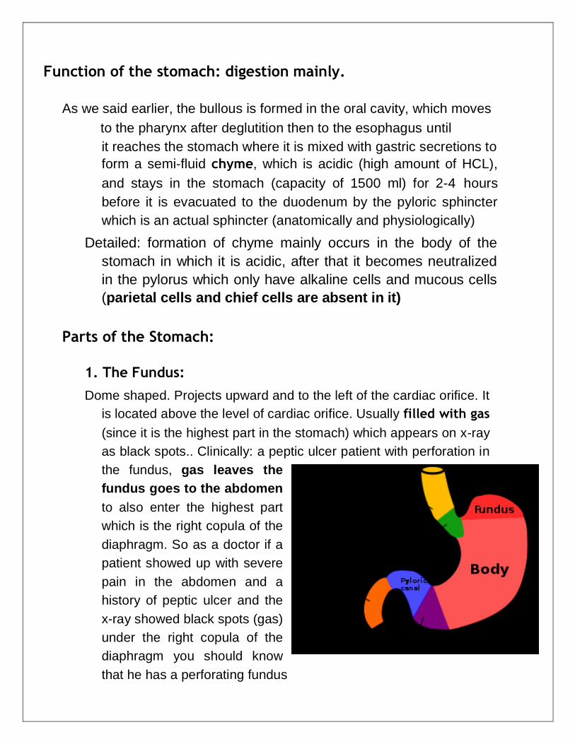
Parts of the Stomach:
1. The Fundus:
Dome shaped. Projects upward and to the left of the cardiac orifice. It
is located above the level of cardiac orifice. Usually filled with gas
(since it is the highest part in the stomach) which appears on x-ray
as black spots.. Clinically: a peptic ulcer patient with perforation in
the fundus, gas leaves the
fundus goes to the abdomen
to also enter the highest part
which is the right copula of the
diaphragm. So as a doctor if a
patient showed up with severe
pain in the abdomen and a
history of peptic ulcer and the
x-ray showed black spots (gas)
under the right copula of the
diaphragm you should know
that he has a perforating fundus

2- The Body (Corpus): Extends from the level of the cardiac orifice (below the fundus) to the level of the incisura angularis (a constant notch in the lower part of the lesser curvature that separates the body from the pylorus).
3.Pyloric Region:
Divided into:
a) Pyloric antrum: extends from the incisura angularis to the pylorus
b) Pyloric canal: the most tubular part of the stomach (1 inch)
c) Pyloric sphincter: anatomical sphincter, which contains a thickening
of the inner circular muscles, controlled by sympathetic and
parasympathetic.
Remember: the cardiac and pyloric orifices:
a) The cardiac orifice where the esophagus enters the stomach, it is a physiological sphincter (no thickening of smooth muscles).
Surface anatomy: at the 7th
costal cartilage, 1 inch to the left from midline at the level of T10, 10 cm from the anterior abdominal wall. 45 cm from incisors.
b)The pyloric orifice is an anatomical sphincter because it contains
thickening of smooth muscles. Surface anatomy: 1 inch to the right
from the midline at the level of L1 [medically known: transpyloric line
passes through the pylorus].
Transpyloric line: located halfway between the
jugular notch and the upper border of the pubic
symphysis.
Structures that passes through it: fundus of
gallbladder, pylorus, 9th costal cartilage, lower
hilum of left kidney, and upper hilum of right
kidney.
Structures above: transverse colon (sometimes,
depends on mesocolon length), celiac trunk.
Structures below: Duodenum, superior mesenteric.
Clinical note: We said that the cardiac orifice is 45 cm away from the incisors. Nowadays, all GIT specialists have gastroscopes in their clinics, which they use for gastroscopy in relation to problems in the upper GIT. The gastroscope is a fibro-optic flexible tube which is inserted through the mouth with light at its tip. The gastroscope has a scale that reads 5 cm, 10 cm, 20 cm… etc. When it reaches 45 cm, the physician will know that they have reached the cardiac orifice and, subsequently, the stomach. It can also be used to inject medicine. Therefore, it is a diagnostic and therapeutic tool. The function of the pyloric sphincter is controlled by:
Nerve fibers:
a) Sympathetic fibers which are motor for the sphincter.
b) The inhibition happens by the vagus nerve for drainage (parasympathetic)
Hormonal influence: which helps in the secretion of cholecystokinin (CCK) from
the duodenum. CCK→ inhibits gastric emptying & causes pyloric contraction.
Curvatures of the stomach:
1) The Lesser Curvature: Forms the right border of the stomach -
Extends from the cardiac orifice to the pylorus. Also has longitudinal
folds for the passage of fluids which goes directly to the duodenum.
Attached to it is the lesser omentum.
2) The Greater Curvature: forms the left border of the stomach- longer,
attached to it is the greater omentum, and in the upper part, the
gastrosplenic ligament (between the stomach{ fundus and part of
greater curvature} and spleen). From greater curvature there is the
gastrocolic ligament (attached to the transverse colon ). We also have
the gastrophrenic ligament (extends to the underside of the left dome
of the diaphragm). All of which help in the fixation of the stomach in its
place, and delivers the blood supply and nerve supply to it.

Histology of the Stomach
The stomach contains 4 layers:
mucosa, submucosa(CT),
muscularis externa and serosa
The layer of serosa is
completely covered by
peritoneum (simple
squamous epithelium)
The muscular layer contains 3 layers:
1) Outer longitudinal.
2) Inner circular(Forms
the sphincter).
3) The most inner, which is
Oblique; absent in the pylorus.
With myenteric (enteric) plexus in
between the outer and inner which is
mainly parasympathetic.
On the inside, the stomach contains rugae, which are foldings of the
Mucosa that submucosa made as it invaginated through the mucosa.
Rugae’s function is to increase the suface area of mucosa for digestion.
They are usually transverse and oblique, but at the lesser curvature they
are longitudinal folds for the fast passage of fluids which goes directly to
the duodenum. This is why the mixing of food and fluid is minimal;
therefor don’t listen to people that tell you to not drink water with food.
The mucosa contains glands and ducts called pits in order for the secretion to reach
the surface, it is composed of three layers: lining epithelium (simple columnar
without goblet cells), lamina propria (filled with gastric glands); contains parietal
cells(secrete HCL), chief cells(secrete pepsinogen) , mucous cells, and endocrine cells (secrete gasrin hormone) and muscularis mucosae

The stomach also contains
mucous cells, parietal cells in
the neck, chief cells at the base,
and enteroendocrine cells.
Peritoneum of the Stomach: The
stomach is completely covered by
peritoneum.
The Lesser Omentum:
The lesser omentum is composed of 2 layers of
peritoneum which extend from the lesser
curvature of the stomach to the porta hepatis of
the liver, and they extend upwards until they
reach the diaphragm. Since there are two layers
between them we are going to have blood
vessels, nerves (branches from vagi-
sympathetic), lymph nodes and lymphatic
drainage and small amount of fat. It is divided to
two parts the part attached to the stomach
hepatogastric and the part attached to the
duodenum hepatoduodenum

Note: space behind stomach: lesser sac//space
in front of stomach: greater sac. Lesser omentum
has a free edge, which contains an opening that
lies deep to it, called the epiploic foramen, or
foramen of Winslow: passage of communication
between lesser and greater sac.
The importance of this foramen is that it reaches the lesser
sac of peritoneum, posterior to the stomach. You can
insert your fingers through it.
The free edge of lesser omentum contains:
1) Common bile duct
2) Hepatic artery
3) And deep to it, the portal vein.
When you insert your fingers in the epiploic foramen, these
structures would be anterior to your fingers. Posterior to your
fingers would be the inferior vena cava.
The Greater Omentum: It is composed of 2 layers of peritoneum,
descending from the greater curvature of the stomach downwards
in the abdominal cavity, and ascends upwards until they reach the
transverse colon (intraperitoneal), surrounding it. The layers of
greater omentum contain the lesser sac. Contains gastroepiploic
blood vessels, veins, nerves, lymph nodes, lymph drainage and
the gastoslpenic ligament, gastrophrenic ligament
Gastrosplenic Ligament: the word ‘ligament’ means 2 layers of
peritoneum which are thickened. So, the gastrosplenic ligament is
located between the fundus of the stomach and a part of the greater
curvature, and goes to the spleen. It contains blood vessels, nerves,
lymph nodes and lymphatics.

Anterior Relations of the
Stomach:
The anterior abdominal wall.
The left costal margin.
The left pleura and lung.
The diaphragm(separates left pleura and lung from the stomach)
The left lobe of the liver.
Posterior Relations of the
Stomach (Stomach Bed)
The lesser sac [space behind
the stomach
The left crus of diaphragm
The spleen (only a small part)most lateral
The body of pancreas
The transverse mesocolon and transverse colon The left suprarenal
gland
The upper part of the left kidney
The splenic artery [which is tortous also for elongation of stomach after food on upper border of pancreas; the same applies for uterine artery and pregnancy.
Note: the position of the transverse colon depends on the length of the
mesocolon. Long mesocolon → transverse colon below the stomach /
short mesocolon →transverse colon behind the stomach
.
The Blood Supply and Venous Drainage of the Stomach:
The GI tract (according to embryology) is divided into 3 parts: 1) Foregut -> takes blood supply from the celiac trunk
It extends from the lower third of the esophagus, the stomach, until the upper half of the duodenum.
2) Midgut -> takes blood supply from the superior mesenteric
artery It extends from the lower half of the duodenum and
ends at the lateral third of the transverse colon.
3) Hindgut -> takes blood supply from the inferior mesenteric
artery It includes the lateral third of transverse colon,
descending colon, rectum, and anal canal.
The Celiac Trunk
It is called a trunk because it is very short (about 1 cm in length).
It originates from the abdominal aorta at the level of T12 (between T12 and
L1). On each side, it contains the celiac ganglia (contain
sympathetic and parasympathetic fibers) and lymph nodes,
which are named after it. Behind it, we have the crus of the
diaphragm.
It gives off 3 main arteries:
1) Left gastric, which goes to the lesser curvature and gives blood
supply to the stomach and the lower third of the esophagus.
2) Splenic, which moves behind the stomach, then on the upper
border of the pancreas and gives blood supply to the stomach. 3) Common Hepatic, which moves towards the liver. It gives
small branches such as:
a) Right gastric artery b) Hepatic Artery Proper
c) Gastro-duodenal artery {passes behind the first part of the
duodenum}
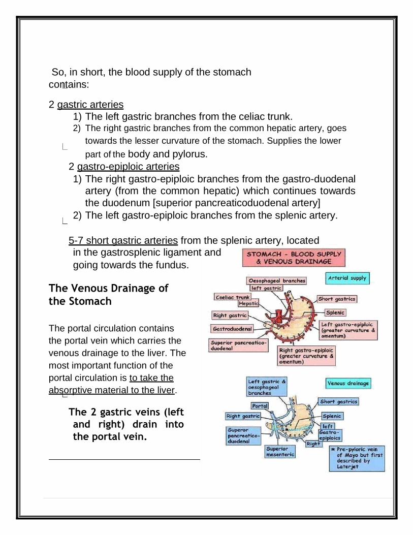
So, in short, the blood supply of the stomach
contains:
2 gastric arteries
1) The left gastric branches from the celiac trunk. 2) The right gastric branches from the common hepatic artery, goes
towards the lesser curvature of the stomach. Supplies the lower
part of the body and pylorus.
2 gastro-epiploic arteries
1) The right gastro-epiploic branches from the gastro-duodenal artery (from the common hepatic) which continues towards
the duodenum [superior pancreaticoduodenal artery]
2) The left gastro-epiploic branches from the splenic artery.
5-7 short gastric arteries from the splenic artery, located in the gastrosplenic ligament and
going towards the fundus.
The Venous Drainage of
the Stomach
The portal circulation contains
the portal vein which carries the
venous drainage to the liver. The
most important function of the
portal circulation is to take the
absorptive material to the liver.
The 2 gastric veins (left
and right) drain into
the portal vein.
The right gastro-epiploic vein drains into the superior mesenteric
vein, and the left gastro-epiploic vein drains into the splenic vein.
The short gastric veins also drain into the splenic vein.
The superior mesenteric and the splenic behind the neck of the pancreas form the portal vein.
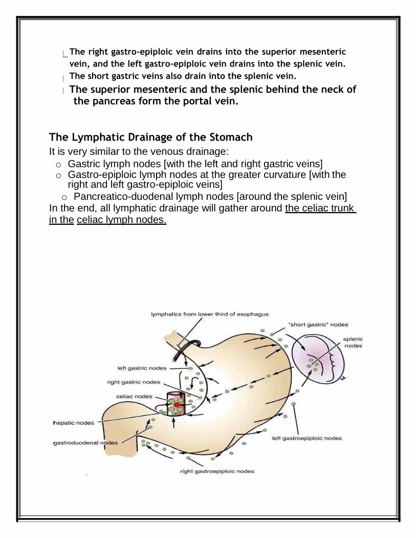
The Lymphatic Drainage of the Stomach
It is very similar to the venous drainage:
o Gastric lymph nodes [with the left and right gastric veins] o Gastro-epiploic lymph nodes at the greater curvature [with the
right and left gastro-epiploic veins]
o Pancreatico-duodenal lymph nodes [around the splenic vein] In the end, all lymphatic drainage will gather around the celiac trunk in the celiac lymph nodes.
The Nerve Supply of the Stomach: We have sympathetic fibers that go towards the celiac plexus of
nerves around the celiac trunk. The sympathetic fibers mainly go
towards the sphincters for contraction (such as the pyloric sphincter),
and they also carry pain sensation.
We also have parasympathetic fibers from the vagus.
The parasympathetic fibers are mainly secretomotor for the gastric
glands, and motor for smooth muscles, so they are responsible for
peristaltic movement.
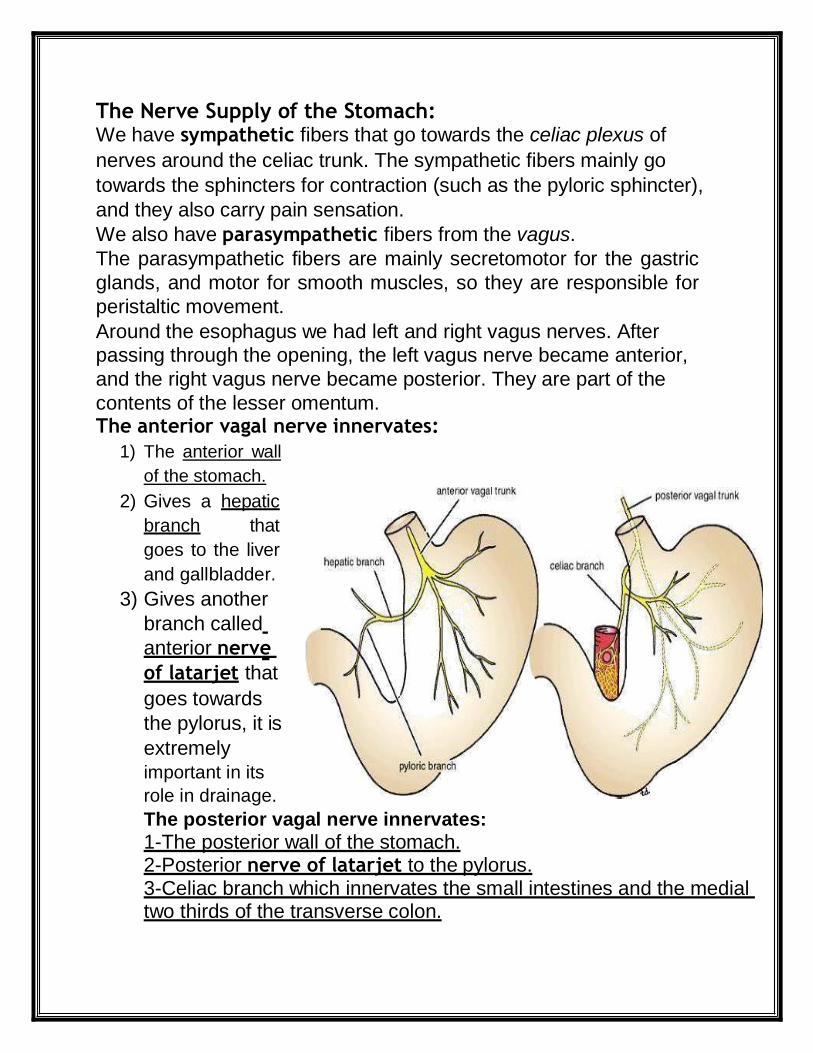
Around the esophagus we had left and right vagus nerves. After
passing through the opening, the left vagus nerve became anterior,
and the right vagus nerve became posterior. They are part of the
contents of the lesser omentum. The anterior vagal nerve innervates:
1) The anterior wall
of the stomach.
2) Gives a hepatic
branch that
goes to the liver
and gallbladder.
3) Gives another
branch called
anterior nerve
of latarjet that
goes towards
the pylorus, it is
extremely
important in its
role in drainage.
The posterior vagal nerve innervates: 1-The posterior wall of the stomach. 2-Posterior nerve of latarjet to the pylorus. 3-Celiac branch which innervates the small intestines and the medial two thirds of the transverse colon.

Clinical Points:
1) Gastric Ulcer
15 years ago, the rule said that hyperacidity meant peptic ulcer, and its treatment was surgery to the vagus nerve.
Now, it has changed completely. The real cause of peptic ulcer is a
bacteria known as Helicobacter pylori. It is present inside the human
body and is usually non-pathogenic. Under certain conditions,
when there is resistance in the body, it may lead to peptic ulcer. Its
treatment consists of a combination of 4 drugs for 21 days. 95% of
the cases are completely cured with this method .Peptic ulcer has
two forms: gastric and duodenal. The gastric ulcer is considered
malignant until proven that it is not malignant. The duodenal ulcer is
a peptic ulcer until proven otherwise (they are the opposite of each
other). It is very common, especially in the first inch, since it
receives acidic chyme which may result in irritation Ulcer. So, a
biopsy needs to be taken to prove malignancy.
Note: common site in stomach: anterior and posterior walls of lesser curvature Remember that: the most common side for peptic ulcer is the first part of the
duodenum, and if progressed it can perforate the gastro-duodenal artery, and
cause severe bleeding, the individual may be prone to death due to shock.
2) Trunkal Vagotomy
We used to cut the vagus nerve below the diaphragm, so the stomach
stopped receiving parasympathetic innervations, which resulted in
bad drainage. Now, we perform a highly selective vagotomy where
we cut all branches of vagi except the nerve of latarjet.
4) Gastroscopy (where we enter an endoscope into the oral
cavity to check the stomach, esophagus, and duodenum, (to
be more specific, until the second part of the duodenum). 5) Pyloroplasty, means drainage, where the pyloric sphincter has a
thickening and continuous contraction that results in no drainage. So
they cut it and add stitches which will lead to the dilatation of the
sphincter