Older adults exhibit a more pronounced modulation …Older adults exhibit a more pronounced...
14
Older adults exhibit a more pronounced modulation of beta oscillations when performing sustained and dynamic handgrips Alba Xifra-Porxas a, b , Guiomar Niso c, d, e , Sara Larivi ere c, f , Michalis Kassinopoulos a , Sylvain Baillet c , Georgios D. Mitsis g , Marie-H el ene Boudrias b, h, * a Graduate Program in Biological and Biomedical Engineering, McGill University, Montr eal, Canada b Center for Interdisciplinary Research in Rehabilitation of Greater Montreal (CRIR), Montr eal, Canada c McConnell Brain Imaging Centre, Montreal Neurological Institute, McGill University, Montr eal, Canada d Center for Biomedical Technology, Universidad Polit ecnica de Madrid, Madrid, Spain e Biomedical Image Technologies, Universidad Polit ecnica de Madrid and CIBER-BBN, Madrid, Spain f Integrated Program in Neuroscience, McGill University, Montr eal, Canada g Department of Bioengineering, McGill University, Montr eal, Canada h School of Physical and Occupational Therapy, McGill University, Montr eal, Canada ARTICLE INFO Keywords: MEG Beta oscillations Motor control Handgrips Aging ABSTRACT Muscle contractions are associated with a decrease in beta oscillatory activity, known as movement-related beta desynchronization (MRBD). Older adults exhibit a MRBD of greater amplitude compared to their younger counterparts, even though their beta power remains higher both at rest and during muscle contractions. Further, a modulation in MRBD has been observed during sustained and dynamic pinch contractions, whereby beta activity during periods of steady contraction following a dynamic contraction is elevated. However, how the modulation of MRBD is affected by aging has remained an open question. In the present work, we investigated the effect of aging on the modulation of beta oscillations and their putative link with motor performance. We collected magnetoencephalography (MEG) data from younger and older adults during a resting-state period and motor handgrip paradigms, which included sustained and dynamic contractions, to quantify spontaneous and motor- related beta oscillatory activity. Beta power at rest was found to be significantly increased in the motor cortex of older adults. During dynamic hand contractions, MRBD was more pronounced in older participants in frontal, premotor and motor brain regions. These brain areas also exhibited age-related decreases in cortical thickness; however, the magnitude of MRBD and cortical thickness were not found to be associated after controlling for age. During sustained hand contractions, MRBD exhibited a decrease in magnitude compared to dynamic contraction periods in both groups and did not show age-related differences. This suggests that the amplitude change in MRBD between dynamic and sustained contractions is larger in older compared to younger adults. We further probed for a relationship between beta oscillations and motor behaviour and found that greater MRBD in primary motor cortices was related to degraded motor performance beyond age, but our results suggested that age-related differences in beta oscillations were not predictive of motor performance. 1. Introduction Aging is a multifaceted process, which involves alterations in brain structure and biochemistry. It is associated with reduced grey matter volume, cortical thinning, decreases of white matter myelination and neurotransmitter depletion (Minati et al., 2007). Motor functions tend to decline in old age in a broad array of motor tasks, manifesting in decline of fine motor control and coordination, slowing of movements, and im- pairments related to gait and balance, which in turn affect quality of life (Maes et al., 2017; Rosso et al., 2013; Seidler et al., 2010). Most common motor tasks require the combination of different types of muscle contraction, in which switches from static to dynamic force production occur frequently. However, age-related effects in brain dynamics during complex contraction sequences remain largely unknown. * Corresponding author. School of Physical and Occupational Therapy, Hosmer House, Room H-206, McGill University, 3654, Prom Sir-William-Osler, Montr eal, Qu ebec, H3G 1Y5, Canada. E-mail address: [email protected] (M.-H. Boudrias). Contents lists available at ScienceDirect NeuroImage journal homepage: www.elsevier.com/locate/neuroimage https://doi.org/10.1016/j.neuroimage.2019.116037 Received 1 April 2019; Received in revised form 12 July 2019; Accepted 19 July 2019 Available online 19 July 2019 1053-8119/Crown Copyright © 2019 Published by Elsevier Inc. All rights reserved. NeuroImage 201 (2019) 116037
Transcript of Older adults exhibit a more pronounced modulation …Older adults exhibit a more pronounced...

NeuroImage 201 (2019) 116037
Contents lists available at ScienceDirect
NeuroImage
journal homepage: www.elsevier.com/locate/neuroimage
Older adults exhibit a more pronounced modulation of beta oscillationswhen performing sustained and dynamic handgrips
Alba Xifra-Porxas a,b, Guiomar Niso c,d,e, Sara Larivi�ere c,f, Michalis Kassinopoulos a,Sylvain Baillet c, Georgios D. Mitsis g, Marie-H�el�ene Boudrias b,h,*
a Graduate Program in Biological and Biomedical Engineering, McGill University, Montr�eal, Canadab Center for Interdisciplinary Research in Rehabilitation of Greater Montreal (CRIR), Montr�eal, Canadac McConnell Brain Imaging Centre, Montreal Neurological Institute, McGill University, Montr�eal, Canadad Center for Biomedical Technology, Universidad Polit�ecnica de Madrid, Madrid, Spaine Biomedical Image Technologies, Universidad Polit�ecnica de Madrid and CIBER-BBN, Madrid, Spainf Integrated Program in Neuroscience, McGill University, Montr�eal, Canadag Department of Bioengineering, McGill University, Montr�eal, Canadah School of Physical and Occupational Therapy, McGill University, Montr�eal, Canada
A R T I C L E I N F O
Keywords:MEGBeta oscillationsMotor controlHandgripsAging
* Corresponding author. School of Physical and OQu�ebec, H3G 1Y5, Canada.
E-mail address: [email protected] (M.-H. B
https://doi.org/10.1016/j.neuroimage.2019.11603Received 1 April 2019; Received in revised form 1Available online 19 July 20191053-8119/Crown Copyright © 2019 Published by
A B S T R A C T
Muscle contractions are associated with a decrease in beta oscillatory activity, known as movement-related betadesynchronization (MRBD). Older adults exhibit a MRBD of greater amplitude compared to their youngercounterparts, even though their beta power remains higher both at rest and during muscle contractions. Further, amodulation in MRBD has been observed during sustained and dynamic pinch contractions, whereby beta activityduring periods of steady contraction following a dynamic contraction is elevated. However, how the modulationof MRBD is affected by aging has remained an open question. In the present work, we investigated the effect ofaging on the modulation of beta oscillations and their putative link with motor performance. We collectedmagnetoencephalography (MEG) data from younger and older adults during a resting-state period and motorhandgrip paradigms, which included sustained and dynamic contractions, to quantify spontaneous and motor-related beta oscillatory activity. Beta power at rest was found to be significantly increased in the motor cortexof older adults. During dynamic hand contractions, MRBD was more pronounced in older participants in frontal,premotor and motor brain regions. These brain areas also exhibited age-related decreases in cortical thickness;however, the magnitude of MRBD and cortical thickness were not found to be associated after controlling for age.During sustained hand contractions, MRBD exhibited a decrease in magnitude compared to dynamic contractionperiods in both groups and did not show age-related differences. This suggests that the amplitude change inMRBD between dynamic and sustained contractions is larger in older compared to younger adults. We furtherprobed for a relationship between beta oscillations and motor behaviour and found that greater MRBD in primarymotor cortices was related to degraded motor performance beyond age, but our results suggested that age-relateddifferences in beta oscillations were not predictive of motor performance.
1. Introduction
Aging is a multifaceted process, which involves alterations in brainstructure and biochemistry. It is associated with reduced grey mattervolume, cortical thinning, decreases of white matter myelination andneurotransmitter depletion (Minati et al., 2007). Motor functions tend todecline in old age in a broad array of motor tasks, manifesting in decline
ccupational Therapy, Hosmer H
oudrias).
72 July 2019; Accepted 19 July 20
Elsevier Inc. All rights reserved.
of fine motor control and coordination, slowing of movements, and im-pairments related to gait and balance, which in turn affect quality of life(Maes et al., 2017; Rosso et al., 2013; Seidler et al., 2010). Most commonmotor tasks require the combination of different types of musclecontraction, in which switches from static to dynamic force productionoccur frequently. However, age-related effects in brain dynamics duringcomplex contraction sequences remain largely unknown.
ouse, Room H-206, McGill University, 3654, Prom Sir-William-Osler, Montr�eal,
19

Table 1Subject characteristics and behavioral scores: mean� SD.
YOUNGER (n¼ 12) OLDER (n¼ 12) p value
Age 24.2� 2.8 67.7� 3.7SEX 4 F/8M 3 F/9MEducation 16.7� 1.9 15.3� 2.8 > 0.1mmse 29.2� 1.0 28.7� 1.3 > 0.19HPT (sec) RH 17.2� 1.8 20.5� 2.1 <0.0005
LH 19.4� 2.4 22.7� 3.4 <0.01BBT (blocks) RH 68.3� 5.6 57.7� 3.9 <0.001
LH 67.1� 6.1 57.4� 4.6 <0.001HGS (kg) RH 48.4� 14.6 39.4� 9.0 > 0.1
LH 39.2� 9.9 35.3� 7.7 > 0.1PPT (pins) RH 16.9� 1.8 13.3� 1.6 <0.001
LH 15.0� 1.5 12.7� 1.8 <0.01RH-LH 12.8� 2.0 10.1� 1.1 <0.005A 42.8� 5.1 28.2� 5.7 <0.0001
F¼ female, M¼male, MMSE¼mini mental state examination, 9HPT¼ ninehole peg test, BBT¼ box and blocks test, HGS¼ hand grip strength, PPT¼ pur-due pegboard test, RH¼ right hand, LH¼ left hand, RH-LH¼ right hand and lefthand, A¼ assembly.
A. Xifra-Porxas et al. NeuroImage 201 (2019) 116037
Understanding how aging affects motor-related neural oscillations isfundamental to better understand the mechanisms of motor control inhumans. A robust brain response induced by motor tasks is the modu-lation of beta sensorimotor rhythms. Beta oscillations are stronger duringrest and are abolished during preparation and execution of motor tasks.This strong decrease in beta power relative to resting levels is known asmovement-related beta desynchronization (MRBD) (Cheyne, 2013), andlasts as long as there is a muscle contraction (Erbil and Ungan, 2007; vanWijk et al., 2012). Several studies have reported age-related changes inbeta oscillations during movement, such as a greater MRBD in bothmotor and premotor areas during right-hand finger extensions (Saileret al., 2000), sequences of finger movements (Heinrichs-Graham et al.,2018; Heinrichs-Graham and Wilson, 2016), cued button presses (Bar-douille et al., 2019), bimanual button presses in a go/no-go task(Schmiedt-Fehr et al., 2016), unimanual hand grips (Rossiter et al.,2014), as well as during a right-hand precision grip force modulation task(Hübner et al., 2018a). Interestingly, despite displaying increased MRBD,older adults exhibit higher absolute beta power during muscle contrac-tions compared to younger adults (Heinrichs-Graham and Wilson, 2016).This is mostly due to the fact that older adults exhibit higher resting-statebeta activity compared to their younger counterparts (G�omez et al.,2013; Heinrichs-Graham et al., 2018; Heinrichs-Graham and Wilson,2016; Hübner et al., 2018a; Koyama et al., 1997; Veldhuizen et al.,1993). Pharmacological manipulations of GABA have shown thatincreased levels of intracortical GABAergic inhibition lead to higherresting beta power and accentuated MRBD during dynamic contractions(Hall et al., 2011, 2010; Jensen et al., 2005; Muthukumaraswamy et al.,2013). These observations are closely related to the ones observed inaging, which seems to indicate that age-related changes are associatedwith changes in GABAergic inhibition. Following a motor task, beta os-cillations exhibit increased amplitude relative to resting levels, known aspost-movement beta rebound (PMBR). PMBR overshoots around 1–2 safter the cessation of a motor task and is stronger over the hemispherecontralateral to the moving limb (Fry et al., 2016; Jurkiewicz et al.,2006). Reduced PMBR has been observed in older adults (Bardouilleet al., 2019; Liu et al., 2017). This suggests that altered brain structuresand biochemistry due to aging have consequences on the observedmotor-related neural activation patterns.
Steady muscle contractions are maintained by a continuous drivefrom the motor cortex to spinal motoneurons (Scott, 2012), during whichthere is a relative increase in beta power compared to dynamic con-tractions (Baker, 2007; Cassim et al., 2000; Espenhahn et al., 2017; Kilneret al., 2003, 1999; Schoffelen et al., 2008; Spinks et al., 2008; van Wijket al., 2012). The functional role of this elevation in beta synchrony re-mains unclear; however, previous studies have suggested that it reflectsthe integration of afferent information to promote a stable motor output(Androulidakis et al., 2007, 2006; Gilbertson et al., 2005; Omlor et al.,2007). A study from Rossiter and colleagues (Rossiter et al., 2014)examined unimanual sustained handgrips in healthy aging, and found anincreased beta suppression with age in the ipsilateral but not in thecontralateral primary motor cortex (M1). This may suggest a heteroge-neous effect of the aging process in different brain regions. However, themodulation of beta activity during sustained muscle contractions has notyet been formally examined in the context of healthy aging.
The aim of the present study was to examine the modulation of betaoscillations during sustained and dynamic contractions in healthy aging.We used a motor paradigm that included periods of steady handgrips andforce modulation, both uni- and bimanual. Exploiting the high spatio-temporal resolution of MEG (Baillet, 2017), we investigated whole-brainage-related changes in spectral dynamics beyond the M1s. We also pro-bed the association between age-induced differences in beta oscillationsand motor performance. Based on previous results, we expected greaterresting beta power in older adults in motor areas (Heinrichs-Graham andWilson, 2016; Rossiter et al., 2014) and hypothesized that age-relatedincreases in resting beta activity would be present beyond the motorcortex since aging is associated with structural alterations in multiple
2
brain regions. We further anticipated that older adults would exhibitincreased MRBD during dynamic contractions (Heinrichs-Graham andWilson, 2016; Hübner et al., 2018a; Sailer et al., 2000; Schmiedt-Fehret al., 2016). In turn, this would indicate that greater beta desynchro-nization is required to produce muscle contractions, compensating forelevated resting-state beta power levels in the older population. Finally,we sought to investigate whether the increase in beta synchrony duringsustained handgrips would exhibit age-specific differences.
2. Materials and methods
2.1. Participants
We studied 12 younger (age range 19–28 years) and 12 older (agerange 60–74 years) healthy individuals recruited via advertisements. Allparticipants were right-handed according to the Edinburgh HandednessInventory (Oldfield, 1971). Subject characteristics are detailed inTable 1. Recruitment criteria included young subjects between 18 and 30years and older adults above 60 years, and excluded subjects with apersonal history of neurological and psychiatric disorder, as well as MEGexclusion criteria related to presence of ferromagnetic material (e.g.dental braces, metal implants and/or crowns). The study was approvedby the McGill University Ethical Advisory Committee. All participantssigned a written informed consent and were compensated for theirparticipation. Measurements were carried out using the MEG facility atthe McConnell Brain Imaging Centre (BIC) of the Montreal NeurologicalInstitute (MNI), McGill University.
At the beginning of the session, participants completed the followingbehavioral assessment tests: Nine Hole Peg Test (9HPT) (Mathiowetzet al., 1985b), Box and Blocks Test (BBT) (Mathiowetz et al., 1985a),Purdue Pegboard Test (PPT) (Lindstrom-Hazel and VanderVlies Veenstra,2015), and Hand Grip Strength (HGS) (Bohannon et al., 2006). All testswere performed using both hands to cover a range of upper limb motorabilities, from manual dexterity to strength. The 9HPT was measured inseconds, reflecting how quickly each participant placed and removednine pegs into the holes of a board. The BBTwas quantified as the numberof blocks moved from one compartment of a box to another of equal sizewithin 60 s. The HGSwas measured in kilograms. The PPTwas quantifiedas the number of pins placed into holes of a board within 30 s (dominant,non-dominant and both hands) or the number of assembled pins, collarsand washers within 60 s (assembly test with both hands). Of note, PPTwas not collected for two older subjects. All participants were screenedfor mental status by means of the mini mental state examination (MMSE)(Folstein et al., 1975). Wilcoxon rank-sum tests were used to determinewhether behavioral assessments were significantly different betweenyounger and older adults.

A. Xifra-Porxas et al. NeuroImage 201 (2019) 116037
2.2. Experimental paradigm
The protocol carried out inside the MEG scanner consisted of twomotor tasks alternated by three 5-min resting-state periods (Fig. 1a).During the resting-state periods, subjects were instructed to stare at awhite cross displayed on a screen in front of them. They were alsoinstructed not to think of anything in particular and not to manipulate thehand grippers. After the 1st rest period, the maximum voluntarycontraction (MVC) was obtained for each participant, using the samehand grippers later employed for the motor tasks. The first motor taskconsisted of a unimanual isometric right handgrip, during which thesubjects had to apply force to track a ramp target as accurately aspossible. At the onset of the trial, an orange circle appeared on the screenand the subjects had 2 s to increase their force to reach a white targetblock at 15% of their MVC. This force was held for 3 s. Subsequently,participants tracked a linear increase of the force to reach 30% of theirMVC over a 3-s period, during which they had to maintain the circleinside the white target block, followed by a 3-s hold at this force (Fig. 1b).A single trial lasted 11 s and was repeated 50 times for a total taskduration of about 13min. The second motor task consisted of bimanualsteady isometric handgrips. At the onset of the trial, two circles (blue andred) appeared on the screen and the subjects had 2 s to increase the forceproduced by both hands to 15% of their MVC. This force was sustainedfor 6 s (Fig. 1c). A single trial lasted 8 s and was repeated 50 times for atotal task duration of about 10min. Visual feedback was providedthroughout the experiment. For both tasks, the inter-trial interval wasjittered between 3 and 5 s, during which subjects stared at a white cross.
Fig. 1. (a) Illustration of the protocol. Participants carried out two motor tasks insifixated on a crosshair for 5min. After the first rest period, the maximum voluntaryticipants fixated on a crosshair for a few seconds, for a jittered period lasting betweenwhere participants had 2 s to apply force to reach 15% of their MVC. A steady grip wparticipants had to apply force to reach 30% of their MVC and sustain this grip strejittered period lasting between 3 and 5 s. Subsequently, two circles (blue and red) aMVC, which they sustained for 6 s.
3
All subjects practised both motor tasks prior to the MEG acquisition tofamiliarise themselves with the experiment. Note that the order of theunimanual and bimanual conditions was not counter-balanced.
2.3. Data acquisition and pre-processing
2.3.1. Hand grippers: grip force fiber optic response padA pair of non-magnetic, non-electronic hand grippers made from
plastic to prevent noise in the MEG environment were used (CurrentDesigns Inc, USA). The hand grippers consisted of a machined blackenclosure with a protruding force bar that moved in when gripped toproduce a linear force measurement output based on the pressureapplied. We used a spring with a range of 500 N. The dimensions of theforce grip were 17.8� 3.2 cm, with a force bar of 12.7� 1.3 cm placed2.5 cm outside the main enclosure. The maximum travel of this bar was0.127 cm. The grippers were connected to a 932 interface through a 3-mlong fiber pigtailed connector, which received the optical signals fromthe hand grippers in the MEG suite, and converted them into electricalsignals that were transferred to a computer.
2.3.2. Neuroimaging data acquisition and pre-processingMEG recordings were acquired with a 275-channel CTF whole-head
system. Participants changed into non-magnetic clothes and performedthe experiment in a seated position while their arms rested on the arm-chairs. Bipolar electrocardiogram (ECG) and vertical bipolar electro-oculogram (EOG) were acquired to correct for cardiac artifacts and eyemovements. All signals were amplified and digitized at a sampling rate of
de the MEG scanner, alternated by three periods of rest, during which subjectscontraction (MVC) was obtained for each participant. (b) Unimanual task. Par-3 and 5 s. This was followed by the appearance of an orange circle on the screen,as then maintained for 3 s, which was followed by a guided ramp period wherength for another 3 s. (c) Bimanual task. Participants fixated on a crosshair for appeared on the screen. Participants had 2 s to apply force to reach 15% of their
A. Xifra-Porxas et al. NeuroImage 201 (2019) 116037
2400 Hz, and MEG files were saved after performing third order gradientcorrection. An empty-room noise recording was collected prior to theacquisition of each session to capture environmental noise conditionsand was used in subsequent offline data analyses. The 3-D digitization ofthe head shape was done with a Polhemus Fastrak device, using around100 head points distributed uniformly. Individual T1-weighted MRIimages were acquired on a 3T MRI scanner (Siemens Prisma;TR¼ 2300m s; TE¼ 2.32m s; field of view¼ 240mm; voxelsize¼ 0.9� 0.9� 0.9mm). The position of the head localization coils(nasion, left and right pre-auricular) and the head-surface points wereused as anatomical references for coregistration between the MEG andMRI coordinate systems.
Offline data were processed using the open-source toolbox Brain-storm (Tadel et al., 2011). Notch filters were applied to remove powerline artifacts around 60Hz and harmonics. MEG data were band-passedfrom 1 to 150Hz. Cardiac and eye movement artifacts were detectedusing the ECG and EOG signals and corrected using signal-space pro-jection (SSP). Artifacts due to external magnetic fields were removedvisually using independent component analysis (ICA). Segments thatpresented motion artifacts or where subjects moved more than 5mmbetween head position measurements were discarded from the analysis.MEG signals were down-sampled to a 120-Hz sampling rate.
Resting-state periods. The 5-min recordings were segmented in epochsof 5 s. Epochs that had previously been found to be contaminated bymotion artifacts were discarded. The average number of epochs afterartifact rejection was 58.6� 1.2/57.6� 4.6 for younger/older adults(Resting-state 1), 58.3� 2.4/56� 5.4 for younger/older adults (Resting-state 2), and 56.3� 9.3/57.4� 2.2 for younger/older adults (Resting-state 3). The difference in the number of epochs between groups was notsignificant across any of the resting-state periods, as assessed using theWilcoxon rank-sum test (Resting-state 1: p¼ 0.473; Resting-state 2:p¼ 0.185; Resting-state 3: p¼ 0.679).
Motor tasks. Data from the unimanual task were epoched from�2.5 toþ14 s, and data from the bimanual task were epoched from �2.5 to þ11s. Time 0 indicates onset of the visual cue for analysis. The averagenumber of trials after artifact rejection was 40.4 � 10.4/40.3 � 9.1 foryounger/older adults (Unimanual task), and 44.3 � 8.4/41.1 � 8.8 foryounger/older adults (Bimanual task). The difference in the number oftrials between groups was not significant for any of the tasks, as assessedusing the Wilcoxon rank-sum test (Unimanual task: p¼ 0.954; Bimanualtask: p¼ 0.277).
2.4. Data analysis
2.4.1. Behavioral analysisThe force exerted by the subjects was recorded using the calibrated
hand grippers. The x and y screen positions of the applied force were alsorecorded for offline analysis. Task accuracy was quantified as the rootmean squared error between the position on the screen and the targetprofile (defined as themiddle of the target ramp), averaged over time andtrials. Trials that exceeded 3 standard deviations were considered out-liers and therefore not used in the computation of task accuracy. This wasthe case for two trials of a younger subject, which were also manuallyrejected in the MEG data.
2.4.2. MRI structural analysisCortical reconstruction and volumetric segmentation were performed
with the FreeSurfer image analysis suite version 5.3.0 (http://surfer.nmr.mgh.harvard.edu/). The technical details of these procedures aredescribed in prior publications (Dale et al., 1999; Dale and Sereno, 1993;Fischl et al., 1999a,b; Fischl et al., 2004, 2002; 2001; Fischl and Dale,2000; Han et al., 2006; Jovicich et al., 2006; Reuter et al., 2012, 2010;S�egonne et al., 2004). Procedures for the measurement of corticalthickness have been validated against histological analysis (Rosas et al.,2002) and manual measurements (Kuperberg et al., 2003; Salat et al.,2004). Thickness measurements were mapped on the inflated surface of
4
each participant’s reconstructed brain and projected to the ICBM152template using Brainstorm (Tadel et al., 2011). Maps were subsequentlysmoothed using a circularly symmetric Gaussian kernel across the surfacewith a full-width-half-maximum (FWHM) of 5mm. Finally, cortical mapswere compared between groups using non-parametric permutation testscombined with independent Student’s t-tests of unequal variance. Thenull distribution was estimated with 10,000 permutations and resultscorrected for multiple comparisons using the false discovery rate (FDR)(number of signals 15,000). The structural analysis was done to identifythe brain areas that presented differences in cortical thickness betweengroups. Particularly, we wanted to assess whether age-related differencesin cortical thickness could have accounted for the differences observed inMRBD, reported in a previous study within the primary motor cortex(Provencher et al., 2016).
2.4.3. MEG source imagingLead fields were obtained using an overlapping spheres head model,
which computes locally-fitted spheres under each sensor (Huang et al.,1999). Source reconstruction was performed using an extension of thelinearly constrained minimum variance (LCMV) beamformer (Van Veenet al., 1997). A set of 15,000 elementary current dipoles distributed overthe cortical surface was used, whereby the dipoles were assumed to beperpendicular to the cortical envelope. The empty room recording of a2-min duration was used to estimate the noise covariance matrix. Thedata covariance matrix was estimated directly from the MEG recordings.The LCVM regularization parameter applied to the data covariance ma-trix was set as its median eigenvalue.
Resting-state periods. Normalized source power was computed usingMorlet wavelets averaged across the 5 s segments (time resolution¼ 3 s,central frequency¼ 1 Hz) over the entire brain volume for the followingfrequency bands: alpha (8–12 Hz) and beta (16–28 Hz). The resultingsource maps were smoothed with a 5mm FWHM circularly symmetricGaussian kernel and projected onto a standard space (ICBM152 tem-plate). Grand-averaged surfaces were computed across subjects for eachgroup and frequency band.
Motor tasks. Single trial source waveforms were extracted per subjectand decomposed to the time-frequency (TF) domain using Morletwavelets (time resolution¼ 3 s, central frequency¼ 1Hz). The evokedresponse was removed from each trial before computing the TF decom-position, a step that has been recommended for the evaluation of the TFdecomposition of neurophysiological signals (Tadel et al., 2011). Anaverage whole-brain TF map across trials was computed and subse-quently averaged within the following frequency bands related tosensorimotor rhythms: alpha (8–12Hz) and beta (16–28Hz). We selectedthe 16–28Hz frequency range to avoid including any power from thecontiguous alpha and gamma bands. For both bands, relative power
ðRP%Þ was calculated as follows: RP%¼ PðtÞ � BB � 100% (Pfurtscheller
and Lopes da Silva, 1999), where PðtÞ is the absolute power at time t andB is the baseline power. B was defined as the mean power obtained fromthe 1st resting-state period (see section 2.4.7. for the effects of usingdifferent baselines). The RP% related to the beta band is denoted asMRBD and PMBR during and after a muscle contraction, respectively.Subsequently, the RP% was averaged across several time windows foreach subject. For the unimanual task, RP% was averaged within three3-sec time windows: sustained contraction at 15% MVC (2–5 s), guideddynamic contraction from 15% MVC to 30% MVC (5–8 s), and sustainedcontraction at 30% MVC (8–11 s). For the bimanual task, the behavioralanalysis showed that task accuracy did not reach the desired thresholdsuntil around 4–5s after the onset of the trial, which suggests that subjectswere not performing a sustained contraction in the first few seconds ofthe trial (Supp. Fig. 1). Hence, RP% for the bimanual task was averagedwithin two 3-sec time windows: unguided dynamic contraction (2–5 s),and sustained contraction at 15% MVC (5–8 s). Cortical surfaces wereobtained per participant, smoothed with a 5mm FWHM circularly sym-metric Gaussian kernel, and projected onto a standard space (ICBM152

A. Xifra-Porxas et al. NeuroImage 201 (2019) 116037
template). Grand-averaged surfaces of each task time window werecomputed across subjects for each group and frequency band.
Statistics. For both rest and task, permutation testing was used to testfor group differences across the whole brain. The test statistic used wasthe independent Student’s t-test of unequal variance. For each compari-son, 10,000 permutations were computed to build the null distribution.Significance testing was performed with a threshold of 5% using FDRcorrection for multiple comparisons (number of signals 15,000).
2.4.4. Modulation of beta oscillationsWe were interested in examining whether the MRBD modulation
observed during sustained and dynamic contractions in young subjectswas altered in older subjects. To this end, regions of interest (ROIs) wereselected for subsequent analysis. The peak MRBD ROIs were identified asthe vertices showing the strongest MRBD (top 5%) within the motorcortex. Since dynamic contractions elicit increased MRBD compared tosustained contractions, the windows containing dynamic contractions(Unimanual: 5–8 s; Bimanual: 2–5 s) were grand-averaged across allsubjects and used to define the ROIs related to MRBD. Supp. Fig. 2 dis-plays the peak MRBD ROIs, located within left and right M1, and Supp.Table 1 provides the coordinates of the peak vertex of each MRBD ROI inMNI space. ROI power time-courses were then extracted and averagedacross vertices. An ROI was also created from the whole-brain analysisthat combined the brain regions identified to exhibit stronger MRBD inolder adults for both unimanual and bimanual tasks, henceforth called“ageMRBD”. The three ROIs are depicted in the first row of Fig. 5.
The following Modulation metrics were used to quantify the depth ofvariations to which subjects modulated their beta power:
Modulation Unimanual¼ abs�β½2;5� � β½5;8�
�þ abs�β½5;8� � β½8;11�
�
Modulation Bimanual ¼ abs�β½2;5� � β½5;8�
�
where β½t1 ;t2 � is the averaged beta activity between time-points t1 and t2.The beta activity used to compute β½t1;t2� was the absolute beta powerinstead of MRBD and was extracted for all three ROIs (left peak MRBD,right peak MRBD, ageMRBD). In this fashion, we can quantify a relativemeasure of how much beta oscillations were modulated without con-founds related to the resting beta power.
Statistics. The Modulation metrics were used to test for age-relateddifferences. The data was transformed using the Box-Cox trans-formation (Box and Cox, 1964) to ensure that the assumption ofnormality was not violated. We conducted two separate mixed-modelANOVA’s for each task, in which “brain region” (left peak MRBD, rightpeak MRBD, ageMRBD) was the within-subjects factor, and “age”(younger, older) was the between-subjects factor. The dependent vari-able was the modulation metric. A Greenhouse–Geisser correction was
Fig. 2. Beta power during the 1st resting-state. Left and middle panels: grand-avedifferences in oscillatory power at rest between groups (FDR-corrected, p< 0.0younger adults.
5
applied whenever Mauchly’s test indicated a lack of sphericity. Post hocBonferroni-adjusted t-tests were performed whenever a main effect wasdetected, with an α-level of 0.05.
2.4.5. PMBR analysisWe were interested in examining whether PMBR exhibited differ-
ences between tasks, hemispheres and/or groups. PMBR is a brainresponse measure strictly localized in the motor cortex after a motor task,thus we did not perform a whole-brain analysis but focused on ROIs inthe motor cortex. Windows starting 1.5 s after each trial and lasting 1 s(Unimanual: 12.5–13.5 s; Bimanual: 9.5–10.5 s), were grand-averagedacross all subjects and used to define the peak ROIs related to PMBR(top 5%). PMBR was localized more anterior than MRBD in both hemi-spheres (Supp. Fig. 2), consistent with previous studies (Fry et al., 2016;Jurkiewicz et al., 2006; Salmelin et al., 1995; Stanc�ak and Pfurtscheller,1995). Supp. Table 1 provides the coordinates of the peak vertex of eachPMBR ROI in MNI space. ROI power time-courses were then extractedand averaged across vertices.
Statistics. PMBR ROI time-courses were averaged within the previ-ously defined 1-sec window for each task. These averaged PMBR valueswere used to test for power differences. The data was transformed usingthe Box-Cox transformation (Box and Cox, 1964) to ensure that theassumption of normality was not violated. Note that the data had to betranslated prior to applying the transformation since the Box-Cox trans-formation cannot handle negative values. We conducted two separatemixed-model ANOVA’s for each task, in which hemisphere (left, right)was the within-subjects factor, and age (younger, older) was thebetween-subjects factor. The dependent variable was the averagedPMBR. Post hoc Bonferroni-adjusted t-tests were performed whenever amain effect was detected, with an α-level of 0.05.
2.4.6. Association between beta oscillations and motor performanceTo examine the relationship between beta oscillations and motor
performance, we carried out separate linear regression analyses, usingtask accuracy and behavioral scores as the dependent variable respec-tively. Linear regression was applied separately for each task (unimanualand bimanual); hence in total 4 regressions were performed. Theexplanatory variables included in all regressions were:
1) Age2) ageMRBD ROI: Modulation metric, averaged MRBD (Unimanual:
5–8 s, Bimanual: 2–5 s), averaged resting-state beta power.3) Peak MRBD ROIs (top 5%): Modulation metric, averaged MRBD
(Unimanual: 5–8 s, Bimanual: 2–5 s), averaged resting-state betapower.
4) Peak PMBR ROIs (top 5%): Averaged PMBR (Unimanual:12.5–13.5 s, Bimanual: 9.5–10.5 s).
raged images across younger and older participants, respectively. Right panel:05). Older adults exhibited greater spontaneous beta power compared to
A. Xifra-Porxas et al. NeuroImage 201 (2019) 116037
Neural features were extracted from both hemispheres separately.Thus, in total 12 and 8 features were used for the unimanual andbimanual tasks respectively. Principal component analysis (PCA) wasused to summarize the behavioral scores that involved unimanual (9HPT,BBT, PPT (Right hand)) and bimanual movements (PPT (Both hands andassembly)). The first PC was used as the dependent variable in theregression. To investigate whether any individual feature was signifi-cantly correlated to motor performance, we first divided the observationsinto two sets: training (90%) and testing (10%). We then permuted thelabels, performed linear regression in the training set, used the linearmodel to predict the motor performance in the testing set, and calculatedthe root-mean-squared-error (RMSE) for the testing set. We carried thisout 5,000 times to build the null distribution of the testing RMSE. Duringthe second stage of analysis, we repeated the same procedure using thecorrect labels, and thus obtained the observed testing RMSE. This cross-validation analysis was done for each of the 4 regressions.
2.4.7. Effect of baseline on relative power calculationAn important step when examining motor-related oscillatory activity
is to express it as a percentage of power change relative to baseline levels.This baseline period is usually defined between 0.5 and 3 s prior to taskonset. However, the duration of the PMBR depends on the motor taskcharacteristics and can last several seconds (Fry et al., 2016), which mayresult in contamination of the baseline if the inter-trial period is not longenough. Careful selection of the baseline is thus a crucial step. Further, ithas been shown that older adults exhibit higher absolute beta powerduring muscle contractions compared to their younger counterparts,despite a larger decrease in beta power relative to baseline (Heinrichs--Graham et al., 2018; Heinrichs-Graham and Wilson, 2016). Therefore, ithas been suggested that, to obtain a more holistic understanding of theage-related power changes during a motor task, both absolute andbaseline-corrected power should be examined (Hübner et al., 2018a). Tothis end, we examined three scenarios: 1) Absolute beta power, 2) RP%with respect to an inter-trial baseline period (�1 to 0 s), 3) RP% withrespect to the 1st resting-state period. The latter is the method used for allthe subsequent analyses presented in this study.
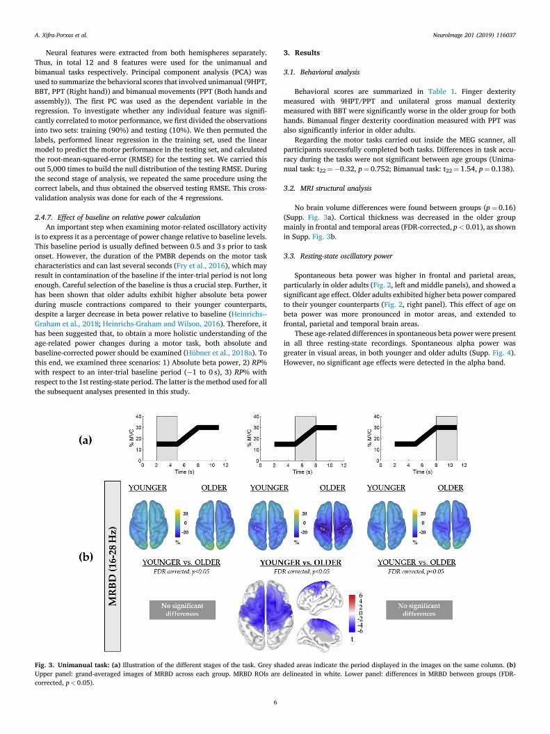
Fig. 3. Unimanual task: (a) Illustration of the different stages of the task. Grey shaUpper panel: grand-averaged images of MRBD across each group. MRBD ROIs arecorrected, p< 0.05).
6
3. Results
3.1. Behavioral analysis
Behavioral scores are summarized in Table 1. Finger dexteritymeasured with 9HPT/PPT and unilateral gross manual dexteritymeasured with BBT were significantly worse in the older group for bothhands. Bimanual finger dexterity coordination measured with PPT wasalso significantly inferior in older adults.
Regarding the motor tasks carried out inside the MEG scanner, allparticipants successfully completed both tasks. Differences in task accu-racy during the tasks were not significant between age groups (Unima-nual task: t22¼�0.32, p¼ 0.752; Bimanual task: t22¼ 1.54, p¼ 0.138).
3.2. MRI structural analysis
No brain volume differences were found between groups (p¼ 0.16)(Supp. Fig. 3a). Cortical thickness was decreased in the older groupmainly in frontal and temporal areas (FDR-corrected, p< 0.01), as shownin Supp. Fig. 3b.
3.3. Resting-state oscillatory power
Spontaneous beta power was higher in frontal and parietal areas,particularly in older adults (Fig. 2, left and middle panels), and showed asignificant age effect. Older adults exhibited higher beta power comparedto their younger counterparts (Fig. 2, right panel). This effect of age onbeta power was more pronounced in motor areas, and extended tofrontal, parietal and temporal brain areas.
These age-related differences in spontaneous beta power were presentin all three resting-state recordings. Spontaneous alpha power wasgreater in visual areas, in both younger and older adults (Supp. Fig. 4).However, no significant age effects were detected in the alpha band.
ded areas indicate the period displayed in the images on the same column. (b)delineated in white. Lower panel: differences in MRBD between groups (FDR-

Fig. 4. Bimanual task: (a) Illustration of the two 3-s subperiods of the task (grey shaded areas). (b) Upper panel: grand-averaged images of MRBD across each group.MRBD ROIs are delineated in white. Lower panel: differences in MRBD between groups (FDR-corrected, p< 0.05).
A. Xifra-Porxas et al. NeuroImage 201 (2019) 116037
3.4. Whole-brain MRBD analysis
Unimanual task. Grand-averaged surfaces displaying MRBD are shownin Fig. 3b (upper panel). We found significant differences in MRBDmagnitude underlying dynamic force production between the two agegroups (Fig. 3b, bottom panel): older adults exhibited increased (i.e.more negative) MRBD during the guided dynamic contraction (5–8 s). Nosignificant differences between groups were found during sustainedcontractions.
Bimanual task. Grand-averaged surfaces showing MRBD are shown inFig. 4b (upper panel). Similarly to the unimanual task, we found signif-icant differences in MRBDmagnitude between the two age groups only atthe beginning of the trial (2–5s) (Fig. 4b, bottom panel), during whicholder adults exhibited greater (i.e. more negative) MRBD. This specifictime interval corresponds to the period when subjects had not yetaccomplished a sustained grip and were thus still performing a dynamiccontraction (Supp. Fig. 1). The peak location of MRBD (denoted in whitein Fig. 4b, top row) did not exhibit significant age-related differences.
Results in the alpha frequency band for the unimanual and bimanualtasks are shown in Supp. Figs. 5 and 6, respectively. Alpha desynchro-nization did not exhibit significant differences between groups.
During the guided dynamic contraction period, older adults exhibiteda significantly greater and more widespread MRBD compared to youngeradults. During sustained contraction periods, no significant differences inMRBD were found between groups.
During the first 3-sec period, older adults exhibited a significantlystronger and more widespread MRBD compared to younger adults.During the second 3-sec period, during which subjects achieved thebimanual sustained contraction, no significant differences were foundbetween groups.
7
3.5. Modulation of beta oscillations
To investigate more precisely the modulation of beta oscillationsbetween different brain regions in younger and older adults, we extracteda power modulation metric from all ROIs (depicted in the first row ofFig. 5) for both task paradigms.
Unimanual task. The unimanual task induced modulations in betapower in several brain regions (Fig. 5). The modulations can be observedboth in the relative (with respect to resting-state power) and absolutepower subfigures. Results of the mixed ANOVA (Table 2) revealed asignificant main effect of “Age”, which suggests an overall difference inthe amplitude of beta power modulation between groups. Post-hoc testingrevealed a significantly larger modulation in older compared to youngeradults (t70¼�3.43, p¼ 0.001). We also observed a significant main ef-fect of “Brain Region”, which suggests that there was an overall differ-ence in beta power modulation between brain regions. Post-hoc testing ofthe “Brain Region” effect showed a significantly greater modulation inthe left and right ROIs (peak MRBD, located at the primary motorcortices) compared to the ageMRBD ROI (left peak MRBD vs. ageMRBD:t23¼�2.79, p¼ 0.010; left peak MRBD vs. ageMRBD: t23¼�3.68,p¼ 0.001), but no significant difference between left and right ROIs(t23¼�0.02, p¼ 0.983). Finally, there was no significant interactionbetween the factors. The statistical analysis quantified through ANOVAcan be evaluated qualitatively in Fig. 5.
Bimanual task. The bimanual task induced weaker modulations inMRBD compared to unimanual muscle contractions (Fig. 5). Nonetheless,the mixed ANOVA (Table 2) revealed the same significant main effects asin the unimanual task. A significant main effect of “Age” was observed,and post-hoc testing again showed significantly greater modulation inolder adults (t70¼�3.56, p< 0.001). We also detected a significant main

Fig. 5. Unimanual and Bimanual tasks: Tempo-ral evolution of the MRBD (upper row) and absolutebeta power response (lower row) in (a) ageMRBDROI, i.e. brain regions identified to exhibit strongerMRBD in older adults, (b) peak MRBD ROI (left M1)and (c) peak MRBD ROI (right M1). Older adultsexhibited higher absolute beta power throughoutthe entire movement execution for both tasks.During the unimanual task, we observed a greater(more negative) MRBD during the guided dynamiccontraction compared to sustained contraction pe-riods (15%MVC and 30%MVC) for both groups.During the bimanual task, older adults exhibitedgreater (more negative) MRBD at the beginning ofthe trial compared to their younger counterparts.
A. Xifra-Porxas et al. NeuroImage 201 (2019) 116037
effect of “Brain region”, and post-hoc testing revealed, as before, asignificantly larger modulation in the left and right ROIs (peak MRBD,located at the primary motor cortices) compared to the ageMRBD ROI(left peak MRBD vs. ageMRBD: t23¼�2.65, p¼ 0.014; left peak MRBDvs. ageMRBD: t23¼�2.69, p¼ 0.013), but no significant difference be-tween left and right ROIs (t23¼ 1.52, p¼ 0.142). Finally, there was nosignificant interaction between the factors. The statistical analysisquantified through ANOVA is illustrated qualitatively in Fig. 5.
3.6. PMBR analysis
We examined possible differences in PMBR between younger andolder adults for both tasks. The ROIs used are depicted in Supp. Fig. 2.
Unimanual task. We found no significant main effect of hemisphere orage; however, there was a significant age-by-hemisphere interaction(Table 3). This interaction indicates that the effect of hemisphere onPMBR was different in younger compared to older adults. To investigatethis interaction, 4 post-hoc tests were conducted using paired and
8
independent t-tests as appropriate, and a Bonferroni correction wasapplied (significance at 0.05/4¼ 0.0125). Paired t-tests between hemi-spheres did not reveal any significant difference (Younger: t11¼ 2.47,p¼ 0.031, Older: t11¼�1.35, p¼ 0.204). Independent t-tests yielded amarginally significant greater PMBR in the right hemisphere (ipsilateral)for the older group compared to the younger group (t22¼�2.66,p¼ 0.014), whereas no significant difference was found in the lefthemisphere (contralateral) (t22¼�0.28, p¼ 0.780).
Bimanual task. We did not find a main effect of hemisphere, age, orany age-by-hemisphere interaction (Table 3).
3.7. Associations between beta oscillations and motor performance
We carried out four linear regression analyses between beta oscilla-tions and motor performance scores. The cross-validation analysis isshown in Supp. Fig. 7. The prediction of task accuracy during theunimanual task was not significantly different compared to usingpermuted labels, hence no further analysis was done. For the other three
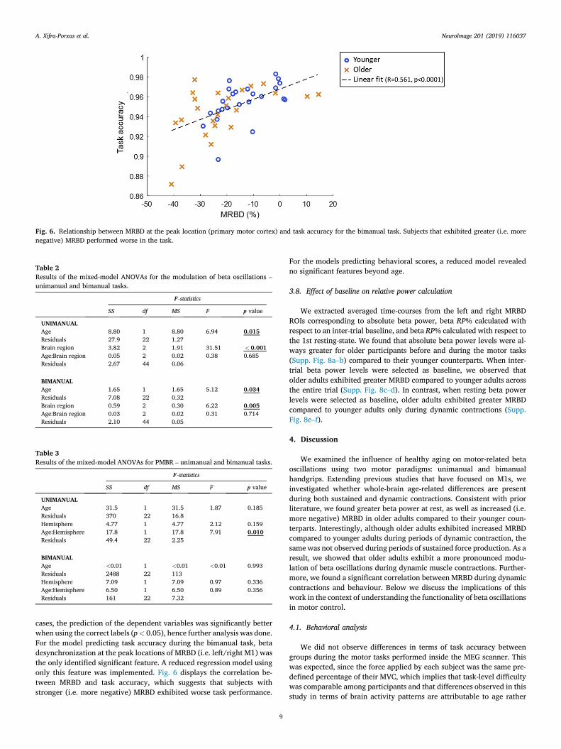
Fig. 6. Relationship between MRBD at the peak location (primary motor cortex) and task accuracy for the bimanual task. Subjects that exhibited greater (i.e. morenegative) MRBD performed worse in the task.
Table 2Results of the mixed-model ANOVAs for the modulation of beta oscillations –
unimanual and bimanual tasks.
F-statistics
SS df MS F p value
UNIMANUALAge 8.80 1 8.80 6.94 0.015Residuals 27.9 22 1.27Brain region 3.82 2 1.91 31.51 < 0.001Age:Brain region 0.05 2 0.02 0.38 0.685Residuals 2.67 44 0.06
BIMANUALAge 1.65 1 1.65 5.12 0.034Residuals 7.08 22 0.32Brain region 0.59 2 0.30 6.22 0.005Age:Brain region 0.03 2 0.02 0.31 0.714Residuals 2.10 44 0.05
Table 3Results of the mixed-model ANOVAs for PMBR – unimanual and bimanual tasks.
F-statistics
SS df MS F p value
UNIMANUALAge 31.5 1 31.5 1.87 0.185Residuals 370 22 16.8Hemisphere 4.77 1 4.77 2.12 0.159Age:Hemisphere 17.8 1 17.8 7.91 0.010Residuals 49.4 22 2.25
BIMANUALAge <0.01 1 <0.01 <0.01 0.993Residuals 2488 22 113Hemisphere 7.09 1 7.09 0.97 0.336Age:Hemisphere 6.50 1 6.50 0.89 0.356Residuals 161 22 7.32
A. Xifra-Porxas et al. NeuroImage 201 (2019) 116037
cases, the prediction of the dependent variables was significantly betterwhen using the correct labels (p< 0.05), hence further analysis was done.For the model predicting task accuracy during the bimanual task, betadesynchronization at the peak locations of MRBD (i.e. left/right M1) wasthe only identified significant feature. A reduced regression model usingonly this feature was implemented. Fig. 6 displays the correlation be-tween MRBD and task accuracy, which suggests that subjects withstronger (i.e. more negative) MRBD exhibited worse task performance.
9
For the models predicting behavioral scores, a reduced model revealedno significant features beyond age.
3.8. Effect of baseline on relative power calculation
We extracted averaged time-courses from the left and right MRBDROIs corresponding to absolute beta power, beta RP% calculated withrespect to an inter-trial baseline, and beta RP% calculated with respect tothe 1st resting-state. We found that absolute beta power levels were al-ways greater for older participants before and during the motor tasks(Supp. Fig. 8a–b) compared to their younger counterparts. When inter-trial beta power levels were selected as baseline, we observed thatolder adults exhibited greater MRBD compared to younger adults acrossthe entire trial (Supp. Fig. 8c–d). In contrast, when resting beta powerlevels were selected as baseline, older adults exhibited greater MRBDcompared to younger adults only during dynamic contractions (Supp.Fig. 8e–f).
4. Discussion
We examined the influence of healthy aging on motor-related betaoscillations using two motor paradigms: unimanual and bimanualhandgrips. Extending previous studies that have focused on M1s, weinvestigated whether whole-brain age-related differences are presentduring both sustained and dynamic contractions. Consistent with priorliterature, we found greater beta power at rest, as well as increased (i.e.more negative) MRBD in older adults compared to their younger coun-terparts. Interestingly, although older adults exhibited increased MRBDcompared to younger adults during periods of dynamic contraction, thesame was not observed during periods of sustained force production. As aresult, we showed that older adults exhibit a more pronounced modu-lation of beta oscillations during dynamic muscle contractions. Further-more, we found a significant correlation between MRBD during dynamiccontractions and behaviour. Below we discuss the implications of thiswork in the context of understanding the functionality of beta oscillationsin motor control.
4.1. Behavioral analysis
We did not observe differences in terms of task accuracy betweengroups during the motor tasks performed inside the MEG scanner. Thiswas expected, since the force applied by each subject was the same pre-defined percentage of their MVC, which implies that task-level difficultywas comparable among participants and that differences observed in thisstudy in terms of brain activity patterns are attributable to age rather
A. Xifra-Porxas et al. NeuroImage 201 (2019) 116037
than other factors, such as increased effort (Aine et al., 2006).On the other hand, older adults exhibited deteriorated fine motor
control in the corresponding behavioral assessments (Table 1), which isin line with the expected motor decline in older adults (Desrosiers et al.,1995; Grice et al., 2003; Lindstrom-Hazel and VanderVlies Veenstra,2015; Mathiowetz et al., 1985a). Handgrip strength was not significantlydifferent between groups due to high variability between individuals.
4.2. Structural analysis
Older adults were characterized by a significant decrease in corticalthickness, particularly in frontal and temporal brain areas (Supp. Fig. 3).The affected regions are in notable agreement with previous studies thatincluded larger sample sizes (Fjell et al., 2009; Hogstrom et al., 2013;Salat et al., 2004). These brain regions were overall in correspondencewith areas that exhibited age-related increases in MRBD (Figs. 3 and 4);however, the magnitude of MRBD and cortical thickness were not foundto be significantly correlated (R¼�0.14, p¼ 0.35), which suggests thatthe observed age-related functional differences may not be directlyassociated with this specific neurodegenerative process.
4.3. Age-related changes in power at rest
We found that older adults exhibited increased resting beta powercompared to younger adults (Fig. 2). We did not find significant differ-ences in resting alpha power between groups. Our results agree withseveral prior studies regarding age-related differences in power at rest,where it was reported that older adults exhibited similar levels of alphapower (Duffy et al., 1984; Heinrichs-Graham and Wilson, 2016; Koyamaet al., 1997; Veldhuizen et al., 1993) and increased beta power (G�omezet al., 2013; Heinrichs-Graham et al., 2018; Heinrichs-Graham and Wil-son, 2016; Hübner et al., 2018a; Koyama et al., 1997; Veldhuizen et al.,1993). However, previous studies only evaluated specific brain areasand/or performed the analysis in sensor space. Our whole-brain analysisdemonstrated that the motor cortex was the area that showed the mostsignificant differences in spontaneous beta power between younger andolder subjects. This aligns with the evidence that beta-band activity ispathologically increased in movement disorders such as Parkinson’sdisease (Brown et al., 2001; Silberstein et al., 2005), which suggests thatincreased beta oscillations at rest may be related with a deterioration offlexible behavioral and cognitive control (Engel and Fries, 2010). How-ever, when we probed whether spontaneous beta power was a goodpredictor of motor performance, we did not find any relationship thatlinked increased spontaneous beta power with poorer motorperformance.
4.4. Whole-brain age-related MRBD changes during muscle contractions
The majority of past studies that examined aging effects on motorcontrol have used motor paradigms whereby the subjects performed adynamic contraction, and they consistently reported age-related in-creases in MRBD – i.e. more negative desynchronization (Bardouilleet al., 2019; Heinrichs-Graham et al., 2018; Heinrichs-Graham and Wil-son, 2016; Hübner et al., 2018a; Rossiter et al., 2014). In line with thesestudies, during periods of dynamic contraction we found a significantincrease in MRBD in older adults compared to younger adults. Ourwhole-brain analysis further revealed a more widespread MRBD in olderadults, in contrast with younger adults, for which the desynchronizationwas mainly located in the M1s (Figs. 3b–4b, upper panel). Specifically,our results suggest a significant age-related increase in MRBD thatcovered frontal and premotor brain regions (Figs. 3b–4b, lower panel).Moreover, we observed that during periods of steady contractions, nodifferences were found between groups across the entire brain(Figs. 3b–4b, lower panel). Thus, our results align with the study fromRossiter and colleagues that reported no differences in MRBD in M1contralateral to the moving hand during steady contractions (Rossiter
10
et al., 2014), however our observations seem to indicate that the ipsi-lateral primary motor cortex does not show differences in MRBD either,in contrast with the study from Rossiter and colleagues (Rossiter et al.,2014).
4.5. Age-related changes in beta power modulation during musclecontractions
Both younger and older adults exhibited the expected modulation ofbeta oscillations that emerges when sequentially performing sustainedand dynamic contractions (Baker, 2007; Cassim et al., 2000; Kilner et al.,1999, 2003; Schoffelen et al., 2008; Spinks et al., 2008; van Wijk et al.,2012). This implies that the motor performance decline observed inhealthy aging is not due to an impairment in the capacity to modulatebeta oscillations. In fact, we observed a larger modulation in oldercompared to younger adults (Table 2). The increase in synchronized betaoscillations that emerges when producing a steady muscle contractionhas been suggested to provide an efficient processing platform for pro-moting the maintenance of a steady motor output whilst compromisinginitiation of newmovements (Androulidakis et al., 2007; Engel and Fries,2010; Gilbertson et al., 2005; Omlor et al., 2007; Pogosyan et al., 2009).Further, it has been recently suggested that absolute beta power needs toreach a certain threshold level in order to initiate a muscle contraction,regardless of age (Heinrichs-Graham andWilson, 2016). Beta oscillationsat rest are greater in older adults; this suggests that an increaseddesynchronization is needed for the required threshold to initiate amuscle contraction to be reached. If we only consider the results weobtained during dynamic contractions, our findings align well with thistheory, since older adults exhibited increased cortical beta suppressionwith respect to resting beta levels compared to younger adults. Yet,considering that we baseline-corrected the motor-related beta powerusing the spontaneous power observed at rest, our results also show thatduring sustained contractions there were no differences between groupsbeyond the ones observed at rest. Our findings may suggest that thethreshold in terms of absolute beta power for the maintenance of a sus-tained contraction is shifted in aging, whereas the threshold for executinga dynamic contraction remains the same.
4.6. Relationship between MRBD and motor performance
Two main theories have aimed to explain over-recruitment in aging:compensation and dedifferentiation (Reuter-Lorenz and Park, 2010). Thebasic idea of compensation is that brain reorganization in older adults is acompensatory mechanism to counterbalance impaired function. Alter-natively, the dedifferentiation hypothesis argues that older adults ineffi-ciently recruit additional brain areas because of less precise brainstructure-function interactions. Hence, this over-activation is not seenas a compensation mechanism to achieve better performance, rather as aless selective activation pattern. Several studies have provided evidenceof a positive correlation between over-recruitment and performanceduring a motor task (Mattay et al., 2002; Heuninckx et al., 2008). Otherstudies have reported that greater brain activity during a cognitive taskwas correlated to poorer performance (Logan et al., 2002; Stebbins et al.,2002). In another study it was reported that there was no correlationbetween brain activity and increased difficulty during a motor task(Riecker et al., 2006). These discrepancies suggest that the associationbetween increased activity in a specific brain region and performance inolder adults may be task-specific or dependent on the task demands andthe behavioral measure used. Therefore, in an attempt to unravelwhether the age-related overactivation of frontal/premotor/motor areasduring dynamic contractions and the increased modulation of betaoscillatory power between sustained and dynamic contractions in agingrepresent a compensation or dedifferentiationmechanism, we examined itsassociation with motor performance.
Features related to the brain regions that showed significantlyincreased MRBD in aging (ageMRBD) did not reveal any association with

A. Xifra-Porxas et al. NeuroImage 201 (2019) 116037
behavioral measures. This suggests that they were recruited in a non-selective fashion. Taken together with the fact that these brain regionsexhibited decreased cortical thickness in the older participants, theoveractivation of these regions in older adults might be indicative of aloss of functional specificity, and therefore supporting the dedifferentia-tion hypothesis. Recent observations that increased prefrontal cortexactivity in healthy aging does not contribute to maintain cognitivefunction (Morcom and Henson, 2018) would align with these results.Further, the modulation metric that quantified the depth of variations ofbeta oscillatory power did not show a relation with behaviour in any ofthe considered regions.
We identified one electrophysiological measure (beta desynchroni-zation at the peak MRBD ROIs) that associated beta oscillations andmotor performance, but only during bimanual muscle contractions insidethe MEG scanner. An explanation could be that the implementedunimanual task was not sensitive enough for the explanatory values tosignificantly predict performance. Participants with stronger (i.e. morenegative) MRBD at the peak location (M1) exhibited worse task perfor-mance. However, these regions did not show significant age-related in-creases in MRBD (Fig. 4b), thus we cannot interpret this association as acompensation or dedifferentiationmechanism. This finding is supported byobservations that after acute exercise, better performance is coupled withdecreased (i.e. less negative) MRBD (Dal Maso et al., 2018; Hübner et al.,2018b). We speculate that, since increased MRBD at the peak location iscorrelated with greater resting-state beta power (Heinrichs-Graham andWilson, 2016), the need to attenuate resting beta power to reach the betathreshold for proper motor execution may cause inferior task perfor-mance. However, further research is needed to understand the underly-ing mechanisms that link beta oscillatory activity and behaviour.
4.7. Age-related changes in PMBR
Recent studies reported that older adults exhibited reduced PMBR inthe contralateral hemisphere to the moving hand during a finger tappingtask compared to younger adults (Bardouille et al., 2019; Liu et al.,2017). On the other hand, our results suggest that older adults did notexhibit significant differences in PMBR in the contralateral (left) hemi-sphere to the moving hand during the unimanual task, but rather anincreased PMBR in the ipsilateral (right) hemisphere (Fig. 5, Table 3).Furthermore, during the bimanual task, no significant differences inPMBR were found between groups. It has been proposed that PMBR re-flects active inhibition of the motor network (Solis-Escalante et al., 2012)and it has been specifically linked to the inhibitory neurotransmitterγ-aminobutyric acid (GABA) (Gaetz et al., 2011; Jensen et al., 2005). Thissuggests that PMBR plays a role in preventing the generation of un-wanted movements. While speculative, our results may reflect a case ofdedifferentiation, whereby inhibition of both cortices after a motor taskoccurs in older adults, in contrast with younger adults for which PMBRoccurs only in the contralateral hemisphere to the executing hand.However, the precise mechanism underlying how PMBR is affected byaging remains to be fully elucidated.
4.8. Effects of baseline on relative power calculation
In agreement with previous studies, absolute beta power levels wereconsistently higher in older participants before and during the motortasks (Supp. Fig. 8a–b) (Heinrichs-Graham et al., 2018; Heinrichs-Gra-ham and Wilson, 2016; Hübner et al., 2018a). When inter-trial betapower levels were used as baseline, older adults exhibited greater MRBDcompared to younger adults across the entire trial (Supp. Fig. 8c–d). Incontrast, when resting beta power levels were used as baseline, olderadults exhibited greater MRBD compared to younger adults only duringdynamic contraction. The reason for this discrepancy is that beta powerlevels during the inter-trial period were significantly higher in bothgroups compared to resting levels (Supp. Fig. 8e–f), an indication thatinter-trial power levels were contaminated by PMBR. This is due to the
11
fact that the rebound effect can last several seconds after the end of amotor task, and has been associated with force output, such that higherforce output results in greater PMBR (Fry et al., 2016). Nevertheless, theinter-trial interval has been traditionally selected as baseline in motorstudies focused on MRBD and PMBR. Our results highlight the impor-tance of investigating whether the inter-trial power levels are artificiallyhigh due to PMBR contamination by comparing with resting powerlevels, as also suggested in recent studies (Heinrichs-Graham et al., 2018;Heinrichs-Graham and Wilson, 2016). In cases where the inter-trial isshort, resulting in PMBR contamination, the usage of a resting-staterecording for baseline normalization is strongly recommended.
4.9. Limitations
It has been suggested that resting beta levels and MRBD are modu-lated by the circadian rhythm (Toth et al., 2007; Wilson et al., 2014). Inthe present experiment, participants were scanned between 10 a.m. and 6p.m. (Morning session: 8 younger/6 older; Afternoon session: 4younger/6 older). Albeit somewhat balanced between groups, we cannotexclude circadian/ultradian effects on the results due to differences in thescanning time.
The inter-trial duration is a crucial parameter to consider whendesigning protocols to study motor-related beta oscillations. As weexemplify in Supp. Fig. 8, PMBR levels may contaminate the inter-trialbaseline, leading to possibly biased results. In this paper, we used theresting-state beta power levels as baseline to take into account this issue.Still, we cannot exclude the possibility that the MRBD was contaminatedby the elevated PMBR, since the inter-trial duration was not long enoughfor the PMBR to fully return to its baseline levels. Nevertheless, the PMBRis mostly localized within the M1s, whereas we observed most of the age-related differences in premotor and pre-frontal areas. This suggests thatthe obtained results are not biased by excessive contamination by theelevated PMBR levels.
The force applied during the experiment was based on each subject’sown MVC, from 0 to 30% MVC (unimanual) and from 0 to 15% MVC(bimanual), to ensure that the required effort, and consequently theresulting fatigue level, was the same across participants. To investigatewhether fatigue modulated the observed age-related differences inMRBD, we repeated our analysis using the initial and final 25 trialscorresponding to each task. We subsequently tested for whole-braindifferences in MRBD between younger and older adults for trials inthe first and second trial set. For the unimanual task, the analysisrevealed age-related differences only during the ramp block andgenerally in the same brain regions as the results using all trials (Supp.Fig. 9). These findings suggest that, for the unimanual task, physicalfatigue was either non-existent or its effect did not differ between agegroups. Specifically, if fatigue did occur, these results suggest that forthe resulting fatigue levels, the corresponding cortical adaptions did notdiffer between age groups. Nevertheless, age-related fatigue modula-tions were out of the scope of this paper, as we did not expect partici-pants to experience fatigue to a large extent based on the low MVClevels used in our paradigms. However, in future studies the use of theBorg scale to monitor fatigue perception could be a good way toquantify fatigue levels (Borg, 1982). For the bimanual task, age-relateddifferences were obtained during the initial 3s segment of the trial, butonly when using the last 25 trials (Supp. Fig. 10). However, it is notlikely that this observation is related to physical fatigue, since thebimanual task required considerably less force (15%MVC) compared tothe unimanual task (30%MVC), and its duration was shorter (6 s/trial)than the unimanual task (9 s/trial). Therefore, the observation seen inSupp. Fig. 10 is more likely related to low statistical power resultingfrom splitting the trials in half. However, because the motor tasks werenot counterbalanced, we cannot discard the possibility that physicaland/or mental fatigue may have had an effect on the results obtainedfor the bimanual task.

A. Xifra-Porxas et al. NeuroImage 201 (2019) 116037
5. Conclusions
Older adults exhibited significantly higher beta oscillations at rest,and our results showed that the motor cortex is the brain area that ex-hibits the highest increase in resting beta oscillatory activity. The presentstudy confirms that older adults produce a larger MRBD during dynamicmuscle contractions compared to younger adults. Our results also suggestthat during sustained contractions, there are no differences in beta powerbetween age groups beyond the ones observed at rest. We further probedthe relationship between motor performance and age-related differencesin beta oscillations during rest and task, but our results suggest that thisaltered beta activity in aging did not carry additional information.
Acknowledgements
We wish to thank Elizabeth Bock for her help during the dataacquisition as well as Arna Ghosh for his support in implementing thevisual feedback for the experiment. This work was supported by fundsfrom Fonds de la Recherche du Qu�ebec – Nature et Technologies(FRQNT; 2016-PR-191780) [MHB, GDM& SB], the Canadian Foundationfor Innovation grant numbers 34277 [MHB] and 34362 [GDM], theNatural Sciences and Engineering Research Council of Canada (NSERC436355-13), the National Institutes of Health (R01 EB026299), the BrainCanada Foundation (PSG15-3755), the Canada Research Chair program[SB], and the Canada First Research Excellence Fund (awarded to McGillUniversity for the Healthy Brains for Healthy Lives (HBHL) initiative).AXP and MK received financial support through funding from McGillUniversity and the Qu�ebec Bio-imaging Network (QBIN). GN receivedfinancial support through a postdoctorate fellowship from the AXAResearch Fund. SL acknowledges funding from FRQ– Sant�e (FRQS). Thefunders had no role in study design, data collection and analysis, decisionto publish, or preparation of the manuscript. The authors declare nocompeting financial or non-financial interests.
Appendix A. Supplementary data
Supplementary data to this article can be found online at https://doi.org/10.1016/j.neuroimage.2019.116037.
References
Aine, C.J., Woodruff, C.C., Knoefel, J.E., Adair, J.C., Hudson, D., Qualls, C., Bockholt, J.,Best, E., Kovacevic, S., Cobb, W., Padilla, D., Hart, B., Stephen, J.M., 2006. Aging:compensation or maturation? Neuroimage 32, 1891–1904. https://doi.org/10.1016/j.neuroimage.2006.05.005.
Androulidakis, A.G., Doyle, L.M.F., Gilbertson, T.P., Brown, P., 2006. Correctivemovements in response to displacements in visual feedback are more effective duringperiods of 13-35 Hz oscillatory synchrony in the human corticospinal system. Eur. J.Neurosci. 24, 3299–3304. https://doi.org/10.1111/j.1460-9568.2006.05201.x.
Androulidakis, A.G., Doyle, L.M.F., Yarrow, K., Litvak, V., Gilbertson, T.P., Brown, P.,2007. Anticipatory changes in beta synchrony in the human corticospinal system andassociated improvements in task performance. Eur. J. Neurosci. 25, 3758–3765.https://doi.org/10.1111/j.1460-9568.2007.05620.x.
Baillet, S., 2017. Magnetoencephalography for brain electrophysiology and imaging. Nat.Neurosci. 20, 327–339. https://doi.org/10.1038/nn.4504.
Baker, S.N., 2007. Oscillatory interactions between sensorimotor cortex and theperiphery. Curr. Opin. Neurobiol. 17, 649–655. https://doi.org/10.1016/j.conb.2008.01.007.
Bardouille, T., Bailey, L., CamCAN Group, 2019. Evidence for age-related changes insensorimotor neuromagnetic responses during cued button pressing in a large open-access dataset. Neuroimage 193, 25–34. https://doi.org/10.1016/j.neuroimage.2019.02.065.
Bohannon, R.W., Peolsson, A., Massy-Westropp, N., Desrosiers, J., Bear-Lehman, J., 2006.Reference values for adult grip strength measured with a Jamar dynamometer: adescriptive meta-analysis. Physiotherapy 92, 11–15. https://doi.org/10.1016/j.physio.2005.05.003.
Borg, G., 1982. Psychophysical bases of perceived exertion. Med. Sci. Sport. Exerc. 14,377–381. https://doi.org/10.1249/00005768-198205000-00012.
Box, G.E.P., Cox, D.R., 1964. An analysis of transformations. J. R. Stat. Soc. Ser. B 26,211–256.
Brown, P., Oliviero, A., Mazzone, P., Insola, A., Tonali, P., Di Lazzaro, V., 2001. Dopaminedependency of oscillations between subthalamic nucleus and pallidum in Parkinson’s
12
disease. J. Neurosci. 21, 1033–1038. https://doi.org/10.1523/JNEUROSCI.21-03-01033.2001.
Cassim, F., Szurhaj, W., Sediri, H., Devos, D., Bourriez, J.L., Poirot, I., Derambure, P.,Defebvre, L., Guieu, J.D., 2000. Brief and sustained movements: differences in event-related (de)synchronization (ERD/ERS) patterns. Clin. Neurophysiol. 111,2032–2039. https://doi.org/10.1016/S1388-2457(00)00455-7.
Cheyne, D.O., 2013. MEG studies of sensorimotor rhythms: a review. Exp. Neurol. 245,27–39. https://doi.org/10.1016/j.expneurol.2012.08.030.
Dal Maso, F., Desormeau, B., Boudrias, M.H., Roig, M., 2018. Acute cardiovascularexercise promotes functional changes in cortico-motor networks during the earlystages of motor memory consolidation. Neuroimage 174, 380–392. https://doi.org/10.1016/j.neuroimage.2018.03.029.
Dale, A.M., Fischl, B., Sereno, M.I., 1999. Cortical surface-based analysis: I. Segmentationand surface reconstruction. Neuroimage 9, 179–194. https://doi.org/10.1006/nimg.1998.0395.
Dale, A.M., Sereno, M.I., 1993. Improved localizadon of cortical activity by combiningEEG and MEG with MRI cortical surface reconstruction: a linear approach. J. Cogn.Neurosci. 5, 162–176. https://doi.org/10.1162/jocn.1993.5.2.162.
Desrosiers, J., H�ebert, R., Bravo, G., Dutil, E., 1995. The purdue pegboard test: normativedata for people aged 60 and over. Disabil. Rehabil. 17, 217–224. https://doi.org/10.3109/09638289509166638.
Duffy, F.H., Albert, M.S., McAnulty, G., Garvey, A.J., 1984. Age-related differences inbrain electrical activity of healthy subjects. Ann. Neurol. 16, 430–438. https://doi.org/10.1002/ana.410160403.
Engel, A.K., Fries, P., 2010. Beta-band oscillations-signalling the status quo? Curr. Opin.Neurobiol. 20, 156–165. https://doi.org/10.1016/j.conb.2010.02.015.
Erbil, N., Ungan, P., 2007. Changes in the alpha and beta amplitudes of the central EEGduring the onset, continuation, and offset of long-duration repetitive handmovements. Brain Res. 1169, 44–56. https://doi.org/10.1016/j.brainres.2007.07.014.
Espenhahn, S., de Berker, A.O., van Wijk, B.C.M., Rossiter, H.E., Ward, N.S., 2017.Movement-related beta oscillations show high intra-individual reliability.Neuroimage 147, 175–185. https://doi.org/10.1016/j.neuroimage.2016.12.025.
Fischl, B., Dale, A.M., 2000. Measuring the thickness of the human cerebral cortex frommagnetic resonance images. Proc. Natl. Acad. Sci. 97, 11050–11055. https://doi.org/10.1073/pnas.200033797.
Fischl, B., Liu, A., Dale, A.M., 2001. Automated manifold surgery: constructinggeometrically accurate and topologically correct models of the human cerebralcortex. IEEE Trans. Med. Imaging 20, 70–80. https://doi.org/10.1109/42.906426.
Fischl, B., Salat, D.H., Busa, E., Albert, M., Dieterich, M., Haselgrove, C., Van DerKouwe, A., Killiany, R., Kennedy, D., Klaveness, S., Montillo, A., Makris, N., Rosen, B.,Dale, A.M., 2002. Whole brain segmentation: automated labeling of neuroanatomicalstructures in the human brain. Neuron 33, 341–355.https://doi.org/10.1016/S0896-6273(02)00569-X.
Fischl, Bruce, Sereno, M.I., Dale, A.M., 1999a. Cortical surface-based analysis: II.Inflation, flattening, and a surface-based coordinate system. Neuroimage 9, 195–207.https://doi.org/10.1006/nimg.1998.0396.
Fischl, B., Sereno, M.I., Tootell, R.B.H., Dale, a M., 1999b. High-resolution inter-subjectaveraging and a surface-based coordinate system. Hum. Brain Mapp. 8, 272–284.https://doi.org/10.1002/(SICI)1097-0193(1999)8.
Fischl, B., Van Der Kouwe, A., Destrieux, C., Halgren, E., S�egonne, F., Salat, D.H., Busa, E.,Seidman, L.J., Goldstein, J., Kennedy, D., Caviness, V., Makris, N., Rosen, B.,Dale, A.M., 2004. Automatically parcellating the human cerebral cortex. Cerebr.Cortex 14, 11–22. https://doi.org/10.1093/cercor/bhg087.
Fjell, A.M., Westlye, L.T., Amlien, I., Espeseth, T., Reinvang, I., Raz, N., Agartz, I.,Salat, D.H., Greve, D.N., Fischl, B., Dale, A.M., Walhovd, K.B., 2009. High consistencyof regional cortical thinning in aging across multiple samples. Cerebr. Cortex 19,2001–2012. https://doi.org/10.1093/cercor/bhn232.
Folstein, M.F., Folstein, S.E., McHugh, P.R., 1975. “Mini-mental state”. A practicalmethod for grading the cognitive state of patients for the clinician. J. psychiat. Res.12, 189–198. https://doi.org/10.1016/0022-3956(75)90026-6.
Fry, A., Mullinger, K.J., O’Neill, G.C., Barratt, E.L., Morris, P.G., Bauer, M., Folland, J.P.,Brookes, M.J., 2016. Modulation of post-movement beta rebound by contractionforce and rate of force development. Hum. Brain Mapp. 37, 2493–2511. https://doi.org/10.1002/hbm.23189.
Gaetz, W., Edgar, J.C., Wang, D.J., Roberts, T.P.L., 2011. Relating MEG measured motorcortical oscillations to resting γ-Aminobutyric acid (GABA) concentration.Neuroimage 55, 616–621. https://doi.org/10.1016/j.neuroimage.2010.12.077.
Gilbertson, T., Lalo, E., Doyle, L., Di Lazzaro, V., Cioni, B., Brown, P., 2005. Existing motorstate is favored at the expense of new movement during 13-35 Hz oscillatorysynchrony in the human corticospinal system. J. Neurosci. 25, 7771–7779. https://doi.org/10.1523/JNEUROSCI.1762-05.2005.
G�omez, C., M P�erez-Macías, J., Poza, J., Fern�andez, A., Hornero, R., 2013. Spectralchanges in spontaneous MEG activity across the lifespan. J. Neural Eng. 10. https://doi.org/10.1088/1741-2560/10/6/066006.
Grice, K.O., Vogel, K.A., Le, V., Mitchell, A., Muniz, S., Vollmer, M.A., 2003. Adult normsfor a commercially available nine hole peg test for finger dexterity. Am. J. Occup.Ther. 57, 570–573. https://doi.org/10.5014/ajot.57.5.570.
Hall, S.D., Barnes, G.R., Furlong, P.L., Seri, S., Hillebrand, A., 2010. Neuronal networkpharmacodynamics of GABAergic modulation in the human cortex determined usingpharmaco-magnetoencephalography. Hum. Brain Mapp. 31, 581–594. https://doi.org/10.1002/hbm.20889.
Hall, S.D., Stanford, I.M., Yamawaki, N., McAllister, C.J., R€onnqvist, K.C., Woodhall, G.L.,Furlong, P.L., 2011. The role of GABAergic modulation in motor function relatedneuronal network activity. Neuroimage 56, 1506–1510. https://doi.org/10.1016/j.neuroimage.2011.02.025.

A. Xifra-Porxas et al. NeuroImage 201 (2019) 116037
Han, X., Jovicich, J., Salat, D., van der Kouwe, A., Quinn, B., Czanner, S., Busa, E.,Pacheco, J., Albert, M., Killiany, R., Maguire, P., Rosas, D., Makris, N., Dale, A.,Dickerson, B., Fischl, B., 2006. Reliability of MRI-derived measurements of humancerebral cortical thickness: the effects of field strength, scanner upgrade andmanufacturer. Neuroimage 32, 180–194. https://doi.org/10.1016/j.neuroimage.2006.02.051.
Heinrichs-Graham, E., McDermott, T.J., Mills, M.S., Wiesman, A.I., Wang, Y.-P.,Stephen, J.M., Calhoun, V.D., Wilson, T.W., 2018. The lifespan trajectory of neuraloscillatory activity in the motor system. Dev. Cogn. Neurosci. 30, 159–168. https://doi.org/10.1016/j.dcn.2018.02.013.
Heinrichs-Graham, E., Wilson, T.W., 2016. Is an absolute level of cortical beta suppressionrequired for proper movement? Magnetoencephalographic evidence from healthyaging. Neuroimage 134, 514–521. https://doi.org/10.1016/j.neuroimage.2016.04.032.
Heuninckx, S., Wenderoth, N., Swinnen, S.P., 2008. Systems neuroplasticity in the agingbrain: recruiting additional neural resources for successful motor performance inelderly persons. J. Neurosci. 28, 91–99. https://doi.org/10.1523/JNEUROSCI.3300-07.2008.
Hogstrom, L.J., Westlye, L.T., Walhovd, K.B., Fjell, A.M., 2013. The structure of thecerebral cortex across adult life: age-related patterns of surface area, thickness, andgyrification. Cerebr. Cortex 23, 2521–2530. https://doi.org/10.1093/cercor/bhs231.
Huang, M.X., Mosher, J.C., Leahy, R.M., 1999. A sensor-weighted overlapping-spherehead model and exhaustive head model comparison for MEG. Phys. Med. Biol. 44,423–440. https://doi.org/10.1088/0031-9155/44/2/010.
Hübner, L., Godde, B., Voelcker-Rehage, C., 2018a. Older adults reveal enhanced task-related beta power decreases during a force modulation task. Behav. Brain Res. 345,104–113. https://doi.org/10.1016/j.bbr.2018.02.028.
Hübner, L., Godde, B., Voelcker-Rehage, C., 2018b. Acute exercise as an intervention totrigger motor performance and EEG beta activity in older adults. Neural Plast. 2018,4756785. https://doi.org/10.1155/2018/4756785.
Jensen, O., Goel, P., Kopell, N., Pohja, M., Hari, R., Ermentrout, B., 2005. On the humansensorimotor-cortex beta rhythm : sources and modeling. Neuroimage 26, 347–355.https://doi.org/10.1016/j.neuroimage.2005.02.008.
Jovicich, J., Czanner, S., Greve, D., Haley, E., Van Der Kouwe, A., Gollub, R., Kennedy, D.,Schmitt, F., Brown, G., MacFall, J., Fischl, B., Dale, A., 2006. Reliability in multi-sitestructural MRI studies: effects of gradient non-linearity correction on phantom andhuman data. Neuroimage 30, 436–443. https://doi.org/10.1016/j.neuroimage.2005.09.046.
Jurkiewicz, M.T., Gaetz, W.C., Bostan, A.C., Cheyne, D., 2006. Post-movement betarebound is generated in motor cortex: evidence from neuromagnetic recordings.Neuroimage 32, 1281–1289. https://doi.org/10.1016/j.neuroimage.2006.06.005.
Kilner, J.M., Baker, S.N., Salenius, S., Jousm€aki, V., Hari, R., Lemon, R.N., 1999. Task-dependent modulation of 15-30 Hz coherence between rectified EMGs from humanhand and forearm muscles. J. Physiol. 516, 559–570. https://doi.org/10.1111/j.1469-7793.1999.0559v.x.
Kilner, J.M., Salenius, S., Baker, S.N., Jackson, A., Hari, R., Lemon, R.N., 2003. Task-dependent modulations of cortical oscillatory activity in human subjects during abimanual precision grip task. Neuroimage 18, 67–73. https://doi.org/10.1006/nimg.2002.1322.
Koyama, K., Hirasawa, H., Okubo, Y., Karasawa, A., 1997. Quantitative EEG correlates ofnormal aging in the elderly. Clin. Electroencephalogr. 28, 160–165.
Kuperberg, G.R., Broome, M.R., McGuire, P.K., David, A.S., Eddy, M., Ozawa, F., Goff, D.,West, W.C., Williams, S.C.R., van der Kouwe, A.J.W., Salat, D.H., Dale, A.M.,Fischl, B., 2003. Regionally localized thinning of the cerebral cortex in schizophrenia.Arch. Gen. Psychiatry 60 (9), 878–888. https://doi.org/10.1001/archpsyc.60.9.878.
Lindstrom-Hazel, D.K., VanderVlies Veenstra, N., 2015. Examining the purdue pegboardtest for occupational therapy practice. Open J. Occup. Ther. 3. https://doi.org/10.15453/2168-6408.1178.
Liu, L., Rosjat, N., Popovych, S., Wang, B.A., Yeldesbay, A., Toth, T.I., Viswanathan, S.,Grefkes, C.B., Fink, G.R., Daun, S., 2017. Age-related changes in oscillatory poweraffect motor action. PLoS One 12, 1–19. https://doi.org/10.1371/journal.pone.0187911.
Logan, J.M., Sanders, A.L., Snyder, A.Z., Morris, J.C., Buckner, R.L., 2002. Under-recruitment and nonselective recruitment: dissociable neural mechanisms associatedwith aging. Neuron 33, 827–840. https://doi.org/10.1016/S0896-6273(02)00612-8.
Maes, C., Gooijers, J., Orban de Xivry, J.J., Swinnen, S.P., Boisgontier, M.P., 2017. Twohands, one brain, and aging. Neurosci. Biobehav. Rev. 75, 234–256. https://doi.org/10.1016/j.neubiorev.2017.01.052.
Mathiowetz, V., Volland, G., Kashman, N., Weber, K., 1985a. Adult norms for the box andblock test of manual dexterity. Am. J. Occup. Ther. 39, 386–391. https://doi.org/10.5014/ajot.39.6.386.
Mathiowetz, V., Weber, K., Kashman, N., Volland, G., 1985b. Adult nonns for the ninehole peg test OfFinger dexterity. Occup. Ther. J. Res. 5, 24–38.
Mattay, V.S., Fera, F., Tessitore, A., Hariri, A.R., Das, S., Callicott, J.H., 2002.Neurophysiological correlates of age-related changes in human. Neurology 58,630–635. https://doi.org/10.1212/WNL.58.4.630.
Minati, L., Grisoli, M., Bruzzone, M.G., 2007. MR spectroscopy, functional MRI, anddiffusion-tensor imaging in the aging brain: a conceptual review. J. Geriatr.Psychiatry Neurol. 20, 3–21. https://doi.org/10.1177/0891988706297089.
Morcom, A.M., Henson, R.N.A., 2018. Increased prefrontal activity with aging reflectsnonspecific neural responses rather than compensation. J. Neurosci. 38 (33),7303–7313. https://doi.org/10.1523/JNEUROSCI.1701-17.2018.
Muthukumaraswamy, S.D., Myers, J.F.M., Wilson, S.J., Nutt, D.J., Lingford-Hughes, A.,Singh, K.D., Hamandi, K., 2013. The effects of elevated endogenous GABA levels onmovement-related network oscillations. Neuroimage 66, 36–41. https://doi.org/10.1016/j.neuroimage.2012.10.054.
13
Oldfield, R.C., 1971. The assessment and analysis of handedness: the Edinburghinventory. Neuropsychologia 9 (1), 97–113. https://doi.org/10.1016/0028-3932(71)90067-4.
Omlor, W., Patino, L., Hepp-Reymond, M.C., Kristeva, R., 2007. Gamma-rangecorticomuscular coherence during dynamic force output. Neuroimage 34,1191–1198. https://doi.org/10.1016/j.neuroimage.2006.10.018.
Pfurtscheller, G., Lopes da Silva, F.H., 1999. Event-related EEG/MEG synchronization anddesynchronization : basic principles. Clin. Neurophysiol. 110, 1842–1857.https://doi.org/10.1016/S1388-2457(99)00141-8.
Pogosyan, A., Gaynor, L.D., Eusebio, A., Brown, P., 2009. Boosting cortical activity atbeta-band frequencies slows movement in humans. Curr. Biol. 19, 1637–1641. https://doi.org/10.1016/j.cub.2009.07.074.
Provencher, D., Hennebelle, M., Cunnane, S.C., Berube-Lauziere, Y., Whittingstall, K.,2016. Cortical thinning in healthy aging correlates with larger motor-evoked EEGdesynchronization. Front. Aging Neurosci. 8, 1–8. https://doi.org/10.3389/fnagi.2016.00063.
Reuter-Lorenz, P.A., Park, D.C., 2010. Human neuroscience and the aging Mind : a newlook at old problems. J. Gerontol. Psychol. Sci. 65B, 405–415. https://doi.org/10.1093/geronb/gbq035.
Reuter, M., Rosas, H.D., Fischl, B., 2010. Highly accurate inverse consistent registration: arobust approach. Neuroimage 53, 1181–1196. https://doi.org/10.1016/j.neuroimage.2010.07.020.
Reuter, M., Schmansky, N.J., Rosas, H.D., Fischl, B., 2012. Within-subject templateestimation for unbiased longitudinal image analysis. Neuroimage 61, 1402–1418.https://doi.org/10.1016/j.neuroimage.2012.02.084.
Riecker, A., Gr€oschel, K., Ackermann, H., Steinbrink, C., Witte, O., Kastrup, A., 2006.Functional significance of age-related differences in motor activation patterns.Neuroimage 32, 1345–1354. https://doi.org/10.1016/j.neuroimage.2006.05.021.
Rosas, H.D., Liu, A.K., Hersch, S., Glessner, M., Ferrante, R.J., Salat, D.H., Van DerKouwe, A., Jenkins, B.G., Dale, A.M., Fischl, B., 2002. Regional and progressivethinning of the cortical ribbon in Huntington’s disease. Neurology 58, 695–701.https://doi.org/10.1212/WNL.58.5.695.
Rossiter, H.E., Davis, E.M., Clark, E.V., Boudrias, M.H., Ward, N.S., 2014. Beta oscillationsreflect changes in motor cortex inhibition in healthy ageing. Neuroimage 91,360–365. https://doi.org/10.1016/j.neuroimage.2014.01.012.
Rosso, A.L., Studenski, S.A., Chen, W.G., Aizenstein, H.J., Alexander, N.B., Bennett, D.A.,Black, S.E., Camicioli, R., Carlson, M.C., Ferrucci, L., 2013. Aging, the central nervoussystem, and mobility. J. Gerontol. Med. Sci. 68, 1379–1386. https://doi.org/10.1093/gerona/glt089.
Sailer, A., Dichgans, J., Gerloff, C., 2000. The influence of normal aging on the corticalprocessing of a simple motor task. Neurology 979–986.
Salat, D.H., Buckner, R.L., Snyder, A.Z., Greve, D.N., Desikan, R.S.R., Busa, E.,Morris, J.C., Dale, A.M., Fischl, B., 2004. Thinning of the cerebral cortex in aging.Cerebr. Cortex 14, 721–730. https://doi.org/10.1093/cercor/bhh032.
Salmelin, R., H€am€al€ainen, M., Kajola, M., Hari, R., 1995. Functional segregation ofmovement-related rhythmic activity in the human brain. Neuroimage. https://doi.org/10.1006/nimg.1995.1031.
Schmiedt-Fehr, C., Mathes, B., Kedilaya, S., Krauss, J., Basar-Eroglu, C., 2016. Agingdifferentially affects alpha and beta sensorimotor rhythms in a go/nogo task. Clin.Neurophysiol. 127, 3234–3242. https://doi.org/10.1016/j.clinph.2016.07.008.
Schoffelen, J.M., Oostenveld, R., Fries, P., 2008. Imaging the human motor system’s beta-band synchronization during isometric contraction. Neuroimage 41, 437–447.https://doi.org/10.1016/j.neuroimage.2008.01.045.
Scott, S.H., 2012. The computational and neural basis of voluntary motor control andplanning. Trends Cogn. Sci. 16, 541–549. https://doi.org/10.1016/j.tics.2012.09.008.
S�egonne, F., Dale, A.M., Busa, E., Glessner, M., Salat, D., Hahn, H.K., Fischl, B., 2004.A hybrid approach to the skull stripping problem in MRI. Neuroimage 22,1060–1075. https://doi.org/10.1016/j.neuroimage.2004.03.032.
Seidler, R.D., Bernard, J.A., Burutolu, T.B., Fling, B.W., Gordon, M.T., Gwin, J.T.,Kwak, Y., Lipps, D.B., 2010. Motor control and aging: links to age-related brainstructural, functional, and biochemical effects. Neurosci. Biobehav. Rev. 34,721–733. https://doi.org/10.1016/j.neubiorev.2009.10.005.
Silberstein, P., Pogosyan, A., Kühn, A.A., Hotton, G., Tisch, S., Kupsch, A., Dowsey-Limousin, P., Hariz, M.I., Brown, P., 2005. Cortico-cortical coupling in Parkinson’sdisease and its modulation by therapy. Brain 128, 1277–1291. https://doi.org/10.1093/brain/awh480.
Solis-Escalante, T., Müller-Putz, G.R., Pfurtscheller, G., Neuper, C., 2012. Cue-inducedbeta rebound during withholding of overt and covert foot movement. Clin.Neurophysiol. 123, 1182–1190. https://doi.org/10.1016/j.clinph.2012.01.013.
Spinks, R.L., Kraskov, A., Brochier, T., Umilta, M.A., Lemon, R.N., 2008. Selectivity forgrasp in local field potential and single neuron activity recorded simultaneously fromM1 and F5 in the awake macaque monkey. J. Neurosci. 28, 10961–10971. https://doi.org/10.1523/JNEUROSCI.1956-08.2008.
Stanc�ak, A., Pfurtscheller, G., 1995. Desynchronization and recovery of β rhythms duringbrisk and slow self-paced finger movements in man. Neurosci. Lett. 196, 21–24.https://doi.org/10.1016/0304-3940(95)11827-J.
Stebbins, G.T., Carrillo, M.C., Dorfman, J., Dirksen, C., Desmond, J.E., Turner, D. a,Bennett, D. a, Wilson, R.S., Glover, G., Gabrieli, J.D.E., 2002. Aging effects onmemory encoding in the frontal lobes. Psychol. Aging 17, 44–55. https://doi.org/10.1037/0882-7974.17.1.44.
Tadel, F., Baillet, S., Mosher, J.C., Pantazis, D., Leahy, R.M., 2011. Brainstorm: a user-friendly application for MEG/EEG analysis. Comput. Intell. Neurosci. 2011. https://doi.org/10.1155/2011/879716.

A. Xifra-Porxas et al. NeuroImage 201 (2019) 116037
Toth, M., Kiss, A., Kosztolanyi, P., Kondakor, I., 2007. Diurnal alterations of brainelectrical activity in healthy adults: a LORETA study. Brain Topogr. 20, 63–76. https://doi.org/10.1007/s10548-007-0032-3.
Van Veen, B.D., van Drongelen, W., Yuchtman, M., Suzuki, A., 1997. Localization of brainelectrical activity via linearly constrained minimum variance spatial filtering. IEEETrans. Biomed. Eng. 44, 867–880. https://doi.org/10.1109/10.623056.
van Wijk, B.C.M., Beek, P.J., Daffertshofer, A., 2012. Differential modulations ofipsilateral and contralateral beta (de)synchronization during unimanual force
14
production. Eur. J. Neurosci. 36, 2088–2097. https://doi.org/10.1111/j.1460-9568.2012.08122.x.
Veldhuizen, R.J., Jonkman, E.J., Poortvliet, D.C., 1993. Sex differences in age regressionparameters of healthy adults–normative data and practical implications.Electroencephalogr. Clin. Neurophysiol. 86, 377–384.
Wilson, T.W., Heinrichs-Graham, E., Becker, K.M., 2014. Circadian modulation of motor-related beta oscillatory responses. Neuroimage 102, 531–539. https://doi.org/10.1016/j.neuroimage.2014.08.013.