OfOLITH ULTRASTRUcrURE OF SMOOTH OREO, MACULATUS, … · black oreo is the most commonly caught...
17
OfOLITH ULTRASTRUcrURE OF SMOOTH OREO, PSEUDOCYTTUS MACULATUS, AND BLACK OREO, ALWCYTTUS SP., SPECIES N. M. DAVIES,! R. W. GAULDIE,I,2 S. A. CRANE,! AND R. K. ABSTRACT The ultrastructure of sagittal otoliths from 14 Pseudocyttrul maculai:'l18 and 25 Allocyttrul sp. individuals were examined to determine their suitability for estimating age in these two species. Scanning electron microscopy revealed high levels of complexity in both external surface topography and internal struc- tural organization in the sagittae of both species. Many different crystal forms were found, including calcite-like prisms. A close similarity in otolith structure exists between the two species. Deposition of cI1eek rings analagous to annual and daily growth increments was found to be irregular with the underlying complexity of crystalline growth obscuring the finer (analogous to daily) growth rings, making their periodicity difficult to validate and implying that with present tecl1niques the sagittal otolit)ls of the orea species PS6'Udocyttus maculat"U8 and Allocyttus sp. are not suitable for age estimation. The smooth oreo, Pseudocyttus maculatus, and the black oreo, Allocyttus sp., are two related species of the family Oreosomatidae. They are both important commercial species in New Zealand. The black oreo is the most commonly caught oreo in New Zealand waters, while the smooth oreo is the second-most commonly caught oreo. Little is known about the biology of these fish. The black oreo is endemic to New Zealand while the smooth oreo occurs in New Zealand, South Australian, South African, and South American waters (Last et aI. 1983). In the waters south of New Zealand, the distributions of the two species overlap (McMillan 1985). The habitat range of the smooth oreo is be- tween 650 and 1,200 m, and that of the black oreo is between 600 and 1,200 m (McMillan 1985). A preliminary examination of the ultrastructure of otoliths [sagittae] of these fish was undertaken as part of a study to establish an ultrastructural basis for a suitable ageing technique. This study describes the external and internal structure and organization of the otoliths in terms of the suitabil- ity of the various check rings of the sagittae for age estimation. MATERIALS AND METHODS Three otoliths (the sagitta, astericus, and lapillus) 'Fisheries Research Centre, Ministry of Agriculture and Fish- eries, Greta Point, Evans Bay Parade, P.O. Box 297, Wellington, New Zealand. ITo whom reprint requests should be sent. IElectron Microscope Unit, Kirk Building, Victoria University of Wellington, Private Bag, Wellington, New Zealand. Manuscript accepted April 1988. FISHERY BULLETIN: VOL. 86, NO.3, 1988. are contained in the endolymphatic sac (Fig. 1a). The sagitta is the largest otolith and is located in the most ventral position in the sac. The arrangement of the three otoliths in the endolymphatic sac ranges between the primitive where a large astericus and sagitta with no lapillus is present (Gauldie et al. 1986) and the typical teleost arrangement where a small astericus is located close to the sagitta, and an even smaller lapillus is displaced into the atrium of the semi-circular canal. The oreosomatids are primitive fishes taxonomically, lying in the order Beryciformes (Nelson 1976), and the arrangement of otoliths reflects the taxonomic position of the fish. The orientation of otoliths described here refers to the orientation in situ. The lateral face is the out- ward (antisulcal) surface; the medial face is the in- ward (sulcal) surface. Investigation was restricted to the sagitta primarily because of the difficulties in establishing homologies for daily and annual type check rings in the astericus and lapillus. Sagittae were dissected from 14 smooth and 25 black oreo individuals caught in bottom trawls off the east coast of New Zealand. These individuals ranged in length from 24.5 to 40.1 cm (black oreo) and 35.1 to 51.2 cm (smooth oreo). Whole otoliths were photographed at 6 x to 20 x using a WILD" photomicroscope. The sagittae were embedded on glass slides in epoxy resin with the antisulcus surface uppermost and finely ground on a Struers Planapol-2 petrographic grinder. The 'Reference to trade names does not imply endorsement by the National Marine Fisheries Service, NOAA. 499
Transcript of OfOLITH ULTRASTRUcrURE OF SMOOTH OREO, MACULATUS, … · black oreo is the most commonly caught...
OfOLITH ULTRASTRUcrURE OF SMOOTH OREO, PSEUDOCYTTUSMACULATUS, AND BLACK OREO, ALWCYTTUS SP., SPECIES
N. M. DAVIES,! R. W. GAULDIE,I,2 S. A. CRANE,! AND
R. K. THOMPSO~
ABSTRACT
The ultrastructure of sagittal otoliths from 14 Pseudocyttrul maculai:'l18 and 25 Allocyttrul sp. individualswere examined to determine their suitability for estimating age in these two species. Scanning electronmicroscopy revealed high levels of complexity in both external surface topography and internal structural organization in the sagittae of both species. Many different crystal forms were found, includingcalcite-like prisms. A close similarity in otolith structure exists between the two species. Deposition ofcI1eek rings analagous to annual and daily growth increments was found to be irregular with the underlyingcomplexity of crystalline growth obscuring the finer (analogous to daily) growth rings, making theirperiodicity difficult to validate and implying that with present tecl1niques the sagittal otolit)ls of the oreaspecies PS6'Udocyttus maculat"U8 and Allocyttus sp. are not suitable for age estimation.
The smooth oreo, Pseudocyttus maculatus, andthe black oreo, Allocyttus sp., are two relatedspecies of the family Oreosomatidae. They are bothimportant commercial species in New Zealand. Theblack oreo is the most commonly caught oreo inNew Zealand waters, while the smooth oreo is thesecond-most commonly caught oreo. Little is knownabout the biology of these fish. The black oreo isendemic to New Zealand while the smooth oreooccurs in New Zealand, South Australian, SouthAfrican, and South American waters (Last et aI.1983). In the waters south of New Zealand, thedistributions of the two species overlap (McMillan1985). The habitat range of the smooth oreo is between 650 and 1,200 m, and that of the black oreo isbetween 600 and 1,200 m (McMillan 1985).
A preliminary examination of the ultrastructureof otoliths [sagittae] of these fish was undertakenas part of a study to establish an ultrastructuralbasis for a suitable ageing technique. This studydescribes the external and internal structure andorganization of the otoliths in terms of the suitability of the various check rings of the sagittae for ageestimation.
MATERIALS AND METHODS
Three otoliths (the sagitta, astericus, and lapillus)
'Fisheries Research Centre, Ministry of Agriculture and Fisheries, Greta Point, Evans Bay Parade, P.O. Box 297, Wellington,New Zealand.
ITo whom reprint requests should be sent.IElectron Microscope Unit, Kirk Building, Victoria University
of Wellington, Private Bag, Wellington, New Zealand.
Manuscript accepted April 1988.FISHERY BULLETIN: VOL. 86, NO.3, 1988.
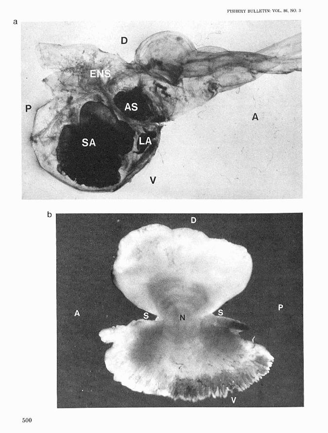
are contained in the endolymphatic sac (Fig. 1a). Thesagitta is the largest otolith and is located in themost ventral position in the sac. The arrangementof the three otoliths in the endolymphatic sac rangesbetween the primitive where a large astericus andsagitta with no lapillus is present (Gauldie et al.1986) and the typical teleost arrangement where asmall astericus is located close to the sagitta, andan even smaller lapillus is displaced into the atriumof the semi-circular canal. The oreosomatids areprimitive fishes taxonomically, lying in the orderBeryciformes (Nelson 1976), and the arrangementof otoliths reflects the taxonomic position of the fish.The orientation of otoliths described here refers tothe orientation in situ. The lateral face is the outward (antisulcal) surface; the medial face is the inward (sulcal) surface. Investigation was restrictedto the sagitta primarily because of the difficultiesin establishing homologies for daily and annual typecheck rings in the astericus and lapillus.
Sagittae were dissected from 14 smooth and 25black oreo individuals caught in bottom trawls offthe east coast of New Zealand. These individualsranged in length from 24.5 to 40.1 cm (black oreo)and 35.1 to 51.2 cm (smooth oreo).
Whole otoliths were photographed at 6 x to 20 xusing a WILD" photomicroscope. The sagittae wereembedded on glass slides in epoxy resin with theantisulcus surface uppermost and finely ground ona Struers Planapol-2 petrographic grinder. The
'Reference to trade names does not imply endorsement by theNational Marine Fisheries Service, NOAA.
499
a
FISHERY BULLETIN: VOL. 86, NO.3
500
b
c
DAVIES ET AL.: OTOLITH ULTRASTRUCTURE OF SMOOTH AND BLACK OR EO
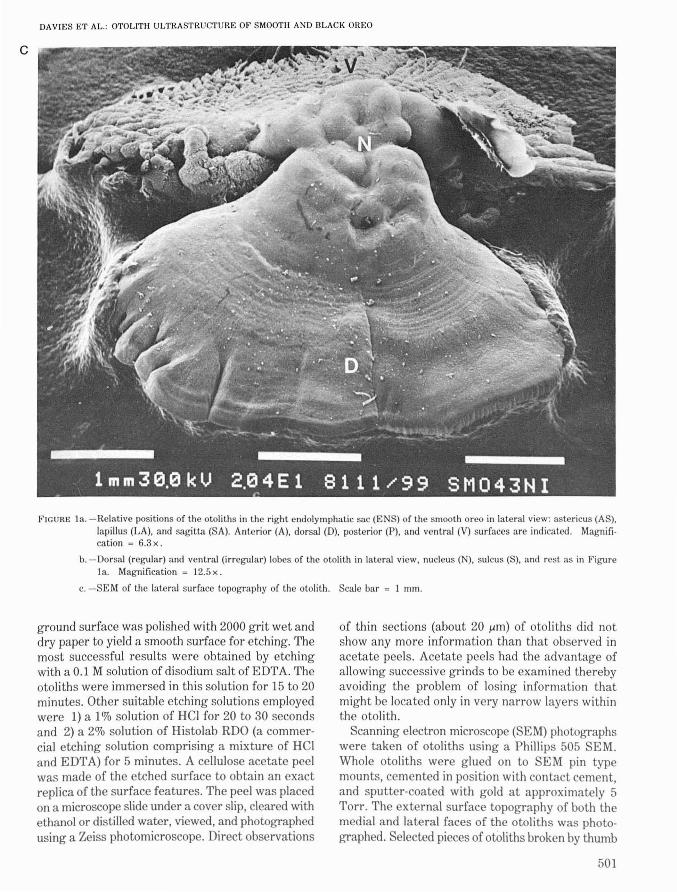
FIGURE 1a. -Relative positions of the otoliths in the right endolymphatic sac (ENS) of the smooth oreo in lateral view: astericus (AS).lapillus (LA), and sagitta (SA). Anterior (A), dorsal (D), posterior (P), and ventral (V) surfaces are indicated. Magnifi·cation = 6.3 x .
b. -Dorsal (regular) and ventral (irregular) lobes of the otolith in lateral view, nucleus (N), sulcus (S), and rest as in Figure1a. Magnification ~ 12.5 x .
c. -SEM of the lateral surface topography of the otolith. Scale bar = 1 mm.
ground sW'face was polished with 2000 grit wet anddry paper to yield a smooth surface for etching. Themost successful results were obtained by etchingwith aO.1 M solution of disodium saltofEDTA. Theotoliths were immersed in this solution for 15 to 20minutes. Other suitable etching solutions employedwere 1) a 1% solution of HCI for 20 to 30 secondsand 2) a 2% solution of Histolab RDO (a commercial etching solution comprising a mixture of HCIand EDTA) for 5 minutes. A cellulose acetate peelwas made of the etched surface to obtain an exactreplica of the surface features. The peel was placedon a microscope slide under a cover slip, cleared withethanol or clistilled water, viewed, and photographedusing a Zeiss photomicroscope. Direct observations
of thin sections (about 20 /Am) of otoliths did notshow any more information than that observed inacetate peels. Acetate peels had the advantage ofallowing successive grinds to be examined therebyavoiding the problem of losing information thatmight be located only in very narrow layers withinthe otolith.
Scanning electron microscope (SEM) photographswere taken of otoliths using a Phillips 505 SEM.WllOle otoliths were glued on to SEM pin typemounts, cemented in position with contact cement,and sputter-coated with gold at approximately 5Torr. The external surface topography of both themedial and lateral faces of the otoliths was photographed. Selected pieces of otoliths broken by thumb
501
pressure were examined and photographed to obtain internal structural information. Finely groundcross-section surfaces were also polished and etchedfor examination with the SEM.
RESULTS
Smooth Oreo Otolith
The sagitta is clearly divided into two distinctstructures: a small, smooth, regular dorsal lobe anda larger, irregular ventral lobe (Fig. Ib). The irregular lobe has branched crystal formations and cleftsat the ventral edge. The lateral face topography iscomplex (Fig. Ic). The central bumpy area containsthe nucleus between the two lobes. Radiating outwards from the nucleus are concentric ridges on theantisulcul surface.
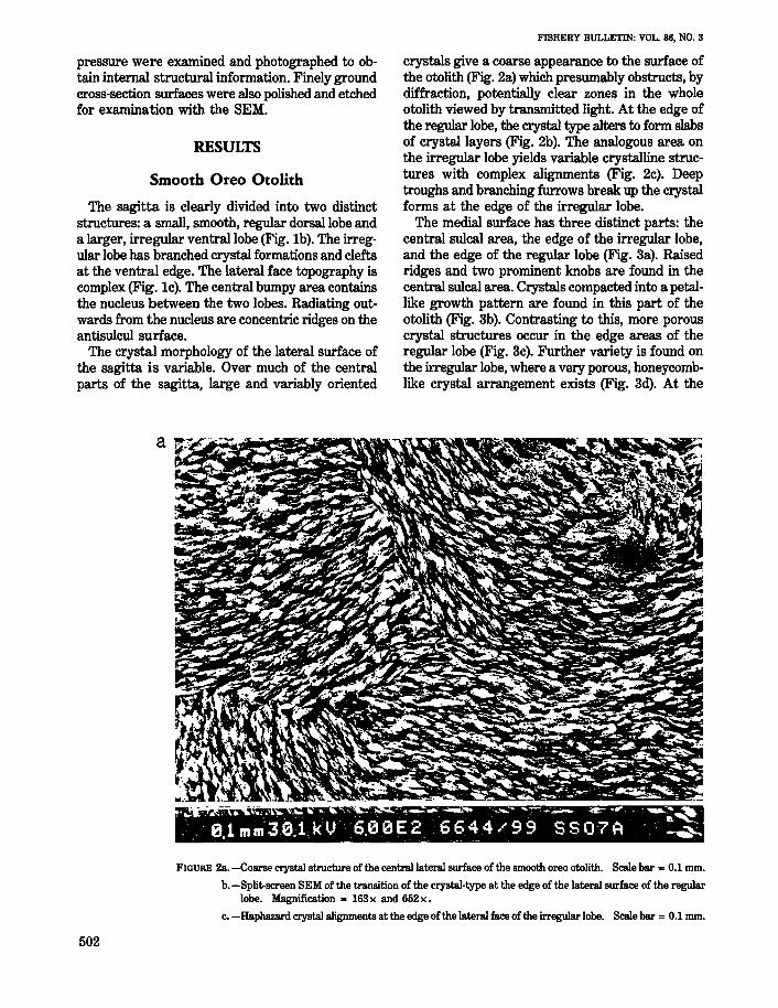
The crystal morphology of the lateral surface ofthe sagitta is variable. Over much of the centralparts of the sagitta, large and variably oriented
FISHERY BULLETIN: VOL. 86, NO.3
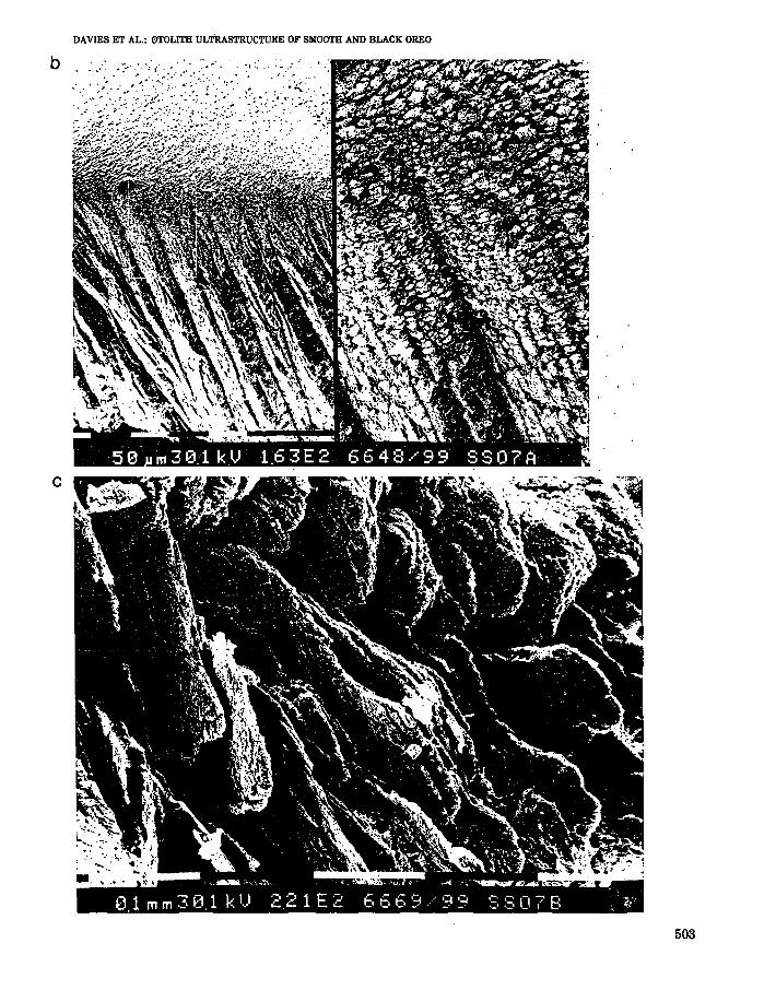
crystals give a coarse appearance to the surface ofthe otolith (Fig. 2a) which presumably obstructs, bydiffraction, potentially clear zones in the wholeotolith viewed by transmitted light. At the edge ofthe regular lobe, the crystal type alters to form slabsof crystal layers (Fig. 2b). The analogous area onthe irregular lobe yields variable crystalline structures with complex alignments (Fig. 2c). Deeptroughs and branching furrows break up the crystalforms at the edge of the irregular lobe.
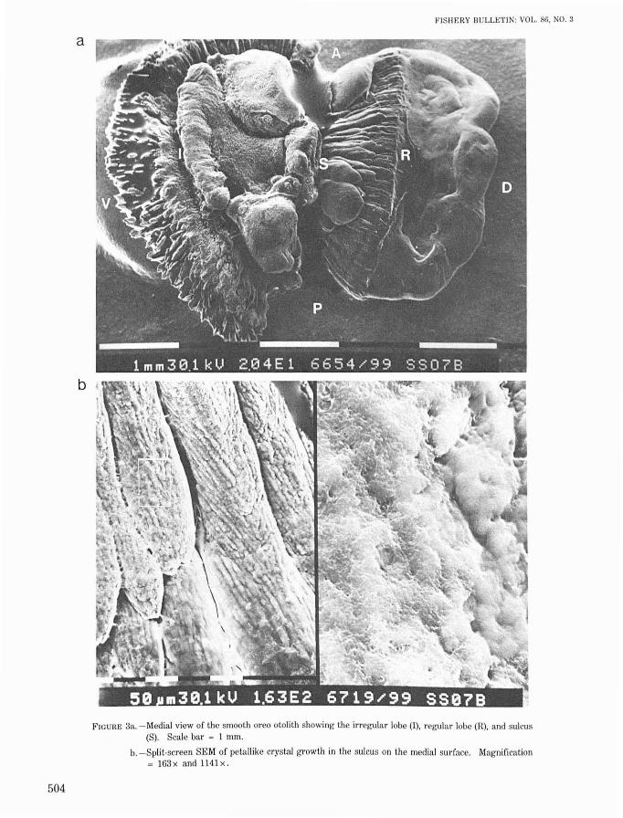
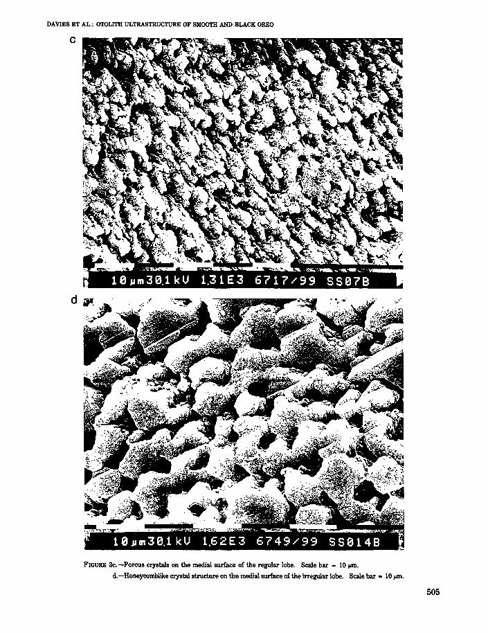
The medial surface has three distinct parts: thecentral sulcal area, the edge of the irregular lobe,and the edge of the regular lobe (Fig. 3a). Raisedridges and two prominent knobs are found in thecentral sulcal area. Crystals compacted into a petallike growth pattern are found in this part of theotolith (Fig. 3b). Contrasting to this, more porouscrystal structures occur in the edge areas of theregular lobe (Fig. 3c). Further variety is found onthe irregular lobe, where a very porous, honeycomblike crystal arrangement exists (Fig. 3d). At the
..... . ;'... <".,.'1 . ,. ~"," ......-.....~~ ........... ....--::,..~ ~ ...,.. ~; a, e.lmm~30.1 kU 6.0~E2 '6~4:4/~99 SS07A ~
FIGURE 2a. -Coarse crystal stmcture of the central lateral surface of the smooth oreo otolith. Scale bar - 0.1 mm.
b. -Split-screen SEM of the transition of the crystal-type at the edge of the lateral surface of the regularlobe. Magnification - 163x and 652x.
c. -Haphazard crystal alignments at the edge of the lateral face of the irregular lobe. Scale bar = 0.1 mm.
502
b
c
DAVIES ET AL.: 0TOLITH ULTRASTRUCTURE OF SMOOTH AND BLACK OREO
503
504
a
b
FISHERY BULLETIN: YOLo 86, NO.3
FIGURE 3a. -Medial view of the smooth oreo otolith showing the irregular lobe (I), regular lobe (R), and sulcus(S). Scale bar = 1 mrn.
b. -Split-screen SEM of petallike crystal growth in the sulcus on the medial surface, Magnification= 163x and 1141 x,
DAVIES ET AL.: OTOLITH ULTRASTRUCTURE OF SMOOTH AND BLACK OREO
c
FIGURE Bc. -Porous crystals on the medial surface of the regular lobe. Scale bar = 10 lAm.
d.-Honeycomblike crystal structure on the medial surface of the irregular lobe. Scale bar - 10 JoIIll.
505
FISHERY BULLETIN: VOL. 86, NO.3
e
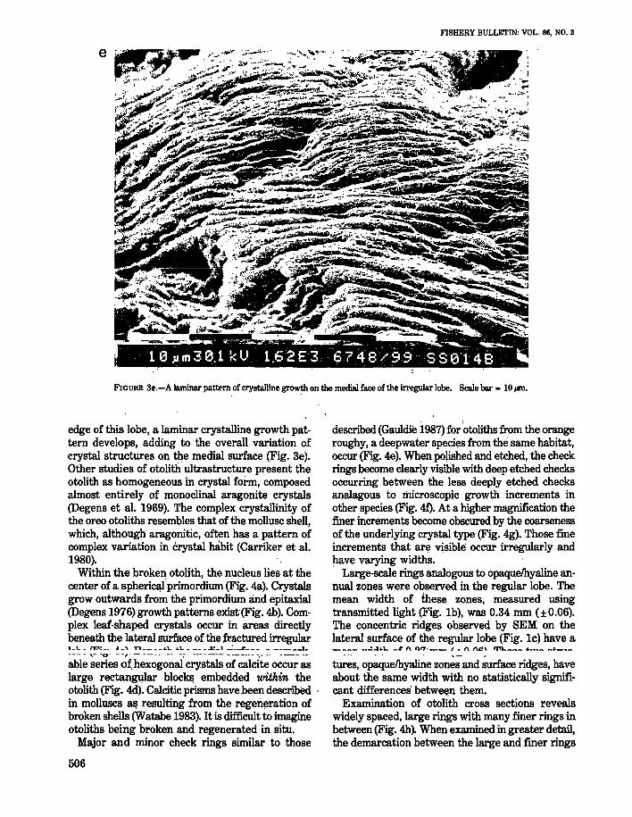
FIGURE 3e.-A laminar pattern of crystalline growth on the medial face of the irregular lobe. Scale bar = 10,..m.
edge of this lobe, a laminar crystalline growth pattern develops, adding to the overall variation ofcrystal structures on the medial surface (Fig. 3e).Other studies of otolith ultrastructure present theotolith as homogeneous in crystal form, composedalmost entirely of monoclinal aragonite crystals(Degens et aI. 1969). The complex crystallinity ofthe oreo otoliths resembles that of the mollusc shell,which, although aragonitic, often has a pattern ofcomplex variation in crystal habit (Carriker et al.1980).
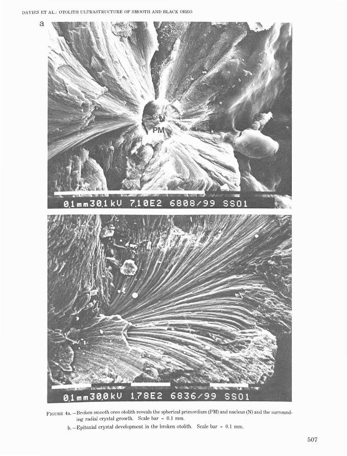
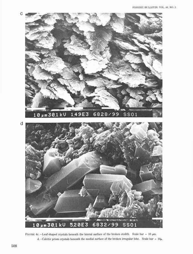
Within the broken otolith, the nucleus lies at thecenter of a spheri~'primordium (Fig. 4a). Crystalsgrow outwards from the primordium and epitaxial(Degens 1976) growth patterns exist(Fig. 4b). Complex leaf-shaped crystals occur in areas directlybeneath the lateral surface of the ;fractured irregular1_1. _ I'r.'! __ ._, T'I .Ll.. ...1.. __ ~_..l.:_l _~_• ..R .... 1_
-_.- - ,- -0" --," - --------- --;-- --------- "-----_. -.,.- ----.--
able series o~ hexogonal crystals of calcite occur aslarge rectangular block~ embedded within theotolith (Fig. 4d). Calcitic prisms have been described .in molluscs aEi resulting from the regeri.~rationofbroken shells (Watabe 1983). It is difficult to imagineotoliths being broken and regenerated in situ.
Major and minor check rings similar to those
506
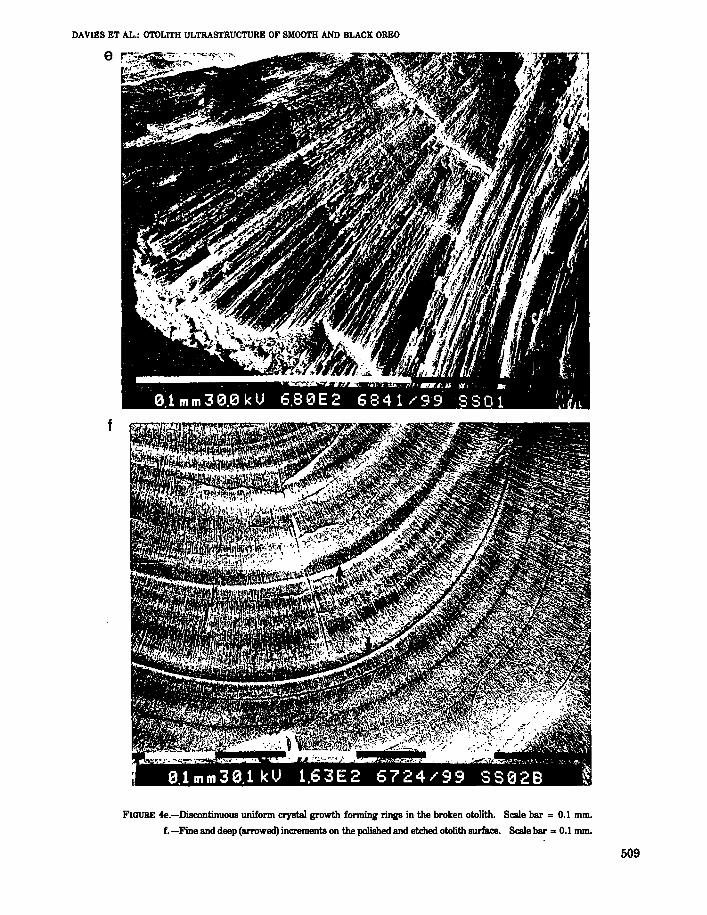
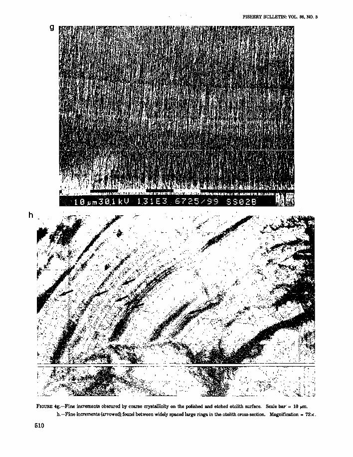
described (Gauldie 1987) for otoliths from the orangeroughy, a deepwater speci~s from the same habitat,occur (Fig. 4e). When polished and etched, the checkrings become clearly visible with deep etched checksoccurring between the less deeply etched checksanalagous to microscopic growth increments inother species (Fig. 4f). At a higher magnification thefiner increments become obscured by the coarsenessof the underlying crystal type (Fig. 4g). Those fineincrements that ar~ visible' occur irregularly andhave varying widths.
Large-scale rings analogous to opaquelhyaline annual zones were observed in the regular lobe. Themean width of these zones, measured usingtransmitted light (Fig. Ib), was 0.34 mm (±0.06).The concentric ridges observed by SEM on thelateral surface of the regular lobe (Fig. Ie) have a_ ..:..J 'L _~ t\ n",__ I • n n~\ '1'11.. _ _ ...
tures, opaquelhyaline zones and surface ridges, haveabout the same width with no statistically significant differences' betwe~n them.
Examination of otolith cross sections revealswidely spaced, large rings with many finer rings inbetween (Fig. 4h). When examined in greater detail,the demarcation between the large and finer rings
DAVIES ET AL.: OTOLITH ULTRASTRUCTURE OF SMOOTH AND BLACK OREO
a
F'ICUIlE 4a. -Broken smooth oreo otolith reveals the spherical primordium (PM) and nucleus (N) and the sllrrounding radial crystal growth. Scale bar = 0.1 mm.
b. -Epitaxial crystal development in the broken otolith. Scale bar = 0.1 mm.
507
508
c
d
1~ISIIERY BULLETIN: VOL. 86. NO.3
FIGURE 4c. -Leaf-shaped crystals beneath the lateral surface of the broken otolith. Scale bar = 10 lIm.
d.-Calcitic prism crystals beneath the medial surface of the broken irregular lobe. Scale bar = 10".
DAVIES ET AL.: OTOLITH ULTRASTRUCTURE OF SMOOTH AND BLACK OREO
e
f
FIGURE 4e.-Diseontinuous uniform crystal growth forming rings in the broken otolith. Scale bar = 0.1 mm.
f. -Fine and deep (arrowed) increments on the polished and etched otolith surface. Scale bar = 0.1 mm.
509
FISHERY BULLETIN: VOL. 86, NO.3
9
.,,/·.ttf·
::",,; .. ..'.'. f'<l~
,;,':,i;t~--I I: " ..~ - .z--- --...:-'--~~..-._-._..--P'!'"~'--:i:t~:~r; ;"'~f~~;"; ~. ,_~~~:'ri§}'c,ji;jFIGURE 4g.-Fine increments obscured by coarse crystallinity on the polished and etched otolith surface. Scale bar' = 10 fIIIl.
h.-Fine increments (arrowed). found between widely spaced large rings in the otolith cross-section. Magnification - 72 x.
h '''J;:;f';;,,~."< .,." . fr / .:....-, .:..~' .. ;": :,;:.." :,'. ( . if':: . ~ I .~:. ~ ••
!' .yrfj.. ',\1..' I·"" ';" . .:......' "";I. ..' .... ", ?'. • ,'. .
510
DAVIES ET AL.: OTOLITH ULTRASTRUCTURE OF SMOOTH AND BLACK OREO
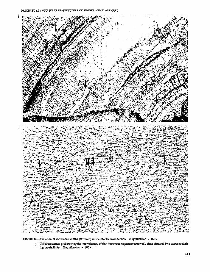
FIGURE 4i.-Variation of increment widths (arrowed) in the otolith cross-section. Magnification = 160x.
j.-Cellulose-acetate peel showing the intennittency of fine increment sequences (arrowed), often obscured by a coarse underlying crystallinity. Magnification = 160x.
511
becomes difficult to determine because of the intermittent nature of the growth increments and thevariety of width-sizes (Fig. 4i). In large areas of theotolith, increments appear to be absent or indeterminate, with a coarse underlying crystal structure(Fig. 4j) making accurate determination of increment sequences difficult. However, the fine increments of the oreo otolith are 3 to 5 jAlIl wide, whichis within the range of daily growth incrementsdescribed for other species (Jones 1986; Gauldie inpress).
Black Oreo Otolith
The black oreo otolith is almost identical to thatfound in the smooth oreo in overall shape, proportion, structure, topography, surface and internalcrystaHinity, and increment pattern. Some minordifferences do, however, exist.
FISHERY BULLETIN: VOL. 86, NO.8
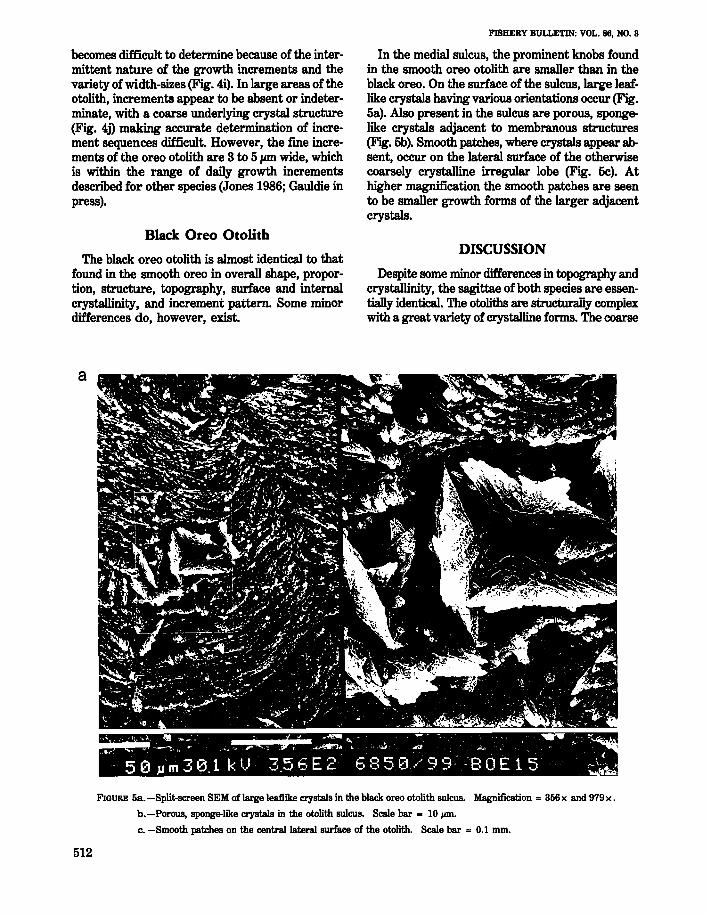
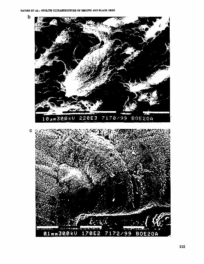
In the medial sulcus, the prominent knobs foundin the smooth oreo otolith are smaller than in theblack oreo. On the surface of the sulcus, large leaflike crystals having various orientations occur (Fig.5a). Also present in the sulcus are porous, spongelike crystals adjacent to membranous structures(Fig. 5b). Smooth patches, where crystals appear absent, occur on the lateral surface of the otherwisecoarsely crystalline irregular lobe (Fig. 5c). Athigher magnification the smooth patches are seento be smaller growth forms of the larger adjacentcrystals.
DISCUSSION
Despite some minor differences in topography andcrystallinity, the sagittae of both species are essentially identicaL The otoliths are structurally complexwith a great variety of crystalline forms. The coarse
a
-, ~ ~ --- - ..~.... •• I. \)., ~ ':>!oo._ L ~ .... -' ~ • -~.., ;) ',. .. k , .... --;. I. ~ - ~ r ~ \, . ~ - ~ ~ .(
5 0 .u m 3 0.1 k IJ 3.5 6 E 2 6 8 5 0 ./ 9 9 -B 0 E 1 5 -, .
FIGURE 5a. -Split-screen SEM of large leaflike crystals in the black oreo otolith sulcus. Magnification = 356 x and 979 x .
b.-Porous, sponge-like crystals in the otolith sulcus. Scale bar - 10,.ro.
c. -Smooth patches on the central lateral surface of the otolith. Scale bar = 0.1 mm.
512
DAVIES ET AL.: OTOLITH ULTRASTRUCTURE OF SMOOTH AND BLACK OREO
b
c
513
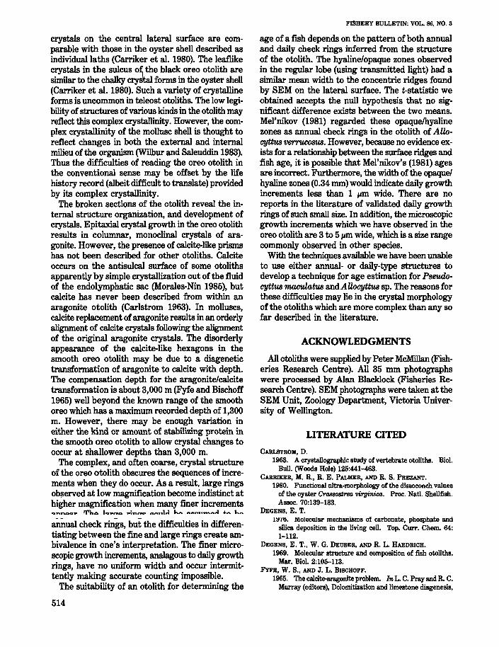
crystals on the central lateral surface are comparable with those in the oyster shell described asindividual laths (Carriker et al. 1980). The leaflikecrystals in the sulcus o~ the black oreo otolith aresimilar to the chalky crys'taI forms in the oyster shell(Carriker et al. 1980). Such a variety of crystallineforms is uncommon in teleost otoliths. The low legibility of structures of various kinds in the otolith mayreflect this complex crystallinity. However, the complex crystallinity of the mollusc shell is thought toreflect changes in both the external and internalmilieu of the organism (Wilbur and Saleuddin 1983).Thus the difficulties of reading the oreo otolith inthe conventional sense may be offset by the lifehistory record (albeit difficult to translate) providedby its complex crystallinity.
The broken sections of the otolith reveal the internal structure organization, and development ofcrystals. Epitaxial crystal growth in the oreo otolithresults in columnar, monoclinal crystals of aragonite. However, the presence of calcite-like prismshas not been described for other otoliths. Calciteoccurs on the antisulcal surface of some otolithsapparently by simple crystallization out of the fluidof the endolymphatic sac (Morales-Nin 1985), butcalcite has never been described from within anaragonite otolith (Carlstrom 1963). In molluscs,calcite replacement of aragonite results in an orderlyalignment of calcite crystals following the alignmentof the original aragop.ite crystals. The disorderlyappearance of the calcite-like hexagons in thesmooth oreo otolith may be due to a diagenetictransformation of aragonite to calcite with depth.The compensation depth for the aragonite/calcitetransformation is about 3,000 m (Fyfe and Bischoff1965) well beyond the known range of the smoothoreo which has a maximum recorded depth of 1,300m. However, there may be enough variation ineither the kind or amount of stabilizing protein inthe smooth oreo otolith to allow crystal changes tooccur at shallower depths than 3,000 m.
The complex, and often coarse, crystal structureof the oreo otolith obscures the sequences of increments when they do occur. As a result, large ringsob!\erved at low magnification become indistinct athigher magnification when many finer incrementsQnT\oa.,. Tho. lq......o. Pi'l"ll"ft:l ",,,,..,1,,.1 l,.,. n",,,,,....-.n,,.) 40ft ...... ",
annual check rings, but the difficulties in differentiating between the fine and large rings create ambivalence in one's interpretation. The finer microscopic growth increments, analagous to daily growthrings, have no uniform width and occur intermittently making accurate counting impossible.
The suitability of an otolith for determining the
514
FISHERY BULLETIN: VOL. 86, NO.3
age of a fish depends on the pattern of both annualand daily check rings inferred from the structureof the otolith. The hyaline/opaque zones observedin the regular lobe (using transmitted light) had asimilar mean width to the concentric ridges foundby SEM on the lateral surface. The t-statistic weobtained accepts the null hypothesis that no significant difference exists between the two means.Mel'nikov (1981) regarded these opaquelhyalinezones as annual check rings in the otolith of AllocytI:us verrucosus. However, because no evidence exists for a relationship between the surface ridges andfish age, it is possible that Mel'nikov's (1981) agesare incorrect. Furthermore, the width of the opaque/hyaline zones (0.34 mm) would indicate daily growthincrements less than 1 Ilm wide. There are noreports in the literature of validated daily growthrings of such small size. In addition, the microscopicgrowth increments which we have observed in theoreo otolith are 3 to 5 Ilm wide, which is a size rangecommonly observed in other species.
With the techniques available we have been unableto use either annual- or daily-type structures todevelop a technique for age estimation for Pseudocyttus maculatus and Allocyttus sp. The reasons forthese difficulties may lie in the crystal morphologyof the otoliths which are more complex than any sofar described in the literature.
ACKNOWLEDGMENTS
All otoliths were supplied by Peter McMillan (Fisheries Research Centre). All 35 mm photographswere processed by Alan Blacklock (Fisheries Research Centre). SEM photographs were taken at theSEM Unit, Zoology Department, Victoria University of Wellington.
LITERATURE CITED
CARLSTROM, D.
1963. A crystallographic study of vertebrate otoliths. BioI.Bull. (Woods Hole) 125:441-463.
CARRIKER, M. R., R. E. PALMER, AND R. S. PREZANT.1980. Functional ultra-morphology of the dissoconch values
of the oyster Crassostrea 'llirginica. Proc. Natl. Shellfish.Assoc. 70:139-183.
DEGENs. E. T.llf/li. M.olecular mecllanisms of carbonate, phosphate and
silica deposition in the living cell. Top. Curro Chem. 64:1-112.
DEGENs, E. T., W. G. DEUSER, AND R. L. HAEDRICH.
1969. Molecular structure and composition of fish otoliths.Mar. BioI. 2:105-113.
FYFE, W. S., AND J. L. BISCHOFF.1965. The calcite-aragonite problem. In L. C. Pray and R. C.
Murray (editors), Dolomitization and limestone diagenesis,
DAVIES ET AL.: OTOLITH ULTRASTRUCTURE OF SMOOTH AND BLACK OREO
p.3-13. Symp. Soc. Econ. Minist. Spec. Pub\. 13.GAULDIE, R. W.
1987. The fine structure of check rings in the otolith of theNew Zealand orange roughy. NZ J. Mar. Freshwater Res.21:267-274.
In press. A study of otolith daily growth rings in the orangeroughy (Hoplostetkus atla:nticus) aimed at resolving problems in age, growth, recruitment and otolith architecture.NZ Fish. Res. Bull.
GAULDIE, R. W., D. DuNLOP, AND J. TSE.1986. The remarkable lungfish otolith. NZ J. Mar. Fresh
water Res. 20:81-92.JONES, C.
1986. Determining age of larval fish with the otolith increment technique. Fish. BuI\., U.S. 84:91-103.
LAST, P. R., E. O. G. SCOTT, AND F. H. TALBOT.1983. Fishes of Tasmania. Hobart, Tasmanian Fisheries
Development Authority.MEL'NIKOV, Y. S.
1981. Size-age composition and growth pattern ofAIWcytt118-verrucosus (Oreosomatidae). Ichthyo\. (Eng\. transl. Vopr.
Ikhtio\.) 21:178-184.MORALES-NIN, B.
1985. Caracteristicas de los otolitos cristalinos de Genypterusoap6lUlis, (Smith, 1847) (pisces: Ophidiidae). Invest. Pesq.49:379-386.
McMILLAN, P. J.1985. Black and smooth oreo dories. b& J. A. Colman, J. L.
McKoy, and G. G. Baird (editors), Background papers forthe 1985 total allowable catch recommendations. (Reportheld in the Fisheries Research Centre Library, P.O. Box 297,Wellington, New Zealand.)
NELSON, J. S.1976. Fishes of the world. John Wiley and Sons. N.Y., 416 p.
WATABE, N.1983. Shell repair. In A. S. M. Saleuddin and K. M. Wilbur
(editors), The mollusca, NoI4, p 289-316. Acad. Press, N.Y.WILBUR, K. M., AND A. S. M. SALEUDDIN.
1983. Shell formation. In A. S. M. Saleuddin and K. M.Wilbur (editors), The mollusca, NoI4, p 236-288. Acad.Press, N.Y.
515