Of Mice and Men and Cattle: Functions of the Pneumovirus ... · Part of this work is or will be...
118
Dissertation der Fakultät für Biologie der Ludwig-Maximilians-Universität München zur Erlangung des Dr. rer.nat. Of Mice and Men and Cattle: Functions of the Pneumovirus Nonstructural Proteins NS1 and NS2 in Interferon Escape vorgelegt von Birgit Bossert am 26. September 2002
Transcript of Of Mice and Men and Cattle: Functions of the Pneumovirus ... · Part of this work is or will be...
Dissertation
der
Fakultät für Biologie
der
Ludwig-Maximilians-Universität München
zur Erlangung des Dr. rer.nat.
Of Mice and Men and Cattle:
Functions of the Pneumovirus Nonstructural
Proteins NS1 and NS2 in Interferon Escape
vorgelegt von
Birgit Bossert
am 26. September 2002

Erstgutachterin: PD Dr. Ruth Brack-Werner
Zweitgutachter: PD Dr. Brian Salmons
Sondergutachter: Prof. Dr. Karl-Klaus Conzelmann
Tag der mündlichen Prüfung: 28.01.2003
Part of this work is or will be published:
Schlender J., B. Bossert, U. Buchholz, and K. K. Conzelmann. 2000. Bovine respiratory
syncytial virus nonstructural proteins NS1 and NS2 cooperatively antagonize alpha/beta
interferon- induced antiviral response. J.Virol. 74: 8234-8242.
Bossert B. and K. K. Conzelmann. 2002. Respiratory syncytial virus (RSV) nonstructural
(NS) proteins as host range determinants: a chimeric bovine RSV with NS genes from
human RSV is attenuated in interferon-competent bovine cells. J.Virol. 76: 4287-4293.
Bossert B. and K. K. Conzelmann. Bovine respiratory syncytial virus nonstructural
proteins NS1 and NS2 inhibit activation of interferon regulatory factor 3 (IRF-3).
Manuscript in preparation.
Other publications:
Gromeier M., S. Mueller, D. Solecki, B. Bossert, G. Bernhardt and E. Wimmer. 1997.
Determinants of poliovirus neurovirulence. J.Neurovirol. 3: 35-38.
Gromeier M., B. Bossert, M. Arita, A. Nomoto and E. Wimmer. 1999. Dual stem loops
within the poliovirus internal ribosomal entry site control neurovirulence. J.Virol. 73: 958-
964.
“…, but was engaged, heart and soul, in the pursuit of some discoveries which I hoped to
make. None but those who have experienced them can conceive of the enticements of
science. In other studies you go as far as others have gone before you, and there is nothing
more to know; but in a scientific pursuit there is continual food for discovery and wonder.”1
1 Mary Shelley, “Frankenstein or, The Modern Prometheus”, 1818

I
TABLE OF CONTENTS
List of tables and figures IV
List of abbreviations VI
INTRODUCTION 1 Respiratory Syncytial Virus 2
Infectious agent 2
The genome 3
Viral proteins 4
Replicative cycle 5
Epidemiology 7
Pathogenesis and immunity 8
2 The IFN-α /β system 9
Virus induction of IFN genes 10
Signal transduction in response to IFNs 12
The antiviral response 13
Viral countermeasures to the IFN response 14
3 The nonstructural proteins NS1 and NS2 17
Overview 17
Reverse genetics as a tool to study viral gene functions 19
BRSV NS proteins are IFN antagonists 20
4 Aim of this study 22
MATERIALS AND METHODS 1 Materials 23
1-1 Chemicals 23
1-2 Enzymes 23
1-3 Kits 23
1-4 Miscellaneous 23
1-5 Bacteria and plasmids 24
II
1-6 Cells and viruses 26
1-7 Cell culture reagents 27
1-8 Serological reagents 28
1-9 Frequently used buffers 28
1-10 Oligonucleotides 29
2 Methods 30
2-1 General cloning procedures 30
2-2 PCR and reverse transcriptase reaction 31
2-3 Generation of recombinant rRVs expressing pneumovirus NS proteins 32
2-4 Generation of recombinant BRSVs expressing NS1ha or NS2fl 34
2-5 Generation of rBRSVs expressing HRSV and PVM NS proteins 35
2-6 Recovery of recombinant rBRSV and rRV 36
2-7 Production of virus stocks 37
2-8 Infection experiments and treatment with IFN 38
2-9 Western blot analysis of pneumovirus NS proteins 39
2-10 Immunofluorescent staining 40
2-11 Monitoring ISG expression 41
2-12 Detection of IFN in supernatants 41
2-13 Transfection of reporter plasmids and luciferase assay 41
2-14 Detection of IRF-3 phosphorylation 43
RESULTS 1 Pneumovirus NS proteins cooperatively confer viral resistance to IFN 44
Construction of recombinant RVs expressing BRSV NS proteins 44
BRSV NS1 and NS2 cooperatively enhance IFN resistance of RV 45
HRSV and PVM NS proteins protect RV from IFN-induced responses 48
HRSV and BRSV NS proteins are able to cooperate 50
A recombinant RV expressing both BRSV NS proteins 51
2 Involvement of pneumovirus NS proteins in determining viral host range 53
Chimeric BRSVs expressing HRSV NS genes 53
Growth of BRSV h1/2 is only attenuated in bovine cells 55
IFN antagonist activity is host cell specific 56
Chimeric BRSVs expressing PVM NS genes 57
BRSV m1/2 is attenuated in human and bovine cells 58
III
3 BRSV NS proteins are required for inhibition of IFN-β induction 61
ISG expression is not upregulated in BRSV wt infected cells 61
IFN is not produced in BRSV wt infected cells 64
BRSV wt inhibits induction of the IFN-β gene promoter 65
BRSV wt prevents activation of IRF-3 67
Both NS proteins are required for inhibition of IFN induction 69
HRSV and BRSV NS proteins cooperated in blocking IFN induction 70
PVM NS proteins cannot prevent induction of IFN 71
4 Excursus: Cellular localization of pneumovirus NS proteins 73
Pneumovirus NS proteins show nuclear localization 73
NS proteins reach nucleus independent of other pneumovirus proteins 77
Pneumovirus NS proteins are retained in the nucleus 78
DISCUSSION Pneumovirus NS proteins are IFN antagonists 80
Involvement of pneumovirus NS proteins in determining host range 82
BRSV NS protein are required for inhibition of IFN-β induction 85
Nuclear localization of pneumovirus NS proteins 90
The frequently asked question: why two proteins? 92
SUMMARY 94
References 96
Curriculum Vitae 103
Danksagung 104
IV
List of tables and figures
Table 1 The paramyxoviridae family and its characteristic members 1
Fig. 1 Organization of a pneumovirus particle 2
Fig. 2 Genome organization of RSV 3
Fig. 3 Transcriptional induction of the IFN-β gene 11
Fig. 4 Signaling pathways activated by IFN-β 13
Fig. 5 Amino acid sequence comparison of pneumovirus NS proteins 18
Fig. 6 NS deletion mutants are more attenuated in MDBK cells than in
BSR cells 20
Fig. 7 All BRSV NS deletion mutants are IFN-α/β sensitive 21
Fig. 8 Cloning diagram for the construction of pNS1haNS2 and pNS1NS2fl 34
Fig. 9 Organization of recombinant RVs expressing tagged BRSV NS genes 44
Fig. 10 IFN resistance in cells coinfected with RVs expressing BRSV NS1
and NS2 45
Fig. 11 RV protein synthesis in IFN challenge experiments 46
Fig. 12 IFN resistance of rBRSV expressing tagged BRSV NS genes is not
altered 47
Fig. 13 rRVs expressing HRSV and PVM NS genes 48
Fig. 14 HRSV and PVM NS proteins confer IFN resistance to RV 50
Fig. 15 Combinations of HRSV and BRSV NS proteins render RV IFN
resistant 51
Fig. 16 Recombinant RV expressing both BRSV NS genes 52
Fig. 17 Construction of chimeric rBRSVs expressing HRSV NS genes 53
Fig. 18 Analysis of chimeric BRSVs expressing HRSV NS genes 54
Fig. 19 Growth of BRSV h1/2 is attenuated in bovine cells 55
Fig. 20 IFN resistance of BRSV h1/2 is cell type dependent 57
Fig. 21 Construction of chimeric rBRSVs expressing PVM NS genes 58
Fig. 22 Analysis of chimeric rBRSVs expressing PVM NS genes 59
Fig. 23 Growth of BRSV m1/2 is attenuated in IFN-competent cells 60
Fig. 24 ISG expression in infected MDBK and HEp-2 cells 62
Fig. 25 Immunofluorescent staining of STAT2 in infected cells 63
Fig. 26 IFN production in infected MDBK and HEp-2 cells 64
V
Fig. 27 Virus-induced activation of the IFN-β promoter 66
Fig. 28 BRSV wt inhibits IFN induction triggered by poly I/C 67
Fig. 29 Phosphorylation of IRF-3 upon viral infection 68
Fig. 30 BRSV wt selectively blocks activation of IRF-3 69
Fig. 31 BRSV NS deletion mutants induce IFN-β promoter 70
Fig. 32 BRSV h1 and BRSV h2 block IFN induction 71
Fig. 33 Antiviral state is established in BRSV m1/2 infected cells 72
Fig. 34 ISG upregulation in infected murine cells 73
Fig. 35 BRSV NS proteins are localized in the nucleus 74
Fig. 36 Cellular localization of PVM NS proteins 75
Fig. 37 Cellular localization of HRSV NS proteins 76
Fig. 38 Localization of pneumovirus NS proteins expressed from rRVs 77
Fig. 39 BRSV NS proteins and PVM NS1 are retained in the nucleus 78
Fig. 40 Pneumovirus NS proteins and the cellular IFN system 86

VI
List of abbreviations
A adenine
aa amino acid
APS ammoniumpersulfate
APV avian pneumovirus
ATP adenosintriphosphate
BRSV Bovine respiratory syncytial virus
C cytosine
cDNA complementary DNA
CPE cytopathic effect
C-terminus carboxy terminus
CoA coenzyme A
DMEM Dulbecco’s Modified Eagle Medium
DNA deoxyribonucleic acid
DOC deoxycorticosterone
EBV Epstein-Barr virus
dNTP deoxyribonucleotide (dATP, dCTP, dGTP, dTTP)
dsRNA double-stranded RNA
DTT dithiothreitol
e.g. for example
et al. et alii
FITC fluoresceinisothiocyanate
ffu focus forming units
G guanine
GFP green fluorescent protein
h, hrs hour(s)
HCV hepatitis C virus
HIV human immunodeficiency virus
HRSV human respiratory syncytial virus
HTLV human T-cell leukemia virus
i.e. id est
IFN interferon
IgG immunoglobulin G
VII
IRF IFN regulatory factor
ISG IFN stimulated gene
IU international units
kD kilo Dalton
l liter
LB Luria Broth
m milli
µ micro
M molar
min minute(s)
MOI multiplicity of infection
mRNA messenger RNA
NS nonstructural
nt nucleotide
ORF open reading frame
PAGE polyacrylamide gel electrophoresis
PBS phosphate-buffered saline
PCNA proliferating cell nuclear antigen
PCR polymerase chain reaction
p.i. post infection
PKR RNA-dependent protein kinase
poly I/C polyriboinosinic acid/polyribocytidilic acid
PRD positive regulatory domain
p.t. post transfection
PVM Pneumonia virus of mice
RLU relative light units
RNA ribonucleic acid
RNase ribonuclease
RNP ribonucleoprotein
rpm rotations per minute
RSV Respiratory syncytial virus
RT reverse transcriptase
RV Rabies virus
SDS sodium dodecylsulfate
VIII
STAT signal transducer and activator of transcription
T thymine
TAE Tris acetate EDTA
TEMED N, N, N’, N’-tetramethylethyldiamin
Tris Tris(hydroxymethyl)aminomethane
U unit(s)
VAK virus activated kinase
wt wild-type
INTRODUCTION
1
INTRODUCTION
The Paramyxoviridae family, order Mononegavirales, is divided into the Paramyxovirinae
and Pneumovirinae subfamilies and includes several important pathogens of humans and
animals (Table 1). The Pneumovirinae are further divided into the Pneumovirus and the
Metapneumovirus genera. The classification of the two genera is based primarily on their
gene constellation. Metapneumoviruses lack the nonstructural proteins NS1 and NS2, and the
gene order is different from that of pneumoviruses (Pneumovirus: 3’-NS1-NS2-N-P-M-SH-G-F-
M2-L-5’; Metapneumovirus: 3’-N-P-M-F-M2-SH-G-L-5’; 93). Human respiratory syncytial virus
(HRSV), the most prominent Pneumovirus, is the leading cause of serious lower respiratory
tract infection in infants and young children worldwide. HRSV accounts for nearly one-
quarter of hospitalizations due to pediatric respiratory tract disease, and is associated with
more than 100,000 hospitalizations annually of infants of less than one year of age in the
United States (57). In addition, HRSV is increasingly recognized as an important disease in
the elderly and in immunocompromised adults. Other members of the Pneumovirus genus
include bovine respiratory syncytial virus (BRSV) which is the major etiological agent of
respiratory disease in calves during their first year of life, ovine respiratory syncytial virus
(ORSV), and pneumonia virus of mice (PVM). Avian pneumovirus (APV) and a newly
discovered human pneumovirus (91) are the only members of the recently assigned
Metapneumovirus genus.
Table 1: The Paramyxoviridae family and its characteristic members
Despite four decades of efforts, there are no effective means to control RSV infections.
Treatment of children hospitalized with RSV is at most supportive, and the development of
Subfamily Genus Human Animal
Paramyxovirinae Respirovirus Parainfluenzavirus types 1, 3 Sendai Virus
Rubulavirus Mumps virus
Parainfluenzavirus types 2, 4
Newcastle Disease Virus
Morbillivirus Measles virus Canine distemper virus
Rinderpest virus
Pneumovirinae Pneumovirus Human respiratory syncytial virus Bovine respiratory syncytial virus
Pneumonia virus of mice
Metapneumovirus Human pneumovirus Avian pneumovirus

INTRODUCTION
2
vaccines has been hampered by the lack of durable immunity, even after natural infection.
Although effective passive immunoprophylaxis involving administration of RSV-neutralizing
antibodies (palivizumab) is now available for high-risk individuals, RSV remains an
important pathogen for vaccine development (41).
1 RESPIRATORY SYNCYTIAL VIRUS
Infectious agent
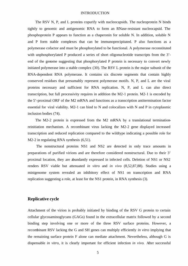
RSV virions consist of a nucleocapsid contained within a lipid envelope and appear as
irregular spherical particles that range in diameter from 150 to 300 nm in the electron
microscope. The nucleocapsid is composed of the major nucleocapsid protein N, the
phosphoprotein P, the antitermination factor M2-1, the large polymerase subunit L, and the
negative-stranded RNA genome. Together they form a symmetrical helical ribonucleoprotein
(RNP) complex.
Fig. 1: Organization of a pneumovirus particle. Left: Electron micrograph of an animal pneumovirus 2. Right: A schematic pneumovirus particle: The RNP complex consists of the nucleocapsid protein N, the phosphoprotein P, the antitermination factor M2, the polymerase subunit L, and a single-stranded RNA genome of negative polarity. Three viral glycoproteins are incorporated into the lipid membrane, the fusion protein F, the glycoprotein G and the small hydrophobic protein SH. The matrix protein M forms an inner layer connecting the viral membrane with the RNP complex.
2 Taken from “The Big Picture Book of Viruses”; the image was kindly provided by Prof. Stewart McNulty, Queen’s University of Belfast, Copyright 1994 Veterinary Science Division
INTRODUCTION
3
The envelope consists of a lipid bilayer derived from the host plasma membrane. It contains
three virally encoded transmembrane surface glycoproteins, the attachment protein G, the
fusion protein F, and the small hydrophobic SH protein. The viral glycoproteins are organized
into virion “spikes” which are visualized as short, closely spaced surface projections. The
matrix protein M is thought to form a layer on the inner envelope thereby connecting the
nucleocapsid with the lipid bilayer (Fig. 1).
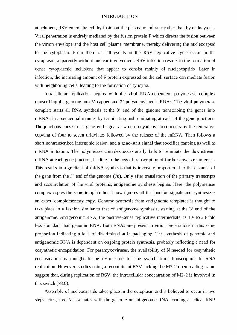
The genome
Infectious RSV contains a nonsegmented, single-stranded RNA genome of negative polarity
tightly contained in an RNase resistant nucleoprotein complex. Antigenomic RNA, the
intermediate in RNA replication, is an exact complementary copy of genomic RNA. Both
RNAs are neither capped nor polyadenylated.
RSV encodes 10 major subgenomic mRNAs (Fig. 2). Each contains a single open reading
frame (ORF) except for M2, which contains the M2-1 and M2-2 ORFs. All mRNAs are
capped at the 5’-end and polyadenylated at the 3’-end. Both modifications are thought to be
mediated by the viral polymerase. Each gene begins with a 10-nucleotide gene-start (GS)
signal that is highly conserved and terminates with a semiconserved 12 to 13 nucleotide (nt)
gene-end (GE) signal that directs polyadenylation and release of the completed mRNA. The
first nine RSV genes are separated by intergenic regions that range in length from 1 to 56
nucleotides and are not well conserved among strains. The last two RSV genes, M2 and L,
overlap by 68 nucleotides leaving the GS signal of the L gene within, rather than downstream
of, the M2 gene. The 3’-ends of genomic (leader, 44 nt) and antigenomic (trailer, 155 nt)
RNA are 81% identical for the first 26 nucleotides, probably representing the major element
of a conserved promoter (74).
Fig. 2: Genome organization of RSV. The location of transcripts (mRNA; shaded bars) and protein-encoding frames (open bars) are shown relative to the viral genome (vRNA; solid bar).
INTRODUCTION
4
Viral proteins
The RSV fusion protein F is a transmembrane glycoprotein responsible for virus penetration
and syncytium formation. Like other paramyxovirus F proteins, RSV F appears to form
trimers. It is synthesized as the precursor F0 that is cleaved by cellular proteases in the trans-
Golgi network to yield the disulfide- linked heterodimer F2-F1. This cleavage liberates the
hydrophobic “fusion peptide” at the N-terminus of the F1 subunit which is thought to be
directly involved in target membrane insertion. Together with the glycoprotein G, the fusion
protein is the major protective antigen of RSV and only antibodies against F or G neutralize
infectivity in vitro and confer resistance to RSV infection when transferred passively to
experimental animals (86,95). Furthermore, the fusion protein was found to mediate T cell
cycle arrest by contact in a species-specific manner, hinting at an immunosuppressive activity
of RSV (81).
The glycoprotein G was described as the major RSV attachment protein because
antibodies specific to G blocked the binding of virions to HeLa cells, whereas antibodies
against F prevented fusion but not binding. G is a type II transmembrane glycoprotein heavily
glycosylated with N- and O-linked sugars. G assembles into homo-oligomers that probably
are trimers or tetramers. In addition, a truncated, soluble G protein is expressed from the G
mRNA arising from translational initiation at the second AUG in the ORF. The significance
of this form is unclear, although it might function as a decoy to trap RSV-neutralizing
antibodies and might influence the immune response. Remarkably, the G protein is not
essential for virion assembly and propagation in vitro or in vivo, although in most cases, it
enhances virus multiplication (74).
The small hydrophobic SH protein is a short integral membrane protein with unknown
function. SH accumulates in multiple glycosylated and nonglycosylated forms. All forms
associate into oligomers. Recombinant RSV from which the SH gene has been deleted is fully
viable in vitro and in vivo. Expression of the SH protein in bacteria increased cell
permeability for small-molecular-weight compounds suggesting that the SH protein forms
membrane channels (73). However, it is unclear what role such an activity would play for
RSV.
The matrix protein M is a nonglycosylated inner virion protein. M plays an important
role in the formation of virus- like particles by mediating the association of the nucleocapsid
with the nascent envelope. A hydrophobic domain in the C-terminal half of the molecule
might be responsible for the interaction with membranes (74).
INTRODUCTION
5
The RSV N, P, and L proteins copurify with nucleocapsids. The nucleoprotein N binds
tightly to genomic and antigenomic RNA to form an RNase-resistant nucleocapsid. The
phosphoprotein P appears to function as a chaperonin for soluble N. In addition, soluble N
and P form stable complexes that can be immunoprecipitated. P also functions as a
polymerase cofactor and must be phosphorylated to be functional. A polymerase reconstituted
with unphosphorylated P produced a series of short oligonucleotide transcripts from the 3’-
end of the genome suggesting that phosphorylated P protein is necessary to convert newly
initiated polymerase into a stable complex (30). The RSV L protein is the major subunit of the
RNA-dependent RNA polymerase. It contains six discrete segments that contain highly
conserved residues that presumably represent polymerase motifs. N, P, and L are the viral
proteins necessary and sufficient for RNA replication. N, P, and L can also direct
transcription, but full processivity requires in addition the M2-1 protein. M2-1 is encoded by
the 5’-proximal ORF of the M2 mRNA and functions as a transcription antitermination factor
essential for viral viability. M2-1 can bind to N and colocalizes with N and P in cytoplasmic
inclusion bodies (74).
The M2-2 protein is expressed from the M2 mRNA by a translational termination-
reinitiation mechanism. A recombinant virus lacking the M2-2 gene displayed increased
transcription and reduced replication compared to the wildtype indicating a possible role for
M2-2 in regulating RNA synthesis (6,51).
The nonstructural proteins NS1 and NS2 are detected in only trace amounts in
preparations of purified virions and are therefore considered nonstructural. Due to their 3’
proximal location, they are abundantly expressed in infected cells. Deletion of NS1 or NS2
renders RSV viable but attenuated in vitro and in vivo (8,52,87,88). Studies using a
minigenome system revealed an inhibitory effect of NS1 on transcription and RNA
replication suggesting a role, at least for the NS1 protein, in RNA synthesis (3).
Replicative cycle
Attachment of the virion is probably initiated by binding of the RSV G protein to certain
cellular glycosaminoglycans (GAGs) found in the extracellular matrix followed by a second
binding step involving one or more of the three RSV surface proteins. However, a
recombinant RSV lacking the G and SH genes can multiply efficiently in vitro implying that
the remaining surface protein F alone can mediate attachment. Nevertheless, although G is
dispensable in vitro, it is clearly important for efficient infection in vivo. After successful
INTRODUCTION
6
attachment, RSV enters the cell by fusion at the plasma membrane rather than by endocytosis.
Viral penetration is entirely mediated by the fusion protein F which directs the fusion between
the virion envelope and the host cell plasma membrane, thereby delivering the nucleocapsid
to the cytoplasm. From there on, all events in the RSV replicative cycle occur in the
cytoplasm, apparently without nuclear involvement. RSV infection results in the formation of
dense cytoplasmic inclusions that appear to consist mainly of nucleocapsids. Later in
infection, the increasing amount of F protein expressed on the cell surface can mediate fusion
with neighboring cells, leading to the formation of syncytia.
Intracellular replication begins with the viral RNA-dependent polymerase complex
transcribing the genome into 5’-capped and 3’-polyadenylated mRNAs. The viral polymerase
complex starts all RNA synthesis at the 3’ end of the genome transcribing the genes into
mRNAs in a sequential manner by terminating and reinitiating at each of the gene junctions.
The junctions consist of a gene-end signal at which polyadenylation occurs by the reiterative
copying of four to seven uridylates followed by the release of the mRNA. Then follows a
short nontranscribed intergenic region, and a gene-start signal that specifies capping as well as
mRNA initiation. The polymerase complex occasionally fails to reinitiate the downstream
mRNA at each gene junction, leading to the loss of transcription of further downstream genes.
This results in a gradient of mRNA synthesis that is inversely proportional to the distance of
the gene from the 3’ end of the genome (78). Only after translation of the primary transcripts
and accumulation of the viral proteins, antigenome synthesis begins. Here, the polymerase
complex copies the same template but it now ignores all the junction signals and synthesizes
an exact, complementary copy. Genome synthesis from antigenome templates is thought to
take place in a fashion similar to that of antigenome synthesis, starting at the 3‘ end of the
antigenome. Antigenomic RNA, the positive-sense replicative intermediate, is 10- to 20-fold
less abundant than genomic RNA. Both RNAs are present in virion preparations in this same
proportion indicating a lack of discrimination in packaging. The synthesis of genomic and
antigenomic RNA is dependent on ongoing protein synthesis, probably reflecting a need for
cosynthetic encapsidation. For paramyxoviruses, the availability of N needed for cosynthetic
encapsidation is thought to be responsible for the switch from transcription to RNA
replication. However, studies using a recombinant RSV lacking the M2-2 open reading frame
suggest that, during replication of RSV, the intracellular concentration of M2-2 is involved in
this switch (78,6).
Assembly of nucleocapsids takes place in the cytoplasm and is believed to occur in two
steps. First, free N associates with the genome or antigenome RNA forming a helical RNP
INTRODUCTION
7
structure. Secondly, P-L protein complexes associate with the nucleocapsid. RSV virion
maturation occurs at the cell surface where progeny virions acquire a lipid envelope enriched
in viral surface glycoproteins by budding at the plasma membrane. M proteins are thought to
play a major role in bringing the assembled RNP to the appropriate patch on the plasma
membrane to form a budding virion. The protein-protein interactions involved in assembling a
virion must be specific, as cellular membrane proteins are largely excluded from the virions.
It is presumed that the glycoprotein cytoplasmic tails make important contacts with the M
protein layer which, in turn, associates with the nucleocapsid. However, the efficient
replication in vitro of ∆SH∆G recombinant RSVs indicates that F is the only viral
transmembrane protein required for efficient virion formation. Disruption of cellular actin
filaments drastically reduce the production of infectious virus implying a role for the
cytoskeleton in assembly (12).
Epidemiology
RSV is the single most important cause of respiratory tract infections in infants and young
children worldwide and is believed to account for approximately 85% of cases in bronchiolitis
and approximately 20% of cases of childhood pneumonia (103). It infects the very young
infant and the neonate despite the presence of maternally derived antibodies, and infection
and reinfection are frequent during the first few years of life. Reinfection of adults is also
common, particularly when exposure to virus is heavy. In addition, RSV is a pathogen of
considerable importance in the elderly. RSV has a worldwide distribution and shows clear
seasonality in temperate zones of the world. In urban centers, epidemics occur yearly in the
late fall, winter or spring but not during the summer. Each RSV epidemics lasts
approximately 5 months, with 40% of infections occurring during the peak month in the
temporal center of the outbreak. It is not known why the virus disappears in the spring, nor
from where it reemerges in the fall or winter. In the United States, RSV was recently
estimated to be responsible for 73,400 to 126,300 hospitalizations annually for bronchiolitis
and pneumonia alone among children younger than 1 year (84). Bronchiolitis or pneumonia
occurs most frequently between ages 6 weeks and 9 months, whereas the peak incidence of
lower respiratory disease is between ages 2 and 7 months corresponding with diminishing
titers of maternal antibodies. The risk of serious RSV disease is increased by prematurity,
young age, chronic cardiac or lung disease, immunodeficiency or immunosuppression, or
familiy history of allergic disease. However, approximately three fourths of hospitalizations
INTRODUCTION
8
for RSV disease occur in infants and children who were previously healthy. However,
mortality due to RSV infection is not common in developed countries (74).
Pathogenesis and immunity
Spread of RSV occurs by direct inoculation of contagious secretions from the hands or by
large-particle aerosols into the eyes and nose, but rarely the mouth. After an incubation period
of two to eight days, RSV replicates in the nasopharyngeal epithelium, with spread to the
lower respiratory tract one to three days later. Pathological characteristics of severe RSV
infection are necrosis and proliferation of the bronchiolar epithelium and destruction of
ciliated epithelial cells (68). A peribronchiolar infiltrate of lymphocytes, plasma cells and
macrophages develops, with migration of the lymphocytes among the mucosal epithelial cells.
Edema develop and secretion of mucus increases, obstructing the bronchioles and alveoli. In
cases of pneumonia, alveolar spaces may fill with fluid and mononuclear cells infiltrate the
interstitium. Immunostaining of infected lungs showed virus-infected cells in the bronchial,
bronchiolar, and alveolar epithelium, and identified numerous syncytial cells (68). Direct
virus-mediated cytopathology and the local inflammatory response initiated by RSV infection
also contributes to RSV pathogenesis, resulting in considerable damage to the epithelium and
to the bronchiolar ciliary apparatus. Complete restoration requires four to eight weeks, in
correlation with the common clinical findings of prolonged cough, wheezing, and altered
pulmonary function (42,74).
RSV is unusual in its ability to infect infants in the presence of moderate titers of
maternal antibodies and to readily reinfect persons of all ages. Naturally acquired immunity is
neither complete nor durable, making the development of an effective vaccine more difficult.
Nevertheless, protection against severe disease develops after primary infection.
The adaptive immune system has the primary role in recovery from RSV infection and
resistance to reinfection (23). For example, immunodeficient children fail to clear RSV, and
severely immunocompromised adults, such as bone marrow transplant recipients, have a very
high incidence of RSV infection leading to serious disease and death (46).
In the BALB/c mouse model, pulmonary NK cells appear in the first few days after
RSV infection followed by pulmonary CD8+ cytotoxic T cells (CTLs) and secretory
antibodies. In addition to being direct effectors against virus- infected cells, NK and CD8+ T
cells modulate the immune response by secretion of lymphokines, especially IFN-γ (74).
INTRODUCTION
9
Studies in which the CD4+ and CD8+ T- lymphocyte subsets were depleted individually or
together demonstrated that both are important for clearing a primary infection (39). Studies in
which mice were depleted of B lymphocytes showed that RSV-specific antibodies are not
required for virus clearance during a primary infection, but are very important in restricting
replication and disease on reinfection (38). Immunization of BALB/c mice with recombinant
vaccinia viruses that express individual RSV proteins showed that F and G are the only viral
proteins that induce RSV-neutralizing antibodies and long- lived resistance to reinfections
with RSV (21). The secretory antibodies produced by infants in response to RSV infection
often fail to neutralize the virus in vitro (65), and this defect may be at least partially
responsible for the incompleteness of natural immunity after infection during infancy and
early childhood. In older individuals, multiple reinfections induce higher levels of IgA
antibodies. Experimental infections of adult volunteers have shown that immunity to infection
correlates better with the level of nasal RSV-neutralizing IgA antibodies than with serum
RSV-neutralizing antibodies (74). To completely eliminate RSV, CD8+ CTLs appear to be
required. However, studies in mice also provide evidence that RSV-specific CTLs may play a
role in the pathogenesis of RSV disease. Passively transferred, stimulated RSV-specific CD8+
CTLs clear virus from the lungs of persistently infected, gamma-irradiated mice. Similarly,
CD8+ CTLs accelerate clearance of RSV from the lungs of infected immunocompetent mice,
but clearance is accompanied by acute pulmonary disease (74). These observations suggest
that a balance exists between the protective and disease-producing effects of CD8+ CTLs.
2 THE INTERFERON-α/β SYSTEM
Interferons (IFNs) are a large family of multifunctional secreted proteins involved in antiviral
defense, cell growth regulation and activation of the immune system. Type I IFNs (IFN-α and
IFN-β) are produced in response to viral infection, double stranded RNA (dsRNA) or tumor
necrosis factor (TNF-) α. Whereas products of the IFN-α multigene family are predominantly
synthesized by leukocytes, IFN-β is produced in most cell types, particularly by fibroblasts.
The expression of the type II IFN gene, IFN-γ, is restricted to activated T lymphocytes and
natural killer (NK) cells. Both types of IFN share no obvious structural homology but a
functional similarity exists due to an extensive overlap in the set of genes they induce. The

INTRODUCTION
10
importance of IFN in mediating responses to viral infections is demonstrated by the fact that
mice lacking IFN receptors are unable to mount effective immune responses to a large
number of different viruses. Both types of IFN stimulate an antiviral state in target cells
whereby replication of the virus is blocked or impaired due to the synthesis of a number of
enzymes that interfere with cellular and viral processes or due to the elimination of the
infected cell via the apoptotic pathway. In addition, both types of IFN possess extensive
immunomodulatory functions, albeit with different specificities, affecting nearly all phases of
the innate and adaptive immune response (37).
Virus induction of IFN genes
Induction of IFN gene transcription is tightly regulated by extra- and intracellular signals
induced at the site of infection. One of the best characterized models of such regulation is the
IFN-β gene promoter/enhancer. This promoter contains an overlapping set of regulatory
elements designated positive regulatory domains (PRDs) I to IV which interact with several
signal-responsive transcription factors including NF-κB, ATF/c-Jun heterodimers, and
interferon regulatory factor 3 or 7 (IRF-3/7) that bind to PRD II, PRD IV, and PRD I-III,
respectively. Together with the chromatin-associated HMG I(Y) proteins, these transcription
factors form a transcriptional enhancer complex, termed the enhanceosome, that stimulates
the high level, transient activation of IFN-β transcription (Fig. 3; 83).
The pathways involved in NF-κB and AP-1 (ATF-2/c-Jun) activation are well
characterized. Following viral infection, treatment with proinflammatory stimuli like TNF-α,
or exposure to dsRNA, these transcription factors are activated through stimulation of distinct
kinase cascades. In uninfected cells, NF-κB is retained in the cytoplasm in association with
inhibitory subunits termed IκBs. Upon viral infection, IκB is phosphorylated at conserved
residues by the IκB kinase (IKK) complex leading to ubiquitin-dependent degradation of IκB
and subsequent nuclear translocation of NF-κB (26). Activation of the IKK complex is
thought to be mediated by the dsRNA-dependent protein kinase (PKR) which activates the
IKKβ subunit of the multicomponent IκB kinase (110). PKR can also phosphorylate IκB
directly (58). However, the biological role for this is unclear. Unlike NF-κB, the heterodimers
ATF-2/c-Jun are expressed as nuclear proteins that are activated by phosphorylation of their
activation domains by c-Jun amino-terminal kinases (JNKs) which are downstream of a well
defined stress-activated kinase cascade (100).
INTRODUCTION
11
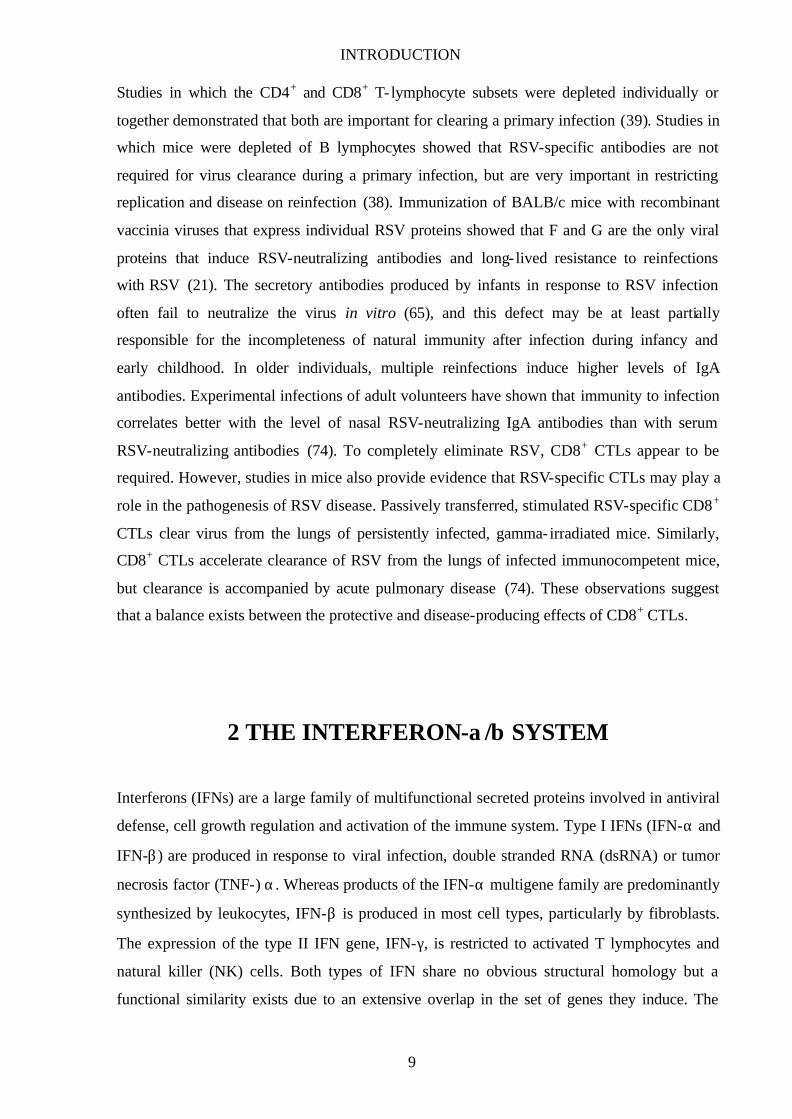
The pathways regulating IRF-3 phosphorylation and activation are under intense
investigation. IRF-3 belongs to the family of interferon regulatory factors (IRFs; 62) and is
expressed constitut ively in a variety of tissues. The relative levels of IRF-3 mRNA do not
change in virus- infected or IFN-treated cells. IRF-3 demonstrates a unique response to viral
infection. Phosphorylation of latent cytoplasmic IRF-3 on serine and threonine residues in the
C-terminal region leads to a conformational change, dimerization, cytoplasmic to nuclear
translocation, association with the p300/CBP coactivator and transcriptional activity. The
rate- limiting step in this process is the C-terminal phosphorylation of IRF-3 by an as of yet
uncharacterized virus activated kinase (VAK) activity (83).
Fig. 3: Transcriptional induction of the IFN-β gene. Virus infection and dsRNA generated during viral replication are able to activate Jun kinases (JNKs), PKR, and perhaps other cellular kinases. PKR in turn activates the IκB kinase leading to the activation of NF-κB. ATF-2/c-Jun and NF-κB, together with a member of the IRF family, assemble on the IFN-β promoter with the help of the accessory factor HMG-I/Y to form a complex called the ‘enhanceosome’. This complex then interacts with factors of the basal transcription machinery resulting in transcription of the IFN-β gene.
The trigger for IRF-3 activation is generally believed to be dsRNA. Indeed, RNA viruses such
as Newcastle disease virus (NDV), Sendai virus (SeV) or vesicular stomatitis virus (VSV) are
INTRODUCTION
12
potent inducers of VAK activity as is the synthetic dsRNA analog polyI/C. However,
tenOever et al. recently demonstrated that the expression of a viral protein, the Measles virus
(MeV) nucleocapsid protein, was sufficient for IRF-3 activation (89). DNA viruses, in
particular herpes simplex virus 1 (HSV-1) and human cytomegalovirus (HCMV), were also
reported to activate IRF-3. Interestingly, whereas binding of HCMV to its cellular receptor
was sufficient to trigger IRF-3 activation, entry of virus particles but not de novo transcription
of viral genes was necessary to activate IRF-3 in the case of HSV-1 (76). Recently, the Rho
GTPases Rac-1 and RhoA were identified to be specifically involved in virus induced gene
expression from IRF-3 responsive promoters since specific inhibitors or dominant negative
forms inhibited virus or dsRNA induced activation of the IFN-β promoter (32). Further
studies will shed more light on the different mechanisms by which cells are able to recognize
viral infections, leading to the activation of IRF-3 and the subsequent establishment of an
antiviral state.
Signal transduction in response to IFNs
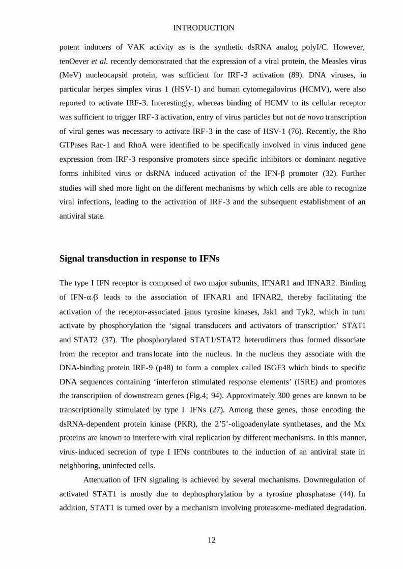
The type I IFN receptor is composed of two major subunits, IFNAR1 and IFNAR2. Binding
of IFN-α/β leads to the association of IFNAR1 and IFNAR2, thereby facilitating the
activation of the receptor-associated janus tyrosine kinases, Jak1 and Tyk2, which in turn
activate by phosphorylation the ‘signal transducers and activators of transcription’ STAT1
and STAT2 (37). The phosphorylated STAT1/STAT2 heterodimers thus formed dissociate
from the receptor and trans locate into the nucleus. In the nucleus they associate with the
DNA-binding protein IRF-9 (p48) to form a complex called ISGF3 which binds to specific
DNA sequences containing ‘interferon stimulated response elements’ (ISRE) and promotes
the transcription of downstream genes (Fig.4; 94). Approximately 300 genes are known to be
transcriptionally stimulated by type I IFNs (27). Among these genes, those encoding the
dsRNA-dependent protein kinase (PKR), the 2’5’-oligoadenylate synthetases, and the Mx
proteins are known to interfere with viral replication by different mechanisms. In this manner,
virus- induced secretion of type I IFNs contributes to the induction of an antiviral state in
neighboring, uninfected cells.
Attenuation of IFN signaling is achieved by several mechanisms. Downregulation of
activated STAT1 is mostly due to dephosphorylation by a tyrosine phosphatase (44). In
addition, STAT1 is turned over by a mechanism involving proteasome-mediated degradation.
INTRODUCTION
13
Moreover, several IRF proteins are known to bind ISREs and negatively regulate expression,
probably preventing expression in the absence of IFN. Also, IFN-induced proteins may play a
role in regulating IFN signaling, i.e. the SOCS/JAB/SSI family induced by IFN-γ was shown
to bind and inhibit activated Jaks, leading to signal downregulation (37).
Fig. 4: Signaling pathways activated by IFN-β. Binding of IFN-β to its receptor activates receptor-associated kinases that phosphorylate members of the STAT family of transcription factors which can then enter the nucleus. In combination with p48, they bind to the ISRE element of target genes and activate their transcription.
The antiviral response
The best-characterized IFN-inducible components of the antiviral response are PKR, the 2’-5’
oligoadenylate synthetases, and the Mx proteins. The RNA-dependent protein kinase PKR is a
serine/threonine kinase with multiple functions in control of transcription and translation. The
PKR protein has two known domains, an N-terminal dsRNA binding site and a C-terminal
catalytic domain that shows protein kinase activity. PKR is activated by binding dsRNA, and
the active form is postulated to be a dimer with two PKR molecules binding one molecule of
dsRNA. These juxtaposed PKR molecules are then able to phosphorylate each other on
several residues. There are no sequence requirements for the dsRNA. However, there are at
least 50 base pairs of duplex necessary for activation. Activated PKR phosphorylates the α

INTRODUCTION
14
subunit of the eukaryotic translation initiation factor eIF2, thereby leading to a shut-down of
protein translation. Phosphorylated eIF2α interacts strongly with the guanine exchange factor
eIF2B which, in turn, cannot mediate the recycling of eIF2-GDP to eIF2-GTP. Since eIF2B is
present in limiting amounts, translation is inhibited (16). PKR also plays a role in mediating
signal transduction events in response to dsRNA and other ligands (102), and in mediating
apoptosis. The downstream targets for PKR-mediated apoptosis remain to be identified but
overexpression of PKR has been shown to induce apoptosis through a Bcl2- and caspase-
dependent mechanism (60).
2’-5’ oligoadenylate synthetases are a group of enzymes that, once activated by binding
to dsRNA, catalyse the synthesis of adenosine oligomers from ATP that are linked by
phosphodiester bonds in the unusual conformation of 2’ to 5’ (2’5’A). The 2’5’A molecules
bind to endoribonuclease L (RNase L) and induce its activation via dimerization (37).
Activated RNase L then catalyses the cleavage of single-stranded RNA including mRNA and
rRNA leading to inhibition of protein synthesis. Since 2’5’A is highly labile, the activation of
RNase L depends upon locally activated 2’-5’oligoadenylate synthetases, thus ensuring that
virus transcripts are destroyed preferentially over cellular mRNAs.
Mx proteins are highly conserved, large GTPases with homology to dynamin, and they
were found in all vertebrate species examined so far (72). Mx proteins are known to impair
the growth of a wide range of RNA viruses, probably at the level of virus transcription or
replication. Recently, Kochs et al. reported that MxA binds to the nucleocapsid protein N of
La Crosse Virus (LACV) and aggregates it into large complexes. Thus, by trapping this
essential virus component in cytoplasmic inclusions viral replication is inhibited (56).
Studies involving the generation of triple knockout mice lacking PKR, RNAse L and
Mx1 indicate that there are additional antiviral effects of IFNs (111). Other factors that clearly
play a role are caspases and the dsRNA-dependent adenosine deaminase (ADAR; 71). In
addition, further functions of IFNs such as their antiproliferative activity, immunomodulatory
function and ability to control apoptosis also contribute to the successful elimination of viral
infections.
Viral countermeasures to the IFN response
To successfully establish infections in vivo, viruses must replicate in the face of antiviral
defense mechanisms induced by IFNs. It therefore seems likely that all viruses must, at least
to a certain extent, have some means of circumventing or blocking the IFN response.

INTRODUCTION
15
However, viral countermeasures rarely achieve absolute inhibition, and the IFN response, by
limiting virus spread, buys time for the generation of an acquired immune response to the
invading virus. On the other hand, the speed and efficiency of the virus in circumventing the
action of IFN may be critical to its host range and pathogenicity.
Viruses vary considerably in their ability to induce IFN. This may simply reflect the
amount of dsRNA produced during infection or the inability of the cell to detect certain viral
infections. However, it may also indicate that viruses are able to inhibit the induction of IFN.
Several viruses have been reported to encode dsRNA-binding proteins, e.g. the reovirus major
outer capsid protein σ3 (5), the NS1 protein of influenza virus (35), the E3L protein of
vaccinia virus (14) as well as the NSP3 gene products of porcine rotavirus (59) were shown to
bind dsRNA. Sequestration of dsRNA may prevent IFN-α/β production, thereby minimizing
the dsRNA-dependent activation of antiviral gene products such as PKR, 2’-5’ oligoadenylate
synthetases, or ADAR. Another common strategy to block IFN-α/β production is to target the
activity of transcription factors necessary for IFN-β promoter induction. African swine fever
virus (ASFV) encodes a homologue of IκB that inhibits the activity of NF-κB (77) whereas
human herpesvirus 8 (HHV-8) encodes a viral IRF-1 homologue that blocks the formation of
transcriptionally competent IRF-3-CBP/p300 complexes (112). The E6 protein of human
papillomavirus type 16 (HPV-16) binds IRF-3 and can inhibit its transcriptional activity (79).
However, induction of IFN-β is never blocked completely, suggesting that other factors can
substitute functionally for IRF-3. These potential substitutes include IRF-1 and ISGF3, but
these factors themselves can be targeted by viral proteins. The plethora of factors that can
bind and activate the IFN-β promoter probably reflects a pivotal need of the cell to be able to
circumvent viral blockades.
Blocking of the IFN signaling pathway can occur at several levels, and there is accumulating
evidence that viruses are able to block most, if not all, stages. Several poxviruses have been
shown to encode soluble IFN-receptor homologues that bind and sequester IFNs to prevent
their biological activity. Inside the cell, human cytomegalovirus (HCMV) inhibits the
phosphorylation of STATs by decreasing the levels of Jak1 and IRF-9 (p48) by a mechanism
involving the proteasome (67) whereas the T antigen of murine polyomavirus (MPyV) binds
to and inactivates Jak1 (99). Further downstream in the signaling cascade, the V protein of
simian virus 5 (SV5) and mumps virus (MV) target STAT1 for proteasome-mediated
degradation (29,105) whereas human parainfluenza virus 2 (hPIV2) targets STAT2, thereby
preventing the formation of ISGF3 complexes (108). Interestingly, at least part of the host

INTRODUCTION
16
range of SV5 appears to be determined by the ability to mediate STAT1 degradation (107).
The adenovirus E1A protein decreases STAT1 and IRF-9 levels, and sequesters the
transcriptional coactivator CBP/p300 (61). Furthermore, the E7 protein of HPV-16 interacts
directly with IRF-9, preventing the formation of ISGF3 and thus the activation of IFN-α/β-
inducible genes (4). It has also been reported that EBNA2 of Epstein-Barr Virus (EBV)
inhibits IFN-α/β signaling by an unknown mechanism that does not prevent the formation of
ISGF3 complexes (53). The block in IFN signaling is advantageous since it prevents the
induction of antiviral genes and the upregulation of class I MHC molecules within infected
cells, making them poorer targets for cytotoxic T cells. In addition, since there are
components in common between IFN-α/β and IFN-γ signaling pathways, it is possible for a
virus to block either one or both pathways. However, it was recently reported that IRF-3 is not
only involved in the induction of the immediate early IFN-α/β genes but also in the direct
activation of a subset of antiviral IFN-stimulated genes, adding to the complexity of the
cellular IFN system encountered by viruses (40).
Another major strategy to overcome the antiviral state is the inhibition of IFN-induced
antiviral enzymes. The importance of PKR in the establishment of antiviral activity can be
inferred from the wide variety of mechanisms that are employed by viruses to inhibit its
activity. Since dsRNA is required to activate PKR, viruses that encode dsRNA-binding
proteins, e.g. influenza virus NS1 and vaccinia virus E3L, simultaneously inhibit both IFN
induction and PKR activation. In addition to binding dsRNA, viral gene products can inhibit
PKR in several other ways. For example, poliovirus induces the degradation of PKR whereas
the nonstructural protein NS5A of hepatitis C virus (HCV) or the baculovirus PK2 protein
bind PKR directly and inhibit its activity (37). The K3L gene product of vaccinia virus has
structural similarity to the N terminus of eIF2α and binds tightly to PKR, preventing its
autophosphorylation and subsequent activation (24). A more indirect method of coping with
PKR is illustrated by the γ1 ICP34.5 protein encoded by herpes simplex virus (HSV). ICP34.5
interacts with the cellular protein phosphatase 1α (PP1) redirecting it to dephosphorylate, and
hence reactivate, eIF2α (45). Moreover, some viruses abundantly produce short RNA
molecules, e.g. the adenovirus VAI transcripts, that bind avidly to the dsRNA-binding site of
PKR and act as competitive inhibitors of PKR activation by dsRNA. Another target for viral
gene products is the 2’-5’ oligoadenylate synthetase/RNase L system. HIV-1 (63) and
encephalomyocarditis virus (EMCV) (64) downregulate RNase L activity by inducing the
expression of the RNase L inhibitor RLI which antagonizes the binding of 2’5’A to RNase L,
thus preventing its activation. Since dsRNA is also required for the activation of RNase L,
INTRODUCTION
17
virus proteins that sequester dsRNA are able to inhibit both PKR and the 2’-5’ oligoadenylate
synthetase/RNase L system.
3 THE NONSTRUCTURAL PROTEINS NS1 AND NS2
Overview
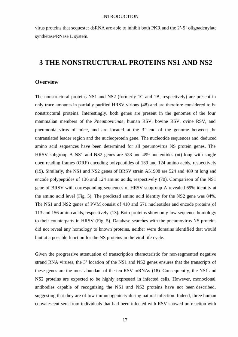
The nonstructural proteins NS1 and NS2 (formerly 1C and 1B, respectively) are present in
only trace amounts in partially purified HRSV virions (48) and are therefore considered to be
nonstructural proteins. Interestingly, both genes are present in the genomes of the four
mammalian members of the Pneumovirinae, human RSV, bovine RSV, ovine RSV, and
pneumonia virus of mice, and are located at the 3’ end of the genome between the
untranslated leader region and the nucleoprotein gene. The nucleotide sequences and deduced
amino acid sequences have been determined for all pneumovirus NS protein genes. The
HRSV subgroup A NS1 and NS2 genes are 528 and 499 nucleotides (nt) long with single
open reading frames (ORF) encoding polypeptides of 139 and 124 amino acids, respectively
(19). Similarly, the NS1 and NS2 genes of BRSV strain A51908 are 524 and 489 nt long and
encode polypeptides of 136 and 124 amino acids, respectively (70). Comparison of the NS1
gene of BRSV with corresponding sequences of HRSV subgroup A revealed 69% identity at
the amino acid level (Fig. 5). The predicted amino acid identity for the NS2 gene was 84%.
The NS1 and NS2 genes of PVM consist of 410 and 571 nucleotides and encode proteins of
113 and 156 amino acids, respectively (13). Both proteins show only low sequence homology
to their counterparts in HRSV (Fig. 5). Database searches with the pneumovirus NS proteins
did not reveal any homology to known proteins, neither were domains identified that would
hint at a possible function for the NS proteins in the viral life cycle.
Given the progressive attenuation of transcription characteristic for non-segmented negative
strand RNA viruses, the 3’ location of the NS1 and NS2 genes ensures that the transcripts of
these genes are the most abundant of the ten RSV mRNAs (18). Consequently, the NS1 and
NS2 proteins are expected to be highly expressed in infected cells. However, monoclonal
antibodies capable of recognizing the NS1 and NS2 proteins have not been described,
suggesting that they are of low immunogenicity during natural infection. Indeed, three human
convalescent sera from individuals that had been infected with RSV showed no reaction with
INTRODUCTION
18
NS1 and NS2 (33). Furthermore, vaccinia virus recombinants expressing the NS proteins did
not confer protective immunity in mice when challenged with live RSV (21), and no murine
helper T cell response to NS2 was elicited by a recombinant vaccinia/NS2 virus (69).
However, a cytotoxic T cell response to the NS2 protein was observed in six of nine human
adult volunteers, but only one of the same nine volunteers reacted to the NS1 protein (15).
Fig. 5: Amino acid sequence comparison of pneumovirus NS proteins. Amino acids identical in all three proteins are marked by black bars. Amino acid identical between two proteins and amino acid similarities are marked in gray.
The first study aimed at elucidating the function of the nonstructural proteins in the viral life
cycle was published in 1995. Here, Yu et al. demonstrated that neither HRSV NS protein is
required for RNA replication in a helper- independent in vitro system (109). It was further
shown that both HRSV NS proteins are synthesized early in infection and are present in
INTRODUCTION
19
multimeric forms in infected cells (33). The HRSV NS1 protein has been shown to
coprecipitate with the matrix protein (33) whereas the HRSV NS2 protein colocalizes in cells
with the N protein (98) but does not coprecipitate with any viral protein. In addition, the
HRSV NS2 protein is unstable with a half life of approximately 30 minutes (33). In a HRSV
minigenome system complemented by plasmid-encoded support proteins, coexpression of
HRSV NS1 cDNA strongly inhibited transcription and RNA replication mediated by the RSV
polymerase even at very low expression levels suggesting a role for the NS1 protein in RNA
synthesis (3).
Reverse genetics as a tool to study viral gene function
The development of a reverse genetic system by which complete infectious recombinant
negative-stranded RNA virus can be recovered from cDNA has provided a new and powerful
tool for the experimental analysis of viral gene functions with molecular approaches (22).
Successful recovery of negative-strand RNA viruses requires the intracellular reconstitution
of RNP complexes from plasmid-derived components. Simultaneous production of full- length
antigenomic RNA and of individual RNP-associated proteins by transfection of cells with
appropriate expression plasmids results in the initiation of an infectious cycle and the
recovery of recombinant virus, as was shown first for rhabdoviruses (82). Whereas the N, P
and L proteins and full- length antigenome RNAs are sufficient to obtain infectious
rhabdovirus and most paramyxoviruses, efficient recovery of recombinant RSV also requires
the expression of the M2 gene (8,10). Using the reverse genetics method for the generation of
RSVs with gene deletions, SH, NS1, NS2, and M2-2 were reported to be dispensable for viral
replication in vitro and in vivo (6,8,11,52,88). Recombinant BRSV lacking the NS2 gene
displayed an attenuated growth phenotype, indicating that NS2 is not essential for virus
replication in cell culture but is required for full replication capacity (8). Similar results were
obtained for an NS2 deletion mutant of HRSV which exhibited small plaque morphology and
reduced replication in vitro (87). HRSV NS1 or NS2 deletion mutants are attenuated in
chimpanzees and show restricted replication in the upper and lower respiratory tract (88,101).
Moreover, studies from Jin et al. showed that recombinant HRSVs with deletions in the NS1,
NS2, SH, and M2-2 genes, and various combinations thereof, are attenuated in vitro and in
vivo (52).

INTRODUCTION
20
BRSV NS proteins are IFN antagonists
To examine more closely the function of the BRSV NS proteins, BRSV deletion mutants
lacking the NS1 or NS2 gene, or both NS1 and NS2, were generated in our laboratory, and
their behaviour in different cell lines was analysed. First, growth characteristics were analysed
in BSR T7/5 cells which had been used for virus recovery. In comparison with the parental
full- length virus, all three mutants were attenuated, suggesting a contribution of both NS
proteins to virus replication (Fig. 6). Similar results were also obtained in Vero cells (data not
shown). Then a cell line of bovine origin was used, MDBK, which had been shown to
optimally support the growth of wt BRSV (8). Surprisingly, in this cell line growth of the NS
deletion mutants was severely impeded whereas in other cells like BSR, the deletion mutants
were only slightly attenuated (Fig. 6; 80).
Fig. 6: NS deletion mutants are more attenuated in MDBK cells (B) than in BSR cells (A). Cells were infected at an MOI of 0.1 with BRSV wt, BRSV ∆NS1, BRSV ∆NS2, or BRSV ∆NS1/2, and virus titers were determined every 2 days. At day 8, infection of BSR cells (and MDBK cells in the case of BRSV wt) led to massive cell destruction. Bars show standard deviation (80).
We therefore postulated the presence of MDBK cell factors, presumably produced upon virus
infection, that are responsible for the selective impediment of the NS deletion mutants but not
wt BRSV. To find out whether soluble molecules produced by MDBK cells are able to
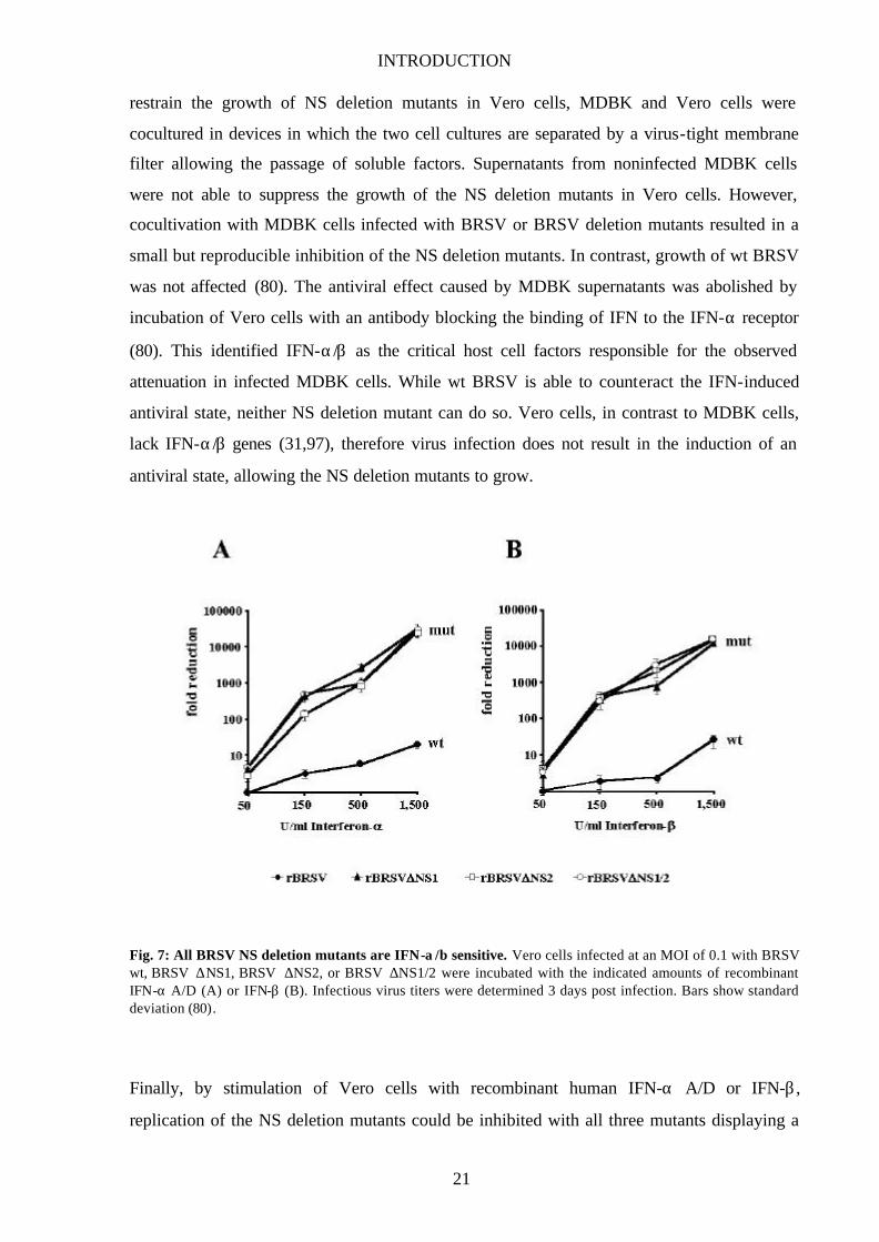
INTRODUCTION
21
restrain the growth of NS deletion mutants in Vero cells, MDBK and Vero cells were
cocultured in devices in which the two cell cultures are separated by a virus-tight membrane
filter allowing the passage of soluble factors. Supernatants from noninfected MDBK cells
were not able to suppress the growth of the NS deletion mutants in Vero cells. However,
cocultivation with MDBK cells infected with BRSV or BRSV deletion mutants resulted in a
small but reproducible inhibition of the NS deletion mutants. In contrast, growth of wt BRSV
was not affected (80). The antiviral effect caused by MDBK supernatants was abolished by
incubation of Vero cells with an antibody blocking the binding of IFN to the IFN-α receptor
(80). This identified IFN-α/β as the critical host cell factors responsible for the observed
attenuation in infected MDBK cells. While wt BRSV is able to counteract the IFN-induced
antiviral state, neither NS deletion mutant can do so. Vero cells, in contrast to MDBK cells,
lack IFN-α/β genes (31,97), therefore virus infection does not result in the induction of an
antiviral state, allowing the NS deletion mutants to grow.
Fig. 7: All BRSV NS deletion mutants are IFN-α/β sensitive. Vero cells infected at an MOI of 0.1 with BRSV wt, BRSV ∆NS1, BRSV ∆NS2, or BRSV ∆NS1/2 were incubated with the indicated amounts of recombinant IFN-α A/D (A) or IFN-β (B). Infectious virus titers were determined 3 days post infection. Bars show standard deviation (80).
Finally, by stimulation of Vero cells with recombinant human IFN-α A/D or IFN-β ,
replication of the NS deletion mutants could be inhibited with all three mutants displaying a
INTRODUCTION
22
highly similar, severe, and dose-dependent sensitivity towards the IFN-induced cellular
response. In contrast, wt BRSV was significantly resistant to IFN treatment suggesting a
function for the NS proteins in mediating virus escape from cellular antiviral mechanisms
induced by IFN (Fig. 7; 80).
4 AIM OF THIS STUDY
Intriguingly, deletion of each NS gene or both NS genes from BRSV leads to deletion mutants
that display approximately equal degrees of IFN sensitivity. This suggests that the presence of
both NS proteins, NS1 and NS2, is required to protect BRSV from IFN-induced antiviral
mechanisms and that each NS protein on its own does not possess IFN antagonist activity. I
therefore wanted to develop an assay system to investigate the role(s) of NS1 and NS2 in
mediating IFN resistance and to address a possible cooperative function of NS1 and NS2.
Furthermore, I was interested to see whether IFN resistance can also be mediated by the NS
proteins of other pneumoviruses such as HRSV and PVM, and whether cooperation of their
NS proteins is necessary for the postulated IFN antagonist function. If so, this would suggest
a common IFN resistance mechanism among Pneumovirinae members carrying NS genes.
Pneumoviruses are known to display a very narrow host range that is only in part determined
by their surface glycoproteins (9) requiring the presence of additional viral proteins that play a
role in determining host range. To investigate whether the NS proteins have the potential to
contribute to the host range restriction of BRSV, I decided to generate recombinant BRSVs
that carry the NS genes of HRSV or PVM, instead of the homologous BRSV NS genes, in an
identical virus background. The evaluation of these chimeric recombinants in cells of different
hosts should allow to monitor possible host specific abilities of different NS proteins in
antagonizing IFN-induced antiviral responses.
Finally, I was also interested in elucidating the molecular mechanism(s) responsible for the
observed IFN resistance of BRSV.

MATERIALS AND METHODS
23
MATERIALS AND METHODS
1 MATERIALS
1-1 Chemicals
Chemicals were purchased from the following companies: Roth (acetone (tech.), DMSO,
glycerol, glycine, HCl, Hepes, methanol, SDS, Tris, Tween 20, urea); Sigma (ATP,
bromophenol blue, CDTA, digitonin, DOC, MgCO3xMg(OH)2, TEMED); Merck (CaCl2,
EDTA, ethidium bromide, CH3COOK, KCl, MgSO4, MgCl2, CH3COONa, NaCl, NH4Cl,
Orange G, paraformaldehyde, Phenol red, propidium iodide, Triton X-100); Riedel-de-Häen
(ethanol, NaOH); Fluka (NP40, tricine); Boehringer Mannheim (DTT); ICN Biomedicals Inc.
(APS); Pharmacia (Ficoll).
1-2 Enzymes
Restriction endonucleases, T4 DNA ligase (New England Biolabs), klenow polymerase (MBI
Fermentas), pfu polymerase, AMV reverse transcriptase (Stratagene), RNase A, RNase I
(Pharmacia), shrimp alkaline phosphatase (Roche Biochemicals).
1-3 Kits
ECL detection kit « Renaissance » NEN
RNeasy kit Qiagen
QIAquick PCR purification/Gel extraction/Nucleotide removal Qiagen
Mammalian Transfection kit Stratagene
Nucleobond AX100, AX500 Macherey & Nagel
1-4 Miscellaneous
AcetylCoA Sigma
Acrylamide/Bisacrylamid solution (29:1) Merck
Agarose Gibco BRL
Ampicillin Gibco BRL
MATERIALS AND METHODS
24
Coomassie Protein Assay Reagent Pierce
DNA 1 kb ladder Gibco BRL
dNTPs NEB
Hyperfilm ECL Amersham
FuGENE Roche
Histogel Linaris Biological Products
IFN-α A/D PBL
Luciferin Sigma
Nitrocellulose membrane (0.5 µm) Schleicher & Schuell
Poly I/C Sigma
Prestained protein ladder Gibco BRL
RNasin Pharmacia
Skim milk powder Merck
Tryptone peptone Difco
Whatman paper Roth
Yeast extract Difco
1-5 Bacteria and plasmids
E.coli XL1 (blue) or XL (gold; Stratagene) were used for preparation of plasmid DNA.
All generated constructs as well as full- length cDNAs of rabies virus and BRSV are based on
the Bluescript SKII- vector obtained from Pharmacia.
pTIT-N, pTIT-P, pTIT-L plasmids containing RV N, P,or L gene
under control of T7 promoter
pTITB-N, pTITB-P, pTITB-M2, pTITB-L plasmids containing BRSV N, P, M2,or L
gene under control of T7 promoter
pSAD VB plasmid containing full- length RV cDNA under T7-promoter
control with additional stop-restart sequence between the G and L
gene
pSAD VB NS1ha pSAD VB harboring BRSV NS1 gene between G and L gene
pSAD VB NS2fl pSAD VB harboring BRSV NS2 gene between G and L gene

MATERIALS AND METHODS
25
pSAD VB hNS1 pSAD VB harboring HRSV NS1 gene between G and L gene
pSAD VB hNS2 pSAD VB harboring HRSV NS2 gene between G and L gene
pSAD VB mNS1ha pSAD VB harboring PVM NS1 gene between G and L gene
pSAD VB mNS2fl pSAD VB harboring PVM NS2 gene between G and L gene
pSAD VB NS2flNS1ha pSAD VB harboring BRSV NS2-stop/restart-BRSV NS1 gene
between G and L gene
pNS1NS2 plasmid containing nt 1 to nt 957 of full length BRSV cDNA
(encompassing the BRSV NS1 and NS2 genes)
pNS1haNS2* pNS1NS2 harboring the BRSV NS1 gene with a C-terminal HA-
tag inserted at the HpaI restriction site (and containing an addition
EcoRI restriction site; see Fig. 8)
pNS1haNS2 pNS1NS2 harboring the BRSV NS1 gene with C-terminal HA-tag
pNS1NS2fl pNS1NS2 harboring the BRSV NS2 gene with a C-terminal
FLAG-tag
phNS1NS2 pNS1NS2 harboring HRSV NS1 gene and BRSV NS2 gene
pNS1hNS2 pNS1NS2 harboring BRSV NS1 gene and HRSV NS2 gene
phNS1hNS2 pNS1NS2 harboring HRSV NS1 and HRSV NS2 genes
pmNS1NS2 pNS1NS2 harboring C-terminally HA-tagged PVM NS1 gene and
BRSV NS2 gene
pNS1mNS2 pNS1NS2 harboring BRSV NS1 gene and C-terminally FLAG-
tagged PVM NS2 gene
pmNS1mNS2 pNS1NS2 harboring C-terminally HA-tagged PVM NS1 gene and
C-terminally FLAG-tagged PVM NS2 gene
prBRSV plasmid containing full- length BRSV cDNA under T7-promoter
control
prBRSV NS1ha prBRSV harboring C-terminal HA-tagged BRSV NS1 gene
prBRSV NS2fl prBRSV harboring C-terminal FLAG-tagged BRSV NS2 gene
prBRSV hNS1NS2 prBRSV harboring HRSV NS1 gene instead of BRSV NS1 gene
prBRSV NS1hNS2 prBRSV harboring HRSV NS2 gene instead of BRSV NS2 gene
prBRSV hNS1hNS2 prBRSV harboring HRSV NS1 and NS2 genes instead of BRSV
NS genes
prBRSV mNS1NS2 prBRSV harboring PVM NS1 gene instead of BRSV NS1 gene
prBRSV NS1mNS2 prBRSV harboring PVM NS2 gene instead of BRSV NS2 gene
MATERIALS AND METHODS
26
prBRSV mNS1mNS2 prBRSV harboring PVM NS1 and NS2 genes instead of BRSV NS
genes
p125Luc luciferase gene under the control of the IFN-β gene
promoter/enhancer (kindly provided by T. Fujita, University of
Kyoto, Japan)
pEF-haIRF358-427 plasmid encoding IRF3 gene with aa 58-427 deleted (dominant
negative form; kindly provided by T. Fujita, University of Kyoto,
Japan)
p55CIBLuc luciferase gene under the control of the positive regulatory domain
I (PRDI) of the IFN-β gene promoter (kindly provided by T.
Fujita, University of Kyoto, Japan)
pNF-κBLuc luciferase gene controlled by a synthetic promoter containing 5
direct repeats of NF-κB binding sites (Stratagene)
pAP-1Luc luciferase gene controlley by a synthetic promoter containing 7
direct repeats of AP-1 binding sites (Stratagene)
1-6 Cells and viruses
Cells Origin
BSR Baby hamster kidney (BHK)
BSR T7/5 BSR cells stably expressing T7 RNA polymerase
Vero African green monkey kidney
MDBK bovine kidney
Klu calf lung
HEp-2 human nasopharyngeal epithelium
293 human kidney (transformed by sheared human adenovirus type 5 DNA)
NIH3T3 murine fibroblasts
L929 murine lung epithelial cells
Viruses
SAD L16 recombinant Rabies Virus carrying nucleotide sequence of Street Alabama
Dufferin B19, an attenuated Rabies Virus strain used for oral
immunization with entire nucleotide sequence determined
MATERIALS AND METHODS
27
SAD VB recombinant Rabies Virus carrying additional stop-start sequence between
G and L gene
rBRSV recombinant BRSV derived from strain A51908 (American Type Culture
Collection) variant ATue51908 (GenBank accession no. AF092942)
HRSV Long subgroup A strain, obtained from G. Herrler, Hannover
Viruses generated during this thesis
VB bNS1 SAD VB harboring BRSV NS1 gene between G and L gene
VB bNS2 SAD VB harboring BRSV NS2 gene between G and L gene
VB hNS1 SAD VB harboring HRSV NS1 gene between G and L gene
VB hNS2 SAD VB harboring HRSV NS2 gene between G and L gene
VB mNS1 SAD VB harboring PVM NS1 gene between G and L gene
VB mNS2 SAD VB harboring PVM NS2 gene between G and L gene
VB 2+1 SAD VB harboring BRSV NS2 and NS1 genes between G and L gene
BRSV ha rBRSV harboring HA-tag at C-terminus of NS1 gene
BRSV fl rBRSV harboring FLAG-tag at C-terminus of NS2 gene
BRSV h1 rBRSV harboring HRSV NS1 gene and BRSV NS2 gene
BRSV h2 rBRSV harboring BRSV NS1 gene and HRSV NS2 gene
BRSV h1/2 rBRSV harboring HRSV NS1 and NS2 genes
BRSV m1 rBRSV harboring PVM NS1 gene and BRSV NS2 gene
BRSV m2 rBRSV harboring BRSV NS1 gene and PVM NS2 gene
BRSV m1/2 rBRSV harboring PVM NS1 and NS2 genes
1-7 Cell culture reagents
Dulbecco’s modified eagle medium (DMEM) Gibco BRL
BHK-21 medium Gibco BRL
Phosphate buffered saline (PBS) Gibco BRL
Penicillin/Streptomycin 100x (P/S) Sigma Cell Culture
MEM amino acids (AA) Gibco BRL
Tryptose phosphate broth 50x (TP) Gibco BRL
Newborn calf serum (CS) Gibco BRL
Fetal calf serum (FCS) Boehringer Mannheim
Trypsin-EDTA 1x Gibco BRL
MATERIALS AND METHODS
28
Tissue culture flasks and plates Nunc
Cell line medium
BSR, BSR T7/5 cells BHK-21 + 10% CS, 2% AA, 2% TP, 1% P/S
VERO, MDBK and HEp-2 cells DMEM + 5% FCS, 1% P/S
A549, 293, Klu and NIH3T3 cells DMEM + 10% FCS, 1% P/S
1-8 Serological Reagents
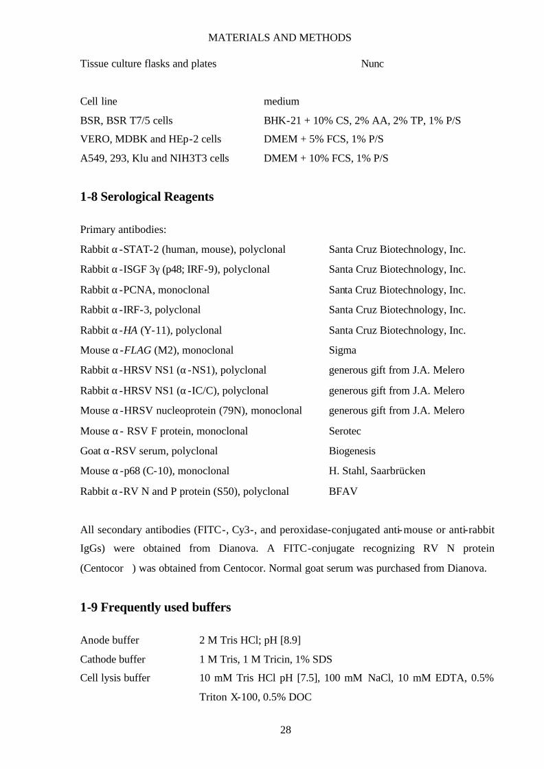
Primary antibodies:
Rabbit α-STAT-2 (human, mouse), polyclonal Santa Cruz Biotechnology, Inc.
Rabbit α-ISGF 3γ (p48; IRF-9), polyclonal Santa Cruz Biotechnology, Inc.
Rabbit α-PCNA, monoclonal Santa Cruz Biotechnology, Inc.
Rabbit α-IRF-3, polyclonal Santa Cruz Biotechnology, Inc.
Rabbit α-HA (Y-11), polyclonal Santa Cruz Biotechnology, Inc.
Mouse α-FLAG (M2), monoclonal Sigma
Rabbit α-HRSV NS1 (α-NS1), polyclonal generous gift from J.A. Melero
Rabbit α-HRSV NS1 (α-IC/C), polyclonal generous gift from J.A. Melero
Mouse α-HRSV nucleoprotein (79N), monoclonal generous gift from J.A. Melero
Mouse α- RSV F protein, monoclonal Serotec
Goat α-RSV serum, polyclonal Biogenesis
Mouse α-p68 (C-10), monoclonal H. Stahl, Saarbrücken
Rabbit α-RV N and P protein (S50), polyclonal BFAV
All secondary antibodies (FITC-, Cy3-, and peroxidase-conjugated anti-mouse or anti-rabbit
IgGs) were obtained from Dianova. A FITC-conjugate recognizing RV N protein
(Centocor) was obtained from Centocor. Normal goat serum was purchased from Dianova.
1-9 Frequently used buffers
Anode buffer 2 M Tris HCl; pH [8.9]
Cathode buffer 1 M Tris, 1 M Tricin, 1% SDS
Cell lysis buffer 10 mM Tris HCl pH [7.5], 100 mM NaCl, 10 mM EDTA, 0.5%
Triton X-100, 0.5% DOC

MATERIALS AND METHODS
29
DNA loading buffer (1x) 15% Ficoll 400, 5x TAE, 0,125% Orange G
Flexi I buffer 100 mM Tris-HCl pH [7.5], 10 mM EDTA pH [8.0], 400 µg/ml
RNase I
Flexi II buffer 200 mM NaOH, 1% SDS
Flexi III buffer 300 mM KCH3COO; pH [5.75]
Gel buffer 3 M Tris HCl pH [8.45], 0.3%SDS,
LB medium 0.5% NaCl, 0.5% yeast extract, 1% tryptone peptone, 1 mM
MgSO4
Luciferase lysis buffer 20 mM Tris HCl pH [7.8], 2 mM DTT, 2 mM CDTA, 10%
glycerol, 1% Triton X-100
Luciferase substrate buffer 20 mM tricine, 2.67 mM MgSO4, 0.1 mM EDTA, 33.3 mM DTT,
1.07 mM MgCO3 x Mg(OH)2 x 5 H2O
Lysis buffer 6.25 mM Tris HCl pH [6.8], 2% SDS, 10% glycerol, 6 M urea, 5%
methanol, 0.01% bromophenol blue, 0.01% phenol red
NP40 buffer 10 mM Hepes pH [7.9], 10 mM NaCl, 3 mM MgCl2, 0.5% NP40
RT buffer (10x) 0.5 M Tris pH [8.3], 0.3 M KCl, 0.1 M MgCl2, 0.05 M DTT
PBS 137 mM NaCl, 2.7 mM KCl, 4.3 mM Na2HPO4, 1.4 mM KH2PO4;
pH [7.5]
SEB (3x) 83.3 mM Tris HCl; pH [6.8], 33.3% glycerol, 6.7% SDS, 16.7% ß-
mercaptoethanol, bromophenol blue
TAE (1x) 40 mM Tris, 5 mM CH3COONa x 3 H2O, 1 mM EDTA
Transfer buffer 48 mM Tris pH [8.6], 39 mM glycine, 18% methanol, 0.005%
SDS
Walter solution 50 mM NH4Cl, 20 mM glycine
1-10 Oligonucleotides
Oligonucleotides were obtained either from MWG Biotechnology Inc. or Metabion. Major
restriction recognition sites are underlined.
NS1HA-EcoRI 3’ 5’-gca ata gaa ttc cta agc gta atc tgg gac atc ata agg ata att cag acc
aag aag agt-3’
NS2FL-EcoRI 3’ 5’-gca ata gaa ttc cta ttt atc gtc atc atc ttt ata atc tgg att taa atc ata
ctt ata-3’
NS1NcoI-5’ 5’-cga ata cca tgg gca gtg aaa cat tga g-3’
MATERIALS AND METHODS
30
NS2BspHI-5’ 5’-gca ata tca tga gca ccc caa atc ccg aa-3’
hNS1-NcoI 5’ 5’-att gac cat ggg cag caa ttc att-3’
hNS2-NcoI 5’ 5’-att gac cat gga cac aac cca ca-3’
hNS1-NotI 5’ 5’-tat gaa gcg gcc gcc ccc tct ctt ctt tct aca gaa aat ggg cag caa ttc
att gag-3’
hNS1-EcoRI 3’ 5’-att gag aat tct tat gga tta aga tca aa-3’
hNS2-AseI 5’ 5’-ata ctt att aat tgg ggc aaa taa atc agt tcc cca acc agc cat gga cac
aac cca caa tg-3’
hNS2-KpnI 3’ 5’-ata aat ggt acc aaa aga taa cac tgt gtg aat taa att ttg aaa agt gct
tat gga ttg aga tca tac ttg-3’
mNS1haEcoRI/V-3’ 5’-aat gat atc gaa ttc tta agc gta atc tgg tac atc ata agg ata acc act
gat cag ctc tac-3’
mNS1NotIEcoRV-5’ 5’-aat gat atc gcg gcc gcc ccc tct ctt ctt tct aca gaa atg ggc tgt aat
gtg atg atg-3’
mNS2flEcoRI/V-3’ 5’-aat gat atc gaa ttc tca ttt atc gtc atc atc ttt ata gtc atc atc atc ctc
atc-3’
mNS2AseIEcoRV-5’ 5’-aat gat atc att aat tgg ggc aaa taa atc agt tcc cca acc agc cat gtc
cac agc tat gaa caa g-3’
mNS2Acc65I 3’ 5’-ata aat ggt acc aaa aga taa cac tgt gtg aat taa att ttg aaa agt gct
cat tta tcg tca tca tct tta tag-3’
bNS2BamHI 5’ 5’-aag cgg atc ccc aac cag cca tga gca cc-3’
bNS1-NotIEcoRV 5’ 5’-aat gat atc gcg gcc gcc ccc tct ctt ctt tct aca gaa atg ggc agt gaa
aca ttg agt g-3’
2 METHODS
2-1 General cloning procedures
Restriction endonuclease digests were done according to the supplier’s manual using 10
units/µg plasmid. Restriction fragments were separated on 1% agarose gels using 1x TAE (40
mM Tris, 5 mM CH3COONa x 3 H2O, 1 mM EDTA) containing 0,1 µg/ml ethidium bromide
as electrophoresis buffer. The samples were supplemented with 1x DNA loading buffer (15%
Ficoll 400, 5x TAE, 0,125% Orange G) prior to loading. To recover a DNA fragment, the

MATERIALS AND METHODS
31
respective band was excised from the gel under UV-light and purified using the QIAquick gel
extraction kit (Qiagen) following the supplier’s manual.
Standard procedures were applied for DNA ligation and transformation of DNA into
competent E.coli XL1 or XL10 prepared by the calcium chloride method.
The following protocol was used to screen for positive clones (miniprep). 1.5 ml of an
overnight culture were centrifuged for 5 min at 7,000 rpm (Eppendorf table centrifuge). The
supernatant was discarded and the pellet resuspended in 0.2 ml Flexi I buffer (100 mM Tris-
HCl pH [7.5], 10 mM EDTA pH [8.0], 400 µg/ml RNase I). 0.2 ml of Flexi II buffer (200 mM
NaOH, 1% SDS) were then added to lyse the cells (5 min at room temperature), followed by
the addition of 0.2 ml of Flexi III buffer (300 mM KCH3COO; pH [5.75]) to precipitate
cellular debris and chromosomal DNA during a 10 min incubation on ice. After centrifugation
for 10 min at 14,000 rpm the supernatant was carefully transferred to a fresh tube and the
plasmid DNA was precipitated with 1 ml ethanol for at least 30 min on ice. The plasmid DNA
was then pelleted by centrifugation and resuspended in dH2O followed by restriction enzyme
analysis to establish the correct clone.
For the preparation of larger amounts of plasmid DNA, overnight cultures grown in 25
ml or 100 ml LB medium (0.5% NaCl, 0.5% yeast extract, 1% tryptone peptone, 1 mM
MgSO4) supplemented with ampicillin (100 µg/ml) were used and plasmid DNA purified
using the Nucleobond plasmid purification kits.
2-2 PCR and reverse transcriptase reaction
In a standard PCR reaction, 100 ng of template DNA were mixed with 250-500 ng of each of
the respective oligonucleotides used as primer, 0.8 µl dNTPs (25 mM of each dATP, dCTP,
dGTP and dTTP), 10 µl DMSO, 10 µl 10x buffer (supplied by the manufacturer), and 0.5 µl
pfu polymerase in a total volume of 100 µl. The reaction was then carried out in a
thermocycler (T3 Thermocycler, Biometra), starting with a 10 min step at 94°C, followed by
35 cycles of the following protocol: a denaturation step for 1 min at 94°C, an annealing step
of 1-2 min at 42-58°C (temperature depending on the length and GC-content of the
oligonucleotides used in the reaction), and a polymerization step of various length at 72°C
(depending on the size of the DNA fragment to be amplified). The resulting DNA fragment
was purified using the Qiagen PCR purification kit.
For reverse transcriptase (RT) reactions, RNA was isolated from infected or mock-
infected cells seeded into 6-well plates using the Qiagen RNeasy kit. 1 µg of RNA was then

MATERIALS AND METHODS
32
mixed with 100 ng of the desired oligonucleotide and 0.3 µl RNasin in a total volume of 23
µl, incubated for 4 min at 65°C followed by 10 min at 37°C. Then 3 µl dNTPs (25 mM each),
3 µl 10x RT buffer (0.5 M Tris pH [8.3], 0.3 M KCl, 0.1 M MgCl2, 0.05 M DTT) and 1 µl
AMV reverse transcriptase were added, and the reaction was incubated for 1 hr at 43°C. 3 µl
of the sample were then used as template in a standard PCR reaction.
2-3 Generation of recombinant RVs expressing pneumovirus NS proteins
Recombinant Rabies Viruses carrying the NS1 or NS2 genes from different pneumoviruses
were constructed on the basis of a full- length RV cDNA (SAD L16) containing an extra
transcriptional stop-restart sequence in the 3’ noncoding sequence of the G gene (SAD VB)
(66).
For the BRSV NS genes, cDNAs encoding C-terminally tagged BRSV NS1 or NS2
proteins were constructed. An additional 27 nt corresponding to an internal region of the
influenza virus hemagglutinin (HA) protein were inserted right before the NS1 stop codon by
PCR using a reverse primer NS1HA-EcoRI 3’ (5’-gca ata gaa ttc cta agc gta atc tgg gac atc
ata agg ata att cag acc aag aag agt-3’) containing an EcoRI recognition sequence (underlined)
and NS1NcoI-5’ (5’-cga ata cca tgg gca gtg aaa cat tga g-3’). For NS2, 24 nt encoding the
synthetic FLAG peptide were inserted C-terminally with reverse primer NS2FL-EcoRI 3’ (5’-
gca ata gaa ttc cta ttt atc gtc atc atc ttt ata atc tgg att taa atc ata ctt ata-3’) and NS2BspHI-5’
(5’-gca ata tca tga gca ccc caa atc ccg aa-3’). The NS1ha PCR fragment was digested with
NotI and EcoRI. After the generation of blunt ends with klenow polymerase, the resulting 475
nt fragment was inserted into the unique SmaI site in pSAD VB immediately downstream of
the extra transcription start signal, leading to pSAD VB NS1ha (VB bNS1). A 470 nt
fragment containing NS2fl was cloned accordingly after digestion with AseI and EcoRI and
filling in with klenow polymerase, resulting in pSAD VB NS2fl (VB bNS2).
For construction of rRVs carrying the HRSV NS1 and NS2 genes, cDNAs of both genes were
obtained by RT-PCR using total RNA isolated from HRSV (Long) infected Vero cells as
template with the following primers for first-strand synthesis: hNS1-NcoI 5’ (5’-att gac cat
ggg cag caa ttc att-3’) for HRSV NS1 and hNS2-NcoI 5’ (5’-att gac cat gga cac aac cca ca-3’)
for HRSV NS2. A PCR fragment encompassing the entire HRSV NS1 gene was then
generated with primers hNS1-NotI 5’ (5’-tat gaa gcg gcc gcc ccc tct ctt ctt tct aca gaa aat ggg
cag caa ttc att gag-3’) and hNS1-EcoRI 3’ (5’-att gag aat tct tat gga tta aga tca aa-3’). For the

MATERIALS AND METHODS
33
HRSV NS2 fragment, primers hNS2-AseI 5’ (5’-ata ctt att aat tgg ggc aaa taa atc agt tcc cca
acc agc cat gga cac aac cca caa tg-3’) and hNS2-KpnI 3’ (5’-ata aat ggt acc aaa aga taa cac tgt
gtg aat taa att ttg aaa agt gct tat gga ttg aga tca tac ttg-3’) were used. The PCR fragments were
digested with NotI and EcoRI in the case of NS1 or digested with AseI and Acc65I in the case
of NS2, followed by generation of blunt ends with klenow polymerase. The resulting
fragments were then inserted into the SmaI site of pSAD VB leading to pSAD VB hNS1 (VB
hNS1) and pSAD VB hNS2 (VB hNS2), respectively.
For the generation of rRVs carrying the PVM NS1 or NS2 gene, cDNAs of PVM NS1 and
NS2 were kindly provided by A. Easton (Warwick, UK). For the NS1 gene, an HA-tag was
inserted right before the NS1 stop codon by PCR using a reverse primer mNS1haEcoRI/V-3’
(5’-aat gat atc gaa ttc tta agc gta atc tgg tac atc ata agg ata acc act gat cag ctc tac-3’) and
mNS1NotIEcoRV-5’ (5’-aat gat atc gcg gcc gcc ccc tct ctt ctt tct aca gaa atg ggc tgt aat gtg
atg atg-3’). In the case of NS2, a FLAG-tag was inserted C-terminally using reverse primer
mNS2flEcoRI/V-3’ (5’-aat gat atc gaa ttc tca ttt atc gtc atc atc ttt ata gtc atc atc atc ctc atc-
3’) and mNS2AseIEcoRV-5’ (5’-aat gat atc att aat tgg ggc aaa taa atc agt tcc cca acc agc cat
gtc cac agc tat gaa caa g-3’). Both PCR fragments were digested with EcoRV and inserted
into the SmaI site of pSAD VB resulting in pSAD VB mNS1ha (VB mNS1) and pSAD VB
mNS2fl (VB mNS2), respectively.
A recombinant RV carrying both NS genes of BRSV was constructed using a plasmid
containing an extra transcription stop-restart sequence (SK9; (66)). A PCR fragment
harboring the NS2fl gene was generated using primers bNS2BamHI 5’ (5’-aag cgg atc ccc aac
cag cca tga gca cc-3’) and NS2FL-EcoRI3’, digested with BamHI and EcoRI and inserted into
SK9 previously digested with BamHI and EcoRI right before the stop-start sequence, giving
rise to SK9 NS2fl. The NS1ha fragment was generated by PCR using primers bNS1-
NotIEcoRV 5’ (5’-aat gat atc gcg gcc gcc ccc tct ctt ctt tct aca gaa atg ggc agt gaa aca ttg agt
g-3’) and NS1HA-EcoRI3’, digested with NotI and EcoRI followed by klenow treatment, and
inserted into SK9 NS2fl digested with BglII and treated with klenow, resulting in SK9
NS2flNS1ha. This plasmid was then digested with NotI and Acc65I followed by the
generation of blunt ends with klenow polymerase, resulting in a large fragment encompassing
NS2-stop/restart-NS1 which was then inserted into the unique SmaI site of pSAD VB
resulting in pSAD VB NS2flNS1ha (VB 2+1).
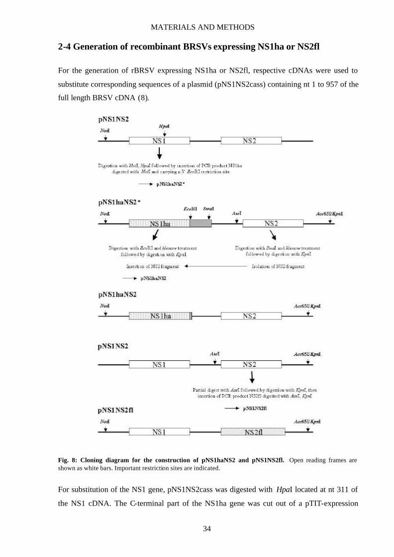
MATERIALS AND METHODS
34
2-4 Generation of recombinant BRSVs expressing NS1ha or NS2fl
For the generation of rBRSV expressing NS1ha or NS2fl, respective cDNAs were used to
substitute corresponding sequences of a plasmid (pNS1NS2cass) containing nt 1 to 957 of the
full length BRSV cDNA (8).
Fig. 8: Cloning diagram for the construction of pNS1haNS2 and pNS1NS2fl. Open reading frames are shown as white bars. Important restriction sites are indicated.
For substitution of the NS1 gene, pNS1NS2cass was digested with HpaI located at nt 311 of
the NS1 cDNA. The C-terminal part of the NS1ha gene was cut out of a pTIT-expression
MATERIALS AND METHODS
35
plasmid (pTIT-NS1ha) with HpaI and BamHI, followed by the conversion of overhanging
DNA ends generated by the restriction digest into blunt ends using klenow polymerase (in the
following referred to as “klenow treatment”), and was then inserted into pNS1NS2cass
resulting in pNS1haNS2*. As a next step, to remove the remaining original NS1 cDNA (nt
313-443), pNS1haNS2* was digested with EcoRI (restriction site was inserted with the
NS1ha cDNA), klenow-treated, and digested with KpnI. Then pNS1NS2cass was digested
with EarI, followed by klenow treatment, and subsequently digested with KpnI. The resulting
fragment encompassing the intergenic region and the entire coding region of NS2 was then
inserted into pNS1haNS2* giving rise to pNS1haNS2 (Fig. 8). This plasmid was digested
with NotI and Acc65I and the resulting fragment inserted into full- length BRSV cDNA
previously digested with NotI and Acc65I, giving rise to pBRSV NS1ha (BRSV ha). For
NS2fl, pNS1NS2cass was digested with ApoI, followed by klenow treatment and a partiell
digest with BsiHKAI. A fragment encompassing almost the entire NS2fl coding region was
obtained by digesting pSKNS2fl with EcoRV and BsiHKAI. This fragment was inserted into
pNS1NS2cass giving rise to pNS1NS2fl (see also Fig. 8). This plasmid was also digested with
NotI and Acc65I, and the resulting fragment was inserted into full- length BRSV cDNA
resulting in pBRSV NS2fl (BRSV fl).
2-5 Generation of rBRSVs expressing HRSV or PVM NS proteins
rBRSVs carrying either HRSV or PVM NS1 were generated using pNS1haNS2* that contains
an additional EcoRI restriction site at nt 311 (see 2-4). Restriction digest with NotI and EcoRI
removes the entire coding region of BRSV NS1ha. PCR fragments containing either the
HRSV or the PVM NS1 gene were generated using primers hNS1-NotI 5’ and hNS1-EcoRI 3’
in case of HRSV NS1, and primers mNS1NotI/EcoRV-5’ and mNS1haEcoRI/V-3’ for PVM
NS1. Both PCR fragments were digested with NotI and EcoRI and inserted into pNS1haNS2
resulting in phNS1NS2 or pmNS1NS2. These plasmids were then digested with NotI and
Acc65I, and the obtained fragments (1094 nt for hNS1NS2, 1043 nt for mNS1NS2) were
inserted into full- length BRSV cDNA, also previously digested with NotI and Acc65I, giving
rise to pBRSV hNS1NS2 (BRSV h1 ) and pBRSV mNS1NS2 (BRSV m1 ).
For generation of rBRSVs carrying either HRSV or PVM NS2, pNS1NS2cass was first
digested with Acc65I, followed by a partial digest with AseI. PCR fragments harboring the
coding region of HRSV or PVM NS2 were generated with primers hNS2-AseI 5’ and hNS2-
Acc65I 3’ for HRSV NS2, and primers mNS2AseIEcoRV-5’ and mNS2Acc65I 3’ (5’-ata aat
ggt acc aaa aga taa cac tgt gtg aat taa att ttg aaa agt gct cat tta tcg tca tca tct tta tag-3’) in the
MATERIALS AND METHODS
36
case of PVM NS2. Both PCR fragments were digested with AseI and Acc65I and inserted into
pNS1NS2cass (949 nt for NS1hNS2, 1069 nt for NS1mNS2), resulting in pNS1hNS2 or
pNS1mNS2. These plasmids were then digested with NotI and Acc65I, and each fragment
was inserted into full- length BRSV cDNA resulting in pBRSV NS1hNS2 (BRSV h2 ) and
pBRSV NS1mNS2 (BRSV m2), respectively.
rBRSVs expressing both NS genes from either HRSV or PVM were generated according
to the protocol described for the generation of pNS1hNS2 or pNS1mNS2. However, instead
of pNS1haNS2*, phNS1NS2 or pmNS1NS2 were used giving rise to phNS1hNS2 and
pmNS1mNS2, respectively. These plasmids were then digested with NotI and Acc65I and the
resulting fragments (1094 nt for hNS1hNS2, 1163 nt for mNS1mNS2) inserted into full-
length BRSV cDNA giving rise to pBRSV hNS1hNS2 (BRSV h1/2) or pBRSV mNS1mNS2
(BRSV m1/2).
2-6 Recovery of recombinant rBRSV and rRV
Recombinant BRSVs were rescued as described previously (8) using plasmids containing the
respective virus cDNA or coding regions for BRSV N, P, L and M2 proteins under the control
of the T7-promoter. BSR T7/5 cells stably expressing T7 polymerase were grown overnight to
80% confluency in 35 mm-diameter dishes in BHK-21 medium supplemented with 10% FCS.
One hour before transfection, cells were washed once with DMEM without FCS, then 1 ml
DMEM was added to each dish. One hour later, cells were transfected with a plasmid mixture
containing 10 µg of full- length plasmid (pBRSV NS1ha, pBRSV NS2fl, pBRSV hNS1NS2,
pBRSV NS1hNS2, pBRSV hNS1hNS2, pBRSV mNS1NS2, pBRSV NS1mNS2, or pBRSV
mNS1mNS2), 4 µg of pTITB-N and pTITB-P, and 2 µg of pTITB-L and pTITB-M2 using the
CaPO4-method (Mammalian transfection kit, Stratagene; plasmids see sections 1-5; 2-4; 2-5).
The transfection medium was removed 4 hours post transfection, and cells were maintained in
BHK-21 medium containing 10% FCS. Three days post transfection, cells were split at a ratio
of 1:3 and maintained in BHK-21 medium supplemented with 2.5% FCS. Cells were then
split every 4-5 days until a cytopathic effect (CPE; for RSV the formation of syncytia) was
observed.
Recombinant RVs were rescued as described previously (34). BSR T7/5 cells were
grown as described above. Plasmids encoding RV proteins N (pTIT-N, 5 µg), P and L (pTIT-
P and pTIT-L, 2.5 µg each) were cotransfected with the respective virus cDNA (pSAD VB
NS1ha, pSAD VB NS2fl, pSAD VB hNS1, pSAD VB hNS2, pSAD VB mNS1ha, pSAD VB
MATERIALS AND METHODS
37
mNS2fl, or pSAD VB NS2flNS1ha, 10 µg each) using the CaPO4-method (Mammalian
transfection kit, Stratagene). The transfection medium was removed 3.5 hours post
transfection, and cells were maintained in BHK-21 medium containing 10% CS. Cell culture
supernatants were harvested 3 and 6 days post transfection and transferred onto fresh BSR
cells. Infectious rRVs were detected by immunostaining of aceton-fixed (80% aceton) cells
with a fluorescein isothiocyanate conjugate (Centocor) recognizing the RV N protein.
2-7 Production of virus stocks
For preparation of RSV stocks, 80% confluent Vero cells grown in medium 80 cm2 tissue
culture flasks were infected at a multiplicity of infection (MOI) of 0.01 to 0.1 in serum free
DMEM. After an incubation time of 1.5 hours, the inoculum was removed, and cells were
incubated in DMEM supplemented with 2.5% FCS until an extensive CPE was observed.
Virus was released by one cycle of freezing and thawing, followed by centrifugation at 3,500
rpm in a centrifuge (Heraeus Varifuge 3.0R) for 5 min to remove cell debris. The supernatant
was then aliquoted (1 ml aliquots) and stored at –70°C. Virus titers were determined on Vero
cells as follows. Vero cells from a confluent 25 cm2 flask were trypsinized and resuspended in
30 ml DMEM containing 5% FCS, and distributed into 96-well microtiter plates (100 µl per
well). Virus stocks were stepwise 10-fold diluted in DMEM and 100 µl of each dilution was
pipetted into the wells. Three days post infection, cells were fixed with 80% acetone for 30
min at 4°C, dried, and afterwards stained with an antibody recognizing the RSV fusion
protein (Serotec, diluted 1:70 to 1:100 in PBS) for 45 min at 37°C. After washing the cells
twice with PBS, a secondary, FITC-conjugated anti-mouse antibody (diluted 1:100 in PBS)
was applied for 45 min at 37°C. The cells were then washed once with PBS and once with
dH2O, and virus titers were determined by counting of infected cell foci using a fluorescent
microscope (Zeiss Axiovert 35).
For production of Rabies virus stocks, 80% confluent BSR cells were infected at an MOI of
0.1 to 1 for 1 hour. Afterwards, the cells were incubated in BHK-21 medium supplemented
with 10% CS, and supernatants were harvested 3 days post infection. If possible, supernatants
were harvested a second time 6 days post infection. The supernatants were centrifuged for 5
min at 3,500 rpm (Haereus Varifuge 3.0R) to remove cell debris, aliquoted (0.4 ml aliquots),
and aliquots were stored at –70°C. To determine virus titers, confluent BSR cells from a 25
cm2 flask were trypsinized and resuspended in 20-25 ml BHK-21 medium containing 10% CS
MATERIALS AND METHODS
38
and distributed into 96-well microtiter plates (100 µl per well). Virus stocks were diluted
stepwise in BHK-21 medium, and 100 µl of each dilution was pipetted into the wells. Two
days post infection, cells were fixed with 80% acetone for 30 min at 4°C, dried, and then
incubated with Centocor (diluted 1:200 in PBS) for at least 45 min at 37°C. Cells were
washed twice with dH2O, and virus titers were determined by counting of infected cells using
a fluorescent microscope (Zeiss, Axiovert 35).
2-8 Infection experiments and treatment with IFN
To determine the growth characteristics of various recombinant BRSVs, different cell lines
were infected in suspension at an MOI of 0.1 for 1 hour in serum free DMEM using 15 ml
falcon tubes for incubation. To keep cells in suspension, tubes were slighty shaken every 15
min. Afterwards, cells were incubated in 12-well plates in 1 ml DMEM containing 2.5% FCS.
Slightly different numbers of cells were used for each cell line in infection experiments
according to cell size (Vero 4x10⁵ cells/well, MDBK 3.5x10⁵ cells/well, HEp-2 and Klu
2.5x10⁵ cells/well). Virus was harvested at the indicated time points by two cycles of freezing
and thawing, and virus titers were determined as described above (2-7).
To measure the effect of IFN-α/β on viral replication, cells were infected at an MOI of
0.1 as described above, and recombinant IFN-α A/D (PBL Biomedical Laboratories) was
added to concentrations of up to 10,000 IU/ml cell culture medium directly after seeding
using a dilution of 100 IU/µl IFN-α in PBS as working concentration. Virus titers were
determined 2 or 3 days post infection depending on the extend of the observed CPE.
Infection of MDBK (Vero) cells with recombinant rRVs expressing pneumovirus NS proteins
were done in suspension at an MOI of 5 (2) for 1 hour in serum free DMEM. For
coinfections, an MOI of 2.5 (1) was used for each recombinant virus. Recombinant IFN-α
A/D was added to concentrations of up to 500 IU/ml immediately after seeding of cells
(MDBK and Vero 4x10⁵ cells/well) into 12-well plates. Cell supernatants were harvested 2
days post infection, centrifuged to remove cell debris, and virus titers were determined as
described above. In addition, cells were harvested with 200 µl lysis buffer, and in the case of
infection with VB bNS1 and VB bNS2, extracts were subjected to SDS-PAGE followed by
detection of RV N and P proteins with antibody S50 (1:20,000 in PBS-T; see also 2-9).
MATERIALS AND METHODS
39
2-9 Western blot analysis of pneumovirus NS proteins
To monitor the expression of pneumovirus NS proteins from recombinant BRSVs, Vero cells
were mock-infected or infected with the respective BRSV at an MOI of 0.1 as described
above and seeded into 6-well plates in 2 ml DMEM supplemented with 2.5% FCS. Three
days post infection, cells were lysed with 500 µl lysis buffer (6.25 mM Tris HCl pH [6.8], 2%
sodium dodecyl sulfate [SDS], 10% glycerol, 6 M urea, 5% methanol, 0.01% bromophenol
blue, 0.01% phenol red). In the case of recombinant RVs expressing pneumovirus NS
proteins, BSR cells were infected with the respective recombinant viruses at an MOI of 1 as
described above and seeded into 6-well plates. Three days post infection, cells were lysed
with 500 µl lysis buffer. Cell extracts were incubated at 95°C for 5 min, and equivalent
amounts (50 µl/well) were loaded onto a 12% gel for sodium dodecyl sulfate-polyacrylamide
gel electrophoresis (SDS-PAGE; Penguin Model P9DS [OWL scientific], 16x14 cm, 40 V
12-14 h run). The proteins were transferred to a nitrocellulose membrane in a semidry transfer
apparatus (ThePanther Model HEP-1 [OWL scientific]) using the following transfer buffer:
48 mM Tris pH [8.6], 39 mM glycine, 18% methanol, 0.005% SDS. After the transfer, the
membrane was incubated in blocking solution (0.05% Tween-20 in phosphate-buffered saline
[PBS-T] supplemented with 5% skim milk powder) for 45 min at room temperature. After
two consecutive washes with PBS-T for 15 min each, the membrane was incubated overnight
at 4°C or 4 hours at room temperature in primary antibody solution (HRSV NS proteins were
detected using α-NS1 and α-IC/C antibodies [1:5,000-1:7,000 in PBS-T; both kindly
provided by J.A. Melero, Madrid, Spain], BRSV and PVM NS1ha were detected with Y-11
antibody [1:1,000 in PBS-T], BRSV and PVM NS2fl with M2 antibody [1:20,000 in PBS-T])
followed by two consecutive washes with PBS-T for 15 min each. Afterwards, the membrane
was incubated with secondary antibody solution for 2 hours at room temperature (peroxidase-
conjugated goat anti-rabbit or anti-mouse IgG; 1:10,000 in PBS-T). The membrane was then
washed twice with PBS-T for 15 min each, followed by incubation of the membrane in ECL
solution (1.5 ml solution #1, 1.5 ml solution #2, 7 ml PBS-T) for 1 min. Exposure times to
Hyperfilm ECL (Amersham) varied depending on signal strength from 5 sec up to several
minutes.
10% gel 12% gel (sufficient for one 14x16 cm gel) polyacrylamide 9.0 ml 10.8 ml gel buffer 12.0 ml 12.0 ml dH2O 12.9 ml 11.1 ml glycerol 2.0 ml 2.0 ml TEMED 0.017 ml 0.017 ml APS 0.175 ml 0.175 ml
MATERIALS AND METHODS
40
Gel buffer (3 M Tris HCl, pH [8.45]; 0.3% SDS)
Anode buffer (2 M Tris HCl, pH [8.9])
Cathode buffer (1 M Tris, 1 M Tricin, 1% SDS)
2-10 Immunofluorescent staining
For immunostaining, cells were seeded out onto coverslips placed into 6-well plates directly
after infection (BRSVs: MOI 0.1, RVs: MOI 0.1-1.0). At most, 4x10⁵ cells were used per
well. At the indicated time points, cells were fixed in PBS containing 3% paraformaldehyde
for 20 min at room temperature, follwed by incubation in Walter solution (50 mM NH4Cl, 20
mM glycine) for 10 min at room temperature. After two washes with PBS, cells were
permeabilized with 0.5% Triton X-100 in PBS for 10 min at room temperature. Next, cells
were washed twice in PBS, followed by a 15 min incubation step in blocking solution (PBS
containing 10% goat normal serum). The cells were then incubated in 100 µl primary
antibody solution for 45 min at room temperature [1.5% goat normal serum in PBS; detection
of BRSV and PVM NS1ha with antibody Y-11 (1:100); BRSV and PVM NS2fl with antibody
M2 (1:2,000); STAT2 with α-STAT2 antibody (1:100); BRSV N protein with antibody 79N
(1:40; kindly provided by J.A. Melero, Madrid, Spain)]. After two washes with PBS, cells
were incubated in secondary antibody solution (1.5% goat normal serum in PBS; FITC- or
Cy3-conjugated anti-rabbit or anti-mouse IgG 1:200) for 45 min at room temperature. Cells
were washed twice with PBS and once with dH2O and then embedded in embedding medium
(50% glycerol or Histogel). Cells were analysed using a laser scan microscope (Leica TCS
NT).
For staining of cell nuclei with propidium iodide, cells were incubated for 45 min with
100 µl of staining solution containing 1 µl propidium iodide (10 mg/ml), 1 µl EDTA 0.5M
and 1 µl RNase A (20 mg/ml) in PBS.
Digitonin treatment of cells was performed as follows. Cells were treated with digitonin
(40 µg/ml in PBS) for 3 min on ice, then washed once with cold PBS. Next, cells were
incubated with 0.025% Triton X-100 in PBS for 5 min on ice before fixation in 3%
paraformaldehyde. Permeabilization using 0.2% Triton X-100 in PBS and staining of cells
were performed as described above. For the GFP control, Vero cells were transfected with 2
µg pEGFP-N3 (Clontech) using the FuGENE 6 transfection reagent (Roche). An antibody
recognizing the nuclear RNA helicase p68, C-10, was used 1:2000 in antibody solution (1.5%
goat normal serum in PBS).
MATERIALS AND METHODS
41
2-11 Monitoring ISG expression
HEp-2, L929 (2.5x10⁵/well), MDBK or NIH3T3 (3x10⁵/well) cells were mock- infected or
infected with the indicated rBRSVs at an MOI of 0.2 as described above (2-8) and seeded into
12-well plates. When indicated, mock- infected MDBK and HEp-2 cells were treated with
2,000 IU IFN-α A/D per ml medium directly after seeding. Cells were lysed in 150-200 µl
cell lysis buffer (10 mM Tris HCl pH [7.5], 100 mM NaCl, 10 mM EDTA, 0.5% Triton X-
100, 0.5% DOC) at indicated time points. Equal amounts of cell extracts (30-50 µl per lane;
determined with Coomassie Protein Assay Reagent, Pierce) were mixed with 25 µl 3x SEB
(83.3 mM Tris HCl pH [6.8], 33.3% glycerol, 6.7% SDS, 16.7% ß-mercaptoethanol,
bromophenol blue) and loaded onto a 10% SDS-polyacrylamide gel. Proteins were transferred
and the membrane was probed with antibodies recognizing STAT-2, PKR, p48 and PCNA
(1:1000 in PBS-T; see also 2-9).
2-12 Detection of IFN in supernatants
Biological active IFN was detected in cell culture supernatants making use of the sensitivity
of RV to IFN in MDBK cells. MDBK cells (4x10⁵/well) were infected in suspension with
SAD VB at an MOI of 5 for 1 hour. Then cells were seeded into 12-well plates in DMEM
supplemented with 2.5% FCS using a total volume of 500 µl per well. In the meantime,
supernatants of mock- infected or infected MDBK or HEp-2 cells were harvested, centrifuged
at 5,000 rpm for 5 min (Eppendorf 5417R) to remove cell debris, transferred to a fresh tube
and incubated at 56°C for 30 min to eliminate replication-competent RSV. Thus treated,
supernatants were then transferred onto RV-infected MDBK cells (400 µl/well). Supernatants
of RV-infected MDBK cells were harvested 2 days post infection and rabies virus titers were
determined by limiting dilution and immunostaining as described above (2-7).
2-13 Transfection of reporter plasmids and luciferase assay
Reporter plasmids containing the luciferase gene under the control of the IFN-β gene
promoter/enhancer (p125Luc) or under the control of the PRDI domain containing the IRF-3
binding sequence (p55CIBLuc), and a plasmid from which a dominant negative form of IRF-
3 is expressed (pEF-haIRF358-427) were kindly provided by Takashi Fujita, Kyoto, Japan.
All transfection experiments were carried out on subconfluent 293 cells seeded into 35 mm
dishes at least 12 hours prior to transfection (2x10⁶ cells/dish). 1 µg of reporter construct
MATERIALS AND METHODS
42
p125Luc and, when indicated, 1.5 µg of expression construct pEF-haIRF358-427 per dish were
transfected using the FuGENE 6 transfection reagent. Ten hours post transfection, cells were
trypsinized, resuspended, and seeded into 24-well plates using 4x10⁵ cells/well. 24 hours post
transfection, cells were infected with the indicated rBRSVs at an MOI of 0.2 (4x10⁵
cells/well) in serum-free DMEM. After an infection step of 90 min, cells were washed once
with DMEM and then incubated in DMEM supplemented with 2.5% FCS. Cells were
harvested 10 hours post infection or at indicated time points with 150-200 µl luciferase lysis
buffer (20 mM Tris HCl pH [7.8], 2 mM DTT, 2 mM CDTA, 10% glycerol, 1% Triton X-
100; incubation 10 min at 37°C), followed by short centrifugation to remove unsoluble parts.
Aliquots were then assayed for reporter gene activity in a luciferase assay. To rule out
differences in gene activity due to varying amounts of transfected cells present in the sample,
the protein content of each sample was determined in parallel using Coomassie Protein Assay
Reagent.
For experiments using poly I/C as trigger for IFN induction, 2x10⁶ 293 cells/ 35mm dish
were mock- infected or infected with BRSV wt at an MOI of 0.25 as described above (one 35
mm dish per amount of poly I/C). 14 hrs post infection cells were transfected with 1 µg
p125Luc/35 mm dish using the FuGENE 6 transfection reagent. 24 hrs post infection, cells
were again transfected with the indicated amounts of poly I/C using the FuGENE 6
transfection reagent. 38 hrs post infection cells were harvested in 300 µl luciferase lysis buffer
and luciferase activity was measured as described below.
For experiments assaying the activities of the transcription factors IRF-3, NF-κB or AP-
1, 2x10⁶ 293 cells were transfected with 1 µg (p55CIBLuc, pAP-1Luc) or 0.5 µg (pNF-
kBLuc) of the indicated reporter construct using the FuGENE transfection reagent. 8 hrs later
cells were trypsinized, seeded into 24-wells using 4x10⁵ cells/well and infected with the
indicated recombinant viruses at an MOI of 0.2. 14-16 hrs post infection cells were harvested
in 120-150 µl luciferase lysis buffer and luciferase activity measured as described below.
Luciferase assays were performed as follows. 20 µl of each cell lysate was mixed with 80 µl
luciferase lysis buffer. 100 µl luciferase substrate buffer (20 mM tricine, 2.67 mM MgSO4,
0.1 mM EDTA, 33.3 mM DTT, 1.07 mM MgCO3 x Mg(OH)2 x 5 H2O) supplemented with
470 µM luciferin, 270 µM AcetylCoA, and 530 µM ATP was added automatically in the
luminometer (Perkin Elmer Wallac GmbH) and light emission was measured as relative light
units (RLU).
MATERIALS AND METHODS
43
2-14 Detection of IRF-3 phosphorylation
For western blot analysis of phosphorylated IRF-3, 2x10⁶ 293 cells were mock-infected or
infected in suspension with the indicated RSVs at an MOI of 0.25-0.3 for 1 hour and then
seeded into 6-well plates. 10 hours post infection, cells were harvested and nuclear extracts
were prepared as follows. Cells were removed from the culture dish using PBS supplemented
with 5 mM EDTA. Cell pellets were resuspended in 150 µl NP40 buffer (10 mM Hepes pH
[7.9], 10 mM NaCl, 3 mM MgCl2, 0.5% NP40) and incubated on ice for 40 min. Afterwards,
the cell suspension was checked for intact nuclei, followed by centrifugation at 7,000 rpm and
4°C for 5 min (Eppendorf 5417R). Supernatants were kept as cytoplasmic extracts, and pellets
containing the intact nuclei were washed once with cold PBS and stored at -20°C. Prior to
SDS-PAGE, pellets were lysed with 100 µl cell lysis buffer containing 1% Triton X-100 and
50 µl of each sample were mixed with 25 µl 3x SEB, incubated for 5 min at 95°C, and applied
onto a large 10% polyacrylamide gel (Penguin Model P10DS, 20x20 cm). The
electrophoresis (80 V, 16-20 h run) was stopped when the 43 kD marker band was migrated
to the lower end of the gel. Proteins were transferred and the membrane probed with an
antibody recognizing human IRF-3 (1:1000 in PBS-T) as described above (2-9).
RESULTS
44
RESULTS
1 PNEUMOVIRUS NS PROTEINS COOPERATIVELY
CONFER VIRAL RESISTANCE TO IFN
Construction of recombinant RVs expressing BRSV NS proteins
BRSV NS deletion mutants that lack either one or both NS genes show approximately equal
degrees of sensitivity against IFN-mediated responses suggesting that both NS proteins are
required to counteract antiviral mechanisms. To verify the obligatory cooperative function of
NS1 and NS2 and to determine whether the two NS proteins are sufficient to protect an
unrelated virus or whether additional factors are needed, we generated rRVs expressing either
NS1 (VB bNS1) or NS2 (VB bNS2). For detection purposes, C-terminally tagged BRSV NS
proteins were generated, with NS1 carrying additional 27 nt corresponding to an internal
region of the influenza virus hemagglutinin (HA) protein, and NS2 carrying additional 24 nt
corresponding to the synthetic FLAG peptide. The additional gene was introduced between
the G and L genes of the attenuated SAD VB, as previously described for successful
expression of other genes (Fig. 9; 66,82). Recombinant viruses were recovered from cDNA in
BSR T7/5 cells transfected with plasmids expressing the RV N, P, and L genes. Synthesis of
the NS proteins was monitored by western blotting (see Figs. 11C, 14B), and no obvious
adverse effect on infectious titers and growth characteristics of the recombinants in BSR cells
was observed (data not shown).
Fig. 9: Organization of recombinant RVs expressing tagged BRSV NS genes. Tagged BRSV NS1 or BRSV NS2 open reading frames (NS1ha or NS2fl, respectively) were inserted at a unique SmaI site located between the RV G and L genes.
RESULTS
45
BRSV NS1 and NS2 cooperatively enhance IFN resistance of RV
To study the activity of the expressed BRSV NS proteins, Vero cells were infected at an MOI
of 5 with parental RV (VB wt) or with each of the recombinants, or were co- infected with
both recombinants, VB bNS1 and VB bNS2, at an MOI of 2.5 each. Directly after infection,
cells were treated with increasing amounts of recombinant IFN-α A/D and the production of
infectious RV titers was analysed 2 days post infection. Growth of the parental RV and of
NS1 or NS2 protein-expressing viruses from single infections was similarly affected (Fig.
10A). On application of 50 IU of IFN-α titers dropped by approximately 1 log unit and then
further decreased very slowly with increasing IFN amounts, indicating either a weak IFN
response of Vero cells or a high intrinsic resistance of RV to IFN-mediated responses in Vero
cells. In cells co- infected with NS1 and NS2-expressing viruses, however, protection of virus
replication could be shown. Virus titers remained significantly higher than in single infections
and decreased slowly in a dose-dependent manner. Similar results were also obtained when
viral protein expression was monitored. Only in cells coinfected with both recombinants, viral
protein synthesis was maintained in the presence of up to 150 IU of IFN-α (Fig. 11A).
Fig. 10: IFN resistance in cells coinfected with RVs expressing BRSV NS1 and NS2. Vero cells (A) or MDBK cells (B) were infected with wild-type VB, VB bNS1, or VB bNS2, or coinfected with VB bNS1 and VB bNS2. Immediately after infection, cells were treated with the indicated amounts of IFN-α A/D. Infectious virus titers were determined 2 days post infection. Results represent the mean values of at least four independent experiments, with error bars indicating standard deviations.

RESULTS
46
In parallel experiments performed in MDBK cells, parental RV and the NS-expressing
recombinants replicated to slightly lower titers in untreated MDBK cells than in Vero cells. In
contrast to Vero cells, treatment with IFN more dramatically reduced infectious titers of wt
RV and of NS-expressing viruses from single infections. An immediate reduction in
infectious titers by 3 log units indicated a highly effective IFN-mediated cell response.
However, in cells co- infected with viruses expressing NS1 and NS2 proteins, virus replication
was completely protected against IFN until more than 150 IU of IFN was applied (Fig. 10B).
These results were reflected by the analysis of RV protein synthesis. In untreated cells, all
recombinants produced substantial and comparable amounts of RV proteins whereas in IFN-
treated cells, only coinfections with VB bNS1 and VB bNS2 led to substantial protein
synthesis until more than 150 to 200 IU was applied (Fig. 11B). These results confirm that the
two BRSV NS proteins are required and sufficient for exerting the observed IFN antagonist
activity. In addition, they show that both NS proteins are able to confer resistance to IFN-
mediated antiviral response not only to BRSV but also to another, unrelated rhabdovirus.
Fig. 11: RV protein synthesis in IFN challenge experiments. Vero (A) or MDBK cells (B) were infected as described in Fig. 8, and cultures were treated with 0, 50, 150, 500 and 2,000 IU (A) or 0, 50, 150, 175, 500 IU of IFN-α (B). Cells were harvested 2 days post infection and extracts were analysed by western blot. RV N protein was detected using a serum raised against RV (S50). (C) Western blot analysis of BRSV NS protein expression in Vero cells infected with VB bNS1 and VB bNS2. Cells were treated with 0, 50 or 150 IU of IFN-α directly after seeding, and harvested 2 days post infection. NS1 protein was detected using an antibody recognizing the HA-tag (Y-11). For detection of NS2, an antibody recognizing the FLAG-tag (M2) was used.
RESULTS
47
To rule out the possibility that the addition of the HA- or FLAG-tag to NS1 and NS2,
respectively, influences their function(s) in the viral life cycle, recombinant BRSVs
expressing either NS1ha or NS2fl were generated (BRSV ha and BRSV fl, respectively; Fig.
12A). Both recombinant BRSVs were recovered by cotransfection of BSR T7/5 cells with
BRSV full- length cDNA with support plasmids encoding BRSV N, P, L, and M2 genes as
described in 2-4, and synthesis of the tagged NS proteins was monitored by Western blotting
(Fig. 12B).
Fig. 12: IFN resistance of rBRSVs expressing tagged BRSV NS genes is not altered. (A) Genomes of rBRSV expressing NS1ha (BRSV ha) or NS2fl (BRSV fl). (B) Western blot analysis of cell lysates from Vero cells infected with BRSV ha and BRSV fl. NS1ha was detected using an antibody recognizing the HA-tag (Y-11). NS2fl was recognized by an antibody raised against the synthetic FLAG epitope (M2). (C) Vero or MDBK cells infected at an MOI of 0.1 with BRSV wt, BRSV ha, or BRSV fl were incubated with the indicated amounts of recombinant IFN-α directly after seeding. Infectious titers were determined 3 days post infection. Results are shown as fold reduction in viral titer and represent the mean values of two independent experiments.
RESULTS
48
IFN challenge experiments were performed in Vero and MDBK cells to determine the
protective capacity of the modified NS proteins towards exogenous IFN. As expected, no
difference in IFN resistance was observed for BRSV ha or BRSV fl when compared with wt
BRSV (Fig. 13). We therefore conclude that the addition of a protein tag to either NS protein
does not compromise its IFN antagonist function in the context of a viral infection.
HRSV and PVM NS proteins protect RV from IFN-induced responses
Having established an assay system based on rRV to monitor IFN resistance function of a
given protein we addressed the question whether the NS proteins of HRSV and PVM also
possess IFN antagonist activity and whether this activity is comparable to that observed with
BRSV NS proteins. RVs expressing either HRSV NS1 or NS2 (VB hNS1 and VB hNS2,
respectively) or PVM NS1 or NS2 (VB mNS1 and VB mNS2, respectively) were generated
by insertion of the additional gene between the G and L genes of the infectious SAD VB full-
length antigenomic cDNA clone (Fig. 13A). Efficient expression of either NS protein was
confirmed by Western blot analysis (Fig.13B).
Fig. 13A: rRVs expressing HRSV and PVM NS genes. Genome organization of rRV containing HRSV or PVM NS open reading frames between the RV G and L genes. For detection purposes, an HA-tag was added at the C-terminus of the PVM NS1 gene, at the C-terminus of the PVM NS2 gene, a FLAG-tag was added. No tag was added to either HRSV NS gene.
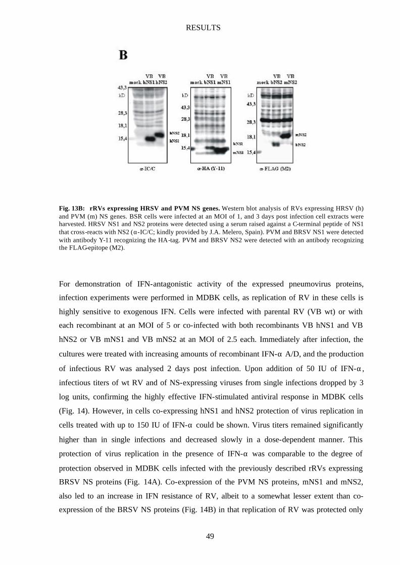
RESULTS
49
Fig. 13B: rRVs expressing HRSV and PVM NS genes. Western blot analysis of RVs expressing HRSV (h) and PVM (m) NS genes. BSR cells were infected at an MOI of 1, and 3 days post infection cell extracts were harvested. HRSV NS1 and NS2 proteins were detected using a serum raised against a C-terminal peptide of NS1 that cross-reacts with NS2 (α-IC/C; kindly provided by J.A. Melero, Spain). PVM and BRSV NS1 were detected with antibody Y-11 recognizing the HA-tag. PVM and BRSV NS2 were detected with an antibody recognizing the FLAG-epitope (M2).
For demonstration of IFN-antagonistic activity of the expressed pneumovirus proteins,
infection experiments were performed in MDBK cells, as replication of RV in these cells is
highly sensitive to exogenous IFN. Cells were infected with parental RV (VB wt) or with
each recombinant at an MOI of 5 or co-infected with both recombinants VB hNS1 and VB
hNS2 or VB mNS1 and VB mNS2 at an MOI of 2.5 each. Immediately after infection, the
cultures were treated with increasing amounts of recombinant IFN-α A/D, and the production
of infectious RV was analysed 2 days post infection. Upon addition of 50 IU of IFN-α,
infectious titers of wt RV and of NS-expressing viruses from single infections dropped by 3
log units, confirming the highly effective IFN-stimulated antiviral response in MDBK cells
(Fig. 14). However, in cells co-expressing hNS1 and hNS2 protection of virus replication in
cells treated with up to 150 IU of IFN-α could be shown. Virus titers remained significantly
higher than in single infections and decreased slowly in a dose-dependent manner. This
protection of virus replication in the presence of IFN-α was comparable to the degree of
protection observed in MDBK cells infected with the previously described rRVs expressing
BRSV NS proteins (Fig. 14A). Co-expression of the PVM NS proteins, mNS1 and mNS2,
also led to an increase in IFN resistance of RV, albeit to a somewhat lesser extent than co-
expression of the BRSV NS proteins (Fig. 14B) in that replication of RV was protected only
RESULTS
50
up to 100 IU of IFN-α. Thus, the NS proteins of HRSV and PVM are also able to antagonize
IFN-mediated antiviral responses and do so only when expressed together.
Fig. 14: HRSV and PVM NS proteins confer IFN resistance to RV. (A) MDBK cells were infected with VB wt, VB hNS1 (H1), VB hNS2 (H2), or coinfected with VB hNS1 and VB hNS2 (H1+H2). As positive control, cells were also coinfected with VB bNS1 and VB bNS2 (B1+B2). Indicated concentrations of recombinant IFN-a were added immediately after seeding, and virus titers were determined 2 days post infection. Error bars indicate standard deviation. (B) Infection of MDBK cells with VB mNS1 (M1) or VB mNS2 (M2), or coinfection with VB mNS1 and VB mNS2 (M1+M2) as described above. Results represent the mean values of three independent experiments, with error bars indicating standard deviations.
HRSV and BRSV NS proteins are able to cooperate
The capacity of all pneumovirus NS proteins to protect against IFN-induced effects prompted
us to further investigate whether combinations of BRSV and HRSV or BRSV and PVM NS
proteins are also functional in mediating IFN resistance. MDBK cells were infected with four
pairs of recombinant viruses expressing single NS proteins from HRSV (h), BRSV (b) or
PVM (m), namely VB bNS1 and VB hNS2, VB hNS1 and VB bNS2, VB bNS1 and VB
mNS2, and VB mNS1 and VB bNS2, and treated with IFN as described above. Whereas no
PVM NS protein was found functional in combination with the complementing BRSV NS
protein (Fig. 15B), both HRSV/BRSV pairs (bNS1/hNS2 and hNS1/bNS2) were able to
increase the IFN resistance of RV (Fig. 15A). However, while co-expression of BRSV NS1
RESULTS
51
and HRSV NS2 was as effective as the two homologous combinations, the inverse
combination of HRSV NS1 and BRSV NS2 did not rescue viral growth in the presence of
more than 50 IU of IFN-α, as observed in several independent experiments. Nevertheless, the
cooperativity of HRSV and BRSV NS proteins in IFN escape strongly suggests that they have
identical function(s) and similar target proteins whereas the highly dissimilar NS proteins of
PVM were unable to cooperate with their BRSV counterparts but might still share a similar
mechanism to antagonize IFN-induced cell responses.
Fig. 15: Combinations of HRSV and BRSV NS proteins render RV IFN resistant. (A) MDBK cells were coinfected with various combinations of rRVs expressing BRSV (b) or HRSV (h) NS1 or NS2. Recombinant IFN-α was added immediately after seeding, and viral titers were determined 2 days post infection. Results represent the mean values of three independent experiments (VBmNS1+VBbNS2 and VBbNS1+VBmNS2 two independent experiments) with error bars indicating standard deviation. (B) Infection of MDBK cells with combinations of recombinant RVs expressing BRSV or PVM nonstructural genes. Results represent the mean values of three independent experiments (VBmNS1+VBbNS2 and VBbNS1+VBmNS2 two independent experiments) with error bars indicating standard deviation.
A recombinant RV expressing both BRSV NS proteins
Since we could show that any IFN antagonist activity requires the presence of both NS
proteins, a recombinant RV expressing both NS proteins of BRSV (VB 2+1) was
subsequently generated allowing us to circumvent the ambiquities related to co-infections,
such as a low percentage of cells expressing both NS proteins or varying protein expression
RESULTS
52
levels, when studying NS protein function. In a first step, an RV stop-restart sequence was
introduced between the NS2 and the NS1 gene of BRSV, then both genes were inserted
between the G and L gene of SAD VB (Fig. 16A). The resulting recombinant (VB 2+1) was
recovered from cDNA as described previously (see section 2-6) and synthesis of both NS
proteins was monitored by Western blotting (Fig. 16B). Even though two additional genes
were inserted into the RV genome, the growth properties of VB 2+1 were not significantly
influenced in BSR cells compared to wt VB (data not shown). However, as expected, VB 2+1
was substantially more IFN resistant than VB wt when challenged with exogenous IFN-α
A/D, with 500 IU reducing viral titers only about 1 log unit (Fig. 16C). The possibility to
generate recombinant RVs expressing several additional proteins without inhibition of virus
replication will provide a useful tool to study IFN antagonist function of various (pneumo-)
virus proteins in vitro and in vivo.
Fig. 16: Recombinant RV expressing both BRSV NS genes. (A) Genome organization of an rRV containing BRSV NS1 and NS2 open reading frames between the RV G and L genes (VB2+1). (B) Western blot analysis of NS protein expression in VB2+1 infected BSR cells using α-HA antibody to detect NS1ha, α-FLAG antibody to detect NS2fl, and S50 serum to detect RV N and P protein. (C) IFN resistance of RV expressing both NS proteins of BRSV. MDBK cells were infected with VB wt (solid square), or VB2+1 (open square), and indicated amounts of recombinant IFN-α were added. Virus titers were determined 2 days post infection. Results represent the mean value of two independent experiments.
RESULTS
53
2 INVOLVMENT OF PNEUMOVIRUS NS PROTEINS
IN DETERMINING VIRAL HOST RANGE
Chimeric BRSVs expressing HRSV NS genes
To investigate NS protein function in its more natural pneumovirus context rather than in the
heterologous rabies virus, we generated recombinant BRSVs possessing either one or both NS
genes from HRSV instead of the homologous NS gene(s) (rBRSV h1, rBRSV h2, or rBRSV
h1/2; Fig. 17). cDNAs were obtained by RT-PCR from Vero cells infected with HRSV strain
Long and were used to replace the BRSV NS genes as described in 2-5. Recombinant viruses
were recovered by co-transfection of BSR T7/5 cells with the BRSV full- length cDNAs with
support plasmids encoding BRSV N, P, L, and M2 genes described previously (8). Virus
stocks were produced in Vero cells, as these lack an intact IFN-α/β system (31,97), and
expression of the NS proteins were monitored by Western blot analysis (Fig. 18A).
Fig. 17: Construction of chimeric rBRSVs expressing HRSV NS genes. The locations of the protein-encoding frames (shaded bars ) are shown relative to the viral genome (vRNA, black bar). In the enlargement, the organization of BRSV wild-type and the recombinant BRSVs carrying one or both HRSV nonstructural proteins, BRSV h1, BRSV h2, and BRSV h1/2, are depicted. Leader RNA is marked by diagonal hatching.
RESULTS
54
The α-IC/C antibody (kindly provided by J.A. Melero) was used for detection of HRSV NS
proteins. α-IC/C serum was raised against a C-terminal peptide of HRSV NS1 and shows
crossreactivity with HRSV NS2 and BRSV NS2 (J.A. Melero, personal communication).
Unfortunately, the HRSV NS2 protein synthesized in BRSV h2 infected cells could not be
detected (Fig. 18A). Therefore, RT-PCR was performed and the presence of virus-encoded
HRSV NS2 mRNA in BRSV h2 infected cells was verified (data not shown). In addition,
immunofluorescent staining of HRSV NS2 in BRSV h2 infected cells also gave a distinct
signal (see Fig. 37). RT-PCR followed by sequencing of the amplified NS2 coding region will
show whether BRSV h2 carries a mutation in its C-terminus responsible for the failure to
detect the HRSV NS2 protein by Western blotting with α-IC/C.
Fig. 18: Analysis of chimeric BRSVs expressing HRSV NS genes. (A) Western Blot analysis of HRSV NS protein synthesis in Vero cells infected with chimeric BRSVs. NS proteins were detected using α-IC/C antibody recognizing HRSV NS proteins and BRSV NS2 (kindly provided by J.A. Melero, Spain). (B), (C) Growth kinetics of chimeric BRSV h1, BRSV h2, BRSV h1/2, and wild-type BRSV in Vero (B) and MDBK cells (C). Cells were infected at an MOI of 0.1 and virus was harvested at indicated time points. Results represent the mean values of three independent experiments, error bars indicate standard deviation.

RESULTS
55
Growth of BRSV h1/2 is attenuated only in bovine cells
The growth behaviour of all three chimeric BRSVs (BRSV h1, BRSV h2, and BRSV h1/2) in
IFN-negative Vero cells was indistinguishable from that of wild-type BRSV. All viruses
produced infectious titers of about 3x10⁵ PFU 2 days after infection at an MOI of 0.1 (Fig.
18B). The identical growth suggests that the HRSV NS proteins are able to entirely fulfill the
function(s) of their bovine counterparts important for efficient RNA synthesis in Vero cells.
Next, a cell line of bovine origin, MDBK, was used which harbors an intact IFN system. In
this cell line that optimally supports the growth of wt BRSV, growth of BRSV h1/2 was
markedly attenuated. Whereas wt BRSV reached titers of 7x10⁵ PFU after 3 days, BRSV h1/2
grew only up to 1x10⁴ PFU (Fig. 18C). In contrast, chimeric BRSV h1 and BRSV h2 were
less attenuated with BRSV h1 reaching titers similar to wild-type BRSV, whereas BRSV h2
growing to slightly lower titers of 1x10⁵ PFU 3 days post infection (Fig. 18C). This indicates
that the presence of at least one NS protein from BRSV can overcome the attenuated growth
phenotype mediated by both HRSV NS proteins, as observed with BRSV h1/2 in bovine cells.
Fig. 19: Growth of BRSV h1/2 is attenuated in bovine cells. Vero (A), HEp-2 (B), MDBK (C) or Klu cells (D) were infected with BRSV wt or BRSV h1/2 at an MOI of 0.1. Virus was harvested at indicated time points. Results represent the mean values of two independent experiments. Bars show range of individual data points.
RESULTS
56
To confirm the attenuated growth of BRSV h1/2 in bovine MDBK cells (see also Fig. 19C)
another bovine cell line isolated from embryonic calf lung tissue, Klu, was subsequently used.
In this cell line, the attenuation of BRSV h1/2 was even more pronounced. Here, BRSV wild-
type grew to highest titers of 1.5x10⁶ PFU 2 days post infection, whereas the chimeric BRSV
h1/2 was barely able to grow, reaching titers of only 3x10² PFU (Fig. 19D). In constrast, in a
human cell line, HEp-2, growth characteristics of wt BRSV and BRSV h1/2 were again
comparable (Fig. 19B). Both viruses reached similar titers of 3x10⁴ PFU after 3 days. This
indicates that in the context of an IFN-competent human cell system, the HRSV NS proteins
may be at least an adequate substitute for their counterparts in BRSV, whereas in bovine cell
IFN systems, they are obviously less efficient.
IFN antagonistic activity is host cell specific
Additional stimulation of infected cells with increasing doses of exogenous recombinant IFN-
α was then used to directly challenge the protective capacity of HRSV and BRSV NS proteins
to cellular IFN-induced responses in cell lines of different origin. Vero cells cannot produce
IFN-α/β , yet they are able to mount an effective antiviral response when exogenous IFN is
added (31,97). In Vero cells infected with BRSV or BRSV h1/2, the replication of both
viruses was largely resistant to high IFN doses (Fig. 20A). Only after application of more than
5,000 IU of IFN-α was an approximately 10-fold reduction in infectious titers observed. In
the bovine MDBK cells, resistance of BRSV wt to IFN-α was even more pronounced than in
Vero cells. After application of 10,000 IU of IFN-α, virus titers were only five-fold reduced
compared to the untreated control. In striking contrast, BRSV h1/2 showed a marked and
dose-dependent sensitivity towards the IFN-induced antiviral response of MDBK cells, with
5,000 IU of IFN-α reducing viral titers about 6-fold and 10,000 IU causing a 80-fold
reduction (Fig. 20B). Thus, while IFN resistance of BRSV h1/2 in primate Vero cells is
comparable to that of wt BRSV, BRSV h1/2 is not able to thoroughly counteract the IFN-
induced antiviral response in cells of bovine origin. On the contrary, in the human HEp-2 cell
line which is derived from a human nasopharyngeal carcinoma, BRSV h1/2 was more
effective in counteracting the IFN-induced antiviral state than wild-type BRSV. Although in
untreated HEp-2 cells growth of both viruses was similar (see Fig. 19B), BRSV h1/2 was
superior to wild-type BRSV in coping with the effects of exogenous IFN (Fig. 20C). The
RESULTS
57
addition of 10,000 IU of IFN-α led to an approximately 10-fold reduction in BRSV h1/2
titers, whereas the same dose reduced the yield of wt BRSV 100-fold. These results indicate
that the NS proteins of BRSV and HRSV are adapted to optimally counteract the cellular IFN
responses of their natural host.
Fig. 20: IFN resistance of BRSV h1/2 is cell type dependent. Vero (A), MDBK (B), or HEp-2 cells (C) were infected at an MOI of 0.1 with the indicated viruses. Recombinant IFN-α A/D was added to concentrations up to 10,000 U/ml directly after seeding. Virus titers were determined 3 days post infection and results are shown as reduction of viral titers (viral titers obtained without IFN treatment were set to 1). Results represent the mean value of two independent experiments with bars showing the range of the individual data points.
Chimeric BRSVs expressing PVM NS genes
Having shown that the NS proteins of PVM are able to confer IFN resistance to an unrelated
rhabdovirus (Fig. 14), we were interested to see whether these only very distantly related
PVM NS genes are also functional in a BRSV background. BRSV recombinants were
therefore generated expressing either PVM NS1 (BRSV m1) or NS2 (BRSV m2), or both
PVM NS proteins (BRSV m1/2; Fig. 21) as described in Materials and Methods (2-5).
Recombinant viruses were recovered as described before, followed by production of virus
stocks in Vero cells. Expression of the PVM and BRSV NS proteins was verified by Western
blotting (Fig. 22A).

RESULTS
58
Fig. 21: Construction of chimeric rBRSVs expressing PVM NS genes. The locations of the protein-encoding frames (shaded bars) are shown relative to the viral genome (vRNA, black bar). In the enlargement, the organization of BRSV wild -type and the recombinant BRSVs carrying one or both PVM nonstructural proteins, BRSV m1, BRSV m2, and BRSV m1/2, are depicted. Leader RNA is marked by diagonal hatching.
BRSV m1/2 is attenuated in human and bovine cells
Growth properties of the different chimeric BRSVs were first analysed in Vero cells that do
not possess an intact IFN system. In comparison with the wild-type BRSV, BRSV m1 and
BRSV m1/2 were slightly attenuated and grew to titers of at most 1x10⁵ PFU/ml, similar to
viral titers obtained with the double deletion mutant BRSV ∆1/2 lacking both NS genes (Fig.
22B). This suggests that, in contrast to the HRSV NS proteins (see Fig. 19), the NS proteins
of PVM cannot take over the function of their bovine counterparts in viral replication and/or
RNA synthesis. In IFN-α challenge experiments, BRSV ∆1/2 was, as expected, highly
sensitive to IFN-induced cellular responses even in the presence of low amounts of IFN-
α (Fig. 22C). However, BRSV m1/2 was significantly resistant to IFN treatment with 5,000
IU reducing titers only about 20-fold demonstrating that the NS proteins of PVM are able to
confer IFN resistance in the context of a pneumovirus infection (Fig. 22C). Interestingly, both
BRSV m1 and BRSV m2 (expressing one BRSV and one PVM NS protein) were highly
sensitive to exogenously added IFN with 5,000 IU resulting in an approximately 1,000-fold
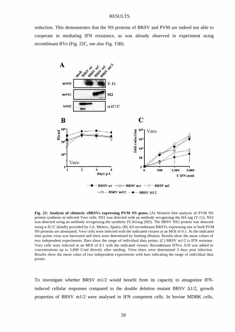
RESULTS
59
reduction. This demonstrates that the NS proteins of BRSV and PVM are indeed not able to
cooperate in mediating IFN resistance, as was already observed in experiment using
recombinant RVs (Fig. 22C, see also Fig. 15B).
Fig. 22: Analysis of chimeric rBRSVs expressing PVM NS genes. (A) Western blot analysis of PVM NS protein synthesis in infected Vero cells. NS1 was detected with an antibody recognizing the HA-tag (Y-11), NS2 was detected using an antibody recognizing the synthetic FLAG-tag (M2). The BRSV NS2 protein was detected using α-IC/C (kindly provided by J.A. Melero, Spain). (B) All recombinant BRSVs expressing one or both PVM NS proteins are attenuated. Vero cells were infected with the indicated viruses at an MOI of 0.1. At the indicated time points virus was harvested and titers were determined by limiting dilution. Results show the mean values of two independent experiments. Bars show the range of individual data points. (C) BRSV m1/2 is IFN resistant. Vero cells were infected at an MOI of 0.1 with the indicated viruses. Recombinant IFN-α A/D was added to concentrations up to 5,000 U/ml directly after seeding. Virus titers were determined 3 days post infection. Results show the mean value of two independent experiments with bars indicating the range of individual data points.
To investigate whether BRSV m1/2 would benefit from its capacity to antagonize IFN-
induced cellular responses compared to the double deletion mutant BRSV ∆1/2, growth
properties of BRSV m1/2 were analysed in IFN competent cells. In bovine MDBK cells,
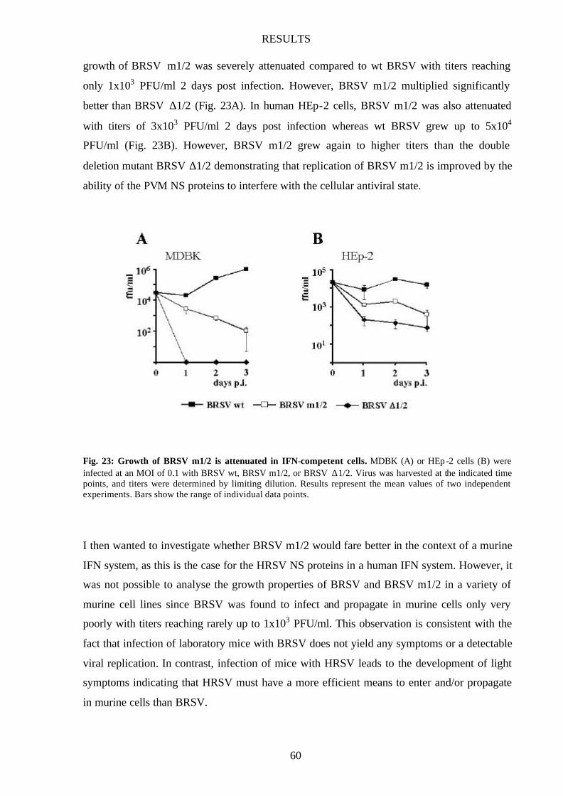
RESULTS
60
growth of BRSV m1/2 was severely attenuated compared to wt BRSV with titers reaching
only 1x10³ PFU/ml 2 days post infection. However, BRSV m1/2 multiplied significantly
better than BRSV ∆1/2 (Fig. 23A). In human HEp-2 cells, BRSV m1/2 was also attenuated
with titers of 3x10³ PFU/ml 2 days post infection whereas wt BRSV grew up to 5x10⁴
PFU/ml (Fig. 23B). However, BRSV m1/2 grew again to higher titers than the double
deletion mutant BRSV ∆1/2 demonstrating that replication of BRSV m1/2 is improved by the
ability of the PVM NS proteins to interfere with the cellular antiviral state.
Fig. 23: Growth of BRSV m1/2 is attenuated in IFN-competent cells. MDBK (A) or HEp -2 cells (B) were infected at an MOI of 0.1 with BRSV wt, BRSV m1/2, or BRSV ∆1/2. Virus was harvested at the indicated time points, and titers were determined by limiting dilution. Results represent the mean values of two independent experiments. Bars show the range of individual data points.
I then wanted to investigate whether BRSV m1/2 would fare better in the context of a murine
IFN system, as this is the case for the HRSV NS proteins in a human IFN system. However, it
was not possible to analyse the growth properties of BRSV and BRSV m1/2 in a variety of
murine cell lines since BRSV was found to infect and propagate in murine cells only very
poorly with titers reaching rarely up to 1x10³ PFU/ml. This observation is consistent with the
fact that infection of laboratory mice with BRSV does not yield any symptoms or a detectable
viral replication. In contrast, infection of mice with HRSV leads to the development of light
symptoms indicating that HRSV must have a more efficient means to enter and/or propagate
in murine cells than BRSV.
RESULTS
61
Since no reverse genetics system is presently available for PVM, other approaches are
necessary to study PVM NS protein function. The replacement of the BRSV surface proteins
F and G with their counterparts from HRSV might improve entry of BRSV into murine cells.
Construction of such “double chimeric” viruses (e.g. BRSV m1/2 FhGh) should then allow us
to analyse the IFN resistance capacity of the PVM NS proteins in their natural environment of
a murine cell and to determine whether the NS proteins of PVM are also involved in
determining host range.
3 BRSV NS PROTEINS ARE REQUIRED FOR
INHIBITION OF IFN-β INDUCTION
ISG expression is not upregulated in BRSV wt infected cells
As demonstrated previously, the presence of both NS proteins increases resistance of BRSV
or an unrelated rhabdovirus against the antiviral action of IFN. However, the molecular events
leading to the observed IFN resistance are unclear. As a first step to elucidating the
underlaying mechanism, I was interested to see whether cells harboring an intact IFN system
would establish an antiviral state after infection with wild-type BRSV or the NS double
deletion mutant. For that purpose, bovine MDBK cells and human HEp-2 cells were infected
with wild-type BRSV, BRSV ∆1/2, and the chimeric BRSV h1/2 at an MOI of 0.2. As
positive control for successful establishment of an IFN-induced antiviral state, mock- infected
cells were treated with 2,000 IU/ml of recombinant IFN-α A/D directly after seeding. Cells
were harvested 24 hrs post infection and equal amounts of whole cell extract were subjected
to SDS-PAGE. As internal loading control, the membranes were probed with an antibody
recognizing the “proliferating cell nuclear antigen” (PCNA), a marker protein for cell
proliferation not influenced by the addition of exogenous IFN-α to both MDBK and HEp-2
cells (Fig. 24).
In HEp-2 cells, infection with BRSV ∆1/2 led to increased expression of IFN stimulated
genes (ISGs), namely STAT2, PKR and IRF-9/p48 (25) indicating that production of IFN and
the subsequent signaling pathway leading to the upregulation of ISGs was successfully
triggered (Fig. 24A/B). To our surprise, however, ISG levels were not upregulated in wt

RESULTS
62
BRSV infected cells suggesting that in the presence of the two NS proteins the pathway
leading to the upregulation of ISGs is blocked. Infection of MDBK cells with the double
deletion mutant and wt BRSV gave similar results showing increased levels of STAT2 and
p48 in BRSV ∆1/2 infected cells but not in MDBK cells infected with the wild-type BRSV
(Fig. 24A/B). However, the antibody recognizing PKR did not give a specific signal when
cell extracts from MDBK cells were used.
Fig. 24: ISG expression in infected MDBK and HEp-2 cells. (A,B) MDBK and HEp-2 cells were infected with the indicated viruses at an MOI of 0.2. 24 hrs post infection, cells were harvested, and whole cell extracts were subjected to SDS-PAGE. As internal control for upregulation of ISGs, mock-infected cells were treated with 2000 U/ml of recombinant IFN-α A/D. After Western blotting, membranes were probed with antibodies recognizing STAT2, p48 (IRF-9), and PCNA as loading control. (C) MDBK and HEp-2 cells were infected with HRSV Long at an MOI of 0.2, cells were harvested 24 hrs post infection, and a Western blot was performed as described in (A).
Interestingly, MDBK or HEp-2 cells infected with the chimeric virus expressing the HRSV
NS genes, BRSV h1/2, also showed increased levels of STAT2, PKR and p48 (Fig. 24A/B)
demonstrating that, in contrast to the BRSV NS proteins, the closely related HRSV NS
proteins are not able to prevent upregulation of ISGs (Fig. 24A). To confirm this finding, both

RESULTS
63
cell lines were infected with the parental HRSV strain Long and assayed for upregulation of
STAT2. Indeed, infection of cells with HRSV led to increased STAT2 levels (Fig. 24C)
suggesting that the HRSV NS proteins do not have the capacity to block upregulation of ISGs
in vitro.
In immunofluorescent studies performed in parallel, infected MDBK and HEp-2 cells were
fixed 2 days post infection and stained with antibodies recognizing BRSV nucleoprotein and
STAT2 as described in Materials and Methods (2-10). Cells treated with exogenous IFN-α
displayed a clear increase in STAT2 levels compared to the untreated control (Fig. 25).
However, a similar increase was not observed in cells infected with wild-type BRSV
indicating that even 2 days after infection, upregulation of ISGs is still blocked. In contrast,
infection with BRSV ∆1/2 or BRSV h1/2 led to upregulated STAT2 levels even in non-
infected cells in the vicinity of productively infected cells demonstrating the establishment of
an antiviral state even in non- infected cells in response to viral infection.
Fig. 25: Immunofluorescent staining of STAT2 in infected cells. MDBK (A) and HEp-2 cells (B) were infected with the indicated viruses at an MOI of 0.2. At 48 hrs post infection cells were fixed with 3% paraformaldehyde and incubated with STAT2 antibody and an antibody recognizing the BRSV N protein (79N; kindly provided by J.A. Melero)
RESULTS
64
IFN is not produced in BRSV wt infected cells
To investigate IFN production in BRSV wt infected cells, an assay system was used based on
the high sensitivity of rabies virus (RV) replication to IFN in MDBK cells. Supernatants of
wild-type BRSV, BRSV h1/2, BRSV ∆1/2 or mock- infected MDBK and HEp-2 cells were
harvested 24, 48 and 72 hrs post infection and incubated at 56°C for 30 minutes to inactivate
replication-competent virus. These supernatants were then added onto MDBK cells previously
infected with RV at an MOI of 5, and RV titers were determined 2 days post infection.
Supernatants from mock- infected MDBK and HEp-2 cells allowed RV to replicate to titers up
to 107 PFU/ml whereas supernatants taken from cells infected with BRSV ∆1/2 or BRSV h1/2
were able to suppress RV replication about 3 log units demonstrating the presence of active
IFN (Fig. 26). However, supernatants from wild-type BRSV infected cells did not influence
RV titers significantly, indicating that the presence of both bovine NS proteins in the
homologous BRSV inhibits production and/or secretion of IFN.
Fig. 26: IFN production in infected MDBK and HEp-2 cells. MDBK or HEp -2 cells were mock-infected or infected with the indicated viruses at an MOI of 0.2. Cell supernatants were harvested 24, 48, and 72 hrs post infection and transferred onto RV-infected MDBK cells. RV titers were determined 2 days post infection. Results represent the mean values of two independent experiments.
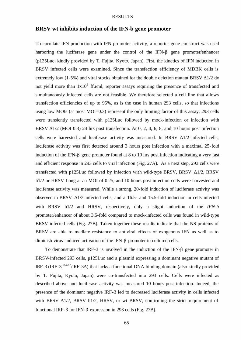
RESULTS
65
BRSV wt inhibits induction of the IFN-β gene promoter
To correlate IFN production with IFN promoter activity, a reporter gene construct was used
harboring the luciferase gene under the control of the IFN-β gene promoter/enhancer
(p125Luc; kindly provided by T. Fujita, Kyoto, Japan). First, the kinetics of IFN induction in
BRSV infected cells were examined. Since the transfection efficiency of MDBK cells is
extremely low (1-5%) and viral stocks obtained for the double deletion mutant BRSV ∆1/2 do
not yield more than 1x10⁵ ffu/ml, reporter assays requiring the presence of transfected and
simultaneously infected cells are not feasible. We therefore selected a cell line that allows
transfection efficiencies of up to 95%, as is the case in human 293 cells, so that infections
using low MOIs (at most MOI=0.3) represent the only limiting factor of this assay. 293 cells
were transiently transfected with p125Luc followed by mock-infection or infection with
BRSV ∆1/2 (MOI 0.3) 24 hrs post transfection. At 0, 2, 4, 6, 8, and 10 hours post infection
cells were harvested and luciferase activity was measured. In BRSV ∆1/2- infected cells,
luciferase activity was first detected around 3 hours post infection with a maximal 25-fold
induction of the IFN-β gene promoter found at 8 to 10 hrs post infection indicating a very fast
and efficient response in 293 cells to viral infection (Fig. 27A). As a next step, 293 cells were
transfected with p125Luc followed by infection with wild-type BRSV, BRSV ∆1/2, BRSV
h1/2 or HRSV Long at an MOI of 0.25, and 10 hours post infection cells were harvested and
luciferase activity was measured. While a strong, 20-fold induction of luciferase activity was
observed in BRSV ∆1/2 infected cells, and a 16.5- and 15.5-fold induction in cells infected
with BRSV h1/2 and HRSV, respectively, only a slight induction of the IFN-β
promoter/enhancer of about 3.5-fold compared to mock-infected cells was found in wild-type
BRSV infected cells (Fig. 27B). Taken together these results indicate that the NS proteins of
BRSV are able to mediate resistance to antiviral effects of exogenous IFN as well as to
diminish virus- induced activation of the IFN-β promoter in cultured cells.
To demonstrate that IRF-3 is involved in the induction of the IFN-β gene promoter in
BRSV-infected 293 cells, p125Luc and a plasmid expressing a dominant negative mutant of
IRF-3 (IRF-358-427/IRF-3∆) that lacks a functional DNA-binding domain (also kindly provided
by T. Fujita, Kyoto, Japan) were co-transfected into 293 cells. Cells were infected as
described above and luciferase activity was measured 10 hours post infection. Indeed, the
presence of the dominant negative IRF-3 led to decreased luciferase activity in cells infected
with BRSV ∆1/2, BRSV h1/2, HRSV, or wt BRSV, confirming the strict requirement of
functional IRF-3 for IFN-β expression in 293 cells (Fig. 27B).
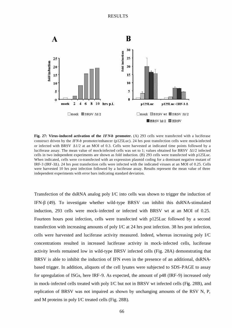
RESULTS
66
Fig. 27: Virus-induced activation of the IFN-β promoter. (A) 293 cells were transfected with a luciferase construct driven by the IFN-β promoter/enhancer (p125Luc). 24 hrs post transfection cells were mock-infected or infected with BRSV ∆1/2 at an MOI of 0.3. Cells were harvested at indicated time points followed by a luciferase assay. The mean value of mock-infected cells was set to 1; values obtained for BRSV ∆1/2 infected cells in two independent experiments are shown as fold induction. (B) 293 cells were transfected with p125Luc. When indicated, cells were co-transfected with an expression plasmid coding for a dominant negative mutant of IRF-3 (IRF-3∆). 24 hrs post transfection cells were infected with the indicated viruses at an MOI of 0.25. Cells were harvested 10 hrs post infection followed by a luciferase assay. Results represent the mean value of three independent experiments with error bars indicating standard deviation.
Transfection of the dsRNA analog poly I/C into cells was shown to trigger the induction of
IFN-β (49). To investigate whether wild-type BRSV can inhibit this dsRNA-stimulated
induction, 293 cells were mock- infected or infected with BRSV wt at an MOI of 0.25.
Fourteen hours post infection, cells were transfected with p125Luc followed by a second
transfection with increasing amounts of poly I/C at 24 hrs post infection. 38 hrs post infection,
cells were harvested and luciferase activity measured. Indeed, whereas increasing poly I/C
concentrations resulted in increased luciferase activity in mock- infected cells, luciferase
activity levels remained low in wild-type BRSV infected cells (Fig. 28A) demonstrating that
BRSV is able to inhibit the induction of IFN even in the presence of an additional, dsRNA-
based trigger. In addition, aliquots of the cell lysates were subjected to SDS-PAGE to assay
for upregulation of ISGs, here IRF-9. As expected, the amount of p48 (IRF-9) increased only
in mock- infected cells treated with poly I/C but not in BRSV wt infected cells (Fig. 28B), and
replication of BRSV was not impaired as shown by unchanging amounts of the RSV N, P,
and M proteins in poly I/C treated cells (Fig. 28B).
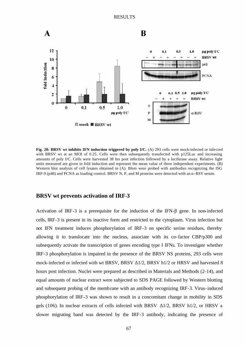
RESULTS
67
Fig. 28: BRSV wt inhibits IFN induction triggered by poly I/C. (A) 293 cells were mock-infected or infected with BRSV wt at an MOI of 0.25. Cells were then subsequently transfected with p125Luc and increasing amounts of poly I/C. Cells were harvested 38 hrs post infection followed by a luciferase assay. Relative light units measured are given in fold induction and represent the mean value of three independent experiments. (B) Western blot analysis of cell lysates obtained in (A). Blots were probed with antibodies recognizing the ISG IRF-9 (p48) and PCNA as loading control. BRSV N, P, and M proteins were detected with an α-RSV serum.
BRSV wt prevents activation of IRF-3
Activation of IRF-3 is a prerequisite for the induction of the IFN-β gene. In non- infected
cells, IRF-3 is present in its inactive form and restricted to the cytoplasm. Virus infection but
not IFN treatment induces phosphorylation of IRF-3 on specific serine residues, thereby
allowing it to translocate into the nucleus, associate with its co-factor CBP/p300 and
subsequently activate the transcription of genes encoding type I IFNs. To investigate whether
IRF-3 phosphorylation is impaired in the presence of the BRSV NS proteins, 293 cells were
mock-infected or infected with wt BRSV, BRSV ∆1/2, BRSV h1/2 or HRSV and harvested 8
hours post infection. Nuclei were prepared as described in Materials and Methods (2-14), and
equal amounts of nuclear extract were subjected to SDS PAGE followed by Western blotting
and subsequent probing of the membrane with an antibody recognizing IRF-3. Virus- induced
phosphorylation of IRF-3 was shown to result in a concomitant change in mobility in SDS
gels (106). In nuclear extracts of cells infected with BRSV ∆1/2, BRSV h1/2, or HRSV a
slower migrating band was detected by the IRF-3 antibody, indicating the presence of
RESULTS
68
substantial amounts of phosphorylated IRF-3 in infected 293 cells (Fig. 29). However, in
BRSV wt infected cells no such band was present indicating that in the context of a viral
infection, the nonstructural proteins NS1 and NS2 are able to inhibit phosphorylation of IRF-3
and subsequent nuclear translocation by an as of yet unknown mechanism, thereby preventing
IFN induction and the subsequent establishment of the antiviral state.
Fig. 29: Phosphorylation of IRF-3 upon viral infection. 293 cells were mock-infected or infected with the indicated viruses at an MOI of 0.25. Ten hours post infection cells were harvested. Nuclear extracts were prepared as described in 2-14 and equal amounts of cell extract were subjected to SDS-PAGE. Nonphosphorylated and phosphorylated forms of IRF-3 were both detected with α-IRF-3 antibody.
To confirm the absence of activated IRF-3 in BRSV wt infected cells, I took advantage of a
reporter construct containing the IRF-3 binding sequence from the IFN-β promoter upstream
of the luciferase gene (kindly provided by T. Fujita, Kyoto, Japan). 293 cells were transfected
with the reporter construct and infected 8 hrs later with BRSV wt, BRSV ∆1/2 or BRSV h1/2
at an MOI of 0.2. 14 hrs post infection cells were harvested and luciferase activity was
measured. As expected, infection with BRSV ∆1/2 and BRSV h1/2 resulted in a more than
30-fold increase in luciferase activity, whereas only a small, 3- fold increase was detected in
wt BRSV infected cells (Fig. 30) demonstrating that the virus-specific activation
(phosphorylation) of IRF-3 is indeed blocked in the presence of the two BRSV NS proteins.
To further investigate whether the two other key transcription factors involved in the
induction of the IFN-β gene, NF-κB and AP-1 (ATF-2/c-Jun) are also not active in BRSV wt
infected cells, 293 cells were transfected with plasmids harboring NF-κB or AP-1 binding
sequences that control expression of the downstream luciferase gene (Stratagene, see also 1-
5). Again, 293 cells were transfected with each reporter construct and mock- infected or
infected with BRSV wt, BRSV ∆1/2 or BRSV h1/2 at an MOI of 0.2. Interestingly, infection
with BRSV wt activated the AP-1 and NF-κB reporter 3-fold and 10-fold, respectively,
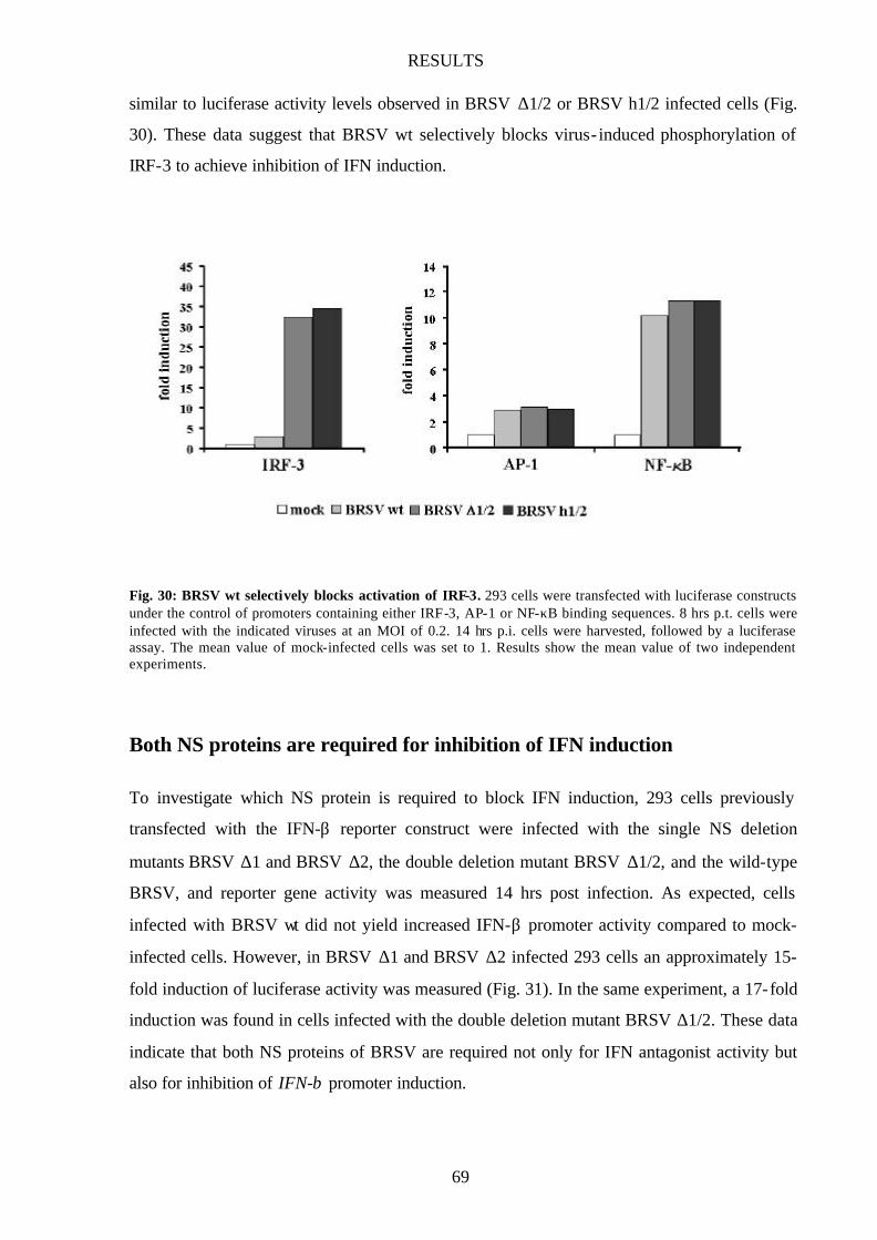
RESULTS
69
similar to luciferase activity levels observed in BRSV ∆1/2 or BRSV h1/2 infected cells (Fig.
30). These data suggest that BRSV wt selectively blocks virus- induced phosphorylation of
IRF-3 to achieve inhibition of IFN induction.
Fig. 30: BRSV wt selectively blocks activation of IRF-3. 293 cells were transfected with luciferase constructs under the control of promoters containing either IRF-3, AP-1 or NF-κB binding sequences. 8 hrs p.t. cells were infected with the indicated viruses at an MOI of 0.2. 14 hrs p.i. cells were harvested, followed by a luciferase assay. The mean value of mock-infected cells was set to 1. Results show the mean value of two independent experiments.
Both NS proteins are required for inhibition of IFN induction
To investigate which NS protein is required to block IFN induction, 293 cells previously
transfected with the IFN-β reporter construct were infected with the single NS deletion
mutants BRSV ∆1 and BRSV ∆2, the double deletion mutant BRSV ∆1/2, and the wild-type
BRSV, and reporter gene activity was measured 14 hrs post infection. As expected, cells
infected with BRSV wt did not yield increased IFN-β promoter activity compared to mock-
infected cells. However, in BRSV ∆1 and BRSV ∆2 infected 293 cells an approximately 15-
fold induction of luciferase activity was measured (Fig. 31). In the same experiment, a 17-fold
induction was found in cells infected with the double deletion mutant BRSV ∆1/2. These data
indicate that both NS proteins of BRSV are required not only for IFN antagonist activity but
also for inhibition of IFN-β promoter induction.

RESULTS
70
Fig. 31: BRSV NS deletion mutants induce IFN-β promoter. 293 cells were transfected with p125Luc, and 8 hrs post transfection cells were infected with the indicated viruses at an MOI of 0.2. 14 hrs post infection cells were harvested and a luciferase assay was performed. Results represent the mean value of three independent experiments with error bars indicating standard deviation.
HRSV and BRSV NS proteins cooperate in blocking IFN induction
Since only the BRSV NS proteins are able to inhibit induction of IFN, I was interested to see
how the chimeric BRSVs carrying one BRSV and one HRSV NS gene (BRSV h1 and BRSV
h2, respectively) would cope with the cellular IFN system. Therefore, MDBK cells were
infected with wt BRSV, BRSV h1, BRSV h2, and BRSV h1/2 and upregulation of STAT2
was monitored 24 hrs post infection.
Surprisingly, infection of MDBK cells with BRSV h1 or BRSV h2 did not lead to
increased STAT2 levels (Fig. 32A) indicating that both combinations of HRSV and BRSV
NS proteins are functional in blocking the upregulation of STAT2 production. In addition,
induction of the IFN-β gene promoter was inhibited in BRSV h1 or BRSV h2 infected 293
cells whereas for BRSV h1/2 or the parental HRSV Long a 16- and 18-fold induction,
respectively, was found (Fig. 32B). To confirm this finding, the presence of a slower
migrating band representing phosphorylated IRF-3 was monitored in infected 293 cells. As
expected, phosphorylated IRF-3 was not found in nuclear extracts of BRSV h1 or BRSV h2-
infected cells (Fig. 32B) suggesting that, intriguingly, both combinations of BRSV and HRSV
NS proteins are successful in preventing the activation of IRF-3. The chimeric BRSV h1 and
BRSV h2 will be a useful tool for the identification of sequences or domains of the BRSV NS
proteins responsible for the block in the induction of the IFN-β promoter and may help
explain how either BRSV NS protein succeeds in forcing the respective other HRSV NS
protein to cooperate in blocking IFN induction.

RESULTS
71
Fig. 32: BRSV h1 and BRSV h2 block IFN induction. (A) MDBK cells were infected with the indicated viruses at an MOI of 0.2. 24 hrs post infection cells were harvested and equal amounts of cell extract were subjected to SDS-PAGE followed by Western blot and detection of STAT2 and PCNA as loading control. (B) 293 cells were transfected with the IFN-β reporter construct p125Luc. Eight hours post infection cells were infected with the indicated viruses at an MOI of 0.2. Cells were harvested 14 hrs post infection followed by luciferase assays. Results represent the mean value of three independent experiments with error bars indicating standard deviation. (C) 293 cells were infected with the indicated viruses at an MOI of 0.25. Nuclear extracts were prepared 8 hrs post infection and equal amounts of extract were subjected to SDS-PAGE. The membrane was probed with an antibody recognizing IRF-3.
PVM NS proteins cannot prevent induction of IFN
To test whether the PVM NS proteins, in contrast to the HRSV NS proteins, are able to
prevent IFN induction, MDBK cells were mock- infected or infected with BRSV wt and
BRSV m1/2 at an MOI of 0.2, and STAT2 levels were determined 24 and 48 hrs post
infection. Similar to BRSV h1/2, STAT2 expression was upregulated in BRSV m1/2 infected
cells indicating that also the PVM NS proteins do not possess the capacity to inhibit ISG
upregulation (Fig. 33A).
These findings were also confirmed in reporter gene assays. 293 cells were transfected
with p125Luc and subsequently mock- infected or infected with BRSV wt or BRSV m1/2 at
an MOI of 0.2. 10 hours post infection cells were harvested and luciferase activity was
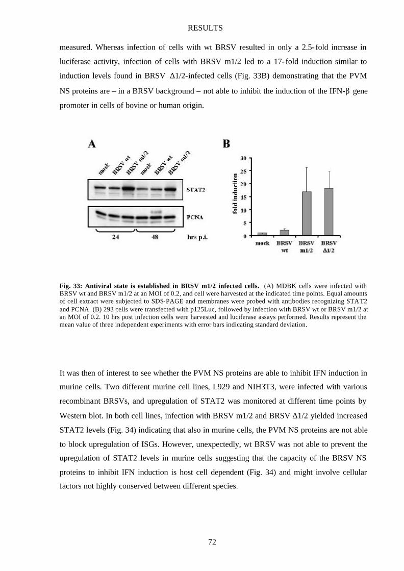
RESULTS
72
measured. Whereas infection of cells with wt BRSV resulted in only a 2.5-fold increase in
luciferase activity, infection of cells with BRSV m1/2 led to a 17-fold induction similar to
induction levels found in BRSV ∆1/2-infected cells (Fig. 33B) demonstrating that the PVM
NS proteins are – in a BRSV background – not able to inhibit the induction of the IFN-β gene
promoter in cells of bovine or human origin.
Fig. 33: Antiviral state is established in BRSV m1/2 infected cells. (A) MDBK cells were infected with BRSV wt and BRSV m1/2 at an MOI of 0.2, and cell were harvested at the indicated time points. Equal amounts of cell extract were subjected to SDS-PAGE and membranes were probed with antibodies recognizing STAT2 and PCNA. (B) 293 cells were transfected with p125Luc, followed by infection with BRSV wt or BRSV m1/2 at an MOI of 0.2. 10 hrs post infection cells were harvested and luciferase assays performed. Results represent the mean value of three independent experiments with error bars indicating standard deviation.
It was then of interest to see whether the PVM NS proteins are able to inhibit IFN induction in
murine cells. Two different murine cell lines, L929 and NIH3T3, were infected with various
recombinant BRSVs, and upregulation of STAT2 was monitored at different time points by
Western blot. In both cell lines, infection with BRSV m1/2 and BRSV ∆1/2 yielded increased
STAT2 levels (Fig. 34) indicating that also in murine cells, the PVM NS proteins are not able
to block upregulation of ISGs. However, unexpectedly, wt BRSV was not able to prevent the
upregulation of STAT2 levels in murine cells suggesting that the capacity of the BRSV NS
proteins to inhibit IFN induction is host cell dependent (Fig. 34) and might involve cellular
factors not highly conserved between different species.
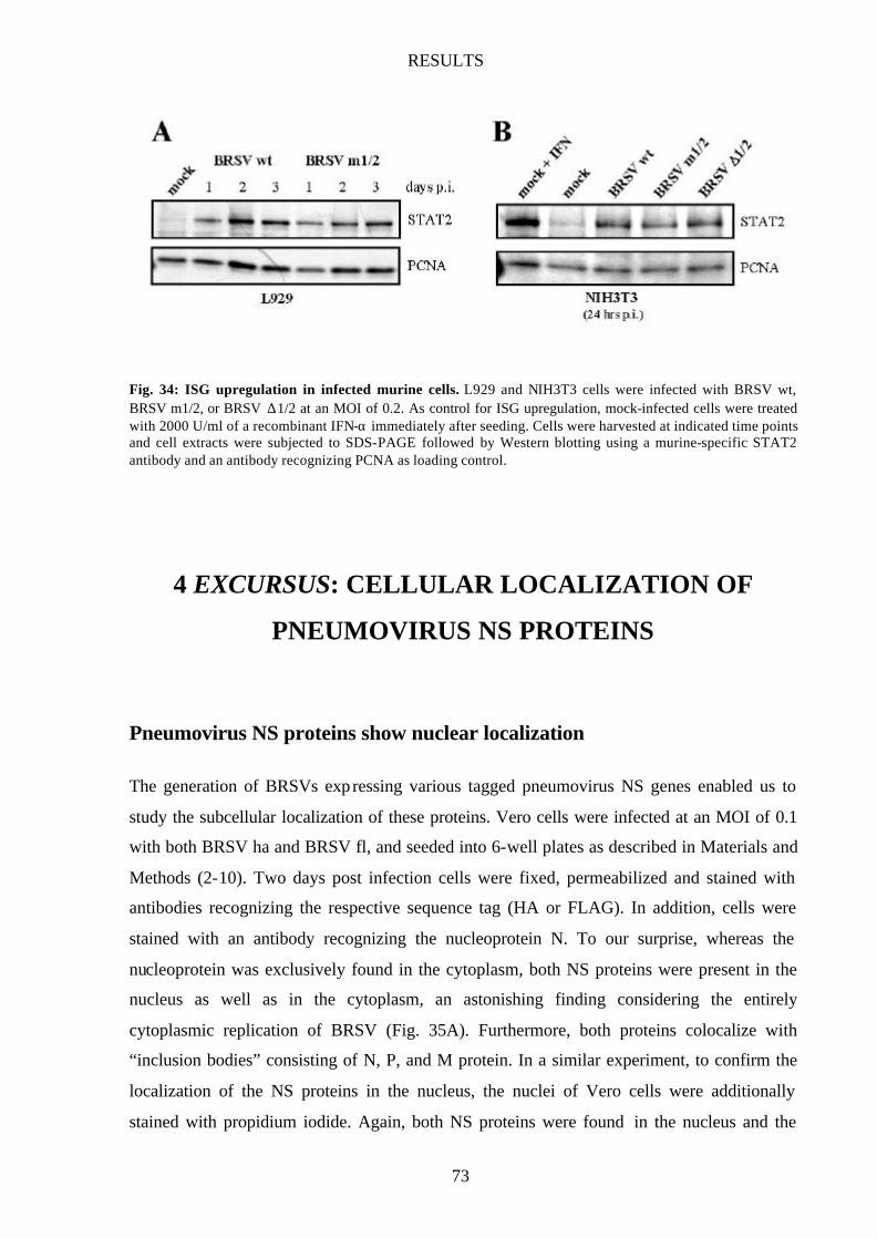
RESULTS
73
Fig. 34: ISG upregulation in infected murine cells. L929 and NIH3T3 cells were infected with BRSV wt, BRSV m1/2, or BRSV ∆1/2 at an MOI of 0.2. As control for ISG upregulation, mock-infected cells were treated with 2000 U/ml of a recombinant IFN-α immediately after seeding. Cells were harvested at indicated time points and cell extracts were subjected to SDS-PAGE followed by Western blotting using a murine-specific STAT2 antibody and an antibody recognizing PCNA as loading control.
4 EXCURSUS: CELLULAR LOCALIZATION OF
PNEUMOVIRUS NS PROTEINS
Pneumovirus NS proteins show nuclear localization
The generation of BRSVs expressing various tagged pneumovirus NS genes enabled us to
study the subcellular localization of these proteins. Vero cells were infected at an MOI of 0.1
with both BRSV ha and BRSV fl, and seeded into 6-well plates as described in Materials and
Methods (2-10). Two days post infection cells were fixed, permeabilized and stained with
antibodies recognizing the respective sequence tag (HA or FLAG). In addition, cells were
stained with an antibody recognizing the nucleoprotein N. To our surprise, whereas the
nucleoprotein was exclusively found in the cytoplasm, both NS proteins were present in the
nucleus as well as in the cytoplasm, an astonishing finding considering the entirely
cytoplasmic replication of BRSV (Fig. 35A). Furthermore, both proteins colocalize with
“inclusion bodies” consisting of N, P, and M protein. In a similar experiment, to confirm the
localization of the NS proteins in the nucleus, the nuclei of Vero cells were additionally
stained with propidium iodide. Again, both NS proteins were found in the nucleus and the
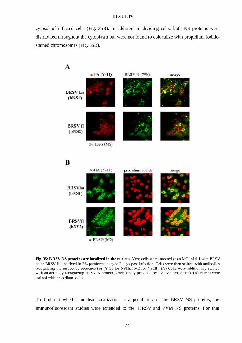
RESULTS
74
cytosol of infected cells (Fig. 35B). In addition, in dividing cells, both NS proteins were
distributed throughout the cytoplasm but were not found to colocalize with propidium iodide-
stained chromosomes (Fig. 35B).
Fig. 35: B RSV NS proteins are localized in the nucleus. Vero cells were infected at an MOI of 0.1 with BRSV ha or BRSV fl, and fixed in 3% paraformaldehyde 2 days post infection. Cells were then stained with antibodies recognizing the respective sequence tag (Y-11 for NS1ha; M2 for NS2fl). (A) Cells were additionally stained with an antibody recognizing BRSV N protein (79N; kindly provided by J.A. Melero, Spain). (B) Nuclei were stained with propidium iodide.
To find out whether nuclear localization is a peculiarity of the BRSV NS proteins, the
immunofluorescent studies were extended to the HRSV and PVM NS proteins. For that
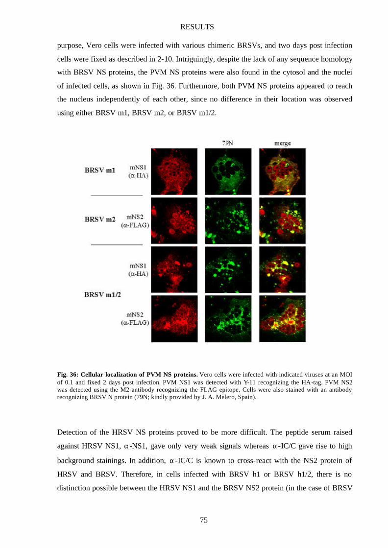
RESULTS
75
purpose, Vero cells were infected with various chimeric BRSVs, and two days post infection
cells were fixed as described in 2-10. Intriguingly, despite the lack of any sequence homology
with BRSV NS proteins, the PVM NS proteins were also found in the cytosol and the nuclei
of infected cells, as shown in Fig. 36. Furthermore, both PVM NS proteins appeared to reach
the nucleus independently of each other, since no difference in their location was observed
using either BRSV m1, BRSV m2, or BRSV m1/2.
Fig. 36: Cellular localization of PVM NS proteins. Vero cells were infected with indicated viruses at an MOI of 0.1 and fixed 2 days post infection. PVM NS1 was detected with Y-11 recognizing the HA-tag. PVM NS2 was detected using the M2 antibody recognizing the FLAG epitope. Cells were also stained with an antibody recognizing BRSV N protein (79N; kindly provided by J. A. Melero, Spain).
Detection of the HRSV NS proteins proved to be more difficult. The peptide serum raised
against HRSV NS1, α-NS1, gave only very weak signals whereas α-IC/C gave rise to high
background stainings. In addition, α-IC/C is known to cross-react with the NS2 protein of
HRSV and BRSV. Therefore, in cells infected with BRSV h1 or BRSV h1/2, there is no
distinction possible between the HRSV NS1 and the BRSV NS2 protein (in the case of BRSV
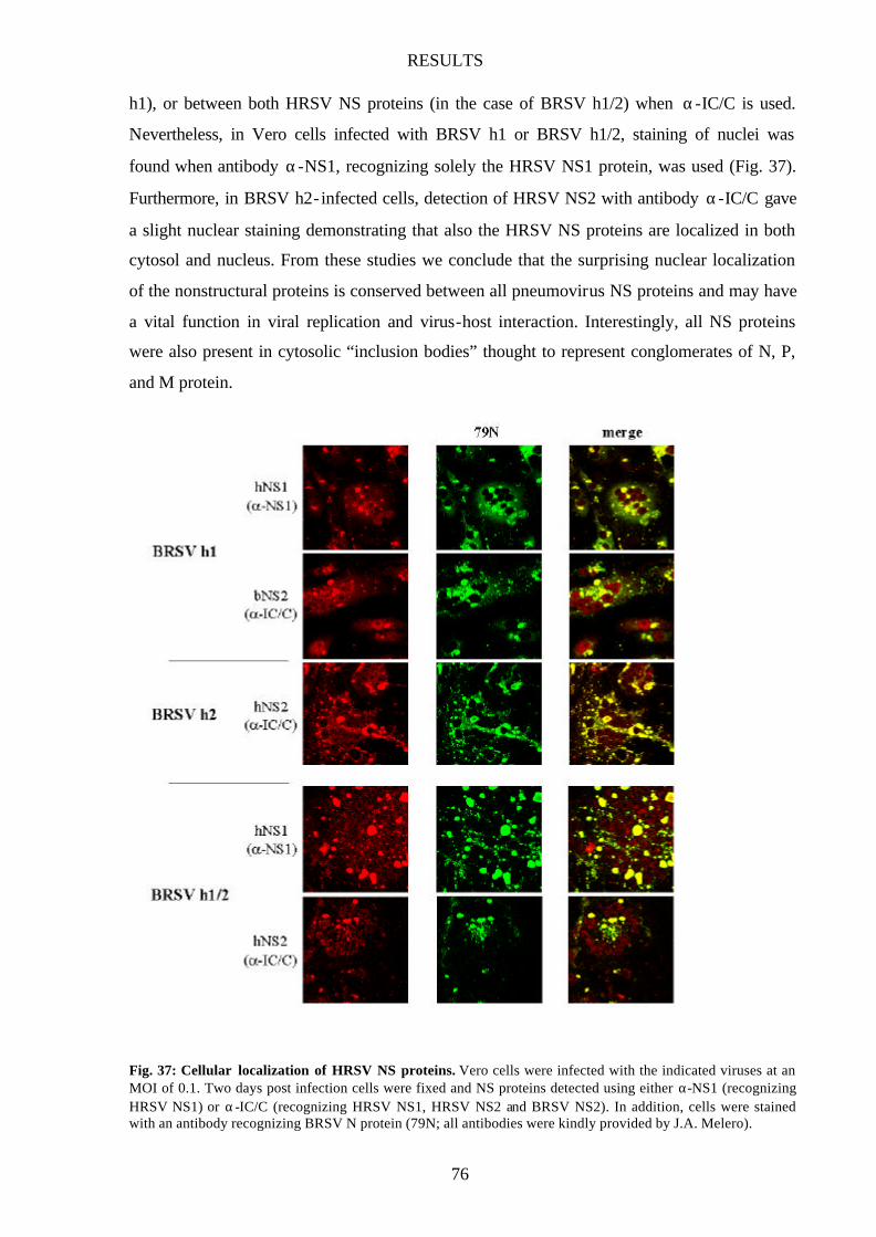
RESULTS
76
h1), or between both HRSV NS proteins (in the case of BRSV h1/2) when α-IC/C is used.
Nevertheless, in Vero cells infected with BRSV h1 or BRSV h1/2, staining of nuclei was
found when antibody α-NS1, recognizing solely the HRSV NS1 protein, was used (Fig. 37).
Furthermore, in BRSV h2- infected cells, detection of HRSV NS2 with antibody α-IC/C gave
a slight nuclear staining demonstrating that also the HRSV NS proteins are localized in both
cytosol and nucleus. From these studies we conclude that the surprising nuclear localization
of the nonstructural proteins is conserved between all pneumovirus NS proteins and may have
a vital function in viral replication and virus-host interaction. Interestingly, all NS proteins
were also present in cytosolic “inclusion bodies” thought to represent conglomerates of N, P,
and M protein.
Fig. 37: Cellular localization of HRSV NS proteins. Vero cells were infected with the indicated viruses at an MOI of 0.1. Two days post infection cells were fixed and NS proteins detected using either α-NS1 (recognizing HRSV NS1) or α-IC/C (recognizing HRSV NS1, HRSV NS2 and BRSV NS2). In addition, cells were stained with an antibody recognizing BRSV N protein (79N; all antibodies were kindly provided by J.A. Melero).

RESULTS
77
NS proteins reach nucleus independent of other pneumovirus proteins
To investigate whether the pneumovirus NS proteins are also present in the nucleus when
expressed independently of the BRSV background, we infected Vero cells with the
recombinant Rabies Viruses expressing single NS proteins. Three days post infection (in the
case of VB bNS1 two days post infection), cells were fixed in 3% paraformaldehyde and
stained for the respective NS protein. In addition, cells were stained with Centocor, a FITC-
conjugated antibody recognizing RV N protein, to demonstrate RV-infection.
Fig. 38: Localization of pneumovirus NS proteins expressed from rRVs. Vero cells were infected with indicated viruses at an MOI of 0.1, and cells were fixed 2 (in the case of VB bNS1) or 3 days post infection (VB mNS1, VB hNS1, VB mNS2). BRSV and PVM NS1 were detected using an antibody recognizing the HA-tag (Y-11); HRSV NS1 was detected with α-IC/C (kindly provided by J.A. Melero, Spain), PVM NS2 was detected with an α-FLAG antibody (M2). Centocor was used to stain RV-infected cells.
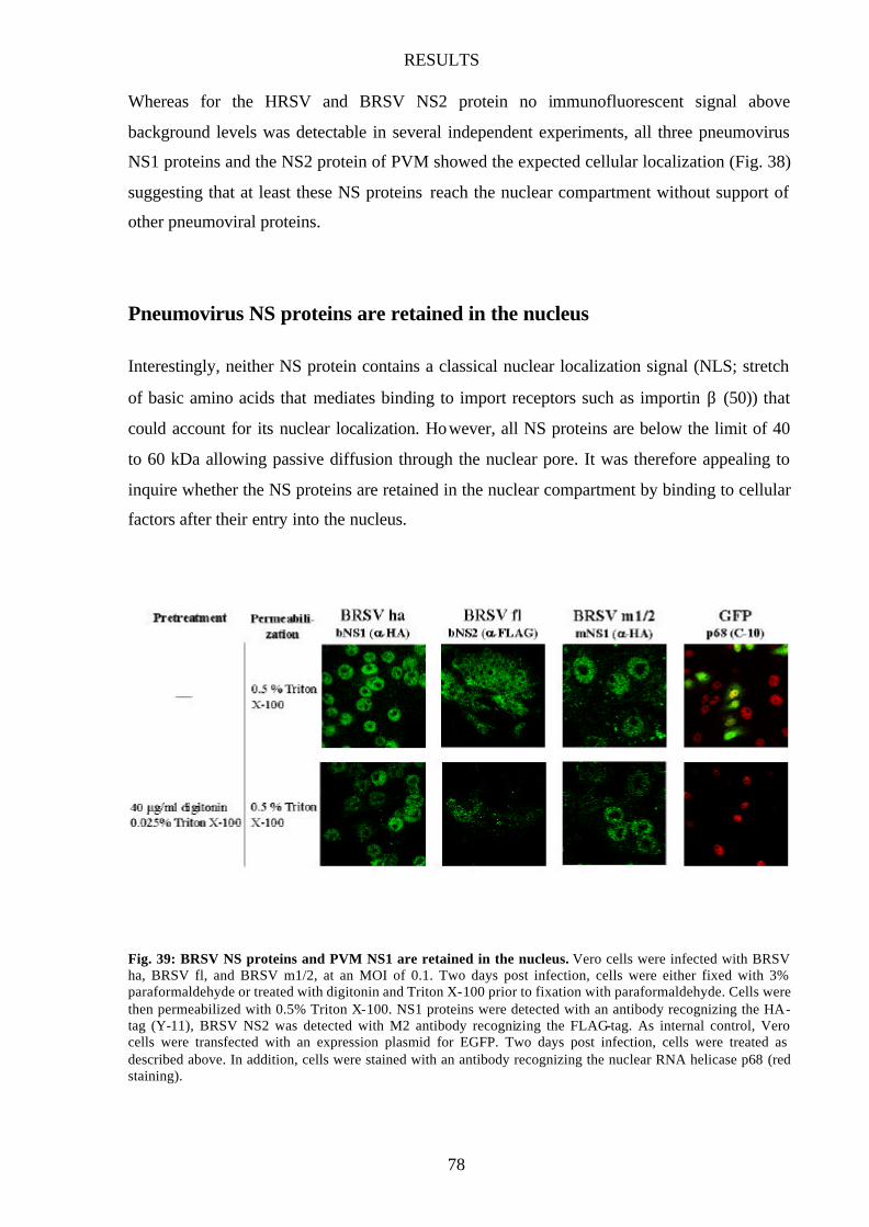
RESULTS
78
Whereas for the HRSV and BRSV NS2 protein no immunofluorescent signal above
background levels was detectable in several independent experiments, all three pneumovirus
NS1 proteins and the NS2 protein of PVM showed the expected cellular localization (Fig. 38)
suggesting that at least these NS proteins reach the nuclear compartment without support of
other pneumoviral proteins.
Pneumovirus NS proteins are retained in the nucleus
Interestingly, neither NS protein contains a classical nuclear localization signal (NLS; stretch
of basic amino acids that mediates binding to import receptors such as importin β (50)) that
could account for its nuclear localization. However, all NS proteins are below the limit of 40
to 60 kDa allowing passive diffusion through the nuclear pore. It was therefore appealing to
inquire whether the NS proteins are retained in the nuclear compartment by binding to cellular
factors after their entry into the nucleus.
Fig. 39: BRSV NS proteins and PVM NS1 are retained in the nucleus. Vero cells were infected with BRSV ha, BRSV fl, and BRSV m1/2, at an MOI of 0.1. Two days post infection, cells were either fixed with 3% paraformaldehyde or treated with digitonin and Triton X-100 prior to fixation with paraformaldehyde. Cells were then permeabilized with 0.5% Triton X-100. NS1 proteins were detected with an antibody recognizing the HA-tag (Y-11), BRSV NS2 was detected with M2 antibody recognizing the FLAG-tag. As internal control, Vero cells were transfected with an expression plasmid for EGFP. Two days post infection, cells were treated as described above. In addition, cells were stained with an antibody recognizing the nuclear RNA helicase p68 (red staining).
RESULTS
79
To wash out nonanchored proteins, Vero cells infected with BRSV ha, BRSV fl or BRSV
m1/2 were treated with digitonin (40 µg/ml in PBS) for 3 min on ice, then washed once with
cold PBS. Afterwards, cells were incubated with 0.025% Triton X-100 for 5 min on ice before
fixation in 3% paraformaldehyde and permeabilization with 0.5% Triton X-100. As control,
infected cells were only fixed in 3% paraformaldehyde. To validate this assay, cells were
transfected with a reporter construct expressing the green fluorescent protein (GFP) and
treated as described above. Normally, GFP is found evenly distributed throughout the cell
including the nuclear compartment and nucleoli. Treatment with digitonin and Triton X-100,
however, eliminated the fluorescence of GFP but not the staining for p68, a nuclear RNA
helicase (Fig. 39). Similar to the nuclear protein p68, the BRSV NS proteins and PVM NS1
are localized in the nucleus in a speckle- like pattern in both untreated and digitonin/Triton X-
100-treated cells (Fig. 39). From that we conclude that the NS proteins are retained in the
nucleus upon entry, probably by interacting with nuclear factors, suggesting a distinct
function for the nuclear NS proteins during viral replication.
DISCUSSION
80
DISCUSSION
Pneumovirus NS proteins are IFN antagonists
A peculiarity of pneumoviruses is the presence of two nonstructural protein genes at the very
3’ end of the genome. Deletion of one or both nonstructural genes leads to attenuation of the
respective pneumovirus in vitro and in vivo indicating that both proteins contribute to optimal
viral replication but are not essential. However, the precise role(s) of the nonstructural
proteins in the viral life cycle remain to be defined. Employing the technique of recovery of
negative-strand RNA viruses from cDNA, several NS deletion mutants of BRSV were
generated in our laboratory. Although only slightly attenuated in IFN-negative Vero cells not
able to produce IFN, growth of these deletion mutants was severely impeded in IFN
producing bovine MDBK cells which are fully permissive for the wild-type virus (80).
Assuming that this severe attenuation was due to missing virus-host interactions, type I
interferons (IFN-α/β) were subsequently identified as the critical host cell factors produced by
infected MDBK cells (80). Intriguingly, however, growth kinetics and IFN sensitivity of
deletion mutants lacking either NS1 or NS2, or both NS genes, were very similar in all cell
lines examined suggesting that, quite unexpectedly, both NS proteins are apparently necessary
to accomplish IFN antagonistic activity whereas each protein on its own does not possess any
activity. To our knowledge, this would be the first example of two virus proteins obligatorily
cooperating to antagonize IFN.
To obtain evidence that both nonstructural proteins are required and sufficient to
interfere with the IFN-induced antiviral state, recombinant RVs expressing individual NS
proteins were generated. The location of the NS genes at the most upstream position of the
RSV genome (Fig. 2) should result in a high and similar level of expression in BRSV-infected
cells. Accordingly, to simulate the conditions in BRSV-infected cells, coinfections with rRV
vectors harboring individual NS genes were done at the same MOI for both recombinants.
Comparable amounts of NS1 and NS2 protein, respectively, should be expressed in cells
infected with the single recombinants and in cells coinfected with both recombinants.
However, only in coinfected cells was a significantly enhanced resistance of the RV vector to
IFN stimulation observed. Whereas standard RV titers dropped by 3 log units in MDBK cells
when only 50 IU of recombinant IFN were applied, complete protection was observed in
coinfected cells treated with IFN doses of up to 150 IU demonstrating that the two BRSV NS
DISCUSSION
81
proteins are indeed able to confer resistance against IFN-mediated antiviral responses (Fig.
10). Furthermore, the results confirm that the presence of both proteins is required for
exerting their IFN antagonist activity. Since the NS proteins are not only effective in a BRSV
background but are also able to protect a heterologous virus from INF-induced responses,
NS1 and NS2 do not require additional viral functions provided by BRSV for their protective
activity.
Since coinfections are prone to some variability and do not result in complete coinfection of
all infected cells, we suspected that a more regulated coexpression of NS proteins might bring
about a greater protective capacity. Therefore, a recombinant RV was generated that
expresses both NS protein of BRSV. As expected, this virus was significantly more IFN
resistant than standard RV even when 500 IU of recombinant IFN were applied (Fig. 16).
Somewhat surprisingly, the introduction of two additional genes into the RV genome did not
attenuate viral replication significantly when compared to wild-type RV. Similarly,
expression of the influenza NS1 protein also showed beneficial effects towards the IFN
resistance of RV vectors without hampering its replication capacity (B. Bossert, unpublished
results). The generation of replication-competent viral vectors with improved IFN resistance
capacities will provide a useful tool to study the in vivo function of a given IFN antagonist
and to identify possible novel IFN antagonists.
Previous studies from Young et al. showed that HRSV is able to replicate in human cells that
produce and respond to IFN, suggesting that HRSV must have some effective means to
replicate in the face of the cellular antiviral response (108). Since the nonstructural proteins of
HRSV and BRSV are highly similar in sequence, with 69 and 84% amino acid identity for the
NS1 and NS2 proteins, respectively, we entertained the idea that the HRSV NS proteins might
also function as IFN antagonists. Using rRVs expressing individual HRSV NS proteins, I
could demonstrate that only in cells coinfected with both recombinants, IFN resistance of the
RV vector towards exogenous IFN was enhanced (Fig. 14). Moreover, their capacity to
mediate virus escape from IFN-α/β-induced antiviral mechanisms was comparable to that of
the BRSV NS proteins. As previously shown for the BRSV NS proteins, the presence of both
HRSV NS proteins is required for their IFN antagonist activity. Identical experiments were
also performed with the nonstructural proteins of the only very distantly related Pneumovirus
of Mice (PVM). Recombinant RVs were generated expressing single PVM NS proteins and
IFN challenge experiments in MDBK cells were carried out. Although the PVM NS proteins
DISCUSSION
82
show only low sequence homology to their human and bovine RSV counterparts (Fig. 5), they
still have the capacity to render the RV vector IFN resistent, albeit to a somewhat lesser extent
than the RSV NS proteins (Fig. 14). Again, the presence of both NS1 and NS2 is required for
the observed IFN antagonist function.
These data clearly show that pneumoviruses share a common IFN resistance mechanism
that requires the presence of both nonstructural proteins and that can be transferred to an
unrelated rhabdovirus. Preliminary studies indicate that Rabies virus is insensitive to MxA
(R. Müller-Waldeck, unpublished results). Thus, I assume that the NS proteins block the
action of specific IFN-induced antiviral gene products other than MxA to accomplish
resistance to IFN. The high sequence similarity of HRSV and BRSV NS proteins and,
moreover, their functional cooperativity (Fig. 15) suggest that BRSV and HRSV interfere
with the same IFN-induced antiviral mechanisms and that they target similar, if not the same,
antiviral gene products. In contrast, the highly dissimilar NS proteins of PVM were not able
to cooperate with their bovine counterparts. Yet, functionally, the presence of both proteins is
required. It is tempting to speculate that the PVM proteins have evolved to better suit their
targets in a murine IFN system, thus conserving the mechanism that renders pneumoviruses
IFN resistant.
Involvement of pneumovirus NS proteins in the determination of host range
Although closely related, bovine and human RSVs display a highly restricted host range in
vivo. One reason might be restricted entry into host target cells due to differences in the viral
surface glycoproteins and in their cellular receptor. In vitro, however, BRSV and HRSV are
able to enter a wide variety of cells of different hosts indicating that other virally encoded
factors may also play a pivotal role in determining host range. Indeed, Buchholz et al.
reported that a chimeric BRSV carrying the HRSV glycoproteins instead of their BRSV
counterparts was somewhat more competent than BRSV for replication in chimpanzees but
remained highly restricted compared to HRSV (9). The authors concluded that the F and G
proteins contribute to the host range restriction of BRSV but are not the major determinants.
In this study we demonstrate that the NS proteins of BRSV and HRSV represent
important determinants of viral host range in that they display a differential ability to
counteract innate responses in cells of different hosts. In Vero cells unable to produce IFN,
viral replication of wild-type BRSV and the various chimeric BRSV h1, BRSV h2, and BRSV
h1/2 was indistinguishable (Fig. 18B). This is notable, as the HRSV NS1 protein was shown

DISCUSSION
83
to associate with the viral matrix protein M (33) and was reported to be a potent inhibitor of
viral transcription and RNA replication in a HRSV minigenome system (3). The virtually
identical growth in Vero cells suggests that these interactions and functions were not largely
affected in the chimeric virus and that the HRSV NS proteins are able to accurately fulfill the
function(s) of their bovine counterparts during viral replication. When growth properties of
different chimeric BRSVs were assessed in cells harboring an intact IFN system, differences
in the ability to counteract host specific IFN-induced antiviral cell responses became obvious
and even more evident in IFN-treated cells. In cells of bovine origin, BRSV was perfectly
protected even against high doses of exogenous IFN-α whereas BRSV h1/2 was severely
attenuated (Fig. 19C) and highly sensitive towards IFN-α (Fig. 20B). In human cells, both
viruses displayed similar growth characteristics (Fig. 19B), but BRSV h1/2 was more resistant
than BRSV wt to antiviral responses triggered by exogenous IFN (Fig. 20C). Although IFN
resistance of BRSV h1/2 was greater than wt BRSV, this did not result in higher titers in the
absence of exogenous IFN. I therefore assume that, most probably, IFN induction and the
activation of antiviral mechanisms are not sufficient to completely overcome the weaker IFN
resistance of wt BRSV. In contrast, infected MDBK and Klu cells are likely to be strong
inducers of IFN so that differences in IFN escape are immediately apparent.
In the case of the recombinant BRSVs expressing one or both PVM NS proteins,
replication was attenuated in Vero cells not able to produce IFN, and resembled replication
levels obtained with the double deletion mutant BRSV ∆1/2 (Fig. 22B). This suggests that the
PVM NS proteins are, unlike their human counterparts, not able to functionally replace the
BRSV NS proteins during the viral life cycle. How the NS proteins contribute in detail to
viral replication is not yet known. Atreya et al. reported an inhibitory effect of NS1 on
transcription and RNA replication in a HRSV minigenome system (3). However, deletion of
NS1 from the HRSV or BRSV genome did not lead to enhanced replication. In contrast, NS
single or double deletion mutants were attenuated even in IFN negative cells suggesting a
supportive rather than an inhibitory function for the NS proteins in viral replication (8,80).
Although the PVM NS proteins cannot replace their bovine counterparts in their
replicative function(s), the recombinant virus expressing both PVM NS proteins, BRSV m1/2,
showed improved growth characteristics compared to BRSV ∆1/2 in IFN-producing cells
(Fig. 23) and was almost as IFN resistant as the wt BRSV (Fig. 22C) indicating that the PVM
NS proteins confer IFN resistance independent of their viral context.
DISCUSSION
84
The contribution of pneumovirus NS proteins to the permissivity of hosts to RSV infection is
supported by the study of Hanada et al. reporting that the markedly restricted growth of
HRSV in mouse embryo cells could be overcome by adding anti-mouse IFN serum to the
medium (43). As a result, HRSV yields were enhanced and the infection spread in the entire
monolayer. Apparently, entry into mouse cells was not the limiting step but rather the
inability of the HRSV NS proteins to efficiently counteract murine IFN responses. However,
infection of murine cells with BRSV is even more inefficient than with HRSV, and mice
infected with BRSV do not develop any symptoms. Therefore, in vivo infection studies with
BRSV have to be carried out in, preferably gnotobiotic, calves. The development of “mouse-
improved” recombinant BRSVs expressing e.g. the HRSV glycoproteins for better entry into
murine cells and the NS proteins of PVM to inhibit murine IFN responses might allow the
study of BRSV pathogenesis in a “small animal” model and might help to gain insight into the
difficult virus – host immune system interactions encountered during pneumovirus infections.
Adaptation of viral proteins to the cellular environment of their natural host may in fact play a
critical role in establishing an infection. The V protein of Simian Virus 5 (SV5), for example,
is very effective in blocking the activation of IFN-responsive genes in primate cells but not in
murine cells (28). A single amino acid substitution, however, renders the V protein fully
functional to block IFN signaling in murine cells (107). Further studies with chimeric RSV
recombinants will reveal whether the observed species specificity in IFN escape can also be
attributed to the PVM NS proteins. Moreover, it will be interesting to see whether the species
specificity can be ascribed to one of the two NS proteins and which sequences are involved.
In addition, mutations in the NS proteins may be identified that only partially knock out the
IFN-antagonist activity without compromising NS protein function necessary for optimal viral
replication. This would lead to the generation of replication competent RSVs that display a
reduced capability of antagonizing IFN in a given host.
The ability to adjust IFN resistance will also have important implications for the development
of an efficacious live attenuated RSV vaccine. The first vaccine evaluated in infants and
children in the mid-1960s consisted of formalin- inactivated and concentrated RSV. However,
the vaccine was not able to induce a protective immune response. Moreover, during
subsequent natural infections, the frequency and severity of RSV disease was increased
(54,55). In contrast, a live attenuated vaccine administered intranasally may have the
advantage of mimicking natural infection, which has never been shown to prime for enhanced
DISCUSSION
85
disease (96), and may be able to replicate in very young infants despite the presence of
maternal antibodies (104). For these reasons, a series of attenuated, immunogenic cold-
passaged, temperature-sensitive RSV mutants were produced and clinically evaluated. The
most attenuated vaccine virus, cpts248/404, was immunogenic and protective against a second
vaccine dose. However, it induced mild nasal congestion indicating the need for additional
attenuation (104). The ability to recover infectious, recombinant RSV from cDNA has greatly
facilitated the characterization and further development of live-attenuated viruses (20) and
might provide the tool for further attenuation (17). The deletion of nonessential genes such as
one or both nonstructural genes, however, resulted in attenuated, highly IFN sensitive virus
with compromised pathogenicity (8,52,87,88,101). In addition, virus replication was also
affected resulting in reduced production of antigen and poor immunogenicity. The challenge
in the development of a live attenuated vaccine will be the elimination of residual virulence
without compromising immunogenicity. BRSV and HRSV vaccines that possess intermediate
ability to escape from the innate bovine or human immune response but are nevertheless able
to keep their replicative capacity are a very promising approach.
BRSV NS proteins are required for inhibition of IFN-β induction
Recently, the accessory V and C proteins of certain members of the Paramyxovirinae
subfamily have been shown to function as antagonists of the interferon system by blocking
signaling pathways induced by IFN (35). In contrast, HRSV (a member of the Pneumovirinae
subfamily) failed to block IFN signaling but was able to replicate in cells pretreated with IFN
indicating that HRSV must have some alternative mechanism for circumventing the IFN
response (108). Subsequently, we were able to demonstrate that the two NS proteins, present
only in the Pneumovirinae subfamily, are able to confer resistance towards IFN-induced
responses. These results suggest that whereas interference with IFN signaling is a common
strategy among the Paramyxovirinae, Pneumovirinae members do not inhibit signaling but
have developed as of yet unknown strategies to block the IFN-induced antiviral response (Fig.
40).
Here, we describe an additional, novel function for the NS proteins of BRSV, namely
their ability to block the early stages of the virus-activated cellular IFN pathway by interfering
with the activation of IRF-3 (Fig. 40). As a result, induction of IFN-β and the subsequent
establishment of the antiviral state are prevented or at least very much delayed.
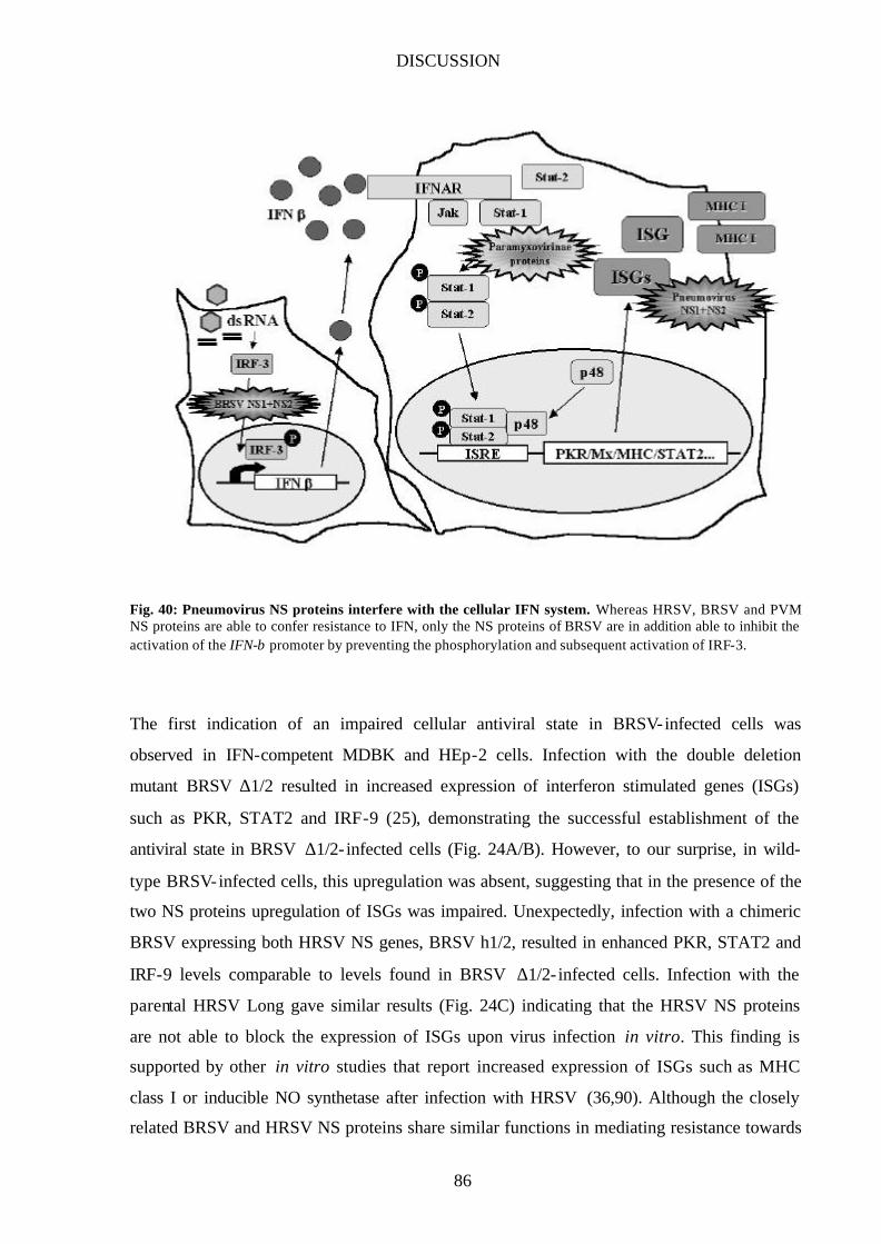
DISCUSSION
86
Fig. 40: Pneumovirus NS proteins interfere with the cellular IFN system. Whereas HRSV, BRSV and PVM NS proteins are able to confer resistance to IFN, only the NS proteins of BRSV are in addition able to inhibit the activation of the IFN-β promoter by preventing the phosphorylation and subsequent activation of IRF-3.
The first indication of an impaired cellular antiviral state in BRSV-infected cells was
observed in IFN-competent MDBK and HEp-2 cells. Infection with the double deletion
mutant BRSV ∆1/2 resulted in increased expression of interferon stimulated genes (ISGs)
such as PKR, STAT2 and IRF-9 (25), demonstrating the successful establishment of the
antiviral state in BRSV ∆1/2- infected cells (Fig. 24A/B). However, to our surprise, in wild-
type BRSV-infected cells, this upregulation was absent, suggesting that in the presence of the
two NS proteins upregulation of ISGs was impaired. Unexpectedly, infection with a chimeric
BRSV expressing both HRSV NS genes, BRSV h1/2, resulted in enhanced PKR, STAT2 and
IRF-9 levels comparable to levels found in BRSV ∆1/2- infected cells. Infection with the
parental HRSV Long gave similar results (Fig. 24C) indicating that the HRSV NS proteins
are not able to block the expression of ISGs upon virus infection in vitro. This finding is
supported by other in vitro studies that report increased expression of ISGs such as MHC
class I or inducible NO synthetase after infection with HRSV (36,90). Although the closely
related BRSV and HRSV NS proteins share similar functions in mediating resistance towards
DISCUSSION
87
IFN-induced cellular antiviral mechanisms and are even able to cooperate for this particular
function, they apparently differ in their ability to block the establishment of an antiviral state.
However, since the ability of HRSV to induce IFN and the IFN-mediated cellular response in
vivo is still a matter of debate, it remains to be investigated whether the inability of HRSV in
preventing the upregulation of ISGs is a particular feature of the HRSV strain Long and
whether other strains or clinical isolates are more inhibitory. The very distant PVM NS
proteins were also found unable to prevent induction of IFN-β when expressed in a BRSV
background (Fig. 33). However, no data are at present available whether infection of murine
cells with the parental PVM induces IFN production or not.
The IFN-α/β system is a powerful and immediate antiviral host response. It is therefore not
surprising that many viruses have evolved mechanisms to counteract this response at every
possible level. Viruses are able to prevent production of IFN-β or prevent binding of IFN to
the IFN receptor, inhibit IFN-mediated signaling and/or selectively block the activity of
distinct ISGs (37). To prevent the stimulation of ISG expression, a virus can either block IFN-
mediated signaling or impair induction/production of IFN. In the case of BRSV, previous
studies suggested that IFN signaling was rather not affected (J. Schlender, personal
communication). In addition, HRSV was reported not to affect IFN signaling (108).
Therefore, as a first step in elucidating the mechanism by which BRSV prevents upregulation
of ISGs, we directly assayed for the presence of IFN in supernatants of cells infected with
various recombinant BRSVs. Whereas IFN was readily detectable in supernatants of MDBK
or HEp-2 cells infected with BRSV ∆1/2 or BRSV h1/2, no significant amount of IFN was
found in BRSV wt-infected cells suggesting that in the presence of the two NS proteins,
induction of IFN may well be inhibited (Fig. 26). To distinguish between a possible block in
IFN induction or secretion we subsequently looked for IFN-β gene promoter activity in
infected cells, making use of a reporter construct expressing the luciferase gene under the
control of the IFN-β promoter/enhancer. While IFN-β promoter activity increased more than
20-fold in cells infected with BRSV ∆1/2, wild-type BRSV elicited only a very slight increase
demonstrating that the NS proteins are able to block the induction of IFN-β promoter activity
in the course of a viral infection (Fig. 27). Moreover, infection of cells with single deletion
mutants BRSV ∆1 and BRSV ∆2 showed that, similar to their IFN resistance function, both
NS proteins are required for the block in IFN induction (Fig. 31). In addition, BRSV wt was
able to inhibit the induction of IFN even in the presence of an additional trigger, namely the
dsRNA analog poly I/C (Fig. 28) demonstrating that in the presence of the two NS proteins
DISCUSSION
88
BRSV can block an as of yet unidentified step in the IFN induction pathway. To our
knowledge, this makes BRSV the first member of the Paramyxovirus family that does not
induce the IFN-β gene promoter.
Induction of IFN-β expression is controlled by an enhanceosome that binds three distinct
transcription factor complexes in the context of chromatin-organizing proteins. Each of these
factors, IRF-3, NF-κB, and ATF/c-Jun (AP-1), becomes activated following protein
phosphorylation events induced in response to viral infection. The virus-specific C-terminal
phosphorylation of IRF-3 is mediated by a unique, as of yet unidentified virus activated
kinase (VAK) activity and leads to conformational changes within the IRF-3 protein that
enable its dimerization, nuclear translocation, association with the CBP/p300 coactivator and
subsequent stimulation of transcriptional activities. Since paramyxoviruses are generally
known to be potent inducers of VAK activity I investigated whether this activity was impaired
in BRSV-infected cells by measuring the amount of phosphorylated IRF-3 available in the
nuclei of cells shortly after viral infection. Indeed, whereas phosphorylated IRF-3 was present
in cells infected with BRSV ∆1/2, BRSV h1/2 or HRSV, I was not able to detect it in wild-
type BRSV infected cells (Fig. 29). This result indicates that the two NS proteins of BRSV,
but not of HRSV, are able to inhibit the induction of IFN-β by preventing the activation of
IRF-3. This finding was further supported by studies using reporter constructs expressing the
luciferase gene under the control of either IRF-3 or NF-κB or AP-1 binding sites. Preliminary
data suggest that whereas IRF-3 activity was greatly compromised in cells infected with
BRSV wt compared to BRSV ∆1/2 infected cells, NF-κB and AP-1 activities were detectable
in both BRSV ∆1/2 and wt BRSV infected cells, (Fig. 30) hinting at a selective block of IRF-
3 activity in wild-type BRSV infected cells.
Surprisingly, both chimeric viruses expressing one HRSV and one BRSV NS protein, BRSV
h1 and BRSV h2, respectively, were also able to block phosphorylation of IRF-3 (Fig. 32). It
is tempting to speculate that this ability to block the establishment of the antiviral state may
explain why both chimeric viruses, in contrast to BRSV h1/2, are not attenuated in bovine
cells (Fig. 18C). Considering BRSV h1/2, we think that its attenuated growth phenotype in
bovine cells can be explained by at least two reasons. Firstly, infection of MDBK cells with
BRSV h1/2 elicits IFN production and the subsequent establishment of an antiviral state.
Secondly, the HRSV NS proteins do not efficiently inhibit the bovine cellular IFN system.
Hence, growth of BRSV h1/2 is impaired. In human cells, BRSV h1/2 infection also leads to

DISCUSSION
89
the production of IFN. However, the HRSV NS proteins are more efficient in inhibiting a
human cellular IFN system and therefore, viral replication is not affected. The generation of
recombinant BRSVs that lack one particular IFN antagonist function (e.g. inhibition of IFN
induction) while keeping the other (e.g. IFN resistance) will be a useful tool to study the in
vivo relevance of RSV IFN antagonist activity.
Activation of IFN gene expression is a cellular response resulting from the innate immune
recognition of virus infections, but how cells sense viral replication is only poorly understood.
It is generally believed that dsRNA generated during certain steps in viral replication is the
principal inducer of IFN. A main sensor for dsRNA seems to be the RNA-dependent protein
kinase PKR which, upon recognition of dsRNA and a subsequent autophosphorylation step,
activates the IKKβ subunit of the multicomponent IκB kinase eventually leading to the
activation of NF-κB. However, PKR is not sufficient to mediate the full antiviral response
since mice with homozygous disruptions of the PKR gene displayed normal antiviral
responses to infection with influenza and vaccinia virus both in mice and in cells lacking PKR
(1). Another protein able to recognize dsRNA is the Toll- like receptor 3 (TLR3) normally
involved in the immune recognition of microbial pathogens. Upon dsRNA-mediated
activation, the receptor was shown to induce the activation of NF-κB and production of type I
IFNs (2). However, whether TLR3 plays a role in the host’s defence against viral infections
remains to be established. Recently, tenOever et al. showed for the first time that direct
recognition of a viral protein, namely the Measles virus nucleocapsid protein, resulted in
activation of IRF-3 and subsequent induction of IFN (89). Furthermore, the nucleocapsid
protein was found associated with both IRF-3 and VAK activity, suggesting that it may be
used in the colocalization of the kinase and its substrate. Future studies will be aimed at
elucidating the trigger provided by BRSV ∆1/2 for efficient IFN induction in infected cells
and the molecular mechanism (and involved targets) by which the BRSV NS proteins prevent
phosphorylation of IRF-3. Interestingly, the inhibition of IRF-3 activation to prevent IFN
induction may be in fact more advantageous for a given virus than e.g. the inhibition of NF-
κB activation. Very recently, IRF-3 was reported to directly induce the expression of various
ISGs via their ISRE elements in an IFN-independent manner (40) and may even be, in certain
cells, able to induce the IFN-β mRNA without involvment of NF-κB or ATF/c-Jun (75).
However, whether BRSV is sensitive to ISGs directly upregulated by IRF-3 and/or whether
these IFN-independent induction pathways have any in vivo relevance awaits further
investigation.

DISCUSSION
90
Interestingly, we were recently able to show that the BRSV NS1 protein can activate the
transcription factor NF-κB (B. Bossert, unpublished results). Activation of NF-κB is a
mechanism employed by multiple families of viruses including HIV-1, HTLV-1, EBV and
HCV to improve viral replication, enhance viral gene expression, and to prevent virus-
induced apoptosis. Although the molecular details remain mostly unclear, certain viral
proteins such as the HIV-1 Tat protein, the HTLV-1 Tax protein, and EBV LMP-1 are able to
activate NF-κB through several distinct mechanism (47). In addition, NF-κB, like IRF-3, is
also activated by synthetic dsRNA, suggesting that viruses that generate dsRNA replicative
intermediates share a common mechanism by which NF-κB is activated. On the other hand,
since activation of NF-κB by infection is a key trigger for the induction of IFN-α/β , several
viruses encode inhibitors of NF-κB activation or function (47). However, it is well established
that NF-κB also induces antiapoptotic genes and any virus that blocks NF-κB activation may
leave itself suceptible to enhanced induction of apoptosis. I therefore hypothesize that BRSV
is able to inhibit the induction of IFN triggered most probably by dsRNA replicative
intermediates at a very early step so that not only activation of IRF-3 but also activation of
NF-κB is prevented. To avert apoptosis in infected cells, BRSV then activates NF-κB by a
dsRNA-independent, NS1-mediated mechanism. However, I do not rule out the possibility
that activated NF-κB may serve additional functions in the BRSV-infected cell. Future
experiments will reveal by which mechanism NS1 activates NF-κB and how this activation
influences the host cell to allow optimal viral replication.
Nuclear localization of pneumovirus NS proteins
Replication of RSV is believed to be accomplished entirely in the cytoplasm. Therefore, the
nuclear localization of both BRSV NS proteins was surprising. Having generated recombinant
BRSVs expressing tagged NS1 or NS2 genes (BRSV ha and BRSV fl, respectively), I was
able to perform immunofluorescent studies to determine the cellular localization of the BRSV
NS proteins. Quite unexpectedly, both proteins were not only found in the cytoplasm,
predominantly colocalizing with inclusion bodies formed by the BRSV N, P, and M proteins,
but also in the nucleus, yet no staining of the nucleoli was observed (Fig. 35). To rule out a
possible staining artefact, nuclear extracts of infected cells were prepared and the presence of
each NS protein in the nucleus was confirmed by Western blot and immunoprecipitation (data
not shown). In subsequent immunofluorescent studies, using chimeric BRSVs expressing
DISCUSSION
91
various combinations of pneumovirus NS proteins, a similar cellular distribution was found
for the HRSV and PVM NS proteins (Figs. 36/37). Moreover, in cells infected with rRVs
expressing individual pneumovirus NS proteins, all three NS1 proteins showed a nuclear
localization indicating that the pneumovirus NS1 proteins do not depend on any other
pneumovirus protein to reach the nuclear compartment (Fig. 38). Unfortunately, we were not
able to detect a specific signal for the BRSV and HRSV NS2 proteins in immunofluorescent
studies when expressed by rRVs, probably due to low expression levels and/or the short half-
life reported for the HRSV NS2 protein (33). However, in cells infected with VB mNS2,
PVM NS2 was readily detectable in the nucleus and the cytosol indicating that at least the
PVM NS2 protein reaches the nucleus independently of another pneumovirus protein (Fig.
38). The conservation of the cellular localization among all pneumovirus NS proteins strongly
suggests that their presence in the nucleus of infected cells must serve an important function
for successful viral replication.
How the pneumovirus NS proteins reach the nuclear compartment is not known. Their amino
acid sequences do not contain short stretches of basic amino acids known to function as
nuclear localization signal (NLS) and which are believed to be recognized by importins,
cytosolic receptor molecules that recognize and dock the NLS-containing proteins at the
nuclear pore. In principal, the NS proteins are small enough (14-18 kD) to enter the nucleus
via diffusion through the nuclear pore. However, when infected cells were treated with
digitonin/Triton X-100 prior to fixation and subsequent immunostaining, the BRSV NS
proteins and PVM NS1 were retained in the nucleus whereas the GFP protein, known to enter
the nucleus via diffusion, was eliminated from the nuclear compartment suggesting that NS
proteins are retained in this specific cellular compartment probably by binding to cellular
factors (Fig. 39). Support for this hypothesis also comes from two-hybrid studies performed
in our laboratory. Several interaction partners for the BRSV NS proteins were identified, most
of which are nuclear proteins involved in transcriptional regulation (M. Wolff, unpublished
results).
Whether the presence of the NS proteins in the nucleus is related to their function as IFN
antagonists is not known at present. Attempts to relocate the BRSV NS1 protein to the
cytoplasm by adding the nuclear export sequence (NES) of protein kinase I (PKI) to either
protein terminus failed so far. However, the use of other nuclear export sequences to exclude
the NS proteins from the nucleus is currently under investigation. The analysis of recombinant
BRSVs expressing NS proteins that are restricted to the cytoplasmic compartment may then

DISCUSSION
92
reveal the contribution of the nuclear NS proteins to their diverse functions in the IFN
pathway and in virus replication.
The frequently asked question: why two proteins?
Many viruses encode one or more proteins that can act as IFN antagonists by inhibiting one or
several steps in the cellular IFN pathway. For example, vaccinia virus encodes two proteins,
E3L and K3L, that bind PKR and, in the case of E3L, also dsRNA thereby preventing the
induction of IFN. In addition, poxviruses secrete soluble IFN-α/β receptors that bind and
sequester IFNs (37). However, each IFN antagonistic protein encoded by vaccinia or any
other virus was found to function individually in blocking the cellular IFN system. Therefore,
the finding that – in the case of BRSV – two viral proteins have to obligatorily cooperate to
block the induction of IFN or mediate resistance to IFN-induced responses is unprecedented
and intriguing.
How do the NS proteins accomplish their IFN antagonist functions? The HRSV NS2 protein
was reported to form dimers and, possibly, tri- and tetramers in infected cells (33). Moreover,
two-hybrid-studies performed in our laboratory showed that the BRSV NS proteins can form
homo- and heterodimers (M. Wolff, unpublished results). Moreover, the BRSV NS proteins
were able to interact with any HRSV NS protein whereas the PVM NS proteins did not
interact with each other or with any other pneumovirus NS protein (M. Wolff, unpublished
results). Interestingly, heterologous NS protein pairs from HRSV and BRSV that interact in
two-hybrid-studies were also found functional in confering IFN resistance whereas
combinations of PVM and BRSV NS proteins that do not interact (and are therefore not able
to cooperate?) cannot mediate resistance to IFN. In view of these data we are tempted to
postulate a heterodimer consisting of NS1 and NS2 proteins (NS1-NS2 or combinations
thereof like e.g. NS1-NS2-NS2) as the active IFN antagonist activity that targets cellular IFN
pathway proteins. That both NS proteins act together to block the IFN system rather than
individually target cellular proteins is also supported by the finding that single expressed NS
proteins do not show any IFN antagonistic activity. However, we do not rule out that the NS
proteins can also have separate functions with individual targets. For example, the NS1
protein of HRSV was shown to bind the RSV matrix protein whereas for the NS2 protein no
viral interaction partner was found (33) suggesting a function for the NS1 protein in viral
replication. However, deletion of either NS protein from the BRSV genome results in similar
DISCUSSION
93
attenuation phenotypes (80) indicating that also for viral replication, the presence of both NS
proteins is required. Interestingly, we were recently able to show that the BRSV NS1 protein
can activate the transcription factor NF-κB whereas expression of NS2 had no effect
demonstrating for the first time a separate function for one of the NS proteins (B. Bossert,
unpublished results). Future studies aimed at elucidating the molecular mechanisms involved
in the manifold functions of the NS proteins such as IFN antagonist activity and a possible
effect on cellular transcription will give more insight into how the NS proteins shape the
cellular environment of their host for the benefit of efficient viral replication.
SUMMARY
94
SUMMARY Bovine respiratory syncytial virus (BRSV), a member of the Paramyxoviridae family,
subfamily Pneumovirinae, is a major etiological agent of respiratory tract disease in calves
(92). A peculiarity of pneumoviruses is the presence of two unique nonstructural (NS) protein
genes, NS1 and NS2. Due to their 3’ proximal location on the genome, the NS genes are
abundantly transcribed. To determine the role of the NS proteins in the viral life cycle, BRSV
deletion mutants that lack either one or both NS genes have been generated (8,80). Growth of
all NS deletion mutants was severely impeded in IFN-producing cells, and subsequent studies
identified type I IFNs as the host cell factors responsible (80). Interestingly, the addition of
exogenous type I IFN suppressed growth of all three NS deletion mutants equally whereas wt
BRSV was not affected. This suggested that the presence of both NS proteins is required for
resistance of BRSV to IFN.
By using recombinant rabies viruses (RVs) expressing either NS gene I was able to
show that only in cells co- infected with NS1- and NS2-expressing RVs, virus replication was
resistant to exogenous IFN (80). Thus, the BRSV NS proteins have the potential to
cooperatively protect an unrelated virus from IFN type I mediated cellular antiviral responses
independent of other viral functions provided by BRSV. In similar experiments, the
nonstructural proteins of two other pneumoviruses, human respiratory syncytial virus (HRSV)
and Pneumovirus of Mice (PVM), were also able to enhance IFN resistance of RV suggesting
that pneumoviruses share a common IFN resistance mechanism mediated by their unique NS1
and NS2 proteins. Furthermore, by employing chimeric BRSVs carrying heterologous NS
genes, the pneumovirus NS proteins were shown to act as host range determinants by
counteracting cellular innate responses in a host cell-specific manner, and confering IFN
resistance most actively in cells derived from their natural host (7).
Production of type I IFNs in response to viral infection is one of the main defense
mechanisms of the innate immune system leading to the establishment of a cellular antiviral
state. Indeed, infection of cells with the NS deletion mutant BRSV ∆1/2 highly stimulated
IFN-β gene promoter activity leading to upregulated expression of IFN-stimulated genes
(ISGs). However, the presence of both NS proteins in wt BRSV-infected cells prevented this
virus- induced activation of the IFN-β gene promoter. Moreover, the inhibitory effect of the
BRSV NS proteins on IFN-β induction was not overcome by the addition of a known IFN
SUMMARY
95
inducer, the dsRNA analog poly I/C. Of the three transcription factors required for activation
of IFN-β gene expression, NF-κB, AP-1, and IRF-3, the activation of IRF-3 was prevented in
BRSV wt infected cells. These results indicate that in the presence of both BRSV NS
proteins, inhibition of virus- induced IFN induction is achieved by the selective block of IRF-3
activation, thus preventing production of IFN-β and the subsequent stimulation of IFN
responsive genes.
The BRSV NS proteins are thus able to interfere with the cellular immune response at two
different levels. Firstly, IFN-β induction is prevented by selectively blocking virus- induced
IRF-3 activation. Secondly, the NS proteins are able to mediate viral resistance to exogenous
IFN. This knowledge on NS protein function and the possibility to generate stable
recombinant RSVs with adjustable IFN escape capacities have important implications for the
development of live-attenuated RSV vaccines.
REFERENCES
96
Reference List
1. Abraham, N., D. F. Stojdl, P. I. Duncan, N. Methot, T. Ishii, M. Dube, B. C. Vanderhyden, H. L. Atkins, D. A. Gray, M. W. McBurney, A. E. Koromilas, E. G. Brown, N. Sonenberg, and J. C. Bell. 1999. Characterization of transgenic mice with targeted disruption of the catalytic domain of the double-stranded RNA-dependent protein kinase, PKR. J.Biol.Chem. 274:5953-5962.
2. Alexopoulou, L., A. C. Holt, R. Medzhitov, and R. A. Flavell. 2001. Recognition of double-stranded RNA and activation of NF-kappaB by Toll- like receptor 3. Nature 413:732-738.
3. Atreya, P. L., M. E. Peeples, and P. L. Collins . 1998. The NS1 protein of human respiratory syncytial virus is a potent inhibitor of minigenome transcription and RNA replication. J.Virol. 72:1452-1461.
4. Barnard, P. and N. A. McMillan . 1999. The human papillomavirus E7 oncoprotein abrogates signaling mediated by interferon-alpha. Virology 259:305-313.
5. Bergeron, J., T. Mabrouk, S. Garzon, and G. Lemay. 1998. Characterization of the thermosensitive ts453 reovirus mutant: increased dsRNA binding of sigma 3 protein correlates with interferon resistance. Virology 246:199-210.
6. Bermingham, A. and P. L. Collins . 1999. The M2-2 protein of human respiratory syncytial virus is a regulatory factor involved in the balance between RNA replication and transcription. Proc.Natl.Acad.Sci.U.S.A 96:11259-11264.
7. Bossert, B. and K. K. Conzelmann . 2002. Respiratory syncytial virus (RSV) nonstructural (NS) proteins as host range determinants: a chimeric bovine RSV with NS genes from human RSV is attenuated in interferon-competent bovine cells. J.Virol. 76:4287-4293.
8. Buchholz, U. J., S. Finke, and K. K. Conzelmann . 1999. Generation of bovine respiratory syncytial virus (BRSV) from cDNA: BRSV NS2 is not essential for virus replication in tissue culture, and the human RSV leader region acts as a functional BRSV genome promoter. J.Virol. 73:251-259.
9. Buchholz, U. J., H. Granzow, K. Schuldt, S. S. Whitehead, B. R. Murphy, and P. L. Collins . 2000. Chimeric bovine respiratory syncytial virus with glycoprotein gene substitutions from human respiratory syncytial virus (HRSV): effects on host range and evaluation as a live-attenuated HRSV vaccine. J.Virol. 74:1187-1199.
10. Bukreyev, A., E. Camargo, and P. L. Collins . 1996. Recovery of infectious respiratory syncytial virus expressing an additional, foreign gene. J.Virol. 70:6634-6641.
11. Bukreyev, A., S. S. Whitehead, B. R. Murphy, and P. L. Collins . 1997. Recombinant respiratory syncytial virus from which the entire SH gene has been deleted grows efficiently in cell culture and exhibits site- specific attenuation in the respiratory tract of the mouse. J.Virol. 71:8973-8982.
12. Burke, E., N. M. Mahoney, S. C. Almo, and S. Barik. 2000. Profilin is required for optimal actin-dependent transcription of respiratory syncytial virus genome RNA. J.Virol. 74:669-675.
13. Chambers, P., C. R. Pringle, and A. J. Easton. 1991. Genes 1 and 2 of pneumonia virus of mice encode proteins which have little homology with the 1C and 1B proteins of human respiratory syncytial virus. J.Gen.Virol. 72 ( Pt 10):2545-2549.
14. Chang, H. W., J. C. Watson, and B. L. Jacobs . 1992. The E3L gene of vaccinia virus encodes an inhibitor of the interferon- induced, double-stranded RNA-dependent protein kinase. Proc.Natl.Acad.Sci.U.S.A 89:4825-4829.
15. Cherrie, A. H., K. Anderson, G. W. Wertz, and P. J. Openshaw. 1992. Human cytotoxic T cells stimulated by antigen on dendritic cells recognize the N, SH, F, M, 22K, and 1b proteins of respiratory syncytial virus. J.Virol. 66:2102-2110.
REFERENCES
97
16. Clemens, M. J. and A. Elia. 1997. The double-stranded RNA-dependent protein kinase PKR: structure and function. J.Interferon Cytokine Res. 17:503-524.
17. Collins, P. L. and B. R. Murphy . 2002. Respiratory syncytial virus: reverse genetics and vaccine strategies. Virology 296:204-211.
18. Collins, P. L. and G. W. Wertz . 1983. cDNA cloning and transcriptional mapping of nine polyadenylylated RNAs encoded by the genome of human respiratory syncytial virus. Proc.Natl.Acad.Sci.U.S.A 80:3208-3212.
19. Collins, P. L. and G. W. Wertz . 1985. Nucleotide sequences of the 1B and 1C nonstructural protein mRNAs of human respiratory syncytial virus. Virology 143:442-451.
20. Collins, P. L., S. S. Whitehead, A. Bukreyev, R. Fearns, M. N. Teng, K. Juhasz, R. M. Chanock, and B. R. Murphy . 1999. Rational design of live-attenuated recombinant vaccine virus for human respiratory syncytial virus by reverse genetics. Adv.Virus Res. 54:423-451.
21. Connors, M., P. L. Collins, C. Y. Firestone, and B. R. Murphy. 1991. Respiratory syncytial virus (RSV) F, G, M2 (22K), and N proteins each induce resistance to RSV challenge, but resistance induced by M2 and N proteins is relatively short-lived. J.Virol. 65:1634-1637.
22. Conzelmann, K. K. 1996. Genetic manipulation of non-segmented negative-strand RNA viruses. J.Gen.Virol. 77 ( Pt 3):381-389.
23. Crowe, J. E., Jr. 1999. Host responses to respiratory virus infection and immunization. Curr.Top.Microbiol.Immunol. 236:191-214.
24. Davies, M. V., H. W. Chang, B. L. Jacobs, and R. J. Kaufman. 1993. The E3L and K3L vaccinia virus gene products stimulate translation through inhibition of the double-stranded RNA-dependent protein kinase by different mechanisms. J.Virol. 67:1688-1692.
25. de Veer, M. J., M. Holko, M. Frevel, E. Walker, S. Der, J. M. Paranjape, R. H. Silverman, and B. R. Williams . 2001. Functional classification of interferon-stimulated genes identified using microarrays. J.Leukoc.Biol. 69:912-920.
26. DeLuca, C., H. Kwon, N. Pelletier, M. A. Wainberg, and J. Hiscott . 1998. NF-kappaB protects HIV-1-infected myeloid cells from apoptosis. Virology 244:27-38.
27. Der, S. D., A. Zhou, B. R. Williams, and R. H. Silverman. 1998. Identification of genes differentially regulated by interferon alpha, beta, or gamma using oligonucleotide arrays. Proc.Natl.Acad.Sci.U.S.A 95:15623-15628.
28. Didcock, L., D. F. Young, S. Goodbourn, and R. E. Randall. 1999. Sendai virus and simian virus 5 block activation of interferon- responsive genes: importance for virus pathogenesis. J.Virol. 73:3125-3133.
29. Didcock, L., D. F. Young, S. Goodbourn, and R. E. Randall. 1999. The V protein of simian virus 5 inhibits interferon signalling by targeting STAT1 for proteasome-mediated degradation. J.Virol. 73:9928-9933.
30. Dupuy, L. C., S. Dobson, V. Bitko, and S. Barik. 1999. Casein kinase 2-mediated phosphorylation of respiratory syncytial virus phosphoprotein P is essential for the transcription elongation activity of the viral polymerase; phosphorylation by casein kinase 1 occurs mainly at Ser(215) and is without effect. J.Virol. 73:8384-8392.
31. Emeny, J. M. and M. J. Morgan. 1979. Regulation of the interferon system: evidence that Vero cells have a genetic defect in interferon production. J.Gen.Virol. 43:247-252.
32. Erhardt C., Wurzer W.J., O. Planz, S. Pleschka, and Ludwig S. 2002. Rho GTPases are critical mediators of virus and dsRNA-induced activation of IRF-3 in the innate immune response to virus infections, abstr. Abstract.
REFERENCES
98
33. Evans, J. E., P. A. Cane, and C. R. Pringle. 1996. Expression and characterisation of the NS1 and NS2 proteins of respiratory syncytial virus. Virus Res. 43:155-161.
34. Finke, S. and K. K. Conzelmann . 1999. Virus promoters determine interference by defective RNAs: selective amplification of mini-RNA vectors and rescue from cDNA by a 3' copy- back ambisense rabies virus. J.Virol. 73:3818-3825.
35. Garcia-Sastre, A. 2001. Inhibition of interferon-mediated antiviral responses by influenza A viruses and other negative-strand RNA viruses. Virology 279:375-384.
36. Garofalo, R., F. Mei, R. Espejo, G. Ye, H. Haeberle, S. Baron, P. L. Ogra, and V. E. Reyes . 1996. Respiratory syncytial virus infection of human respiratory epithelial cells up-regulates class I MHC expression through the induction of IFN- beta and IL-1 alpha. J.Immunol. 157:2506-2513.
37. Goodbourn, S., L. Didcock, and R. E. Randall. 2000. Interferons: cell signalling, immune modulation, antiviral response and virus countermeasures. J.Gen.Virol. 81 Pt 10:2341-2364.
38. Graham, B. S., L. A. Bunton, P. F. Wright, and D. T. Karzon. 1991. Reinfection of mice with respiratory syncytial virus. J.Med.Virol. 34:7-13.
39. Graham, B. S., L. A. Bunton, P. F. Wright, and D. T. Karzon. 1991. Role of T lymphocyte subsets in the pathogenesis of primary infection and rechallenge with respiratory syncytial virus in mice. J.Clin.Invest 88:1026-1033.
40. Grandvaux, N., M. J. Servant, B. tenOever, G. C. Sen, S. Balachandran, G. N. Barber, R. Lin, and J. Hiscott. 2002. Transcriptional profiling of interferon regulatory factor 3 target genes: direct involvement in the regulation of interferon-stimulated genes. J.Virol. 76:5532-5539.
41. Hall, C. B. 1994. Prospects for a respiratory syncytial virus vaccine. Science 265:1393-1394.
42. Hall, C. B. 2001. Respiratory syncytial virus and parainfluenza virus. N.Engl.J.Med. 344:1917-1928.
43. Hanada, N., T. Morishima, K. Nishikawa, S. Isomura, and Y. Nagai . 1986. Interferon-mediated self-limiting growth of respiratory syncytial virus in mouse embryo cells. J.Med.Virol. 20:363-370.
44. Haque, S. J., V. Flati, A. Deb, and B. R. Williams . 1995. Roles of protein-tyrosine phosphatases in Stat1 alpha-mediated cell signaling. J.Biol.Chem. 270:25709-25714.
45. He, B., M. Gross, and B. Roizman. 1997. The gamma(1)34.5 protein of herpes simp lex virus 1 complexes with protein phosphatase 1alpha to dephosphorylate the alpha subunit of the eukaryotic translation initiation factor 2 and preclude the shutoff of protein synthesis by double-stranded RNA-activated protein kinase. Proc.Natl.Acad.Sci.U.S.A 94:843-848.
46. Hertz, M. I., J. A. Englund, D. Snover, P. B. Bitterman, and P. B. McGlave . 1989. Respiratory syncytial virus-induced acute lung injury in adult patients with bone marrow transplants: a clinical approach and review of the literature. Medicine (Baltimore) 68:269-281.
47. Hiscott, J., H. Kwon, and P. Genin. 2001. Hostile takeovers: viral appropriation of the NF-kappaB pathway. J.Clin.Invest 107:143-151.
48. Huang, Y. T., P. L. Collins, and G. W. Wertz . 1985. Characterization of the 10 proteins of human respiratory syncytial virus: identification of a fourth envelope-associated protein. Virus Res. 2:157-173.
49. Jacobs, B. L. and J. O. Langland. 1996. When two strands are better than one: the mediators and modulators of the cellular responses to double-stranded RNA. Virology 219:339-349.
50. Jans, D. A., C. Y. Xiao, and M. H. Lam. 2000. Nuclear targeting signal recognition: a key control point in nuclear transport? Bioessays 22:532-544.

REFERENCES
99
51. Jin, H., X. Cheng, H. Z. Zhou, S. Li, and A. Seddiqui . 2000. Respiratory syncytial virus that lacks open reading frame 2 of the M2 gene (M2-2) has altered growth characteristics and is attenuated in rodents. J.Virol. 74:74-82.
52. Jin, H., H. Zhou, X. Cheng, R. Tang, M. Munoz, and N. Nguyen . 2000. Recombinant respiratory syncytial viruses with deletions in the NS1, NS2, SH, and M2-2 genes are attenuated in vitro and in vivo. Virology 273:210-218.
53. Kanda, K., T. Decker, P. Aman, M. Wahlstrom, A. von Gabain, and B. Kallin. 1992. The EBNA2-related resistance towards alpha interferon (IFN-alpha) in Burkitt's lymphoma cells effects induction of IFN-induced genes but not the activation of transcription factor ISGF-3. Mol.Cell Biol. 12:4930-4936.
54. Kapikian, A. Z., R. H. Mitchell, R. M. Chanock, R. A. Shvedoff, and C. E. Stewart. 1969. An epidemiologic study of altered clinical reactivity to respiratory syncytial (RS) virus infection in children previously vaccinated with an inactivated RS virus vaccine. Am.J.Epidemiol. 89:405-421.
55. Kim, H. W., J. G. Canchola, C. D. Brandt, G. Pyles, R. M. Chanock, K. Jensen, and R. H. Parrott. 1969. Respiratory syncytial virus disease in infants despite prior administration of antigenic inactivated vaccine. Am.J.Epidemiol. 89:422-434.
56. Kochs, G., C. Janzen, H. Hohenberg, and O. Haller. 2002. Antivirally active MxA protein sequesters La Crosse virus nucleocapsid protein into perinuclear complexes. Proc.Natl.Acad.Sci.U.S.A 99:3153-3158.
57. Krilov, L. R. 2001. Respiratory Syncytial Virus: Update on Infection, Treatment, and Prevention. Curr.Infect.Dis.Rep. 3 :242-246.
58. Kumar, A., Y. L. Yang, V. Flati, S. Der, S. Kadereit, A. Deb, J. Haque, L. Reis, C. Weissmann, and B. R. Williams . 1997. Deficient cytokine signaling in mouse embryo fibroblasts with a targeted deletion in the PKR gene: role of IRF-1 and NF-kappaB. EMBO J. 16:406-416.
59. Langland, J. O., S. Pettiford, B. Jiang, and B. L. Jacobs . 1994. Products of the porcine group C rotavirus NSP3 gene bind specifically to double-stranded RNA and inhibit activation of the interferon-induced protein kinase PKR. J.Virol. 68:3821-3829.
60. Lee, S. B., D. Rodriguez, J. R. Rodriguez, and M. Esteban . 1997. The apoptosis pathway triggered by the interferon-induced protein kinase PKR requires the third basic domain, initiates upstream of Bcl- 2, and involves ICE-like proteases. Virology 231:81-88.
61. Leonard, G. T. and G. C. Sen. 1996. Effects of adenovirus E1A protein on interferon-signaling. Virology 224:25-33.
62. Mamane, Y., C. Heylbroeck, P. Genin, M. Algarte, M. J. Servant, C. LePage, C. DeLuca, H. Kwon, R. Lin, and J. Hiscott. 1999. Interferon regulatory factors: the next generation. Gene 237:1-14.
63. Martinand, C., C. Montavon, T. Salehzada, M. Silhol, B. Lebleu, and C. Bisbal . 1999. RNase L inhibitor is induced during human immunodeficiency virus type 1 infection and down regulates the 2-5A/RNase L pathway in human T cells. J.Virol. 73:290-296.
64. Martinand, C., T. Salehzada, M. Silhol, B. Lebleu, and C. Bisbal . 1998. RNase L inhibitor (RLI) antisense constructions block partially the down regulation of the 2-5A/RNase L pathway in encephalomyocarditis - virus-(EMCV)-infected cells. Eur.J.Biochem. 254:248-255.
65. McIntosh, K., H. B. Masters, I. Orr, R. K. Chao, and R. M. Barkin. 1978. The immunologic response to infection with respiratory syncytial virus in infants. J.Infect.Dis. 138:24-32.
66. Mebatsion, T., M. J. Schnell, J. H. Cox, S. Finke, and K. K. Conzelmann. 1996. Highly stable expression of a foreign gene from rabies virus vectors. Proc.Natl.Acad.Sci.U.S.A 93:7310-7314.
REFERENCES
100
67. Miller, D. M., B. M. Rahill, J. M. Boss, M. D. Lairmore, J. E. Durbin, J. W. Waldman, and D. D. Sedmak . 1998. Human cytomegalovirus inhibits major histocompatibility complex class II expression by disruption of the Jak/Stat pathway. J.Exp.Med. 187:675-683.
68. Neilson, K. A. and E. J. Yunis . 1990. Demonstration of respiratory syncytial virus in an autopsy series. Pediatr.Pathol. 10:491-502.
69. Openshaw, P. J., R. M. Pemberton, L. A. Ball, G. W. Wertz, and B. A. Askonas . 1988. Helper T cell recognition of respiratory syncytial virus in mice. J.Gen.Virol. 69 ( Pt 2):305-312.
70. Pastey, M. K. and S. K. Samal . 1995. Nucleotide sequence analysis of the non-structural NS1 (1C) and NS2 (1B) protein genes of bovine respiratory syncytial virus. J.Gen.Virol. 76 ( Pt 1):193-197.
71. Patterson, J. B., D. C. Thomis, S. L. Hans, and C. E. Samuel. 1995. Mechanism of interferon action: double-stranded RNA-specific adenosine deaminase from human cells is inducible by alpha and gamma interferons. Virology 210:508-511.
72. Pavlovic, J., A. Schroder, A. Blank, F. Pitossi, and P. Staeheli. 1993. Mx proteins: GTPases involved in the interferon-induced antiviral state. Ciba Found.Symp. 176:233-243.
73. Perez, M., B. Garcia-Barreno, J. A. Melero, L. Carrasco, and R. Guinea. 1997. Membrane permeability changes induced in Escherichia coli by the SH protein of human respiratory syncytial virus. Virology 235:342-351.
74. Peter L.Collins, Robert M. Chanock, and Brian R. Murphy. “Respiratory Syncytial Virus” in Fields Virology, Fourth edition (2001), 1313-1352. David M. Knipe, Peter M. Howley (eds). Lippincott Williams&Wilkins.
75. Peters, K. L., H. L. Smith, G. R. Stark, and G. C. Sen. 2002. IRF-3-dependent, NFkappa B- and JNK-independent activation of the 561 and IFN-beta genes in response to double-stranded RNA. Proc.Natl.Acad.Sci.U.S.A 99:6322-6327.
76. Preston, C. M., A. N. Harman, and M. J. Nicholl. 2001. Activation of interferon response factor-3 in human cells infected with herpes simplex virus type 1 or human cytomegalovirus. J.Virol. 75:8909-8916.
77. Revilla, Y., M. Callejo, J. M. Rodriguez, E. Culebras, M. L. Nogal, M. L. Salas, E. Vinuela, and M. Fresno. 1998. Inhibition of nuclear factor kappaB activation by a virus-encoded IkappaB-like protein. J.Biol.Chem. 273:5405-5411.
78. Robert A.Lamb and Daniel Kolakofsky. “Paramyxoviridae: The Viruses and Their Replication” in Fields Virology, Fourth edition (2001), 1305-1340. David M. Knipe, Peter M. Howley (eds). Lippincott Williams&Wilkins.
79. Ronco, L. V., A. Y. Karpova, M. Vidal, and P. M. Howley. 1998. Human papillomavirus 16 E6 oncoprotein binds to interferon regulatory factor-3 and inhibits its transcriptional activity. Genes Dev. 12:2061-2072.
80. Schlender, J., B. Bossert, U. Buchholz, and K. K. Conzelmann. 2000. Bovine respiratory syncytial virus nonstructural proteins NS1 and NS2 cooperatively antagonize alpha/beta interferon-induced antiviral response. J.Virol. 74:8234-8242.
81. Schlender, J., G. Walliser, J. Fricke, and K. K. Conzelmann. 2002. Respiratory syncytial virus fusion protein mediates inhibition of mitogen-induced T-cell proliferation by contact. J.Virol. 76:1163-1170.
82. Schnell, M. J., T. Mebatsion, and K. K. Conzelmann . 1994. Infectious rabies viruses from cloned cDNA. EMBO J. 13:4195-4203.
83. Servant, M. J., B. ten Oever, C. LePage, L. Conti, S. Gessani, I. Julkunen, R. Lin, and J. Hiscott. 2001. Identification of distinct signaling pathways leading to the phosphorylation of interferon regulatory factor 3. J.Biol.Chem. 276:355-363.
REFERENCES
101
84. Shay, D. K., R. C. Holman, R. D. Newman, L. L. Liu, J. W. Stout, and L. J. Anderson. 1999. Bronchiolitis -associated hospitalizations among US children, 1980-1996. JAMA 282:1440-1446.
85. Stope, M. B., A. Karger, U. Schmidt, and U. J. Buchholz. 2001. Chimeric bovine respiratory syncytial virus with attachment and fusion glycoproteins replaced by bovine parainfluenza virus type 3 hemagglutinin -neuraminidase and fusion proteins. J.Virol. 75:9367-9377.
86. Taylor, G., E. J. Stott, M. Bew, B. F. Fernie, P. J. Cote, A. P. Collins, M. Hughes, and J. Jebbett. 1984. Monoclonal antibodies protect against respiratory syncytial virus infection in mice. Immunology 52:137-142.
87. Teng, M. N. and P. L. Collins . 1999. Altered growth characteristics of recombinant respiratory syncytial viruses which do not produce NS2 protein. J.Virol. 73:466-473.
88. Teng, M. N., S. S. Whitehead, A. Bermingham, M. St Claire, W. R. Elkins, B. R. Murphy, and P. L. Collins . 2000. Recombinant respiratory syncytial virus that does not express the NS1 or M2-2 protein is highly attenuated and immunogenic in chimpanzees. J.Virol. 74 :9317-9321.
89. TenOever, B. R., M. J. Servant, N. Grandvaux, R. Lin, and J. Hiscott. 2002. Recognition of the measles virus nucleocapsid as a mechanism of IRF-3 activation. J.Virol. 76:3659-3669.
90. Tsutsumi, H., R. Takeuchi, M. Ohsaki, K. Seki, and S. Chiba . 1999. Respiratory syncytial virus infection of human respiratory epithelial cells enhances inducible nitric oxide synthase gene expression. J.Leukoc.Biol. 66:99-104.
91. van den Hoogen, B. G., J. C. de Jong, J. Groen, T. Kuiken, R. de Groot, R. A. Fouchier, and A. D. Osterhaus . 2001. A newly discovered human pneumovirus isolated from young children with respiratory tract disease. Nat.Med. 7:719-724.
92. Van der Poel, W. H., A. Brand, J. A. Kramps, and J. T. Van Oirschot. 1994. Respiratory syncytial virus infections in human beings and in cattle. J.Infect. 29:215-228.
93. van Regenmortel M.H. , Fauquet C.M., and Bishop D.H. Seventh report of the International Committee on Taxonomy of Viruses. 551, 557, 559-560. 2000. San Diego, Academic 2000.
94. Veals, S. A., C. Schindler, D. Leonard, X. Y. Fu, R. Aebersold, J. E. Darnell, Jr., and D. E. Levy. 1992. Subunit of an alpha-interferon-responsive transcription factor is related to interferon regulatory factor and Myb families of DNA-binding proteins. Mol.Cell Biol. 12:3315-3324.
95. Walsh, E. E., J. J. Schlesinger, and M. W. Brandriss. 1984. Protection from respiratory syncytial virus infection in cotton rats by passive transfer of monoclonal antibodies. Infect.Immun. 43:756-758.
96. Waris, M. E., C. Tsou, D. D. Erdman, D. B. Day, and L. J. Anderson . 1997. Priming with live respiratory syncytial virus (RSV) prevents the enhanced pulmonary inflammatory response seen after RSV challenge in BALB/c mice immunized with formalin-inactivated RSV. J.Virol. 71:6935-6939.
97. Wathelet, M. G., P. M. Berr, and G. A. Huez. 1992. Regulation of gene expression by cytokines and virus in human cells lacking the type-I interferon locus. Eur.J.Biochem. 206:901-910.
98. Weber, E., B. Humbert, H. J. Streckert, and H. Werchau. 1995. Nonstructural protein 2 (NS2) of respiratory syncytial virus (RSV) detected by an antipeptide serum. Respiration 62:27-33.
99. Weihua, X., S. Ramanujam, D. J. Lindner, R. D. Kudaravalli, R. Freund, and D. V. Kalvakolanu. 1998. The polyoma virus T antigen interferes with interferon-inducible gene expression. Proc.Natl.Acad.Sci.U.S.A 95:1085-1090.
100. Weston, C. R. and R. J. Davis . 2002. The JNK signal transduction pathway. Curr.Opin.Genet.Dev. 12:14-21.
REFERENCES
102
101. Whitehead, S. S., A. Bukreyev, M. N. Teng, C. Y. Firestone, M. St Claire, W. R. Elkins, P. L. Collins, and B. R. Murphy. 1999. Recombinant respiratory syncytial virus bearing a deletion of either the NS2 or SH gene is attenuated in chimpanzees. J.Virol. 73:3438-3442.
102. Williams, B. R. 1999. PKR; a sentinel kinase for cellular stress. Oncogene 18:6112-6120.
103. Wright, P. F. and F. T. Cutts. Generic protocol to examine incidence of RSV-LRI. Department of Vaccines and Biologicals, WHO Geneva 2000.
104. Wright, P. F., R. A. Karron, R. B. Belshe, J. Thompson, J. E. Crowe, Jr., T. G. Boyce, L. L. Halburnt, G. W. Reed, S. S. Whitehead, E. L. Anderson, A. E. Wittek, R. Casey, M. Eichelberger, B. Thumar, V. B. Randolph, S. A. Udem, R. M. Chanock, and B. R. Murphy . 2000. Evaluation of a live, cold-passaged, temperature-sensitive, respiratory syncytial virus vaccine candidate in infancy. J.Infect.Dis. 182:1331-1342.
105. Yokosawa, N., T. Kubota, and N. Fujii. 1998. Poor induction of interferon-induced 2',5'-oligoadenylate synthetase (2- 5 AS) in cells persistently infected with mumps virus is caused by decrease of STAT-1 alpha. Arch.Virol. 143:1985-1992.
106. Yoneyama, M., W. Suhara, Y. Fukuhara, M. Fukuda, E. Nishida, and T. Fujita. 1998. Direct triggering of the type I interferon system by virus infection: activation of a transcription factor complex containing IRF-3 and CBP/p300. EMBO J. 17:1087-1095.
107. Young, D. F., N. Chatziandreou, B. He, S. Goodbourn, R. A. Lamb, and R. E. Randall. 2001. Single amino acid substitution in the V protein of simian virus 5 differentiates its ability to block interferon signaling in human and murine cells. J.Virol. 75:3363-3370.
108. Young, D. F., L. Didcock, S. Goodbourn, and R. E. Randall. 2000. Paramyxoviridae use distinct virus-specific mechanisms to circumvent the interferon response. Virology 269:383-390.
109. Yu, Q., R. W. Har dy, and G. W. Wertz . 1995. Functional cDNA clones of the human respiratory syncytial (RS) virus N, P, and L proteins support replication of RS virus genomic RNA analogs and define minimal trans-acting requirements for RNA replication. J.Virol. 69:2412-2419.
110. Zamanian-Daryoush, M., T. H. Mogensen, J. A. DiDonato, and B. R. Williams . 2000. NF-kappaB activation by double-stranded-RNA-activated protein kinase (PKR) is mediated through NF-kappaB-inducing kinase and IkappaB kinase. Mol.Cell Biol. 20:1278-1290.
111. Zhou, A., J. M. Paranjape, S. D. Der, B. R. Williams, and R. H. Silverman . 1999. Interferon action in triply deficient mice reveals the existence of alternative antiviral pathways. Virology 258:435-440.
112. Zimring, J. C., S. Goodbourn, and M. K. Offermann. 1998. Human herpesvirus 8 encodes an interferon regulatory factor (IRF) homolog that represses IRF-1-mediated transcription. J.Virol. 72:701-707.
103
Curriculum Vitae
Birgit Bossert
Diplom-Biologin
* 23.05.1973 in Schorndorf
Schulbildung
1979-1983 Grundschule Rudersberg
1983-1992 Max-Planck-Gymnasium Schorndorf, Abitur (Note 1,6)
Sonderpreis in Erdkunde/Gemeinschaftskunde
Studium
10/1992 Studienbeginn an der Universität Konstanz, Studiengang Biologie
(Diplom)
10/1993-7/1995 Fachschaftsvertreterin im Fakultätsrat der Fakultät für Biologie
6/1995 Betreuung des Biochemie-Grundpraktikums für Biologen am Lehrstuhl
für Biochemie
8/1995-5/1996 Auslandsaufenthalt an der State University of New York (SUNY) in
Stony Brook, New York, USA (DAAD-Stipendiatin)
7/1997 Diplomprüfung in den Fächern Genetik bei Prof. Dr. Rolf Knippers und
Entwicklungs-/Zellbiologie bei Prof. Dr. Pierluigi Nicotera
9/1997-8/1998 Diplomarbeit bei Prof. Dr. Eckard Wimmer in Stony Brook, New York,
USA
„Molecular Aspects of IRES Function and Virus Virulence“
10/1998 Erhalt des Diplomprüfungszeugnisses, Gesamtnote „sehr gut“
Promotion
1/1999 Beginn der Promotion bei Prof. Dr. Karl-Klaus Conzelmann am Max-
von-Pettenkofer Institut der LMU München
9/2002 Einreichen der Dissertation
„Of Mice and Men and Cattle: Functions of the Pneumovirus
Nonstructural Proteins NS1 and NS2 in Interferon Escape“
104
DANKSAGUNG
Der hier vorliegenden, im Angesicht wissenschaftlichen Ernstes abgefassten Dissertation
möchte ich eine etwas leichter gehaltene Danksagung hintanstellen.
Zuallererst möchte ich meinen Eltern für ihre immerwährende Unterstützung auf allen
erdenklichen Ebenen und in jeder Lebenslage danken. Zusätzlich danke ich insbesondere
meinem Vater für den Erhalt derjenigen Gene, die für das geschriebene und gesprochene
Wort zuständig sind. Sie haben mir die rege Vortragstätigkeit während meiner Doktorarbeit
und die Erstellung der Dissertation sehr erleichtert. Auch bin ich froh, die praktische Natur
und Konsequenz meiner Mutter geerbt zu haben, wobei ich mir nicht anmaßen möchte,
glauben zu machen, ich würde auch nur in die Nähe ihrer Konsequenzkapazität gelangen. Im
übrigen scheine ich eine exakte Mischung der Eigenschaften meiner Eltern erhalten zu haben,
was ich meistens mit den Worten meines Vaters („du bist wie deine Mutter“) oder meiner
Mutter („du bist schon wie dein Vater“) quittiert bekomme. Aber das nur nebenbei.
Weiterhin bin ich ihnen dankbar für ihre ungemeine Großzügigkeit, die mir ein sehr sorgloses
und von zahlreichen Freizeitbeschäftigungen gekennzeichnetes Doktorandendasein beschert
hat. Letztere haben wahrscheinlich nicht immer zur Freude meines Chefs beigetragen, da sie
unter anderem zum ständigen Späterscheinen am Montagvormittag geführt haben. Dennoch
gab es nie Probleme und dafür möchte ich ihm herzlich danken.
Meinem Chef Prof. Dr. Klaus Conzelmann danke ich weiterhin für die fortwährende
Unterstützung und Betreuung meiner Doktorarbeit am Max-von-Pettenkofer Institut für
Virologie am Genzentrum, für seine immer offenstehende Bürotür (einzige Ausnahme: akuter
Nikotinentzug, aber man darf trotzdem reinkommen) und seine schwäbisch-herzliche Art.
Ein ganz herzlicher Dank geht an Frau PD Dr. Brack-Werner für die Betreuung der
Doktorarbeit an der Fakultät für Biologie, für ihre vielzähligen Ideen für mein Projekt und
ihre Anregung, die Dinge auch aus einem anderen Blickwinkel zu betrachten.
Herrn PD Dr. Salmons danke ich für seine spontane und unkomplizierte Bereitschaft, die
Erstellung des Zweitgutachtens zu übernehmen.
Der gesamten AG Conzelmann gebührt ein großes Dankeschön für die angenehme
Arbeitsatmosphäre und gute Zusammenarbeit in unserem abgeriegelten „Tollwut-Labor“.
105
Stefan Finke bin ich dankbar für die ausführliche Anleitung im Umgang mit Tollwut-Viren
und für die vielen hilfreichen Diskussionen, Jörg Schlender für die Einarbeitung auf dem
Gebiet der respiratorischen Syncytialviren und seine Sprüche. Nadine Hagendorf sei herzlich
gedankt für ihre stete Hilfsbereitschaft, auch wenn sie selbst viel zu tun hatte, und für ihre
erstklassigen Kuchen. Meinen Mitdoktoranden Gunther Walliser und Michael Wolff ein
großes Dankeschön für ihre Unterstützung. Gunther danke ich insbesondere für seine
ausgesprochene Gutmütigkeit (ohne ihn würde das Labor im Chaos versinken!) und für seinen
herrlichen schwäbischen Spruch, der so gut unsere Arbeitsbesprechungen beschreibt :o).
Meinem Mitstreiter im Streben nach der wahren Bestimmung der NS Proteine Michael bin
ich dankbar für die vielen Gespräche über die Wissenschaft und die Welt im allgemeinen und
besonderen. Ausserdem danke ich ihm für das Korrekturlesen dieser Arbeit.
Dem gesamten Max-von-Pettenkofer Institut im Genzentrum danke ich für die überaus
freundliche Atmosphäre auf unserem Stockwerk. Weiterhin danke ich der AG Schätzl unter
anderem für die Demonstration, wie problemlos sich Essen und Forscherdrang miteinander
verbinden lassen. Ein herzlicher Dank geht auch an unsere zwei Sekretärinnen, Margret Brand
und Heike Sperling, für ihre stete Hilfsbereitschaft und Anteilnahme am harten Forscherleben
einer Doktorandin. Ausserdem danke ich Margret für mehrere hochgeschätzte Kuchenrezepte.
Last, but not least: meinem Freund Tobl gebührt der Orden des offenen Ohres. Nicht nur, dass
er immer ein solches hatte (um genau zu sein, er hatte und hat immer noch zwei davon) und
mir mit Rat und Tat zur Seite stand, er hat auch geduldig – zur Freude der Telekom - endloses
Lamentieren über verunglückten Titrationen, Infektionen, Klonierungen, ... ertragen. Dafür
und für noch viel mehr ein riesiges Dankeschön!

106
Hiermit bestätige ich, dass ich die hier vorliegende Arbeit selbständig verfasst und keine
anderen als die hier angegebenen Quellen und Hilfsmittel verwendet habe.
------------------------------ ---------------------------------
Ort/Datum Unterschrift