
of Lilium longiftorum Thunb'
5
Plant Physiol. (1984) 76, 40-44 0032-0889/84/76/0040/05/$0 1.00/0 Further Studies on myo-Inositol-1-phosphatase from the Pollen of Lilium longiftorum Thunb' Received for publication February 27, 1984 and in revised form April 30, 1984 SUBHASH C. GUMBER, MARY W. LOEWUS, AND FRANK A. LOEWUS* Program in Biochemistry/Biophysics and the Institute of Biological Chemistry, Washington State University, Pullman, Washington 99164-6340 ABSTRACT myo-Inositol-l-phosphatase has been purified to homogeneity from Lilium longiflorum pollen using an alternative procedure which includes pH change and phenyl Sepharose column chromatography. Sodium do- decyl sulfate-polyacrylamide gel electrophoretic analysis shows that the enzyme is a dimer (subunit molecular weight, 29,000 daltons). The enzyme is stable at low pH values and is inactivated only below pH 3.0. In addition to IL-and ID-myo-inositol-l-phosphate, it shows high speci- ficity for IL-chiro-inositol-3-phosphate. As observed earlier with other primary phosphate esters, D-glucitol-6phosphate and D-maanitol-6- phosphate are hydrolyzed very slowly. No activity is observed with inorganic pyrophosphate or myo-inositol pentaphosphate as substrate. The enzyme is inhibited by fluoride, sulfate, molybdate, and thiol-directed reagents. Partial protection against N-ethylhaleimide inhibition by sub- strate and Mg2+ together suggests sulfhydryl involvement at the active site. MI- I -phosphatase2 from lily pollen readily hydrolyzes the enantiomeric forms of MI- 1-P (16) and in this respect resembles similar MI-1-phosphatases from yeast (4), rat testis (7), and bovine brain (11). It also hydrolyzes MI-2-P as does the MI- I-P phosphatase from chick erythrocytes (20). In so far as all these Mg2"-dependent, alkaline phosphatases have been examined, their molecular and catalytic properties are quite similar; the notable exception being specificity toward MI-2-P as substrate (16). Here, we describe an alternative method for purification to homogeneity of MI- I -phosphatase from lily pollen and.examine subunit structure, substrate specificity, response to inhibitors, and stability of enzyme to heat and pH. Studies on sulfhydryl involvement at the active site are also presented. MATERIALS AND METHODS Chemicals. fl-Glycerol-P, i-glucose-6-P, i-glucitol-6-P, D- mannitol-6-P, phenyl Sepharose and polyethylene glycol (P- 2139) were purchased from Sigma Chemical Co. PHMB, NEM, and 2-mercaptoethanol were purchased from Aldrich Chemical 'Supported in part by Grant GM-22427 from the National Institute of Health, United States Public Health Service. Scientific Paper No. 6763, Project 0266, College of Agriculture Research Center, Washington State University, Pullman, WA 99164. 2 Abbreviations: MI, myo-inositol; MI-I-P, myo-inositol-I-phosphate; MI-1-phosphatase, myo-inositol-l-phosphatase (EC 3.1.3.25); NEM, N- ethylmaleimide; PHMB, p-hydroxymercuribenzoate. Co and MI-pentaphosphate from Calbiochem-Behring. 1 L-chiro- Inositol-3-P was kindly supplied by Professor William R. Sher- man, Washington University, St. Louis, MO. All other chemicals used in this study were analytical grade. Tris-acetate buffer consisted of 0.02 M Tris-acetate, pH 8.0. Purification of MI-1-Phosphatase. The procedure previously described (16) was used through the heat treatment step that followed DEAE-cellulose chromatography to obtain a partially purified MI- I -phosphatase. The DEAE-cellulose step was slightly modified in that the enzyme-loaded DEAE-cellulose column was washed with 0.12 M NaCl in Tris-acetate buffer (300 ml) prior to gradient elution. This pregradient wash removed considerable inactive protein previously encountered in gradient fractions. The gradient was initiated with buffer containing 0.12 M NaCl instead of 0.10 M NaCl as previously described. Heat-treated enzyme (80 ml, 60°C, 20 min) was dialyzed against 10 mm citrate buffer (2 L, pH 3.5) overnight at 4°C. The dialyzed suspension was centrifuged (25,000g, 15 min, 4°C) to remove precipitated protein. NaCl was added to a final concen- tration of 0.5 M. The enzyme was loaded on a phenyl Sepharose column (8 x 0.6 cm) which had been preequilibrated with 0.5 M NaCl in Tris-acetate. The column was washed with 0.1 M NaCl in Tris-acetate buffer (25 ml) and then the enzyme was eluted with straight buffer. Active fractions were pooled, concentrated by osmodialysis with polyethylene glycol (average mol wt 8,000) to 5 ml, loaded on a column of Ultrogel AcA 34 (90 x 1.2 cm), and washed with 50 mM Tris-acetate, pH 8.0 (150 ml). The MI- I-phosphatase obtained from this column was homogenous by electrophoretic standards. Enzyme Assay. The assay described in our earlier paper was modified slightly. Tris-acetate buffer was 20 mm rather than 100 mm and MgCl2 was 2 mM rather than 4 mM in order to avoid the slight inhibition encountered at the higher concentrations. The reaction was stopped by addition of 6% SDS rather than 30% TCA as used previously. This modification avoided precip- itation of protein and allowed an immediate analysis of phos- phatase activity. A typical assay (0.5 ml) contained 1.2 umol 13- glycerol-P, 1 ,mol MgC92, 10 ,mol Tris-acetate, pH 8.6, and enzyme. The reaction was run 30 min at 37°C, stopped with 0.1 ml of 6% SDS, and analyzed for Pi (5). When PPi, D-mannitol- 6-P, or MI-pentaphosphate was tested as substrate, the reaction was terminated by addition of HC104 (final concentration, 0.05 M; total volume of reaction mixture, 0.9 ml). Pi was measured (5) under conditions that did not hydrolyze the substrate (9). Gel Electrophoresis. Enzyme at each step of purification was analyzed on 10% polyacrylamide slab gels by method 1 of Maurer (17). To detect MI- 1-phosphatase, the gel was immersed in 50 mM 13-glycerol-P containing 10 mM MgC92, (pH 8.0), for 15 min at 37°C. This was followed by incubation of the gel in phosphomolybdate reagent for 15 to 30 min at 37°C (5) to detect Pi in the region of MI-1-phosphatase activity as a sharp blue 40
Transcript of of Lilium longiftorum Thunb'
Plant Physiol. (1984) 76, 40-440032-0889/84/76/0040/05/$01.00/0
Further Studies on myo-Inositol-1-phosphatase from the Pollenof Lilium longiftorum Thunb'
Received for publication February 27, 1984 and in revised form April 30, 1984
SUBHASH C. GUMBER, MARY W. LOEWUS, AND FRANK A. LOEWUS*Program in Biochemistry/Biophysics and the Institute ofBiological Chemistry, Washington StateUniversity, Pullman, Washington 99164-6340
ABSTRACT
myo-Inositol-l-phosphatase has been purified to homogeneity fromLilium longiflorum pollen using an alternative procedure which includespH change and phenyl Sepharose column chromatography. Sodium do-decyl sulfate-polyacrylamide gel electrophoretic analysis shows that theenzyme is a dimer (subunit molecular weight, 29,000 daltons). Theenzyme is stable at low pH values and is inactivated only below pH 3.0.In addition to IL-and ID-myo-inositol-l-phosphate, it shows high speci-ficity for IL-chiro-inositol-3-phosphate. As observed earlier with otherprimary phosphate esters, D-glucitol-6phosphate and D-maanitol-6-phosphate are hydrolyzed very slowly. No activity is observed withinorganic pyrophosphate or myo-inositol pentaphosphate as substrate.The enzyme is inhibited by fluoride, sulfate, molybdate, and thiol-directedreagents. Partial protection against N-ethylhaleimide inhibition by sub-strate and Mg2+ together suggests sulfhydryl involvement at the activesite.
MI- I -phosphatase2 from lily pollen readily hydrolyzes theenantiomeric forms of MI- 1-P (16) and in this respect resemblessimilar MI-1-phosphatases from yeast (4), rat testis (7), andbovine brain (11). It also hydrolyzes MI-2-P as does the MI- I-Pphosphatase from chick erythrocytes (20). In so far as all theseMg2"-dependent, alkaline phosphatases have been examined,their molecular and catalytic properties are quite similar; thenotable exception being specificity toward MI-2-P as substrate(16).
Here, we describe an alternative method for purification tohomogeneity of MI- I -phosphatase from lily pollen and.examinesubunit structure, substrate specificity, response to inhibitors,and stability of enzyme to heat and pH. Studies on sulfhydrylinvolvement at the active site are also presented.
MATERIALS AND METHODS
Chemicals. fl-Glycerol-P, i-glucose-6-P, i-glucitol-6-P, D-
mannitol-6-P, phenyl Sepharose and polyethylene glycol (P-2139) were purchased from Sigma Chemical Co. PHMB, NEM,and 2-mercaptoethanol were purchased from Aldrich Chemical
'Supported in part by Grant GM-22427 from the National Instituteof Health, United States Public Health Service. Scientific Paper No.
6763, Project 0266, College of Agriculture Research Center, WashingtonState University, Pullman, WA 99164.
2 Abbreviations: MI, myo-inositol; MI-I-P, myo-inositol-I-phosphate;MI-1-phosphatase, myo-inositol-l-phosphatase (EC 3.1.3.25); NEM, N-
ethylmaleimide; PHMB, p-hydroxymercuribenzoate.
Co and MI-pentaphosphate from Calbiochem-Behring. 1 L-chiro-Inositol-3-P was kindly supplied by Professor William R. Sher-man, Washington University, St. Louis, MO. All other chemicalsused in this study were analytical grade. Tris-acetate bufferconsisted of 0.02 M Tris-acetate, pH 8.0.
Purification of MI-1-Phosphatase. The procedure previouslydescribed (16) was used through the heat treatment step thatfollowed DEAE-cellulose chromatography to obtain a partiallypurified MI- I -phosphatase. The DEAE-cellulose step was slightlymodified in that the enzyme-loaded DEAE-cellulose column waswashed with 0.12 M NaCl in Tris-acetate buffer (300 ml) priorto gradient elution. This pregradient wash removed considerableinactive protein previously encountered in gradient fractions.The gradient was initiated with buffer containing 0.12 M NaClinstead of 0.10 M NaCl as previously described.
Heat-treated enzyme (80 ml, 60°C, 20 min) was dialyzedagainst 10 mm citrate buffer (2 L, pH 3.5) overnight at 4°C. Thedialyzed suspension was centrifuged (25,000g, 15 min, 4°C) toremove precipitated protein. NaCl was added to a final concen-tration of 0.5 M. The enzyme was loaded on a phenyl Sepharosecolumn (8 x 0.6 cm) which had been preequilibrated with 0.5 MNaCl in Tris-acetate. The column was washed with 0.1 M NaClin Tris-acetate buffer (25 ml) and then the enzyme was elutedwith straight buffer. Active fractions were pooled, concentratedby osmodialysis with polyethylene glycol (average mol wt 8,000)to 5 ml, loaded on a column of Ultrogel AcA 34 (90 x 1.2 cm),and washed with 50 mM Tris-acetate, pH 8.0 (150 ml). The MI-I-phosphatase obtained from this column was homogenous byelectrophoretic standards.Enzyme Assay. The assay described in our earlier paper was
modified slightly. Tris-acetate buffer was 20 mm rather than 100mm and MgCl2 was 2 mM rather than 4 mM in order to avoidthe slight inhibition encountered at the higher concentrations.The reaction was stopped by addition of 6% SDS rather than30% TCA as used previously. This modification avoided precip-itation of protein and allowed an immediate analysis of phos-phatase activity. A typical assay (0.5 ml) contained 1.2 umol 13-glycerol-P, 1 ,mol MgC92, 10,mol Tris-acetate, pH 8.6, andenzyme. The reaction was run 30 min at 37°C, stopped with 0.1ml of 6% SDS, and analyzed for Pi (5). When PPi, D-mannitol-6-P, or MI-pentaphosphate was tested as substrate, the reactionwas terminated by addition of HC104 (final concentration, 0.05M; total volume of reaction mixture, 0.9 ml). Pi was measured(5) under conditions that did not hydrolyze the substrate (9).Gel Electrophoresis. Enzyme at each step of purification was
analyzed on 10% polyacrylamide slab gels by method 1 ofMaurer (17). To detect MI- 1-phosphatase, the gel was immersedin 50 mM 13-glycerol-P containing 10 mM MgC92, (pH 8.0), for15 min at 37°C. This was followed by incubation of the gel inphosphomolybdate reagent for 15 to 30 min at 37°C (5) to detectPi in the region of MI-1-phosphatase activity as a sharp blue
40

mvo-INOSITOL-1-PHOSPHATASE FROM LILY POLLEN
band. Subsequently, the gel was stained for protein (3).SDS-PAGE analysis followed the procedure of King and La-
emmli (13) with minor changes. A 6% stacking gel and 12.6%separation gel were used. Separations were run at 150 v for 4 hat 23°C. Gels were fixed overnight in 20% sulfosalicylic acid, aprocess that leached out the SDS (6). Gels were stained forprotein with Coomassie Blue G250 (3) and glycoprotein withperiodate-Schiffs reagent (23).
Soluble protein was measured with Coomassie Blue G250 (21).Reaction with Thiol-Reactive Reagents. To study the protec-
tive effect of substrates on inhibition of MI-1-phosphatase byNEM, the enzyme was first incubated with Mg2" and/or thephosphate-ester for 5 min at 30C and then exposed to NEM.Reaction with NEM was carried out at 30C but residual enzymeactivity was determined at 37C under standard conditions.Reaction mixtures containing both Mg2" and substrate wereassayed for release of Pi during the period of incubation withNEM. Residual enzyme activity was corrected for this value.
RESULTSPurification of lily pollen MI- I-phosphatase is summarized in
Table I. Modifications to the original procedure (16) are: (a)deletion of chromatofocusing following recovery of heat-treatedMI-1-phosphatase from the DEAE-cellulose step; (b) addition oftwo steps involving a treatment at pH 3.5 followed by hydropho-bic affinity chromatography of heat-treated MI-1-phosphatasefrom DEAE-cellulose; and (c) postponement of gel filtrationuntil after affinity chromatography. Since MI-I-phosphatase isheat stable (4, 7, 16, 19, 20), the effects of this treatment wereexamined briefly. At the DEAE-cellulose step, lily pollen enzymewas stable at 60C for 30 min and then slowly lost activity (tm,2 =108 min) (Fig. 1). Further purification increased this rate. Mag-nesium chloride provided some protection against inactivation.Results obtained with 2 mM MgCI2 were similar to data shownin Figure I at 10 mm. At 50C, enzyme from the Ultrogel AcAstep lost 25% of its activity in the first 30 min with no furtherloss (data not shown). When the pH was lowered to 3.5, consid-erable protein was precipitated while 90% of the MI- 1 -phospha-tase remained soluble (Fig. 2). Additional purification wasachieved by loading the pH 3.5-treated enzyme on a phenylSepharose column that had been equilibrated with buffered 0.5M NaCl. A small amount of MI-1-phosphatase activity wasreleased by buffered 0.1 M NaCl but the bulk of the enzyme waseluted by straight buffer (Fig. 3). Other hydrophobic affinitycolumns (ethyl, butyl, hexyl, and octyl) were tested but noneretained the enzymic activity. The enzymically active peak thateluted from phenyl Sepharose contained two PAGE-detectableprotein bands (Fig. 4, gel no. 3) of which the faster band ac-counted for all MI-1-phosphatase activity. Gel filtration on Ul-trogel AcA 34 separated the contaminating higher mol wt com-ponent from the enzyme (Fig. 4, gel no. 4). A test for glycoproteinby periodic acid-Schiffs reagent on a gel containing approxi-
Table I. Purification ofMI-1-phosphataseTreatment Protein Activity Specific Activity
mg nkais nkats/mgHomogenatea 2829 NDbCrude extract 2560 NDAmmonium sulfate (35-55%) 876 NDDEAE-cellulose 49 18.87 0.385pH (3.5) 7 15.09 2.16Phenyl sepharose 0.5 7.55 15.10Ultrogel AcA 34 0.2 6.04 30.2a Freezer-stored mature pollen (30g) from Lilium longiflorum Thunb.
cv Nellie White, harvested in 1981.
* 60
0
41
-0 20 40 60
Time, min.FIG. 1. Heat stability of MI-1-phosphatase. In each determination,
the enzyme was held at the selected temperature and aliquots wereassayed at intervals for activity at 37°C standard assay. DEAE-cellulose-treated enzyme was held at 60'C (0) or 70°C (0). Pure enzyme fromUltrogel AcA 34 was held at 60°C with (A) or without (A) 10 mM MgCJ2.
-
'-p
0
00
0
0
0
0
0.m6
ow0
CO
01.
cn
0
0
0
Co
pHFIG. 2. pH stability of MI-I-phosphatase. For each point, DEAE-
cellulose enzyme (5 ml) was dialyzed overnight at 4°C against 10 mMcitrate (pH 3.0-6.0) or glycine-HCI (pH 2.2 and 2.65) buffer of desiredpH (250 ml). The enzyme was centrifuged (25,000g, 20 min, 4C) andprecipitate was discarded. Supernatant was readjusted to pH 8.0 andassayed for activity and protein. Plot shows MI-1-phosphatase activity(0) as per cent of control (activity at pH 8 = 100%) and as specificactivity (0).
mately 20 ,lg of MI- 1-phosphatase was negative. Under the sameconditions, a positive test for glycoprotein was observed in theband corresponding to contaminating higher mol wt proteinfrom the phenyl Sepharose step.
Analysis of MI- I-phosphatase by SDS-PAGE after UltrogelAcA 34 column chromatography revealed a single band with amol wt of 29,000 ± 2,000 D when log mol wt was plotted againstelectrophoretic mobility for a set of mol wt markers.bNot determined due to the presence of nonspecific phosphatases.
Plant Physiol. Vol. 76, 1984
0.20
1 0.16
0.12
8 0.08
0.04
I-I
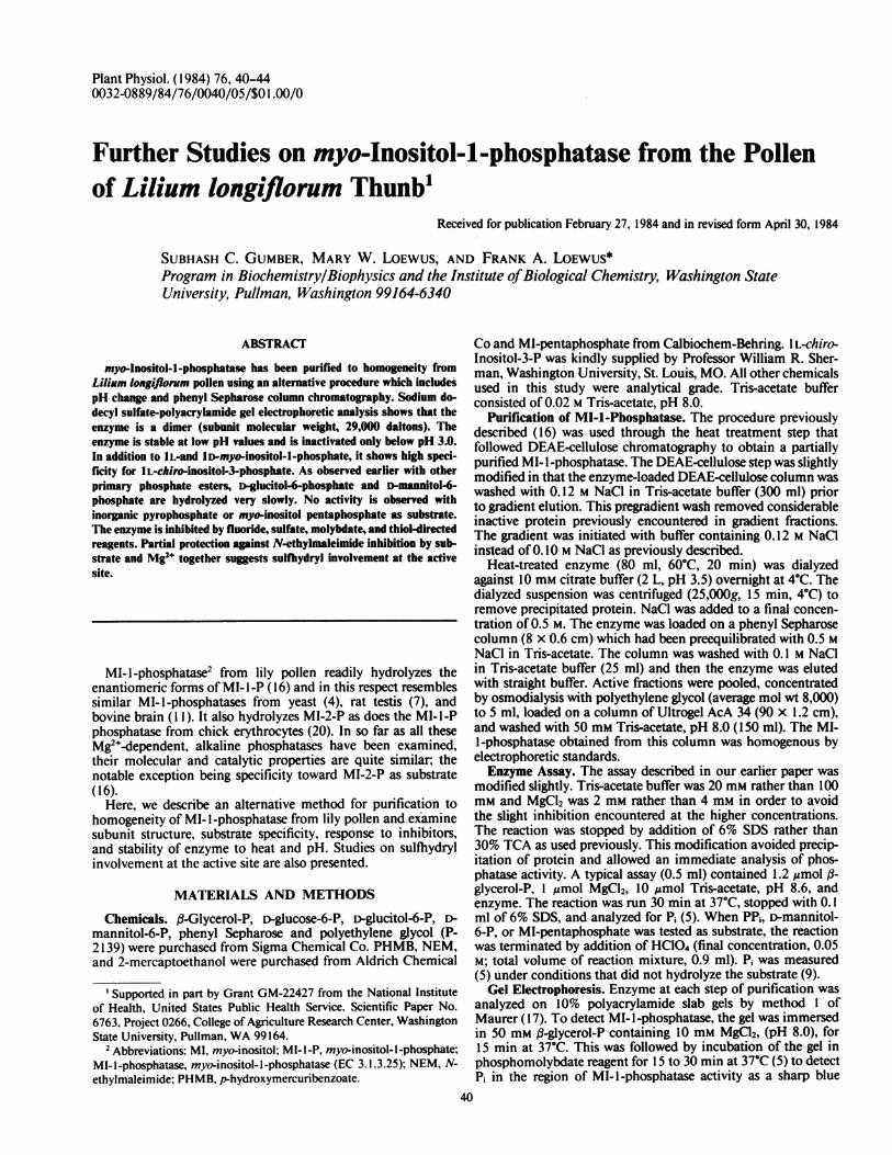
Fraction NLmberFIG. 3. Purification of MI-I-phosphatase on a phenyl Sepharose col-
umn. Arrows mark starting points for washing the column with 0.5 Mand 0.1 M NaCl in Tris-acetate buffer and with straight buffer. The bulkof inactive protein (0) eluted ahead of MI-I-phosphatase (0).
Pure enzyme stored in 50 mM Tris-acetate, pH 8.0, for 1month at 4°C lost no activity. When the enzyme solutions thatwere used to obtain data for Figure 2 were stored at theirrespective pH values for I month at 4°C, samples held at pH 4to 5 lost 50% of their total activity. The remaining samples lostno activity. Attempts to store purified enzyme in the presence of1 mM 2-mercaptoethanol or DTT at 4°C resulted in completeloss of activity within 4 d. Fifty mM 2-mercaptoethanol or DTTfailed to reactivate the inactive enzyme. Enzyme from the phenylSepharose step was stable overnight at 23°C. The activity wasnot significantly affected by ionic strength in 20 to 300 mm Tris-acetate, pH 8.6.
In addition to substrates whose Km and relative Vm.. valueswere determined previously (16), other substrates were tested.IL-chiro-Inositol-3-P had a Km of 0.044 mm and a V,..v of 54relative to IL-MI-i-P as 100. The corresponding values for D>glucitol-6-P were 2 mm and 25, and for D-mannitol-6-P, 0.82
I 3
mm and 20, respectively. PP, and MI-pentaphosphate were in-active as substrates.Under standard assay conditions, lily pollen MI-l -phosphatase
was inhibited 45% by I mM NaF, 95% by 1 mM HgCl2, and 30%by 10 mM iodoacetate, 10 mM (NHI)2SO4, or 20 mm Na2MoO4,respectively. Sulfate inhibited competitively (data not shown).Other compounds that failed to inhibit or stimulate phosphataseactivity included MI, myo-inosose, D-chiro-inositol, L-chiro-ino-sitol, DL-bornesitol, sequoyitol, scylo-inositol, deoxy-scyilo-ino-sitol, L-phenylalanine, L-glutamic acid, glycine, Na2CO3, H3BO3,and D-glucuronic acid. Cyclitols were tested at 20 mm and othercompounds at 10 mM.
Inhibition by HgCI2, iodoacetate, 2-mercaptoethanol, andDTT revealed a sulthydryl requirement for enzymic activity.This effect was investigated further by reaction of the enzymewith NEM and PHMB. Figure 5 shows the pseudo-first orderinactivation of MI-1-phosphatase at several concentrations ofNEM. At the higher concentrations, 0.6 and 1.17 mM, a biphasicinactivation is observed. The enzyme always exhibits a residualactivity (10-15%) even after treatment with 9 mm NEM. Theapparent first order rate constant (k') can be calculated from t',of inactivation, for the appropriate NEM concentration. Theinset of Figure 5 shows a plot ofk' versus log [NEM] which givesa straight line of slope 1. Studies involving the protection fromNEM inactivation ofenzyme by preincubation with substrates)were also conducted (Table II). Mg2e alone gave no protection.PGlycerol-P alone accelerated inactivation *hile IL-MI-1-Palone gave some protection. PGlycerol-P or I L-MI- I-P providedsubstantial protection in the presence of Mg2+. MI-2-P was lesseffective than either (i-glycerol-P or IL-MI-I-P.PHMB inactivated the enzyme so rapidly that kinetic studies
were impossible. Preincubation ofenzyme with ,B-glycerol-P and/or Mg2+ gave no protection against inhibition by PHMB. Toshow that NEM and PHMB reacted with the same groups, theenzyme was treated first with 100 iM PHMB and then with 5mM NEM (Fig. 6). Addition of 16 mM L-cysteine restored activitycompletely whether the enzyme was treated with PHMB aloneor with PHMB followed by NEM.
4:.I War E0
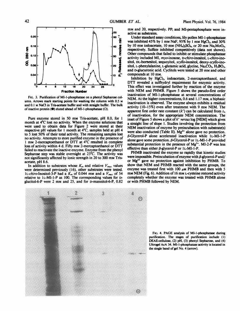
FIG. 4. PAGE analysis of MI-I-phosphatase duringpurification. The stages of purification include (1)DEAE-cellulose, (2) pH, (3) phenyl Sepharose, and (4)Ultrogel AcA 34. MI- I -phosphatase activity is located inthe single band of gel No. 4 (arrow).
0
42 GUMBER ET AL.

myo-INOSITOL-1-PHOSPHATASE FROM LILY POLLEN
80
60
;I
asw
cc20,
0 10 20TIME, mi
30
FIG. 5. Concentration dependence of MI-I-phosphatase inactivationby NEM. Purified enzyme (150 p1. 1.5 pg) was incubated in 50 mm Tris-acetate, pH 7.0 at 30°C. NEM was added to required concentration andaliquots (25 ul) were withdrawn to test for residual activity. The concen-trations (in mM) of NEM used were 0.00 (U), 0.09 (X), 0.18 (0), 0.29(A), 0.60 (0), and 1. 17 (0). Inset: Order ofMI- I -phosphatase inactivationwith respect to NEM concentration. The apparent first-order rate con-stant (k') for the inactivation was calculated from slopes of the lines inFigure 5 for each NEM concentration employed.
Table II. Protection ofMJ-1-phosphatasefrom NEM Inactivation bySubstrate(s)
Experiment 1: Enzyme (2.7 pg protein) from phenyl Sepharose step,pH 7.0, was incubated for 5 min at 30 C in the presence of substrate(s)and NEM was then added to a final concentration of 2.4 mM. Aliquots(0.5 pg protein) were withdrawn at selected intervals to test for residualactivity. Experiment 2: The procedure and conditions of this experimentwere as described in experiment I except that enzyme (I pg protein)from Ultrogel AcA 34 step was used and aliquots containing 0.2 pgprotein were withdrawn to test for residual activity.
Experiment Substrate(s) Used Per Cent Activity Remaining after'for Protection 3 min 12 min
I None 44 19Mg2+ (50)b 43 18O%Glycerol-P (60) 16 12S-Glycerol-P (60) andMg2+ (50) 75 64
2 None 22 15I L-MI- I -P (8.7) 34 1 7I L-MI- I -P (8.7) andMg2+ (52) 49 36
'The control (100% activity) was incubated under similar conditionsexcept that no protecting substrate or NEM was added. The activity ofthis enzyme was monitored at various intervals.bThe numbers in parenthesis indicate the final concentration of
substrate (in mM) in the incubation mixture.
16
Tk, mhFiG. 6. Sequential inactivation of MI-1-phosphatase by PHMB and
NEM. Six hundred ;d of enzyme (approximately 6.1 ug), pH 8.0, wastreated with PHMB (final concentration, 89 pM) at 30C and aliquots(75 A1) were withdrawn to test for residual enzyme activity (0). After 6min, a 150-pd aliquot was removed and held at 30C. To remainingenzyme was added NEM (final concentration, 3.8 mM) and again aliquots(75 p1) were withdrawn for assay (0). At 20 min, L-cysteine was added(final concentration, 16 mM) to NEM-treated (A) and untreated (A)enzyme, and aliquots were removed for assay of MI-I-phosphatase. Acontrol to which nothing was added is included (x).
DISCUSSION
The purification procedure for MI-l-phosphatase from lilypollen is modified to eliminate the chromatofocusing step andto introduce two new steps, a pH 3.5 treatment that takesadvantage of the stability of the enzyme in a relatively acidenvironment and hydrophobic chromatography on phenyl Seph-arose. The new procedure provides a relatively simple route tohomogeneous enzyme in quantities sufficient for further studies.On SDS gel, the enzyme has a single protein band correspondingto 29,000 + 2,000 D. This is approximately one-half of the molwt of 54,000 D reported for active lily pollen MI-l-phosphatase(16) and leads to the conclusion that this enzyme is a dimer.The stability of lily pollen enzyme to low pH is much greater
than that of bovine brain which is inactivated at pH 5 (1 1), butits stability to heat treatment (60°C for 15-30 min) is lower thanvalues (70°C for 15-30 min) reported for enzyme from mam-malian or avian sources (7, 11, 19, 20).The ratio V,gz/Km provides a means of measuring specificity
in terms of discrimination among substrates (22). In addition tosubstrates tested previously (16; see Table II), IL-chiro-inositol-3-P (also identified as (-)-inositol-3-P) (4, 7, 1 1) and D-glucitol-6-P were tested as substrate for lily pollen MI-l-phosphatase.Ratios of V,MJ/Km for these substrates are: lL-chiro-inositol-3-P,1227; lL-MI-I-P, 1220; lD-MI-l-P, 1038; aglycerol-P, 693; MI-2-P, 64; a-L-glycerol-P, 32; D-glucitol-6-P, 13; and D-glucose-6-P, 5. Clearly, the first three substrates are best. lL-MI-l-P hasmetabolically significant roles in plants such as intermediate inthe MI oxidation pathway and in phytic acid biosynthesis (14,15). Hallcher and Sherman (I 1) found ID-MI-I-P in brain tissuewhere it too may have a metabolic role. The recent discoverythat Ml- 1,4,5-trisP acts as second messenger in the mobilizationofintracellularcalcium in mammalian tissues (12)and undergoessequential loss of phosphate via MI-I ,4-bisP and ID-MI-I-P toform free MI (2) has yet to be repeated in plant tissues. Shouldthis process be present in lily pollen, the MI-1-phosphatasedescribed here may have an essential role in the cyclic processdescribed by Berridge. I L-chiro-Inositol-3-P, which was preparedsynthetically, does not appear to be a naturally occurring com-pound.
o \
0
\: 2.241.6 .
I.. a . .20 2. 2.8
Log [(NEM4X °°°l
w 9 I
DO_- v -
pj:P6
43

Plant Physiol. Vol. 76, 1984
When models of these three substrates are compared by super-imposing phosphate groups, only one additional equatorial hy-droxy group (equivalent to the one at C-5 in 1 L-MI- l-P) has aconfiguration common to all three structures. The absence of athird polar binding site common to these substrates may explainits lack of enantiomeric specificity. Of course, the possibilityexists that MI- I-phosphatase has multiple binding sites whichaccomodate different hydroxyl groups but this is less likely.
Alternatively, these models superimpose with an axial hy-droxyl group above the ring and two equatorial hydroxyl groupsbelow the ring but separated by one ring carbon from the axialhydroxyl group. Here, the phosphate is above the ring, eitheradjacent (1 L- and 1 D-MI- 1-P) or opposite (1 L-chiro-inositol-3P)to the carbon bearing the axial hydroxyl. A binding site thatcatalyzes the cleavage of phosphate ester at any one of threeseparate positions is indicated in this model.An interesting property of MI-l-phosphatase is its hydrolysis
of MI-2-P. Although the Km for this substrate is 9-fold greaterthan those of IL- or lI>MI-l-P, hydrolysis of MI-2-P proceedsat 50% of the rate of either enantiomer of MI- I-P. Similarkinetics for hydrolysis of MI-2-P were found in MI- l-phosphatasefrom chick erythrocytes (20). While the higher Km may be dueto the axial orientation of phosphate in MI-2-P, it can be shownthat higher reaction rates are achieved if the enzyme uses itsbinding energy to increase its Vmax rather than to lower its Km(8). To fully understand the structural requirements of the sub-strate, more detailed studies involving the use of mono- andpolyphosphate esters of the inositols are needed.A Mg2e-dependent specific D-glucitol-6-phosphatase has been
isolated from plants (10). This enzyme differs from lily pollenMI-1-phosphatase which hydrolyzes D-glucitol-6-P very slowlyas previously noted for other primary phosphate esters (16).Primary phosphates have lower free energy of hydrolysis andthus a lower phosphate transfer potential (18). The nature of thenucleophilic attack on phosphorus of the phosphate group at theactive site will, therefore, determine whether an enzyme catalyzesthe hydrolysis of primary phosphate as efficiently as it doessecondary phosphate.
Fluoride, a common inhibitor of metal-activated enzymes (1),inhibits MI- 1-phosphatase from yeast (4), mammalian and aviansources (1 1, 19, 20), and lily pollen. Although this inhibition hasbeen ascribed to removal of Mg2" (I 1), the stoichiometry suggeststhat inhibition may include other effects such as binding withactive site groups or replacement of H20 in the coordinationsphere of Mg2+ where H20 may be required for phosphatehydrolysis (1).
Sulfate and molybdate also inhibit MI-l-phosphatase. Thecompetitive nature of S42- inhibition suggests that it binds tothe enzyme in the same way as phosphate of the substrate.
MI-l-phosphatase is inactivated by thiol-directed reagents,PHMB and NEM. The inactivation reaction exhibits first orderkinetics with respect to NEM concentration (Fig. 5, inset) whichsuggests that the modification of a single sulfhydryl group causesthe observed loss of activity. Whether the residual activity ob-served after modification is due to activity of the modifiedenzyme or a form of the enzyme which is resistant to modifica-tion is not clear. Partial protection of the enzyme from NEMinactivation by j3-glycerol-P or 1L-MI-I-P in the presence ofMg2+ indicates the presence of an essential sulfhydryl at or nearthe active site. Accelerated inactivation in the presence of,-glycerol-P alone is probably due to increased reactivity of essen-tial sulfhydryl upon binding of a-glycerol-P or to increased
accessibility ofNEM to this group (24) following such treatment.Restoration of full activity following sequential treatment of
MI-l-phosphatase with PHMB and NEM suggests that NEM isreacting with the same groups as PHMB and provides addedsupport for the view that inactivation by PHMB and NEM isdue to modification of a sulfhydryl. Complete reactivation by L-cysteine also indicates that no other irreversible side reactionoccurred.
Acknowledgments-lL-chiro-lnositol-3-P was generously provided by Dr. Wil-liam R. Sherman, Washington University, St. Louis, MO. We wish to thank Dr.Thomas Okita for his suggestion concerning the use of hydrophobic affinitychromatography. The pollen used in this study was obtained through the coopera-tion of Mr. Norman Yock, Oregon Propagating Co., Brookings, OR.
LITERATURE CITED
1. BAYKOV AA, AA ARTJUKOV, SM AVAEVA 1976 Fluoride inhibition ofinorganicpyrophosphatase. I. Kinetic studies in a Mg2'-PPi system using a newcontinuous enzyme assay. Biochim Biophys Acta 429: 982-992
2. BERRIDGE MJ 1983 Rapid accumulation of inositol triphosphate reveals thatagonists hydrolyze polyphosphoinositides instead of phosphatidylinositol.Biochem J 212: 849-858
3. BLASKESLY RW, JA BOEZI 1977 A new staining technique for proteins inpolyacrylamide gels using Coomassie Brilliant Blue G250. Anal Biochem82: 580-587
4. CHEN I-W, FC CHARALAMPOUS 1966 Biochemical studieson inositol. X. Partialpurification of yeast inositol l-phosphatase and its separation from glucose6-phosphate cyclase. Arch Biochem Biophys 117: 154-157
5. CHEN JR PS, TY TORIBARA, H WARNER 1956 Microdetermination of phos-phorus. Anal Chem 28: 1726-1758
6. DUNKER AK, RR RUECKERT 1969 Observations on molecular weight deter-minations on polyacrylamide gels. J Biol Chem 244: 5074-5080
7. EISENBERG JR F 1967 D-Myo-inositol 1-phosphate as product of cyclization ofglucose 6-phosphate and substrate for a specific phosphatase in rat testis. JBiol Chem 242: 1375-1382
8. FERSHT A 1977 Enzyme Structure and Mechanism. WH Freeman, San Fran-cisco, pp 247-254
9. FURCHGOTT RF, T GUBAREFF 1956 The determination of inorganic phosphateand creatine phosphate in tissue extracts. J Biol Chem 233: 377-388
10. GRANT CR, T APREES 1981 Sorbitol metabolism by apple seedlings. Phyto-chemistry 20: 1505-151 1
11. HALLCHER LM, WR SHERMAN 1980 The effects of lithium ion and otheragents on the activity of myo-inositol-l-phosphatase from bovine brain. JBiol Chem 255: 10896-10901
12. JOSEPH SK, AP THOMAS, RJ WILLIAMS, RF IRVINE, JR WILLIAMSON 1984myo-Inositol 1,4,5-triphosphate. A second messenger for the hormonal mo-bilization of intracellular Ca2' in liver. J Biol Chem 259: 3077-3081
13. KING J, UK LAEMMLI 1971 Polypeptides ofthe tail fibers of bacteriophage T4.J Mol Biol 62: 465-477
14. LOEwus FA 1983 Phytate metabolism with special reference to its myo-inositolcomponent. Recent Adv Phytochem 17: 173-192
15. LOEWUS FA, MW LOEWUS 1983 myo-Inositol: Its biosynthesis and metabolism.Annu Rev Plant Physiol 34: 137-161
16. LOEWUS MW, FA LOEWUS 1982 myo-Inositol-l-phosphatase from the pollenof Lilium longiflorum Thunb. Plant Physiol 70: 765-770
17. MAURER HR 1971 Disc Electrophoresis and Related Techniques of Polyacryl-amide Gel Electrophoresis. Walter de Gruyter, New York
18. MEYERHOF 0, H GREEN 1949 Synthetic action of phosphatases. I. Equilibriaof biological esters. J Biol Chem 178: 655-667
19. NACCARATO WF, RE RAY, WW WELLS 1974 Biosynthesis of myo-inositol inrat mammary gland. Isolation and properties of the enzymes. Arch BiochemBiophys 164: 194-201
20. ROTH SC, DR HARKNESS, RE ISAACKS 1981 Studies on avian erythrocytemetabolism: purification of myo-inositol l-phosphatase from chick erythro-cytes. Arch Biochem Biophys 210: 465-473
21. SEDMARK JJ, SE GROSSBERG 1977 A rapid, sensitive and versatile assay forprotein using Coomassie Brilliant Blue G250. Anal Biochem 79: 544-552
22. SEGEL IH 1976 Biochemical Calculations. John Wiley and Sons, New York pp208
23. SEGREST VP, RL JACKSON 1972 Molecular weight determination of glycopro-teins by polyacrylamide gel electrophoresis in sodium dodecyl sulfate. Meth-ods Enzymol 28: 54-63
24. WHITAKER JR 969 Ficin- and papain-catalyzed reactions. Changes in reactivityof essential sulfhydryl group in the presence of substrate and competitiveinhibitors. Biochemistry 8: 4591-4597
44 GUMBER ET AL.


