OF BAYO BEAN · PDF fileKeywords: bayo bean, protein hydrolysate, Eudragitfi L 30 D-55,...
12
Revista Mexicana de Ingeniería Química ISSN: 1665-2738 [email protected] Universidad Autónoma Metropolitana Unidad Iztapalapa México Tovar-Benítez, T.; Jiménez-Martínez, C.; Perea-Flores, M.J.; Téllez-Medina, D.I.; Dávila- Ortiz, G. MICROENCAPSULATION OF BAYO BEAN (Phaseolus vulgaris) PROTEIN HYDROLYSATE WITH INHIBITORY ACTIVITY ON ANGIOTENSIN-I CONVERTING ENZYME THROUGH FREEZE-DRYING Revista Mexicana de Ingeniería Química, vol. 15, núm. 3, 2016, pp. 797-807 Universidad Autónoma Metropolitana Unidad Iztapalapa Distrito Federal, México Available in: http://www.redalyc.org/articulo.oa?id=62048168011 How to cite Complete issue More information about this article Journal's homepage in redalyc.org Scientific Information System Network of Scientific Journals from Latin America, the Caribbean, Spain and Portugal Non-profit academic project, developed under the open access initiative
Transcript of OF BAYO BEAN · PDF fileKeywords: bayo bean, protein hydrolysate, Eudragitfi L 30 D-55,...
Revista Mexicana de Ingeniería Química
ISSN: 1665-2738
Universidad Autónoma Metropolitana
Unidad Iztapalapa
México
Tovar-Benítez, T.; Jiménez-Martínez, C.; Perea-Flores, M.J.; Téllez-Medina, D.I.; Dávila-
Ortiz, G.
MICROENCAPSULATION OF BAYO BEAN (Phaseolus vulgaris) PROTEIN
HYDROLYSATE WITH INHIBITORY ACTIVITY ON ANGIOTENSIN-I CONVERTING
ENZYME THROUGH FREEZE-DRYING
Revista Mexicana de Ingeniería Química, vol. 15, núm. 3, 2016, pp. 797-807
Universidad Autónoma Metropolitana Unidad Iztapalapa
Distrito Federal, México
Available in: http://www.redalyc.org/articulo.oa?id=62048168011
How to cite
Complete issue
More information about this article
Journal's homepage in redalyc.org
Scientific Information System
Network of Scientific Journals from Latin America, the Caribbean, Spain and Portugal
Non-profit academic project, developed under the open access initiative
Vol. 15, No. 3 (2016) 797-807
Ingeniería de alimentos Revista Mexicana de Ingeniería Química
CONTENIDO
Volumen 8, número 3, 2009 / Volume 8, number 3, 2009
213 Derivation and application of the Stefan-Maxwell equations
(Desarrollo y aplicación de las ecuaciones de Stefan-Maxwell)
Stephen Whitaker
Biotecnología / Biotechnology
245 Modelado de la biodegradación en biorreactores de lodos de hidrocarburos totales del petróleo
intemperizados en suelos y sedimentos
(Biodegradation modeling of sludge bioreactors of total petroleum hydrocarbons weathering in soil
and sediments)
S.A. Medina-Moreno, S. Huerta-Ochoa, C.A. Lucho-Constantino, L. Aguilera-Vázquez, A. Jiménez-
González y M. Gutiérrez-Rojas
259 Crecimiento, sobrevivencia y adaptación de Bifidobacterium infantis a condiciones ácidas
(Growth, survival and adaptation of Bifidobacterium infantis to acidic conditions)
L. Mayorga-Reyes, P. Bustamante-Camilo, A. Gutiérrez-Nava, E. Barranco-Florido y A. Azaola-
Espinosa
265 Statistical approach to optimization of ethanol fermentation by Saccharomyces cerevisiae in the
presence of Valfor® zeolite NaA
(Optimización estadística de la fermentación etanólica de Saccharomyces cerevisiae en presencia de
zeolita Valfor® zeolite NaA)
G. Inei-Shizukawa, H. A. Velasco-Bedrán, G. F. Gutiérrez-López and H. Hernández-Sánchez
Ingeniería de procesos / Process engineering
271 Localización de una planta industrial: Revisión crítica y adecuación de los criterios empleados en
esta decisión
(Plant site selection: Critical review and adequation criteria used in this decision)
J.R. Medina, R.L. Romero y G.A. Pérez
MICROENCAPSULATION OF BAYO BEAN (Phaseolus vulgaris) PROTEINHYDROLYSATE WITH INHIBITORY ACTIVITY ON ANGIOTENSIN-I
CONVERTING ENZYME THROUGH FREEZE-DRYING
MICROENCAPSULACION DE UN HIDROLIZADO PROTEINICO DE FRIJOL BAYO(Phaseolus vulgaris) CON ACTIVIDAD INHIBITORIA SOBRE LA ENZIMACONVERTIDORA DE ANGIOTENSINA-I MEDIANTE LIOFILIZACION
T. Tovar-Benıtez1, C. Jimenez-Martınez1, M.J. Perea-Flores2, D.I. Tellez-Medina1, G. Davila-Ortiz1*1Escuela Nacional de Ciencias Biologicas, Instituto Politecnico Nacional, Av. Wilfrido Massieu, Esq. Cda. Miguel Stampa S/N,
Unidad Profesional Adolfo Lopez Mateos, Zacatenco, Del. Gustavo A. Madero, C.P. 07738, Ciudad de Mexico, Mexico.2Centro de Nanociencias y Micro y Nanotecnologıas, Instituto Politecnico Nacional, Av. Luis Enrique Erro S/N, Unidad
Profesional Adolfo Lopez Mateos, Zacatenco, Del. Gustavo A. Madero, C.P. 07738, Ciudad de Mexico, Mexico.Received May 17, 2016; Accepted July 6, 2016
AbstractThe aim of this study was to obtain microcapsules of a bayo bean protein hydrolysate (BBPH) with antihypertensive activityusing Eudragit® L 30 D-55 as wall material (EGLD) through freeze-drying processing. The BBPH was obtained usingsequential pepsin-pancreatin enzymatic system. The ACE-I inhibitory activity was measured using tripeptide hippuryl-histidyl leucine (HHL) as model peptide. Three microcapsule formulations were prepared containing BBPH and EGLD atratios of 1:20, 1:4 and 1:1, respectively. The physicochemical characteristics of microcapsules were evaluated by optical(OM) and scanning electronic microscopy (SEM), confocal laser scanning microscopy (CLSM), differential scanningcalorimetry (DSC), X-ray diffraction (XRD) and Fourier transform infrared spectroscopy (FT-IR). ACE-I inhibitory activityof BBPH was IC50=0.42 mg/mL. All microcapsules showed irregular shapes. The BBPH was distributed homogeneouslyin all formulations. The DSC and XRD analysis revealed a uniform dispersion of the BBPH and partially crystallinestructures of EGLD and BBPH. The FT-IR confirmed the chemical stability of BBPH in the microcapsules. In conclusion,the EGLD microcapsules containing BBPH were prepared successfully by freeze-drying processing.Keywords: bayo bean, protein hydrolysate, Eudragit® L 30 D-55, microcapsules, freeze-drying.
ResumenEl objetivo de este estudio fue obtener microcapsulas de un hidrolizado proteınico de frijol bayo (BBPH) con actividadantihipertensiva utilizando Eudragit® L 30 D-55 como material de pared (EGLD) por el metodo de liofilizacion. El BBPHse obtuvo usando el sistema enzimatico secuencial pepsina-pancreatina. La actividad inhibidora sobre la ECA-I se midioutilizando hipuril-histidil leucina (HHL) como peptido modelo. Se prepararon tres formulaciones conteniendo BBPH yEGLD en proporciones de 1:20, 1:4 y 1:1, respectivamente. Las caracterısticas fisicoquımicas de las microcapsulas seevaluaron por microscopıa optica (OM) y microscopıa electronica de barrido (SEM), microscopıa confocal de barridolaser (CLSM), calorimetrıa diferencial de barrido (DSC), difraccion de rayos X (XRD) y espectroscopıa infrarroja contransformada de Fourier (FT-IR). La actividad inhibitoria del BBPH sobre la ACE-I presento un valor de IC50=0.42 mg/mL.Todas las formulaciones mostraron microencapsulados con forma irregular. El BBPH se distribuyo homogeneamente entodas las formulaciones. El analisis por DSC y XRD mostro que EGLD y BBPH presentaron una distribucion uniforme delBBPH y estructuras parcialmente cristalinas. El analisis FT-IR confirmo la estabilidad del BBPH en las microcapsulas. Enconclusion, las microcapsulas de EGLD conteniendo BBPH fueron preparadas satisfactoriamente por liofilizacion.Palabras clave: frijol bayo, hidrolizado proteico, Eudragit® L 30 D-55, microcapsulas, liofilizacion.
1 Introduction
Angiotensin-I converting enzyme (ACE-I; EC3.4.15.1) play an important role in regulating blood
pressure. This enzyme catalyzes the conversionof angiotensin-I (DRVYIHPFHL) into a potentvasoconstrictor, the angiotensin-II (DRVYIHPF)and inactive the vasodilatory peptide bradykinin
* Corresponding author. E-mail: [email protected]. +52 (55)57296000x57870; fax: +52 (55)57296000.
Publicado por la Academia Mexicana de Investigacion y Docencia en Ingenierıa Quımica A.C. 797

Tovar-Benıtez et al./ Revista Mexicana de Ingenierıa Quımica Vol. 15, No. 3 (2016) 797-807
(RPPGFSPFR). Inhibition of ACE-I activity is thusconsidered to be a pivotal therapeutic approach fortreating hypertension (Udenigwe and Mohan, 2014).ACE-I synthetic inhibitors such as captopril, lisinopriland enalapril are used as treatment to control highblood pressure. However, prolonged use of suchdrugs can produce side effects, for instance vertigo,cough, agiodemas and decreased kidney function(Regulska et al., 2014). Currently the bioactivepeptides isolated from different protein sources ofanimal or vegetable origin may inhibit the activity ofACE-I, becoming an alternative for the prevention andcontrol of hypertension (Hernandez-Ledesma et al.,2011).
Protection of bioactive peptides fromphysiological changes is essential in the translationof in vitro activity into animal and human models;therefore, microencapsulation could be used as analternative to minimize factors that might interferewith their bioavailability. According to Nesterenkoet al., (2013) microencapsulation is a process bywhich an encapsulated substance (solid, liquid orgaseous), also called active compound, active, internalor core phase, is covered by embedded in anothersubstance called encapsulating coating, membrane,wall material, external phase or matrix; resulting inthe formation of microcapsules which aim to providea protective barrier to the active compound againstfactors that may affect its activity or structure.
The correct choice of wall material directlyaffects the encapsulation efficiency, stability anddegree of protection of the active compound.Materials commonly used are synthetic polymersand copolymers, carbohydrates (starch, maltodextrin,gum arabic), fats, waxes, animal (gelatin, casein)and plant (soy protein) proteins, petroleum-basedpolymers used commonly in pharmacy and medicineas a matrix (polystyrenes, polyamides, polyurethanes,polyacrylates, and polyethylene glycols), amongothers (Nesterenko et al., 2013; Ray et al.,2016). Eudragits®, as the well-known pharmaceuticalexcipients, have been widely used for the formation ofdifferent sustained and controlled release formations.Eudragit® L 30 D-55 is an anionic copolymer basedon methacrylic acid and ethyl acrylate. The ratioof the free carboxyl groups to the ester groups isapproximately 1:1. It is characterized by a fastdissolution in the upper Bowel (duodenum) to pH 5.5(El-Malah and Nazzal, 2008).
There are many techniques available to carryout the production of microencapsulates, such asspray-drying, spray-cooling/chilling, freeze-drying,
fluidized bed, coacervation/phase separation, gelation,solvent evaporation, supercritical fluid expansion,interfacial polymerization (polycondensation),emulsion polymerization and extrusion (Nesterenkoet al., 2013). The choice of the convenientmicroencapsulation technique depends on size,biocompatibility and biodegradability of desiredmicrocapsules, the physicochemical properties of theactive compound and the wall material, the proposedmechanism for the release of the active compoundand process costs. Freeze-drying is used for waterremoval with a minimum of associated heat damageto the solid matrix and is based on the sublimationof the ice fraction of the product. Mass transferoccurs by diffusion of vapour through the dry layerof the specimen under the action of a pressuredifference (Viveros-Contreras et al., 2013). From thephenomenon of sublimation, a process of successfullyophilization retains most of the initial properties ofraw materials such as shape, size, appearance, taste,color, texture and biological activity (Ceballos et al.,2012). Based on this, the aim of this study was tomicroencapsulate a bayo bean protein hydrolysatewith ACE-I inhibitory activity using Eudragit® L30 D-55 as wall material through freeze-dryingprocessing.
2 Material and methods
2.1 Material
Phaseolus vulgaris L. com. var. Bayo seedscrop 2012, were donated by CEVAMEX (CampoExperimental Valle de Mexico) at Santa Lucıa,Texcoco, Estado de Mexico, Mexico. Pepsin fromporcine gastric mucosa (P7125), Pancreatin fromporcine pancreas (P1750) and Angiotensin ConvertingEnzyme (ACE) from rabbit lung (A6778) werepurchased from Sigma-Aldrich (St. Louis, MO,USA). Eudragit® L 30 D-55 was provided byHELM (Naucalpan de Juarez, Edo. de Mexico,Mexico) and was used as received. All otherreagents were analytical grade and purchased from J.T.Baker (Phillipsburg, NJ, USA), Sigma-Aldrich (SigmaChemical Co., St. Louis, MO, USA) and Bio-Rad(Bio-Rad Laboratories, Inc. Hercules, CA, USA).
2.2 Preparation of protein concentrate
Bayo bean protein concentrate (BBPC) was preparedaccording to the method described by Betancur-Ancona et al., (2004). Whole seeds were ground to
798 www.rmiq.org
Tovar-Benıtez et al./ Revista Mexicana de Ingenierıa Quımica Vol. 15, No. 3 (2016) 797-807
powder and passed through a 0.2 mm mesh sieve. Theresulting flour was mixed with distilled water (1:10w/v) adjusted to pH 11 (NaOH 1 N) and stirred for1 h. This suspension was allowed to sediment for 1 hat room temperature to recover the starch and proteinfractions. The protein fraction was adjusted to theirisoelectric point (pH 4.5) with HCl 1 N in order toprecipitate proteins. The precipitated proteins wererecovered by centrifugation at 1,317 g for 15 min andfreeze-dried at -47 ºC and 1.3 Pa. The protein contentwas analyzed according to the method 954.01 fromAOAC (AOAC, 1997).
2.3 Enzymatic hydrolysis
Enzymatic hydrolysis was performed with thesequential pepsin-pancreatin system according toMegıas et al., (2004) with modifications. TheBBPC was mixed with distilled water to produce aprotein solution to 5% (w/v) which was equilibratedat optimum temperature (37 °C for both enzymes)and pH (2.0 for pepsin and 7.5 for pancreatin) foreach protease before adding the respective enzyme.Protease was then added to the protein solution at aratio 1:10 (enzyme/substrate). The hydrolysis with thesequential pepsin-pancreatin system was done for 60min: predigestion with pepsin for 30 min followed byincubation with pancreatin for 30 min. The hydrolysisreaction was stopped by heating at 80 °C for 20min, followed by centrifuging at 10,000 g for 30 minto remove the insoluble portion. The supernatants(protein hydrolysate) were freeze-dried at -47 ºC and1.3 Pa.
2.4 Degree of hydrolysis
Degree of hydrolysis (DH) was calculatedby determining free amino groups with O-phthaldialdehyde following as described by Nielsenet al., (2001) using Ec. (1).
DH =h
htot× 100 (1)
Where h is the number of hydrolyzed bonds, andhtot is the total number of peptide bonds per proteinequivalent.
2.5 ACE-I inhibitory activity
Angiotensin-I converting enzyme inhibitory activity inthe samples was determined by following the method
described by Hayakari et al., (1978). Hippuryl-L-histidyl-L-leucine (HHL) is hydrolyzed by ACE-I to yield hippuric acid and histidyl-leucine. Thismethod relies on the colorimetric reaction of hippuricacid with 2,4,6-trichloro-triazine (TT) in a 0.5 mLincubation mixture containing 40 µM potassiumphosphate buffer (pH 8.3), 300 µM sodium chloride,3% HHL in potassium phosphate buffer (40 µM,pH 8.3), and 100 mU/mL of ACE-I. The mixturewas incubated at 37 ºC for 45 min and the reactionfinished by adding TT (3%, v/v) in dioxane and 3mL of potassium phosphate buffer (0.2 M, pH 8.3).After centrifuging the reaction mixture at 10,000g for 10 min, enzymatic activity was determinedin the supernatant by measuring absorbance at 382nm. ACE-I inhibitory activity was quantified by aregression analysis of ACE-I inhibitory activity (%)versus protein hydrolysate concentration and definedas an IC50 value, that is, the protein hydrolysateconcentration (mg protein/mL) required to produce50% ACE-I inhibition under the described conditions.
2.6 Amino acid composition
Protein sample (2 mg) was hydrolysed withHCl 6 N (4 mL) at 110 ºC for 24 h in tubessealed under nitrogen. After derivatization withdiethyl ethoxymethylenemalonate, amino acidswere determined by high-performance liquidchromatography (HPLC) according to the methoddescribed by Alaiz et al., (1992) using D,L-α-aminobutyric acid as an internal standard and aNovapack C18 reverse phase column (300 x 3.9mm i.d., 4 µm film thickness; Waters, Milford, MA,USA). Tryptophan was analyzed by HPLC after basichydrolysis according to Yust et al., (2004).
2.7 Preparation of microcapsules
The microcapsules were prepared in the followingway: Eudragit® L 30 D-55 as wall material(EGLD) was dissolved in 50 mL of phosphatebuffer at pH 4.2 with constant stirring for 30minutes. Similarly, the protein hydrolysate (activecompound) was dissolved and stirred under thesame experimental conditions. These solutions weremixed at 13,000 rpm for 1 min using an Ultra-Turrax® (T25Basic, IKA Labortechnik, Wilmington,Delaware, USA). The Eudragit® L 30 D-55-proteinhydrolysate dispersions were processed for two cyclesthrough a M-110Y microfluidizer (Microfluidics®,Newton, Massachusetts, USA) with two interaction
www.rmiq.org 799

Tovar-Benıtez et al./ Revista Mexicana de Ingenierıa Quımica Vol. 15, No. 3 (2016) 797-807
chambers: the primary one of the type “Y” (F20Y,φ=75 µm) and the secondary one of type “Z” (H30Z,φ =200 µm). The air chamber of the equipmentoperated at room temperature and 70 MPa. Finally,the dispersions obtained were frozen at -80 °C for 24h and lyophilized (Virtis, Consol 255L, France) to -47°C and 1.3 Pa. Three formulations were prepared fromprotein hydrolysate and EGLD with the followingratios: 1:20, 1:4 and 1:1, respectively.
2.8 Characterization of microcapsules
2.8.1. Efficiency of encapsulation (EE)
The encapsulation efficiency (EE) was determinedaccording to the protein content in 10 mg of sampledetermined by the colorimetric method of Biuretusing a calibration curve for BSA (0.5-10 mg/mL,R2=0.998). The EE was calculated in agree the Ec.(2).
RE =CPECPT
× 100 (2)
Where CPE corresponds to experimental proteincontent and the content CPT theoretical protein.
2.8.2. Optical (OM) and scanning electronic (SEM)microscopy
The shape and surface morphology of the sampleswere observed with an optical microscope NikonEclipse 50i (Tokyo, Japan), a ring light microscopeAmscope Led-144 (CA, USA) was used as lighting.Images were acquired at 150x of total amplification.SEM was also used (EVO LS10, ZEISS, Munich,Germany) at an accelerating voltage of 6 kV. It wasused an EDS detector of the brand Bruker QUANTAX200. The samples were fixed on an aluminum supportand processed in environmental mode. Images wereacquired at 500x of total amplification.
2.8.3. Confocal laser scanning microscope (CLSM)
The samples were mounted on glass slides andviewed under CLSM (LSM 710, Carl Zeiss,Germany). The laser wavelengths excitation were405, 488 and 561 nm. A spectral canal wasused to detect autofluorescence signals, whichcomes from microencapsulated components, in wherethe measurements of fluorescence intensity wereperformed using the built ZEN software of the LSM710. The intensity peak characteristic of fluorescenceemission signal was 463 nm for EGLD and 472 nm forBBPH.
2.8.4. Differential scanning calorimetry (DSC)
The thermal properties of the samples were studiedwith a Perkin-Elmer Differential scanning calorimeter(DSC). Samples (5 mg) were weighed and sealed in analuminum DSC pan. Scanning was performed from 30to 200 °C at a heating rate of 10 °C/min with a nitrogenflow of 40 mL/min. The temperature at the onset (To),the temperature at peak (Tp), the temperature at theend (T f ), and the enthalpy (∆H) were determined.
2.8.5. X-ray powder diffraction (XRD)
The samples were analyzed in an X-ray diffractometer(MiniFlex 600, Rigaku, Tokyo, Japan) equipped witha CuKα radiation. The X-ray tube was run at a powerof 40 kV and 15 mA. The scanning was operated inthe range between 5 and 70° 2Θ with a step angle of0.02° at a scan rate of 1°/min.
2.8.6. Fourier transform infrared spectroscopy (FT-IR)
The FT-IR spectra of samples were obtained usinga Fourier transform infrared (FT-IR) spectrometer(Perkin-Elmer, Waltham, Massachusetts, USA) in theregion of 400 to 4000 cm−1. Each spectrum wasobtained at a resolution of 1 cm−1.
2.9 Statistical analysis
Results were presented as mean ± standard deviation.A Duncan multiple range test was used to estimatesignificant differences among the mean values at 2α =
0.05 significance level. All analyses were processedwith the Minitab 17 software.
3 Results and discussion
3.1 Enzymatic hydrolysis
The protein content in the concentrate obtained frombayo bean seeds was 63.18 g/100 g of sample, thisvalue is similar to that reported by Valdez-Ortiz etal., (2012) for sulfured bean protein concentrates (61-66 g/100 g sample), but lower than that reported byGuzman-Mendez et al., (2014) for P. vulgaris (76.66g/100 g of sample) and P. lunatus (71.88 g/100 g ofsample) concentrate. According to Torruco-Uco etal., (2009) the protein content of BBPC is suitableto be enzymatically hydrolyzed and produce bioactivepeptides with possible antihypertensive activity.
800 www.rmiq.org

Tovar-Benıtez et al./ Revista Mexicana de Ingenierıa Quımica Vol. 15, No. 3 (2016) 797-807
The hydrolysis of BBPC with sequential pepsin-pancreatin system produced a DH value of 24.29%,which was lower than 35.74% in 90 min reported for aV. unguiculata protein hydrolysate (Segura-Campos etal., 2010), and to 28.47% for a hardened bean proteinhydrolysate produced in 90 min (Betancur-Ancona etal., 2014), both obtained with the same sequentialenzyme system. According to Pedroche et al., (2002)the sequential use of proteases with different or samecatalytic activity can generate protein hydrolysateshaving a degree of hydrolysis greater than 10%. Thesehydrolysates may be useful to obtain biologicallyactive peptides for the treatment or prevention ofchronic degenerative diseases. Furthermore, Segura-Campos et al., (2010) reported that the production ofprotein hydrolysates with sequential pepsin-pancreatinsystem produces a group of peptides similar to thosegenerated during gastrointestinal digestion in vivo.
Therefore, the resulting peptides are resistant to theseenzymes, suggesting that can be absorbed by thedigestive epithelial cells in the small intestine andreach the blank site exerting its biological activity.
3.2 ACE-I inhibitory activity
The IC50 value of BBPH produced in 60 min withsequential pepsin-pancreatin system was 0.42 mg/mL.This value is within the range reported for otherprotein hydrolysates obtained with the same enzymesystem, and from vegetable protein sources, suchas chickpea (0.14-0.23 mg/mL), pea (0.16 mg/mL)(Barbana and Boye, 2010), lentil (0.44 mg/mL) (Boyeet al., 2010), azufrado beans (0.06-0.32 mg/mL)(Valdez-Ortiz et al., 2012), lupin (0.23-0.27 mg/mL)and soybean (0.22 mg/mL) (Boschin et al., 2014).
Table 1. Amino acid composition of bayo bean protein hydrolysate (BBPH).
Amino acids BBPH FAO/WHO (1991)
Asp+Asn 12.5 ± 0.01Glu+Gln 15.8 ± 0.01
Ser 6.6 ± 0.01His* 3.0 ± 0.07 1.9Gly 4.8 ± 0.01Thr* 4.2 ± 0.14 3.4Arg 5.8 ± 0.07Ala 4.5 ± 0.01Pro 5.0 ± 0.35Tyr* 2.8 ± 0.07Val* 5.1 ± 0.01 3.5Met* 0.9 ± 0.01 2.5Cys* 0.5 ± 0.01Ile* 4.5 ± 0.01 2.8Trp* 1.5 ± 0.01 1.1Leu* 8.7 ± 0.07 6.6Phe* 6.2 ± 0.07 6.3Lys* 8.1 ± 0.07 5.8
Amino acid distribution
Hydrophobic 36.4Hydrophilic 45.2
Neutral 18.9Values correspond to mean ± standard deviation of tworeplicates and expressed in g/100g of sample on dry base.*Essential amino acids, FAO/WHO: Met±Cys, Phe±Tyr.Hydrophobic (Ala, Val, Met, Phe, Leu, Ile, Pro, Trp)Hydrophilic (Arg, Asp, His, Lys, Glu) Neutral (Ser, Gly, Thr,Tyr, Cys)
www.rmiq.org 801

Tovar-Benıtez et al./ Revista Mexicana de Ingenierıa Quımica Vol. 15, No. 3 (2016) 797-807
Table 2. Values of encapsulation efficiency (EE) of all microcapsule formulations.
Formulation BBPH (mg) EGLD (mg) Ratio EE (%)
EGHP01 100 2000 1:20 35.95 ± 3.50a
EGHP02 250 1000 1:4 79.34 ± 2.55b
EGHP03 500 500 1:1 96.83 ± 1.93c
Values correspond to mean ± standard deviation of three replicates.
The IC50 value obtained in the present researchindicates that the hydrolysis with sequential pepsin-pancreatin system was efficient to release ACE-Iinhibitory peptides. However, Betancur-Ancona etal., (2014) indicated that the ability of a proteinhydrolysate to inhibit the activity of ACE-I cannot becompletely attributed to the value of IC50, since itis mainly due to the amino acid composition of thepeptides in the hydrolysate.
The amino acid analysis showed that BBPHcontains a high proportion of hydrophobic aminoacids (Table 1). A study on the structure-activity relationship of ACE-I inhibitory peptides haveconfirmed that tripeptides composed of hydrophobicamino acids at their C- and N-terminal positionshave potent ACE-I inhibitory activity (Ondetti andCushman, 1982). Hwang and Ko (2004) mention thattripeptides and peptides with high inhibitory activityhave Trp, Phe, Tyr or Pro in position C-terminal, andbasic amino acids (Lys, Hys, Arg) in position N-terminal. Other authors have suggested that aminoacids as Leu, Ile and Val can contribute to increasedACE-I inhibition activity (Ruiz et al., 2004). BBPHhas a high proportion of some of the above amino acidsthat may be contributing to inhibition of the activity ofACE-I.
3.3 Characterization of microcapsules
3.3.1. Encapsulation efficiency (EE)
Table 2 shows the values of EE, being the EGHP03formulation the one with the highest efficiency(96.83%), a value similar to that reported fordiclofenac sodium-Eudragit® RS100 nanoparticlesand solid dispersions prepared by single-emulsionsolvent diffusion and solvent co-evaporation. Thediclofenac sodium encapsulation efficiency for allprepared nanoparticles and solid dispersions was over99.8% (Barzegar-Jalali et al., 2012). According to theresults obtained in this study the proportion of EGLDand BBPH are directly related to the encapsulationefficiency. Similar behavior was reported by Sansone
et al., (2011), the authors studied the encapsulation ofnaringin and quercetin in a phythalate cellulose acetateemulsion system and the encapsulation efficiencyranged 62-94% depending on the combination used.Also, Joshi et al., (2013) found that the encapsulationefficiency depends on the nature of polymer used inthe microsphere preparation with Eudragit RS100 andRL100.
3.3.2. Optical (OM) and scanning electronic (SEM)microscopy
The shape and surface morphology of all microcapsuleformulations determined by OM and SEM arepresented in Fig. 1. The images obtained byoptical microscopy (Fig. 1A-C) show microcapsuleswith irregular shapes and sizes greater than 100µm. Furthermore, compact structures, formation ofagglomerates with subparticles of about 1 µm wereobserved in the SEM images (Fig. 1D-F). Generally,the microencapsulates had similar morphologicalcharacteristics despite of the concentration of EGLDand BBPH used in each formulation. The conservationof a uniform size of microencapsulated after freezedrying process is considered a good indication of asuccessful freeze drying cycle. Molina Ortiz et al.,(2009) reported a similar behavior when preparingmicrocapsules of a casein hydrolysate by spray dryingwith soybean protein isolate as wall material.
3.3.3. Confocal laser scanning microscope (CLSM)
Fig. 2 shows the spatial distribution of BBPH (greenfluorescence) inside the microcapsules EGLD (redfluorescence) obtained by freeze-drying and analyzedby CLSM. Regarding the images corresponding toEGHP01 (Fig. 2-1) and EGHP02 (Fig. 2-2) formulations, it was observed that EGLD andBBPH were homogeneously distributed in themicrocapsules, the EGLD fluorescence intensity wasgreater compared to the obtained for BBPH. Thisbehavior indicates that the greatest amount of activecompound was within the microcapsules.
802 www.rmiq.org

Tovar-Benıtez et al./ Revista Mexicana de Ingenierıa Quımica Vol. 15, No. 3 (2016) 797-807
Manuscrito sometido a la Revista Mexicana de Ingeniería Química 2
Table 3
Samples To (°C) Tp (°C) Tf (°C) ΔH (J/g) EGLD 57.77 63.32 66.82 220.41 BBPH 55.58 58.27 60.11 205.88
EGHP03 53.46 58.28 61.01 162.44 To: onset temperature; Tp: peak temperature; Tf: final temperature; ΔH: gelatinisation enthalpy. Figure 1
Figure 2
Fig. 1: Micrographs corresponding to EGHP01 (A), EGHP02 (B) and EGHP03 (C) obtained by optical microscopy(150x), and micrographs of EGHP01 (D), EGHP02 (E) and EGHP03 (F) obtained by SEM (500x).
Manuscrito sometido a la Revista Mexicana de Ingeniería Química 2
Table 3
Samples To (°C) Tp (°C) Tf (°C) ΔH (J/g) EGLD 57.77 63.32 66.82 220.41 BBPH 55.58 58.27 60.11 205.88
EGHP03 53.46 58.28 61.01 162.44 To: onset temperature; Tp: peak temperature; Tf: final temperature; ΔH: gelatinisation enthalpy. Figure 1
Figure 2
Fig. 2: Micrographs corresponding to EGHP01 (A), EGHP02 (B) and EGHP03 (C) obtained by CLSM (150x).
Nevertheless, in the EGHP03 formulation (Fig. 2-3) it was observed accumulation of BBPH on themicrocapsule surface, indicated by an increasedfluorescence of the active compound.
3.3.4. Differential scanning calorimetry (DSC)
The thermal properties of EGLD, BBPH and EGHP03determined by DSC are shown in Table 3. Resultsindicate that EGLD had a melting enthalpy of 220.41J/g, which was higher than the BBPH. For EGHP03formulation, the melting enthalpy was 162.44 J/g.According to Attama et al., (2006), a high value ofmelting enthalpy suggests high organization in thecrystalline reticulum, because the fusion of a highlyorganized crystal requires more energy to rupture theforces of cohesion of the crystalline reticulum thanthe fusion of a slightly ordered or amorphous crystal.The Eudragit® L 30 D-55 is an anionic copolymerbased on methacrylic acid and ethyl acrylate (1:1). Itslinear structure gives certain crystalline characteristic
while BBPH being a denatured protein (enzymatichydrolysis) crystalline structure becomes amorphous.The results obtained in the EGHP03 formulationindicate an interaction of the components, and that theactive compound (BBPH) is uniformly dispersed in theEGLD microcapsules. Similar behavior was reportedby Xu et al., (2015) in microspheres of clarithromycin-Eudragit® L-100.
3.3.5. X-ray pattern diffraction (XRD)
The X-ray pattern diffraction (XRD) of EGLD, BBPHand EGHP03 showed in Fig. 3 reported greaterintensity peaks at 2°Θ: 11.5° and 29.8°, and weakersignals to 2°Θ: 9.0°, 11.5°, 17.1°, 19.2°, 22.6°, 25.2°,27.1°, 28.3°, 31.1°, 32.7°, 33.7°, 35.7° and 36.7°.These results reflect a partially crystalline structure ofwall material and active compound. XRD of EGHP03formulation (Fig. 3-3) showed a decrease in the
www.rmiq.org 803
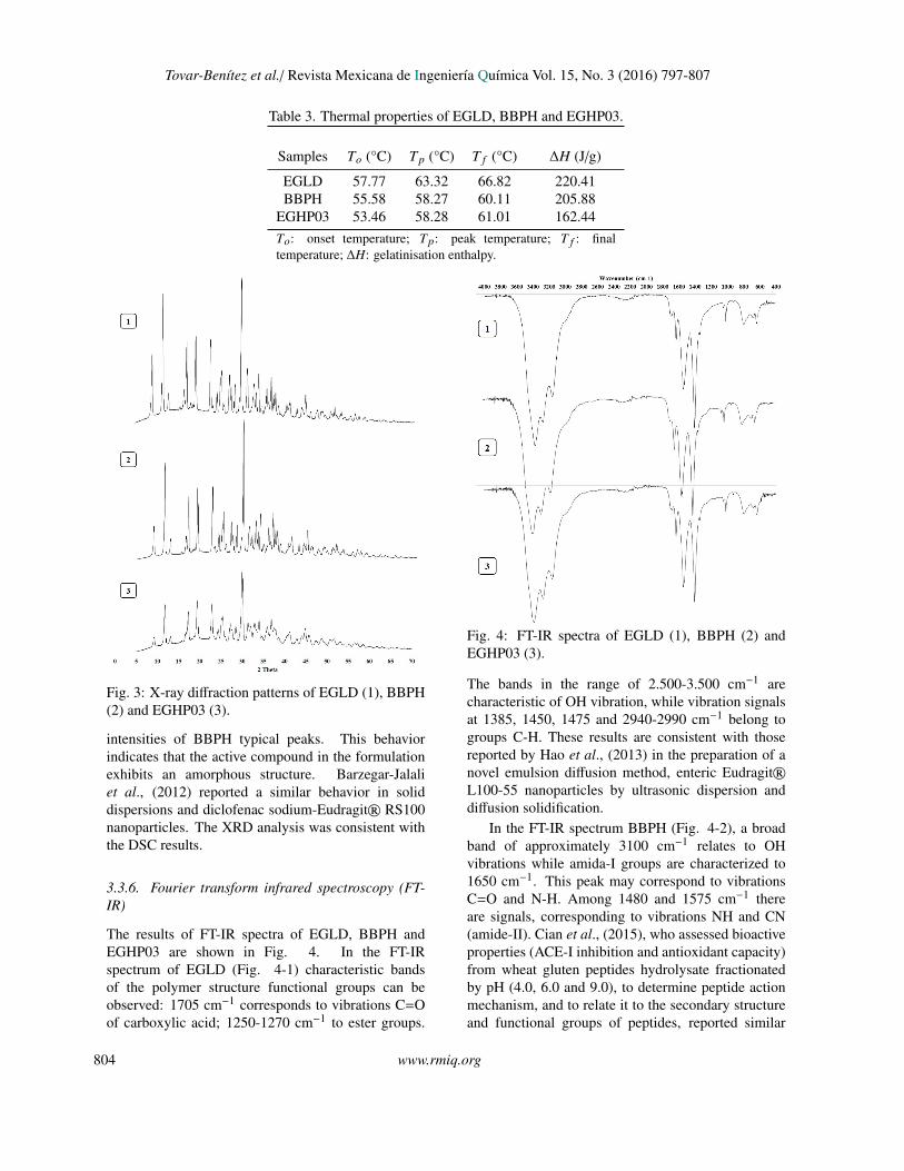
Tovar-Benıtez et al./ Revista Mexicana de Ingenierıa Quımica Vol. 15, No. 3 (2016) 797-807
Table 3. Thermal properties of EGLD, BBPH and EGHP03.
Samples To (°C) Tp (°C) T f (°C) ∆H (J/g)
EGLD 57.77 63.32 66.82 220.41BBPH 55.58 58.27 60.11 205.88
EGHP03 53.46 58.28 61.01 162.44To: onset temperature; Tp: peak temperature; T f : finaltemperature; ∆H: gelatinisation enthalpy.
Manuscrito sometido a la Revista Mexicana de Ingeniería Química 3
Figure 3
Fig. 3: X-ray diffraction patterns of EGLD (1), BBPH(2) and EGHP03 (3).
intensities of BBPH typical peaks. This behaviorindicates that the active compound in the formulationexhibits an amorphous structure. Barzegar-Jalaliet al., (2012) reported a similar behavior in soliddispersions and diclofenac sodium-Eudragit® RS100nanoparticles. The XRD analysis was consistent withthe DSC results.
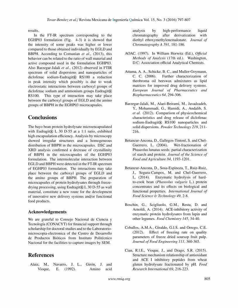
3.3.6. Fourier transform infrared spectroscopy (FT-IR)
The results of FT-IR spectra of EGLD, BBPH andEGHP03 are shown in Fig. 4. In the FT-IRspectrum of EGLD (Fig. 4-1) characteristic bandsof the polymer structure functional groups can beobserved: 1705 cm−1 corresponds to vibrations C=Oof carboxylic acid; 1250-1270 cm−1 to ester groups.
Manuscrito sometido a la Revista Mexicana de Ingeniería Química 4
Figure 4
Fig. 4: FT-IR spectra of EGLD (1), BBPH (2) andEGHP03 (3).
The bands in the range of 2.500-3.500 cm−1 arecharacteristic of OH vibration, while vibration signalsat 1385, 1450, 1475 and 2940-2990 cm−1 belong togroups C-H. These results are consistent with thosereported by Hao et al., (2013) in the preparation of anovel emulsion diffusion method, enteric Eudragit®L100-55 nanoparticles by ultrasonic dispersion anddiffusion solidification.
In the FT-IR spectrum BBPH (Fig. 4-2), a broadband of approximately 3100 cm−1 relates to OHvibrations while amida-I groups are characterized to1650 cm−1. This peak may correspond to vibrationsC=O and N-H. Among 1480 and 1575 cm−1 thereare signals, corresponding to vibrations NH and CN(amide-II). Cian et al., (2015), who assessed bioactiveproperties (ACE-I inhibition and antioxidant capacity)from wheat gluten peptides hydrolysate fractionatedby pH (4.0, 6.0 and 9.0), to determine peptide actionmechanism, and to relate it to the secondary structureand functional groups of peptides, reported similar
804 www.rmiq.org
Tovar-Benıtez et al./ Revista Mexicana de Ingenierıa Quımica Vol. 15, No. 3 (2016) 797-807
results.In the FT-IR spectrum corresponding to the
EGHP03 formulation (Fig. 4-3) it is showed thatthe intensity of some peaks was higher or lowercompared to those obtained individually by EGLD andBBPH. According to Comunian et al., (2013), thisbehavior can be related to the ratio of wall material andactive compound used in the formulation EGHP03.Also Barzegar-Jalali et al., (2012) observed in FTIRspectrum of solid dispersions and nanoparticles ofdiclofenac sodium-Eudragit® RS100 a reductionin peak intensity which possibly is due to weakelectrostatic interactions between carboxyl groups ofdiclofenac sodium and ammonium groups Eudragit®RS100. This type of interaction may take placebetween the carboxyl groups of EGLD and the aminogroups of BBPH in the EGHP03 microcapsules.
ConclusionsThe bayo bean protein hydrolysate microencapsulatedwith Eudragit® L 30 D-55 at a 1:1 ratio, exhibitedhigh encapsulation efficiency. Analysis by microscopyshowed irregular structures and a homogeneousdistribution of BBPH in the microcapsules. DSC andXRD analysis confirmed a decrease of crystallinityof BBPH in the microcapsules of the EGHP03formulation. The intermolecular interaction betweenEGLD and BBPH were detected in the FT-IR spectrumof EGHP03 formulation. The interactions may takeplace between the carboxyl groups of EGLD andthe amino groups of BBPH. The preparation ofmicrocapsules of protein hydrolysates through freeze-drying processing, using Eudragit® L 30 D-55 as wallmaterial, constitute a new route for the developmentof innovative new delivery systems and/or functionalfood products.
AcknowledgementsWe are grateful to Consejo Nacional de Ciencia yTecnologıa (CONACYT) for financial support throughscholarship for doctoral studies and to the Laboratorio-microscopia-electronica of the Centro de Desarrollode Productos Bioticos from Instituto PolitecnicoNacional for the facilities to capture images by SEM.
ReferencesAlaiz, M., Navarro, J. L., Giron, J. and
Vioque, E. (1992). Amino acid
analysis by high-performance liquidchromatography after derivatization withdiethyl ethoxymethylenemalonate. Journal ofChromatography A 591, 181-186.
AOAC. (1997). In William Horwitz (Ed.), OfficialMethods of Analysis (17th ed.). Washington,D.C: Association official Analytical Chemists.
Attama, A. A., Schicke, B. C., and Muller-Goymann,C. C. (2006). Further characterization oftheobroma oil beeswax admixtures as lipidmatrices for improved drug delivery systems.European Journal of Pharmaceutics andBiopharmaceutics 64, 294-306.
Barzegar-Jalali, M., Alaei-Beirami, M., Javadzadeh,Y., Mohammadi, G., Hamidi, A., Andalib, S.et al. (2012). Comparison of physicochemicalcharacteristics and drug release of diclofenacsodium-Eudragit® RS100 nanoparticles andsolid dispersions. Powder Technology 219, 211-216.
Betancur-Ancona, D., Gallegos-Tintore, S. and Chel-Guerrero, L. (2004). Wet-fractionation ofPhaseolus lunatus seeds: partial characterizationof starch and protein. Journal of the Science ofFood and Agriculture 84, 1193-1201.
Betancur-Ancona, D., Sosa-Espinoza, T., Ruiz-Ruiz,J., Segura-Campos, M. and Chel-Guerrero,L. (2014). Enzymatic hydrolysis of hard-to-cook bean (Phaseolus vulgaris L.) proteinconcentrates and its effects on biological andfunctional properties. International Journal ofFood Science & Technology 49, 2-8.
Boschin, G., Scigliuolo, G.M., Resta, D. andArnoldi, A. (2014). ACE-inhibitory activity ofenzymatic protein hydrolysates from lupin andother legumes. Food Chemistry 145, 34-40.
Ceballos, A.M.A., Giraldo, G.I.S. and Orrego, C.E.(2012). Effect of freezing rate on qualityparameters of freeze dried soursop fruit pulp.Journal of Food Engineering 111, 360-365.
Cian, R.l.E., Vioque, J., and Drago, S.R. (2015).Structure mechanism relationship of antioxidantand ACE I inhibitory peptides from wheatgluten hydrolysate fractionated by pH. FoodResearch International 69, 216-223.
www.rmiq.org 805
Tovar-Benıtez et al./ Revista Mexicana de Ingenierıa Quımica Vol. 15, No. 3 (2016) 797-807
Comunian, T.A., Thomazini, M., Alves, A.J.G.A.,de Matos Junior, F.E., de Carvalho Balieiro,J.l.C. and Favaro-Trindade, C.S. (2013).Microencapsulation of ascorbic acid bycomplex coacervation: Protection andcontrolled release. Food Research International52, 373-379.
El-Malah, Y. and Nazzal, S. (2008). Novel useof Eudragit® NE 30D/Eudragit® L 30 D-55blends as functional coating materials in time-delayed drug release applications. InternationalJournal of Pharmaceutics 357, 219-227.
Guzman-Mendez, B., Jaramillo-Flores, M.E., Chel-Guerrero, L. and Betancur-Ancona, D. (2014).Comparison of physicochemical properties,antioxidant and metal-chelating activitiesof protein hydrolysates from Phaseoluslunatus and hard-to-cook Phaseolus vulgaris.International Journal of Food Science &
Technology 49, 1859-1868.
Hao, S., Wang, B., Wang, Y., Zhu, L., Wang, B., andGuo, T. (2013). Preparation of Eudragit L 100-55 enteric nanoparticles by a novel emulsiondiffusion method. Colloids and Surfaces B:Biointerfaces 108, 127-133.
Hayakari, M., Kondo, Y. and Izumi, H. (1978).A rapid and simple spectrophotometric assayof angiotensin-converting enzyme. AnalyticalBiochemistry 84, 361-369.
Hernandez-Ledesma, B., Del Mar Contreras, M. andRecio, I. (2011). Antihypertensive peptides:Production, bioavailability and incorporationinto foods. Advances in Colloid and InterfaceScience 165, 23-35.
Hwang, J.S. and Ko, W.C. (2004). Angiotensin I-converting enzyme inhibitory activity of proteinhydrolysates from tuna broth. Journal FoodDrug Anal 12, 232-237.
Joshi, A.S., Patil, C.C., Shiralashetti, S.S., andKalyane, N.V. (2013). Design, characterizationand evaluation of Eudragit microspherescontaining glipizide. Drug Invention Today 5,229-234.
Megıas, C., Yust, M.d.M., Pedroche, J., Lquari,H., Giron-Calle, J., Alaiz, M. et al. (2004).Purification of an ACE Inhibitory peptide afterhydrolysis of sunflower (Helianthus annuus L.)
protein isolates. Journal of Agricultural andFood Chemistry 52, 1928-1932.
Molina Ortiz, S.E., Mauri, A., Monterrey-Quintero, E.S., Trindade, M.A., Santana,A.S., and Favaro-Trindade, C.S. (2009).Production and properties of casein hydrolysatemicroencapsulated by spray drying withsoybean protein isolate. LWT-Food Science andTechnology 42, 919-923.
Nesterenko, A., Alric, I., Silvestre, F. andDurrieu, V. (2013). Vegetable proteinsin microencapsulation: A review of recentinterventions and their effectiveness. IndustrialCrops and Products 42, 469-479.
Nielsen, P., Petersen, D. and Dammann, C. (2001).Improved method for determining food proteindegree of hydrolysis. Journal of Food Science66, 642-646.
Ondetti, M.A. and Cushman, D.W. (1982). Enzymesof the Renin-Angiotensin System and theirinhibitors. Annual Review of Biochemistry 51,283-308.
Pedroche, J., Yust, M.M., Giron-Calle, J., Alaiz, M.,Millan, F. and Vioque, J. (2002). Utilisationof chickpea protein isolates for production ofpeptides with angiotensin I-converting enzyme(ACE)-inhibitory activity. Journal of theScience of Food and Agriculture 82, 960-965.
Ray, S., Raychaudhuri, U. and Chakraborty, R.(2016). An overview of encapsulation of activecompounds used in food products by dryingtechnology. Food Bioscience 13, 76-83.
Regulska, K., Stanisz, B., Regulski, M. and Murias,M. (2014). How to design a potent, specific, andstable angiotensin-converting enzyme inhibitor.Drug Discovery Today 19, 1731-1743.
Ruiz, J.A.G., Ramos, M. and Recio, I.(2004). Angiotensin converting enzyme-inhibitory activity of peptides isolated fromManchego cheese. Stability under simulatedgastrointestinal digestion. International DairyJournal 14, 1075-1080.
Sansone, F., Picerno, P., Mencherini, T., Villecco,F., D´Ursi, A.M., Aquino, R.P. et al. (2011).Flavonoid microparticles by spray-drying:Influence of enhancers of the dissolution rate
806 www.rmiq.org
Tovar-Benıtez et al./ Revista Mexicana de Ingenierıa Quımica Vol. 15, No. 3 (2016) 797-807
on properties and stability. Journal of FoodEngineering 103, 188-196.
Segura-Campos, M.R., Chel-Guerrero, L.A. andBetancur-Ancona, D.A. (2010). Angiotensin-I converting enzyme inhibitory and antioxidantactivities of peptide fractions extracted byultrafiltration of cowpea Vigna unguiculatahydrolysates. Journal of the Science of Food andAgriculture 90, 2512-2518.
Torruco-Uco, J., Chel-Guerrero, L., Martınez-Ayala, A., Davila-Ortız, G. and Betancur-Ancona, D. (2009). Angiotensin-I convertingenzyme inhibitory and antioxidant activities ofprotein hydrolysates from Phaseolus lunatusand Phaseolus vulgaris seeds. LWT-FoodScience and Technology 42, 1597-1604.
Udenigwe, C.C. and Mohan, A. (2014). Mechanismsof food protein-derived antihypertensivepeptides other than ACE inhibition. Journalof Functional Foods 8, 45-52.
Valdez-Ortiz, A., Fuentes-Gutierrez, C.I., German-Baez, L.J., Gutierrez-Dorado, R. and Medina-Godoy, S. (2012). Protein hydrolysates
obtained from Azufrado (sulphur yellow)beans (Phaseolus vulgaris): Nutritional, ACE-inhibitory and antioxidative characterization.LWT - Food Science and Technology 46, 91-96.
Viveros-Contreras, R.; Tellez-Medina, D.I.; Perea-Flores, M.J.; Alamilla-Beltran, L.; Cornejo-Mazon, M.; Beristain-Guevara, C. I.; Azuara-Nieto, E.; Gutierrez-Lopez, G.F. (2013).Encapsulation of ascorbic acid into calciumalginate matrices through coacervation coupledto freeze-drying. Revista Mexicana deIngenierıa Quımica 12, 29-39
Xu, J., Li, W., Liu, Z., Li, J., Zhao, X., Li, D. etal. (2015). Preparation, characterization andpharmacokinetics evaluation of clarithromycin-loaded Eudragit® L-100 microspheres.European Journal of Drug Metabolism andPharmacokinetics, 1-7.
Yust, M. M., Pedroche, J., Giron-Calle, J., Vioque, J.,Millan, F. and Alaiz, M. (2004). Determinationof tryptophan by high-performance liquidchromatography of alkaline hydrolysates withspectrophotometric detection. Food Chemistry85, 317-320.
www.rmiq.org 807