
OET flashBAC Use Guide · 8.2 Test expression by infecting cells with high titre virus stocks 49...
90
baculoCOMPLETE A Complete Laboratory Guide to the Baculovirus Expression System and Insect Cell Culture User Guide 2017-18
Transcript of OET flashBAC Use Guide · 8.2 Test expression by infecting cells with high titre virus stocks 49...
baculoCOMPLETE
A Complete Laboratory Guide to the Baculovirus
Expression System and Insect Cell Culture
User Guide
2017-18

OET baculoCOMPLETE flashBAC™ & Insect Cell Culture User Guide 2017-18
2
This User Guide comprises two separate OET manuals
that have been combined into one convenient
document:
1 flashBACTM and BacPAK6 baculovirus
expression manual
2 Insect cell culture manual
OET baculoCOMPLETE flashBAC™ & Insect Cell Culture User Guide 2017-18
3
flashBAC™ and BacPAK6 Baculovirus
Expression Manual 2017-18
Contents
1.0 Limited Use License 5
2.0 Kit Contents 7
3.0 Essential Information and Technical Assistance 8
4.0 Safety Requirements 8
5.0 Products Ordering Information 9
6.0 Introduction to the Baculovirus Expression System 13
6.1 Baculoviruses 13
6.2 The baculovirus expression system 15
6.3 The BacPAK6 system 18
6.4 The flashBAC™ system 19
7.0 Making Recombinant Baculoviruses Using Either
BacPAK6 or flashBAC™ 25
7.1 Choice of transfer plasmid 25
7.2 Cotransfection of insect cells with BacPAK6/
flashBAC™ DNA and transfer plasmid 27
7.3 Plaque purification of recombinant BacPAK6 virus 32
7.4 Amplification of recombinant baculoviruses 39
7.5 Titration of recombinant virus by plaque assay 42
7.6 Using flashBACTM in 24 well plate format 48
8.0 Analysis and Optimisation of Gene Expression 49
8.1 Quick check for gene expression 49
8.2 Test expression by infecting cells with high titre
virus stocks 49
8.3 Optimisation of expression 51

OET baculoCOMPLETE flashBAC™ & Insect Cell Culture User Guide 2017-18
4
8.4 Scaling up production 51
9.0 Trouble Shooting and FAQ 52
10.0 References 55

OET baculoCOMPLETE flashBAC™ & Insect Cell Culture User Guide 2017-18
5
1. Limited Use Licence for flashBAC™ Virus DNA
I. In the License the following expressions shall have the following
meanings:
DNA shall mean deoxyribonucleic acid;
Fee shall mean the fee invoiced for the Materials by the
Licensor to the Licensee;
Licensee shall mean the purchaser of the Materials;
Licensor shall mean Oxford Expression Technologies Ltd;
Material shall mean the Licensor’s product known as
flashBAC™ comprising either or both an agreed
quantity of DNA and the relevant User Guide;
Purpose shall mean the use by the Licensee of the Materials
for the production of recombinant proteins and/or
viruses for Research purposes only;
Research shall mean the Licensor’s systematic search or
investigation towards increasing the sum of
knowledge in the production of recombinant proteins
and/or viruses;
User Guide shall mean the instructions provided with flashBAC™
to enable the Licensee to produce recombinant
proteins and/or viruses from the DNA.
II. The Licensor and the Licensee have agreed to enter into this Licence on
the following terms and conditions.
III. The Licensee acknowledges and accepts that by opening and/or using the
Materials it is agreeing to and accepting these terms and condition. If the
Licensee does not agree to these terms and conditions it must
immediately return all the Materials unused to the Licensor who shall
issue a refund for the fee.
IV. The Licensor has certain know-how and has developed a product that can
be used to produce recombinant proteins and/or viruses and has the
right to exploit the product under, inter alia, patent applications
numbered EP1144666, WO0112829 and AU6460800.

OET baculoCOMPLETE flashBAC™ & Insect Cell Culture User Guide 2017-18
6
V. This Licence shall commence on the date hereof and continue until the
DNA has been used or destroyed.
VI. The Licensor hereby grants to the Licensee and the Licensee hereby
accepts a limited, non-exclusive, non-transferable, licence to use the
Materials for the Purpose and as otherwise set out in this licence.
VII. The Licensee warrants to the Licensor that:
a) it shall only use the Materials for the purpose of Research; and
b) it shall not alter, reverse engineer, produce, manufacture or amplify
the DNA; and
c) it shall not sell any protein and/or virus created pursuant to this
Licence to any third party; and
d) it shall not provide any services to any third party using the
Materials; and
e) if the Licensee desires to the Materials for any purpose other than
the Purpose, it shall notify the Licensor accordingly and procure a
suitable licence prior to any such use.
VIII. The Licensee shall keep the DNA in accordance with the directions
contained in the User Guide.
IX. The Licensor shall raise an invoice to the Licensee for the Fee and the
Licensee agrees to pay the same to the Licensor within thirty (30) day of
receipt of the invoice (unless otherwise agreed in writing).
X. The Materials are provided as is and neither the Licensor nor any staff
acting on its behalf accepts any liability whatsoever for any of the
Materials or in connection with the Licensee’s possession, handling or use
of the Materials.
XI. The Licensee’s remedy pursuant to this Licence shall be limited at the
Licensor’s option to the replacement of the Materials or a refund of the
Fee paid by the Licensee.
XII. Ownership of the Materials shall pass to the Licensee upon dispatch of
the Materials by the Licensor to the Licensee.
XIII. The Licensee shall indemnify the Licensor for any loss suffered by the
Licensor as a result of the Licensee’s breach of this licence and/or third
party’s intellectual property rights.
XIV. This Licence is personal to the parties and shall not be assigned or
otherwise transferred in whole or in part by either party.

OET baculoCOMPLETE flashBAC™ & Insect Cell Culture User Guide 2017-18
7
XV. This Licence constitutes the entire agreement and understanding
between the parties in respect of the Materials and supersedes all
previous agreements, understandings and undertakings in this respect
and all obligations implied by the law to the extent that they conflict with
the express provisions of this Licence.
XVI. The invalidity, illegality or unenforceability of a provision of this Licence
shall not affect or impair the continuation in force of the remainder of
this Licence.
XVII. The Licensor reserves the right to revoke this permission and may require
the Licensee to return or destroy any remaining DNA and/or the User
Guide.
XVIII. Clauses 1, 3, 7, 9, 10, 13, 16, 18-20 shall survive any termination or expiry
of this Licence.
XIX. The interpretation construction and effect of this Licence shall be
governed and construed in all respects in accordance with the laws of
England and the parties hereby submit to the non-exclusive jurisdiction of
the English courts.
XX. The Contracts (Rights of Third Parties) Act 1999 shall have no application
to this Licence whatsoever and the parties do not intend hereunder to
benefit any third party.
End of Limited Use Licence.

OET baculoCOMPLETE flashBAC™ & Insect Cell Culture User Guide 2017-18
8
2. Kit Contents
All reagents and materials provided and referred to in this User Guide are
for Research Purposes only.
a) flashBAC™ DNA (or BacPAK6 DNA). Store at 4⁰C.
b) Control transfer plasmid DNA (containing lacZ reporter gene).
Store at -20⁰C. (flashBAC™ kits only)
c) Baculovirus Expression System User Guide. Download from
www.oetltd.com.
d) Certificate of Analysis. Download from www.oetltd.com.
e) MSDS. Download from www.oetltd.com.
Note
Transfection reagent and insect cells are NOT supplied as part of this kit
unless it is part of a baculoCOMPLETE kit.
3. Essential Information and Technical Assistance
The information given in this User Guide is accurate to the best of our
knowledge. It is a practical guide to allow researchers to use the
flashBAC™ (and BacPAK6) technology to produce recombinant
baculoviruses. It is not intended as a comprehensive guide to the
baculovirus expression system or insect cell culture. Those experienced
with the baculovirus expression system may find that they are already
familiar with much of the information provided.
Users are reminded that they may require other licences to use the
baculovirus expression system or types of insect cells and it is the
responsibility of the user to ascertain and act on this information.

OET baculoCOMPLETE flashBAC™ & Insect Cell Culture User Guide 2017-18
9
For additional help or guidance please refer to the Trouble Shooting
Section of this Guide and/or the Frequently Asked Questions (FAQ) section
of our website (www.oetltd.com). If these resources are unable to help,
please contact us at [email protected] and we will be pleased to help where
possible. All technical assistance provided is given in good faith; we cannot
take any responsibility whatsoever for any results you obtain by relying on
our assistance. We make no warranties of any kind with respect to
technical assistance or advice we provide.
4. Safety Requirements
These research products have not been approved for human or animal
diagnostic or therapeutic use.
Procedures described within this User Guide should only be carried out by
qualified persons trained in appropriate laboratory safety procedures.
Always use good laboratory practice when handling this product.
WARNING: SAFETY PRECAUTIONS MAY BE NECESSARY WHEN HANDLING
SOME OF THE REAGENTS DESCRIBED IN THIS USER GUIDE. PLEASE REFER
TO THE MATERIAL SAFETY DATA SHEETS SUPPLIED BY THE APPROPRIATE
MANUFACTURER.

OET baculoCOMPLETE flashBAC™ & Insect Cell Culture User Guide 2017-18
10
5. Products Ordering Information
flashBAC
TM Kits
Product Details Price Catalogue number
flashBAC™ 5 reactions £159.00 100150
flashBAC™ 24 reactions £685.00 100151
flashBAC™ 96 reactions £2,725.00 100152
flashBAC™ Bulk POA 100153
flashBAC GOLD 3 reactions £235.00 100200
flashBAC GOLD 5 reactions £369.00 100201
flashBAC GOLD 24 reactions £999.00 100202
flashBAC GOLD 96 reactions £3,250.00 100203
flashBAC GOLD Bulk POA 100204
flashBAC ULTRA 3 reactions £265.00 100304
flashBAC ULTRA 5 reactions £420.00 100300
flashBAC ULTRA 24 reactions £1,185.00 100301
flashBAC ULTRA 96 reactions £3,880.00 100302
flashBAC ULTRA Bulk POA 100303
flashBAC selection box 1 3 x 3 reactions £685.00 100400
flashBAC selection box 2 4 x 3 reactions £865.00 100401
flashBAC PRIME 5 reactions £369.00 100500
flashBAC PRIME 24 reactions £999.00 100501
flashBAC PRIME Bulk POA 100502
BacPAK6 Kits
BacPAK6 Linearised DNA 5 reactions £265.00 101101
BacPAK6 Linearised DNA 24 reactions £789.00 101102
BacPAK6 Linearised DNA 96 reactions £2,830.00 101103
BacPAK6 Sec+ Linearised DNA 5 reactions £265.00 101104
BacPAK6 Sec+ Linearised DNA 24 reactions £789.00 101105
BacPAK6 Sec+ Linearised DNA 96 reactions £2,830.00 101106

OET baculoCOMPLETE flashBAC™ & Insect Cell Culture User Guide 2017-18
11
Transfection Reagents
Product Size Price Cat. No.
baculoFECTIN II 150 µl £79.00 300105 baculoFECTIN II 1 ml £369.00 300106
Transfer Plasmids
Product Details Price Cat. No.
pOET1 (10 µg) Polyhedrin gene promoter with multiple cloning site (MCS)
£139.00 200101
pOET1N_6xHis (10 µg) Polyhedrin gene promoter, MCS with N-terminal 6xHis tag
£139.00 2001011
pOET1C_6xHis (10 µg) Polyhedrin gene promoter, MCS with C-terminal 6xHis tag
£139.00 2001012
pOET2 (10 µg) As pOET1 but with reversed MCS
£139.00 200103
pOET2N/C_6xHis (10 µg)
Polyhedrin gene promoter, MCS with N- and C-terminal 6xHis tags and thrombin cleavage site
£139.00 2001031
pOET2C_6xHis (10 µg) As pOET1C_6xHis but with reversed MCS £139.00
2001032
pOET3 (10 µg) P6.9 gene promoter for late phase expression £139.00
200104
pOET4 (10 µg) As pOET3 but with reversed MCS £139.00
200105
pOET5 (10 µg)
Dual expression with polyhedrin and p10 gene promoters £139.00
200106
pOET6 BacMAM (10 µg)
CMV promoter for BacMam – mediated transduction of mammalian cells
£139.00 200107
pOET1 Gateway™ (6µg)
pOET1 transfer plasmid with Gateway® technology
£310.00 200108
pOET6 BacMAM Gateway™ (6µg)
pOET6 transfer plasmid with Gateway® technology
£310.00 200109
pOET sequencing primers (2 x 100µl)
Can be used with any pOET transfer vector
£45.00 200100

OET baculoCOMPLETE flashBAC™ & Insect Cell Culture User Guide 2017-18
12
baculoQUANTTM
and baculoCOMPLETE Kits
Product Details Price Cat. No. baculoCOMPLETE protein expression kit 100 reactions £579.00 400100 baculoCOMPLETE protein expression kit + baculoQUANT all-in-one kit 5 + 100 reactions £940.00 400101 baculoQUANT All-in-one virus extraction & titration kit 100 reactions £385.00 100602
titrePLUS flashBAC all-in-one 5 + 100 reactions £489.00 100710
titrePLUS flashBAC all-in-one 24 + 100 reactions £960.00 100711
titrePLUS flashBAC GOLD all-in-one 5 + 100 reactions £675.00 100712
titrePLUS flashBAC GOLD all-in-one 24 + 100 reactions £1,245.00 100713
titrePLUS flashBAC ULTRA all-in-one 5 + 100 reactions £725.00 100714
titrePLUS flashBAC ULTRA all-in-one 24 + 100 reactions £1,410.00 100715
titrePLUS flashBAC PRIME all-in-one 5 + 100 reactions £675.00 100716
titrePLUS flashBAC PRIME all-in-one 24 + 100 reactions £1,245.00 100717
Insect Cell Lines
Product Frozen Live culture Price Medium Cat. No.
Sf9 cells ≥1 x 10⁷
cells per ampoule
On request £185.00 Serum-free 600100
Sf21 cells for plaque-assay
≥1 x 10⁷
cells On request £185.00 TC100 with
10% serum 600105
Super Sf91
≥1 x 10⁷
cells On request £579.00 Serum-free 600102
Super Sf92
≥1 x 10⁷
cells On request £579.00 Serum-free 600103
Super Sf93
≥1 x 10⁷
cells On request £579.00 Serum-free 600104

OET baculoCOMPLETE flashBAC™ & Insect Cell Culture User Guide 2017-18
13
Insect Cell Culture Media Product Size Shipping
conditions Price Medium Cat. No.
BaculoGROW II
500 ml Room temperature
£31.00 Serum-free 500200
ESF 921 insect cell media
1 L
Room temperature
£52.00
Serum-free and protein-free
500300
3 x 1 L £150.00 500301 5 x 1 L £240.00 500302 10 x 1 L £450.00 500304 Bulk POA 500305
ES custom
media
production
Min. 20L
Room temperature
POA Custom to order
500306
ES production boost additive
100 ml Room temperature
£48.00
Serum-free, nutrient additive
500307
ES transfection medium
20 ml Room temperature
£40.00 Serum and animal free
500308
ES transfection medium
100 ml Room temperature
£105.00 Serum and animal free
500309
ESF 921 delta series methionine deficient
1 L Room temperature
£55.00
Serum-free, methionine free
500310
ESF 921 delta
series all
amino acid
deficient
1 L Room temperature
£65.00 Serum and amino acid free
500311
ESF AF insect
cell media
1L
Room temperature
£58.00
Animal componentfree
500400
3 x 1L £165.00 500401
5 x 1L £255.00 500402
10 x 1L £440.00 500403
Bulk POA 500404
OET baculoCOMPLETE flashBAC™ & Insect Cell Culture User Guide 2017-18
14
6. Introduction to the Baculovirus Expression System and
flashBAC™/BacPAK6 Technology
6.1 Baculoviruses
Baculoviruses are insect viruses, predominantly infecting insect larvae of
the order Lepidoptera (butterflies and moths)1. A baculovirus expression
vector is a recombinant baculovirus that has been genetically modified to
contain a foreign gene of interest, which can then be expressed in insect
cells under control of a baculovirus gene promoter. The most commonly
used baculovirus for foreign gene expression is Autographa californica
nucleopolyhedrovirus (AcMNPV)2,3
. AcMNPV has a circular, double-
stranded, super-coiled DNA genome (133894 bp; Accession NC_001623)4,
packaged in a rod-shaped nucleocapsid. The nucleocapsid can be
extended lengthways and thus the DNA genome can accommodate quite
large insertions of DNA. The AcMNPV genome forms the basis of the
flashBAC™ or BacPAK6 DNA provided in this kit.
AcMNPV has a bi-phasic life cycle (Figure 1) resulting in the production of
two virus phenotypes: budded virus (BV) and occlusion-derived virus
(ODV). BV contain single, rod-shaped nucleocapsids enclosed by an
envelope, derived from the plasma membrane of insect cells, containing a
membrane-fusion protein GP64 (Figure 2 A). GP64 is acquired when the
nucleocapsids bud through the host cell plasma membrane5. The BV form
of the virus is 1000-fold more infectious for cultured insect cells6,
compared to the ODV phenotype, and is responsible for cell-cell
transmission in the early stages of infection7. It is the BV form of the virus
that delivers the foreign gene into the host insect cell for expression.

OET baculoCOMPLETE flashBAC™ & Insect Cell Culture User Guide 2017-18
15
Figure 1. A schematic
representation of the
bi-phasic life cycle of
baculoviruses resulting
in budded virus and
occlusion-derived virus.
P/PDV = polyhedra
with occlusion derived
virus
ECV = extracellular
virus = budded virus
In the later stages of the infection cycle large numbers of occlusion bodies
(OB) or polyhedra are formed inside the nuclei of cells (Figure 2 B & C).
These consist of multiple rod-shaped nucleocapsids enclosed within an
envelope, acquired de novo in the nucleus of cells, which then become
embedded within a para-crystalline matrix of the OB/polyhedra. The
major component of the OB matrix is comprised of a single protein –
polyhedrin (29 kDa)8,9
, which is produced by the powerful transcriptional
activity of the polyhedrin gene (polh) promoter13
. OBs protect the virus
and allow them to survive between hosts in the environment. Most
baculovirus expression vectors do not produce polyhedra (see below for
details), because the coding sequence for polyhedrin has been replaced by
that of the foreign gene being expressed under control of the polh

OET baculoCOMPLETE flashBAC™ & Insect Cell Culture User Guide 2017-18
16
promoter. This is a useful safety feature because recombinant virus
cannot persist in the environment in the absence of polyhedra.
Figure 2. (A) Rod-shaped baculovirus particle. (B) Section through a
polyhedron showing occlusion-derived virus particles embedded in a
matrix of polyhedrin protein. (C) Infected cell in culture showing polyhedral
in the enlarged nuclei.
6.2 The Baculovirus expression system
The baculovirus polyhedrin gene is non-essential for virus replication in
insect cells grown in culture and this has led to the development of the
widely-used baculovirus expression vector system, first described in 19833.
The coding sequence of the polyhedrin gene is replaced by the coding
region of the gene to be expressed, to produce a recombinant baculovirus
in which the powerful polyhedrin gene promoter drives expression of the
foreign gene. Recombinant baculoviruses produced in this way are
polyhedrin or polyhedral-negative viruses (Figure 3).
A B C

OET baculoCOMPLETE flashBAC™ & Insect Cell Culture User Guide 2017-18
17
Figure 3. SDS-PAGE analysis of cell
extracts from (1) non-infected insect
cells (2) wild-type virus infected cells
showing polyhedrin protein at 29kDa
and (3) recombinant virus infected cells
expressing lacZ (beta-galactosidase) –
note no polyhedrin protein is made.
Cells expressing beta-galactosidase are
also shown.
Expression of foreign genes in insect cells using recombinant baculoviruses
has become one of the most widely used eukaryotic expression systems.
The BEVS, as it is called, has several advantages over other expression
systems:
Safe to use – baculoviruses only infect insects and polyhedrin-
negative viruses cannot survive in the environment
OET baculoCOMPLETE flashBAC™ & Insect Cell Culture User Guide 2017-18
18
Can accommodate large genes or multiple genes – as the rod
shaped nucleocapsid can increase in length
Wide variety of promoters can be used – not just polyhedrin – to
control level of expression and or temporal aspects of expression
Proteins made are usually functional and are cleaved/processed
correctly
Can be used to transduce mammalian cells and achieve gene
expression by replacing polyhedrin gene promoter with a
mammalian-specific promoter
Insect cells are easy to grow and scale up at lower temperatures
than mammalian cells and without the need for CO2 incubators
However, the BEVS is not without its disadvantages and these lie mainly in
the labour intensive and technically demanding steps needed to produce
and isolate recombinant viruses and the fact that glycosylation differs from
mammalian cells; the latter often has no effect on function but is
important in considering therapeutic proteins.
The following section outlines the development of the BEVS over time and
the fine tuning that has been achieved to improve the system over the last
few years. Our focus is on the improvements made with the system we
call flashBAC™, which was developed to make it easier and quicker to
make recombinant viruses and to help achieve better expression with
‘difficult’ to express proteins.
Generally, the baculovirus genome is considered too large to insert genes
directly (although one commercial product BaculoDirectTM
achieves this).
Instead, foreign genes are cloned into a transfer plasmid, which contains
sequences that flank the polyhedrin gene in the virus genome. The virus
genome and transfer plasmid are simultaneously introduced into insect
cells (co-transfection) and homologous recombination, between the

OET baculoCOMPLETE flashBAC™ & Insect Cell Culture User Guide 2017-18
19
flanking sequences of the polh in the plasmid and genome, results in
exchange of DNA resulting in a recombinant baculovirus (Figure 3). The
virus genome then replicates and produces recombinant virus which can
be harvested as budded virus in the culture medium.
In most available BEVS using the homologous recombination method, this
results in a mixture of recombinant virus and recirculation of the parental
virus DNA to produce non-recombinant virus. These are separated by
plaque-purification to produce a stock of pure recombinant virus. Plaque-
purification is time consuming and technically demanding to the non-
virologist. Many developments have attempted to improve the method by
which recombinant and parental virus may be separated. The frequency of
recombinant efficacy in the BEVS is low, less than 1%, so recombinant virus
plaques can often be obscured by parental virus plaques. This problem
was partially addressed by inserting a copy of the lacZ gene into the virus
genome so that recombinant virus plaques would stain blue after the
addition of X-gal11
. However, this did not address the fact that only 1% of
plaques went blue and also resulted in contamination of the expressed
protein with beta-galactosidase.
6.3 The BacPAK6 system
The efficiency with which recombinant viruses could be recovered was
improved by the addition of a unique restriction enzyme site (Bsu36I) at
the polh locus. Linearization of the virus genome prior to homologous
recombination reduced the infectivity of the parental virus DNA;
recombinant virus genomes become circular and can replicate. This
resulted in the recovery of about 30% recombinant virus. LacZ was then
introduced into the parental virus genome to replace the polh coding
sequence, resulting in 3 Bsu36I sites at the polh locus12
. Triple digestion of
the resulting virus genome with Bsu36I removed a section of virus DNA
coding for lacZ and part of the essential gene ORF 162912
, resulting in a

OET baculoCOMPLETE flashBAC™ & Insect Cell Culture User Guide 2017-18
20
linear virus DNA (BacPAK6) that cannot replicate in insect cells. Co-
transfection of insect cells with linearised BacPAK6 DNA and a transfer
plasmid with foreign gene under control of polh, creates recombinant virus
DNA in which ORF1629 is restored and the recircularised DNA can replicate
to produce recombinant budded virus12
. This reduced even further the
chance of parental virus replicating and resulted in an increase in the
recovery of recombinant virus to more than 90%*. It also introduced a
useful blue-white selection system – with non-recombinant virus giving rise
to blue plaques and recombinant virus to white plaques. It was thus easier
to achieve purified virus with a single round of plaque-purification. *It is
not 100% because it is impossible to ensure that every molecule of DNA is
triple-digested and any circular DNA remaining can replicate and produce
non-recombinant virus.
NOTE
The triple-cut linear BacPAK6 virus DNA is available from OET (see page
10). We are also pleased to offer BacPAK6-Sec+, which has a deletion in
the chitinase gene to aid expression of membrane targeted and secreted
proteins. Practical techniques to make recombinant BacPAK6 viruses are
included in this User Guide.
Despite this fine tuning and optimisation of the system, a number of steps
are still required to make recombinant baculoviruses, thus making it more
time consuming than bacterial expression systems and less amenable to
scale up and high throughput automation.
6.4 The flashBAC™ system
The flashBACTM
system is a new platform technology for the production
and isolation of recombinant baculoviruses. Importantly, flashBACTM
has
been designed to remove the need for separation of recombinant virus
from parental virus, so no plaque-purification steps are needed. The
production of recombinant virus has been simplified to a single stage

OET baculoCOMPLETE flashBAC™ & Insect Cell Culture User Guide 2017-18
21
procedure that is fully amenable to high throughput manipulations –
multiple recombinant viruses can be made at one time using 24 well plates
either manually or using simple robotic systems.
The flashBACTM
technology builds on the BacPAK6 technology. At the
heart of the new system is an AcMNPV genome that lacks part of the
essential gene ORF 1629 and contains a bacterial artificial chromosome
(BAC) at the polh locus, replacing the polh coding sequence. The essential
gene deletion prevents virus replication in insect cells and the BAC allows
the virus genome to be maintained in bacterial cells as a bacmid. Circular
virus DNA is isolated from bacterial cells and purified ready for use in
flashBACTM
kits and co-transfections to make recombinant viruses.
A recombinant baculovirus is produced simply by co-transfecting insect
cells with flashBACTM
DNA and a transfer plasmid containing the gene to be
expressed (Figure 4). Homologous recombination within the insect cells
(1) restores ORF 1629 allowing the recombinant virus to replicate (2)
removes the BAC sequences and (3) inserts the foreign gene under control
of the polh promoter (or other promoter chosen that is in the transfer
plasmid).
The recombinant virus budded virus is harvested from the co-transfection
medium and becomes the seed stock (P0) of recombinant virus. No
selection systems are needed. However, the virus stock is not
homogeneous in the way plaque-purified virus is and for very large scale
applications or for work that may be taken through regulatory processes,
we recommend a single round of plaque-purification. For most purposes,
however, plaque-purification is not necessary.
This single step procedure greatly facilitates the high throughput
production of baculovirus expression vectors via automated systems

OET baculoCOMPLETE flashBAC™ & Insect Cell Culture User Guide 2017-18
22
(Figure 5). However, it is just as useful for a research lab making one or
two viruses in individual dishes. It is very useful for the novice.
The flashBACTM
system is back-compatible with all transfer plasmids based
on homologous recombination at the polh locus. The OET website has
details of most of these and they include single, dual, triple and quadruple
expression plasmids, those with purification tags at N and C termini, and
other promoters including p10, p6.9, ie-1 and, CMV (for mammalian cells).
It is not compatible with pFASTBacTM
vectors and the Bac-toBac® system14
.
Since the launch of the original flashBACTM
DNA, we have made further
modifications to help express difficult to express proteins and the different
flashBACTM
variants are now described:
flashBAC™ Backbone virus DNA has a chiA deletion, which prevents
production of virus chitinase. This enzyme blocks the
secretory pathway and its absence helps improve
membrane and secreted protein production15-18
.
flashBAC
GOLD
Backbone virus DNA has gene deletions for chiA and v-
cath19
. This avoids production of chitinase and cathepsin, a
viral protease that may otherwise degrade susceptible
target proteins. See Figure 6.
flashBAC
ULTRA
Backbone virus DNA has deletions of chiA, v-cath and
p10/p26/p74. Deletion of p10 results in delayed cell lysis
(particularly noticeable) in TnHi5 cells and thus can extend
protein production times. It also reduces the metabolic
burden on the cell of producing high levels of P10 protein.
flashBAC
PRIME
No gene deletions in the virus back bone. Useful if the
proteins being expressed form complexes inside the
cytoplasm or nucleus that need to be purified. We find that
the relatively early cell lysis associated with PRIME makes it
easier to purify these complexes e.g. VLPs.

OET baculoCOMPLETE flashBAC™ & Insect Cell Culture User Guide 2017-18
23
Advantages of the flashBACTM
system:
Simple to use
One step procedure that does not require plaque-purification
(Figure 4)
Amenable to making many viruses simultaneously – manually or
using a robot in 24 well plates (Figure 4, 5)
Amenable to high throughput systems
Maximise secreted or membrane targeted proteins (Figure 6)
Maximise difficult protein production
Maximise VLP production and release from cells
Back compatible with a large range of transfer plasmids
Now compatible with Gateway® cloning system

OET baculoCOMPLETE flashBAC™ & Insect Cell Culture User Guide 2017-18
24
Figure 4. Overview of how the flashBACTM
system works in practice. The
co-transfection mix comprises flashBACTM
DNA, transfer plasmid with gene
to be expressed and transfection reagent.
Up to 24 viruses can be made at one time using a 24 well plate dish either
manually or using a simple liquid handing robotic platform.

OET baculoCOMPLETE flashBAC™ & Insect Cell Culture User Guide 2017-18
25
Figure 5. Production and analysis of a number of secreted recombinant
proteins using flashBAC viruses (P1 stock) in Sf9 cells and probed with anti-
His antisera. Thanks to Dr Ray Owens Oxford Protein Production Facility
for beta-testing flashBAC.

OET baculoCOMPLETE flashBAC™ & Insect Cell Culture User Guide 2017-18
26
Figure 6. Expression of secreted proteins 1-6 using flashBAC (FB) or
flashBAC GOLD (FBG). Western blots probed with anti-His antisera are
shown as are densitometry results to semi-quantify expression levels. In
most cases FBG improves secretion levels of proteins.
OET baculoCOMPLETE flashBAC™ & Insect Cell Culture User Guide 2017-18
27
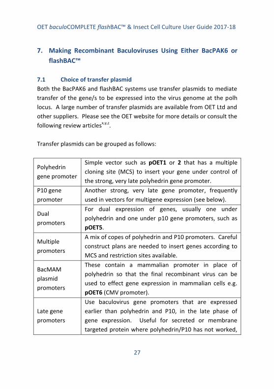
7. Making Recombinant Baculoviruses Using Either BacPAK6 or
flashBAC™
7.1 Choice of transfer plasmid
Both the BacPAK6 and flashBAC systems use transfer plasmids to mediate
transfer of the gene/s to be expressed into the virus genome at the polh
locus. A large number of transfer plasmids are available from OET Ltd and
other suppliers. Please see the OET website for more details or consult the
following review articlesx,y,z
.
Transfer plasmids can be grouped as follows:
Polyhedrin
gene promoter
Simple vector such as pOET1 or 2 that has a multiple
cloning site (MCS) to insert your gene under control of
the strong, very late polyhedrin gene promoter.
P10 gene
promoter
Another strong, very late gene promoter, frequently
used in vectors for multigene expression (see below).
Dual
promoters
For dual expression of genes, usually one under
polyhedrin and one under p10 gene promoters, such as
pOET5.
Multiple
promoters
A mix of copes of polyhedrin and P10 promoters. Careful
construct plans are needed to insert genes according to
MCS and restriction sites available.
BacMAM
plasmid
promoters
These contain a mammalian promoter in place of
polyhedrin so that the final recombinant virus can be
used to effect gene expression in mammalian cells e.g.
pOET6 (CMV promoter).
Late gene
promoters
Use baculovirus gene promoters that are expressed
earlier than polyhedrin and P10, in the late phase of
gene expression. Useful for secreted or membrane
targeted protein where polyhedrin/P10 has not worked,
OET baculoCOMPLETE flashBAC™ & Insect Cell Culture User Guide 2017-18
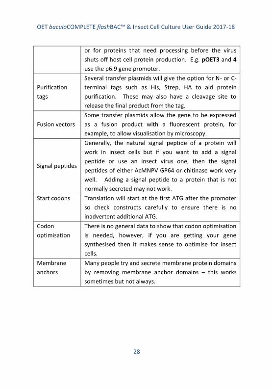
28
or for proteins that need processing before the virus
shuts off host cell protein production. E.g. pOET3 and 4
use the p6.9 gene promoter.
Purification
tags
Several transfer plasmids will give the option for N- or C-
terminal tags such as His, Strep, HA to aid protein
purification. These may also have a cleavage site to
release the final product from the tag.
Fusion vectors
Some transfer plasmids allow the gene to be expressed
as a fusion product with a fluorescent protein, for
example, to allow visualisation by microscopy.
Signal peptides
Generally, the natural signal peptide of a protein will
work in insect cells but if you want to add a signal
peptide or use an insect virus one, then the signal
peptides of either AcMNPV GP64 or chitinase work very
well. Adding a signal peptide to a protein that is not
normally secreted may not work.
Start codons Translation will start at the first ATG after the promoter
so check constructs carefully to ensure there is no
inadvertent additional ATG.
Codon
optimisation
There is no general data to show that codon optimisation
is needed, however, if you are getting your gene
synthesised then it makes sense to optimise for insect
cells.
Membrane
anchors
Many people try and secrete membrane protein domains
by removing membrane anchor domains – this works
sometimes but not always.

OET baculoCOMPLETE flashBAC™ & Insect Cell Culture User Guide 2017-18
29
When cloning genes into transfer plasmids note:
Check the gene is in the correct orientation with respect to the
promoter
Check that the first ATG after the promoter is the start codon you
want to initiate translation in the mRNA
Check you have a stop codon
Check that any fusion or purification tags are in frame and after
any signal peptide sequence (that will be cleaved off)
Sequence any gene that has been cloned via PCR or gene
synthesis. Check cloning junctions of genes cloned in using RE
digestion and ligation.
Ensure final plasmid is sterile as it will be used to transfect insect
cells – you don’t want your cells getting bugged
Mini-prep type DNA works OK in transfections
Contact us on [email protected] if you need advice or help with transfer
plasmids.
7.2 Co-transfection of insect cells with BacPAK6 or flashBAC™ DNA
and a transfer plasmid to make a seed stock (P0) of recombinant
baculovirus
This method uses cells prepared in individual 35mm cell culture dishes or
12 well plates. Protocol 7.6 provides an adaptation of this method for
making multiple viruses using 24 well plates. This method must be carried
out using aseptic technique as the DNA complexes will be introduced in
insect cells in the absence of antibiotics. Read through the whole protocol
before starting to check you have all the reagents and equipment needed.
Check safety advice and MSDS data sheets where appropriate. We
recommend wearing PPE such as lab coats and gloves at all times.

OET baculoCOMPLETE flashBAC™ & Insect Cell Culture User Guide 2017-18
30
Provided in the kit:
flashBAC™ DNA (any type) or BacPAK6 DNA (use 100 ng [5µl] DNA
per co-transfection)
Positive control transfer plasmid DNA (expressing lacZ) (use 500
ng [5µl] per co-transfection) [flashBAC™ kit only]
Also needed:
12 well plate or 35mm tissue culture dish seeded with a sub-
confluent monolayer of Sf21 or Sf9 cells – one dish/well for each
co-transfection and one for a control
NOTE
(See OET’s Cell Culture Manual for details on insect cell culture; it is vital
for transfection success that cells used are taken from a culture that is in
log phase growth – virus can only replicate when cells are in log phase! A
sub-confluent monolayer is one in which there are spaces around each
cell so there is room for each cell to divide in the 24 hours after co-
transfection)
Serum-free insect cell culture medium (we recommend using
TC100 medium as a transfection medium but most serum-free
medium will also work)
Growth medium (serum-free or TC100 with 10% serum, as
preferred)
Sterile transfer plasmid DNA containing gene to be expressed (see
7.1 for details) (500 ng per transfection)
Transfection reagent such as OET’s baculoFECTIN II or flashFECTIN
(volume as indicated by the manufacturer)
Incubator set at 28⁰C
1% Virkon (Amtec) or other suitable disinfectant
Inverted phase contrast microscope

OET baculoCOMPLETE flashBAC™ & Insect Cell Culture User Guide 2017-18
31
Plastic box to house dishes in the incubator
Sterile pipettes and bijoux or other polystyrene containers to
make up the transfection mix; do not use micro-centrifuge tubes
made of polypropylene.
Method:
1. For each co-transfection you require one 35mm dish, or one well
of a 12 well plate, containing sub-confluent Sf9 or Sf21 cells. If you
are making a virus with the control vector provided in the
flashBAC™ kit, add an extra dish/well of cells. It is also good
practice to have one dish/well for a mock-transfection in which no
DNA is added.
NOTE
Do not use TniHi5 cells to make viruses as they are prone to mutations
that affect gene expression.
Seed the dishes/wells with cells at least one hour before use to
allow cells to attach and recover. Ensure cells were taken from a
log-phase culture of cells that were at least 90% viable. As a
rough guide you need about 1.5 x 10⁶
Sf21 or 1 x 10⁶ Sf9 cells per
35mm dish to form a sub-confluent monolayer. For 12-well plates,
add 0.4 x 10⁶ Sf9/Sf21 cells per well. The volume of medium
should be 2 ml in 35mm dishes and 1 ml in 12-well plates. Ensure
cells are evenly distributed over the surface of the dish/well.
2. During the 1 hour incubation period above, prepare the co-
transfection mix of DNA and transfection reagent. For each co-
transfection in either a 35mm dish or well of a 12-well plate, you
need to mix in a polystyrene tube, in the following order:
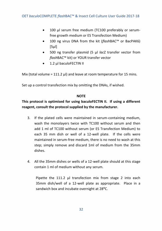
OET baculoCOMPLETE flashBAC™ & Insect Cell Culture User Guide 2017-18
32
100 µl serum free medium (TC100 preferably or serum-
free growth medium or ES Transfection Medium)
100 ng virus DNA from the kit (flashBAC™ or BacPAK6)
[5µl]
500 ng transfer plasmid (5 µl lacZ transfer vector from
flashBAC™ kit) or YOUR transfer vector
1.2 µl baculoFECTIN II
Mix (total volume = 111.2 µl) and leave at room temperature for 15 mins.
Set up a control transfection mix by omitting the DNAs, if wished.
NOTE
This protocol is optimised for using baculoFECTIN II. If using a different
reagent, consult the protocol supplied by the manufacturer.
3. If the plated cells were maintained in serum-containing medium,
wash the monolayers twice with TC100 without serum and then
add 1 ml of TC100 without serum (or ES Transfection Medium) to
each 35 mm dish or well of a 12-well plate. If the cells were
maintained in serum-free medium, there is no need to wash at this
step; simply remove and discard 1ml of medium from the 35mm
dishes.
4. All the 35mm dishes or wells of a 12-well plate should at this stage
contain 1 ml of medium without any serum.
Pipette the 111.2 µl transfection mix from stage 2 into each
35mm dish/well of a 12-well plate as appropriate. Place in a
sandwich box and incubate overnight at 28⁰C.

OET baculoCOMPLETE flashBAC™ & Insect Cell Culture User Guide 2017-18
33
5. After overnight incubation, add one extra ml of growth medium to
the 35mm dishes or replace the 1 ml medium in the 12-well plates
with 1 ml growth medium*. Continue the incubation for 4 more
days (5 days in total).
*Growth medium may either be serum-free medium or TC100 with
10% serum.
NOTE
Cells in which virus has replicated appear different from mock-
transfected cells so comparing mock-transfected cells with experimental
dishes can be a useful indicator that the transfection has worked;
infected cells appear more grainy with swollen nuclei.
6. Harvest the culture medium containing budded recombinant virus
into a sterile container and store in the dark at 4⁰C.
NOTE
If you prepared a control virus with the lacZ transfer plasmid in the
flashBAC™ kit, you can check for transfection success by staining the
monolayer of cells left after harvesting the P0 virus; add 1 ml of growth
medium containing 15µl 2% v/v X-gal to the cell monolayer and leave for
a few hours to overnight for the blue colour to develop.
7. The next step depends on whether you have used BacPAK6 or
flashBAC™ DNA.
flashBAC™ DNA: Your 1-2 ml stock of virus is your seed stock (P0),
you now need to amplify this to obtain a 50 ml P1 stock of virus
for experimental work and freezing down (go to 7.4).
BacPAK6 DNA: You now need to plaque-purify your recombinant
virus to obtain your seed stock (P0) (go to 7.3)

OET baculoCOMPLETE flashBAC™ & Insect Cell Culture User Guide 2017-18
34
NOTE
You can also plaque-purify virus produced using flashBAC™ DNA if
required (go to 7.3)
7.3 Plaque-purification of recombinant BacPAK6 virus
The budded virus harvested after the co-transfection with BacPAK6 virus
DNA will contain a mixture of parental virus (about 10% blue) and
recombinant virus (about 90% clear/colourless). These need to be
separated by performing a plaque-assay and picking individual plaques to
amplify pure virus stocks. As long as well isolated plaques are picked, only
one round of plaque-purification is needed.
This is a multi-step method that enables you to isolate plaques and then
amplify plaque-picked virus to produce a P0 seed stock of virus. Read
through the whole method before starting to ensure you are aware of time
scales and reagents/equipment needed at each stage.
The OET Cell Culture Manual has details of insect cell culture.
Required:
Virus harvested from a co-transfection (see 7.2)
TC100 growth medium with serum (best; or serum free growth
medium) - antibiotics (Penicillin, 10000 units/ml and
Streptomycin, 10000 µg/ml in 0.85% saline; dilute 1:50 for use)
may be added to plaque-assay medium to help reduce the chance
of contamination
Culture of Sf21 cells (preferred; or Sf9 cells) that are in log phase
of growth and at least 90% viable
35 mm dishes and T25 flasks
Low temperature gelling (Sea-plaque) agarose for overlay (Sigma;
2% w/v solution in d.water). It is convenient to make up small

OET baculoCOMPLETE flashBAC™ & Insect Cell Culture User Guide 2017-18
35
batches (15 ml) of agarose overlay by melting the agarose using a
boiling water bath or microwave oven (take care). Solidified
agarose can be stored and re-melted prior to use. (Larger
volumes may also be prepared and melted multiple times). Cool
to ‘hand hot’ before making up final overlay.
Sterile pipettes and bijoux or similar containers for making virus
dilutions
Sterile Pasteur pipettes
Beaker with hand hot water as a temporary water bath
Plastic box
Incubator at 28⁰C
Phosphate-buffered saline (PBS)
Neutral Red stain (Sigma; 5 mg/ml in d.water, filter sterilize and
store at room temperature). For use dilute 1 in 20 with PBS
solution. Do not store diluted stain.
X-gal (2% v/v in DMF) to stain for blue plaques
1% Virkon (Amtec) or similar disinfectant
Inverted phase contrast light microscope
Lightbox to view plaques
Method:
1. Seed 35mm cell culture dishes with a sub-confluent monolayer of
healthy Sf21 cells (or Sf9 cells if Sf21 are not available). See Cell
Culture Manual for more details. Allow the cells to settle for at
least one hour.
You need 12 dishes per virus.
NOTE
Sf21 cells in TC100 with 10% serum give the largest easy-to-spot plaques
because these cells have a well-defined CPE (see Figure 7). Sf9 cells will

OET baculoCOMPLETE flashBAC™ & Insect Cell Culture User Guide 2017-18
36
also yield plaques but they are smaller, take longer to develop and are
not quite so easy to define. We have also noted that plaque assays
conducted with Sf9 cells and serum free medium produce plaques that
quickly fade after staining with Neutral Red.
2. Make 1 in 10 dilutions of your transfection virus stock from 1 in 10
(10-1
) to 1 in 106 (10
-6). Use 50 µl virus and 450 µl growth medium
as diluent at each step. Mix the virus and diluent between each
step and change tip/pipette each time to avoid carry-over.
3. Remove the medium from the dishes of cells using aseptic
technique and add 100 µl of diluted virus drop wise to the centre
of each dish. Plate a range of dilutions and two plates per dilution
– the aim is to get well isolated plaques on at least one dilution.
We normally plate the 1 in 100 (10-2
) to 10-6
dilutions in duplicate
dishes and use two dishes as mock infected controls (use medium
only).
NOTE
It is important that the cell monolayers do not dry out during this process
of virus inoculation. Do not leave lids off dishes for long periods. If
working in a class 2 hood be aware the air flow can dry plates very
quickly. If, after staining, your monolayer appears with a shiny red patch
devoid of cells, you have allowed the monolayer to dry out.
4. Allow the virus to adsorb and be taken up into the cells at room
temperature for 45 - 60 min. Rock the dishes every few minutes
to ensure even coverage of the inoculum. Do NOT put the cells in
the incubator as they will dry out.
5. During this time prepare the overlay. Dissolve agarose in water to
2% w/v by boiling (water bath or microwave oven – take

OET baculoCOMPLETE flashBAC™ & Insect Cell Culture User Guide 2017-18
37
appropriate safety precautions). You need 1 ml per dish of cells.
Cool the overlay to hand hot (about 50-55⁰C) and add an equal
volume of pre-warmed growth medium (28⁰C). Keep warm to
prevent setting (we use a temporary clean water bath comprising
a beaker of hot tap water). You need 2 ml final overlay per dish.
NOTE
If the agarose in water sets, it is easy to melt again by boiling. If the
agarose overlay with growth medium sets, you cannot re-melt. You have
to start again. We often prepare several small batches of agarose in
water and let them set and then melt each aliquot as we need it (15 ml is
convenient).
6. At the end of the incubation period (4), remove the inoculum
using a pipette and discard into Virkon or other disinfectant.
Working quickly, add 2 ml warm overlay to each dish allowing the
agarose to flow down the side of the dish and spread slowly over
the monolayer of cells. Do NOT pipette into the centre of the
dish.
NOTE
Process one set of dishes per virus sample at a time. If working in a hood,
keep the agarose overlay in a beaker or sandwich box filled with warm
water to delay solidification. If the agarose sets prematurely, you can
leave the dishes with virus inoculums for longer than 60 min without
adverse effects. If you have removed the virus and then find that your
overlay medium has set, just add 0.1-0.2ml fresh medium to each plate
to prevent drying of cells. Prepare more agarose overlay medium and
carry on, but don’t forget to remove the extra medium you added to each
dish!

OET baculoCOMPLETE flashBAC™ & Insect Cell Culture User Guide 2017-18
38
7. Allow the agarose overlay to set at room temperature. Then add
a 1 ml liquid overlay of growth medium to feed the cells and
prevent them from drying out.
8. Place the dishes in a plastic box and incubate at 28⁰C for 3-4 days.
Three days for Sf21 and four days for Sf9 cells.
9. Add 1 ml growth medium containing 15 µl 2% v/v X-gal in DMF to
each dish to stain for blue (parental) plaques. Incubate at 28⁰C
for 5-6 h.
Conveniently, this is done in the course of a normal working day.
Blue plaques should start to develop during this time.
10. Prepare the Neutral Red stain in water to 5 mg/ml d.water and
filter sterilize or purchase ready made from Sigma, for example.
Dilute 1 in 20 with sterile PBS for use.
NOTE
Different batches of Neutral Red may differ in their efficacy. Sometimes
1 in 40 dilutions give better results. Do not store diluted stain, it will
form a precipitate. The concentrated stock is stable at room temperature
for several months (if sterile).
11. Add 1 ml diluted neutral red stain to each dish. Do not remove
the X-gal already added. Incubate at 28⁰C for 16 hours
(overnight).
12. Decant all liquid and view plaques on a light box. Recombinant
virus plaques will appear clear in a sea of red healthy cells.
Parental, non-recombinant plaques will stain blue with X-gal.

OET baculoCOMPLETE flashBAC™ & Insect Cell Culture User Guide 2017-18
39
13. You need to pick 3-6 plaques for each virus. Select well isolated
plaques from a dish where there are no blue plaques (see Figure
7).
NOTE
If the dilutions were unsuitable (i.e. too few or to many plaques per
dish), you may have to redo the plaque assay adjusting the dilutions to
obtain dishes with well isolated plaques and no blue plaques.
With experience you can cut down the range of dilutions plated once you
know the general titre of virus that you obtain from a co-transfection.
We recommend starting with a wide range, as transfection efficiency
varies considerably.
14. To pick a plaque, you need to take up a plug of agarose from the
centre of a plaque using a Pasteur pipette or Gilson tip. Disperse
the plug of agarose into 500 µl growth medium in a micro
centrifuge tube and vortex to release the virus from the agarose
into the medium. Store in the dark at 4⁰C.
15. Amplify the plaque-picked virus by inoculating either a 35mm dish
or a T25 flask of Sf21 or Sf9 cells using 100 µl (35 mm dish) or 250
µl (T25 flask) of your 500 µl as inoculum.
Figure 7. Plaque-
assay in Sf21 cells
stained with Neutral
red to show well
isolated plaques (a),
crowded plaques (b)
and merged plaques
(c).

OET baculoCOMPLETE flashBAC™ & Insect Cell Culture User Guide 2017-18
40
To do this, seed a 35 mm dish or T25 flask with cells to form a sub-
confluent monolayer and after an hour or so, remove the medium
and replace with the inoculum for 45-60 mins. Then add 2 ml
(dish) or 5 ml (T25 flask) growth medium (no need to remove the
inoculum) and incubate for 4-5 day at 28⁰C.
The cells should be well infected under the microscope at the end
of the infection period.
16. Harvest the 2 ml or 5 ml of medium containing your P0 seed stock
virus. Store at 4⁰C in the dark. Use this to amplify a P1 working
stock of recombinant virus to test gene expression (see 7.4).
17. The cell monolayers from the dish or flask used to amplify virus
can be harvested and used to test for gene expression or to
isolate DNA to do a PCR to check that the gene has gone into the
virus genome.
7.4 Amplification of recombinant baculoviruses to produce high titre
stocks
This is a generic method to amplify recombinant baculovirus from P0 to P1,
or P1 to P2 etc. We do not recommend serial passage of the virus stock
because mutations can and do arise. These can sometimes lead to
reduced expression levels or loss of the gene. Good practice is to amplify
a 50 to 200 ml P1 stock for initial test of gene expression and optimisation
of expression. Some of this virus should also be frozen down at -80⁰C for
long term storage. Do not store virus at -20⁰C. Virus can be stored in the
dark at 4⁰C for a few months but in the absence of serum, the titre can
start to drop after a few weeks. We recommend adding serum to 5% for
all viruses stored at 4⁰C. If you cannot do this, then freeze aliquots of P1
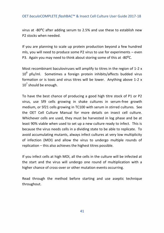
OET baculoCOMPLETE flashBAC™ & Insect Cell Culture User Guide 2017-18
41
virus at -80⁰C after adding serum to 2.5% and use these to establish new
P2 stocks when needed.
If you are planning to scale up protein production beyond a few hundred
mls, you will need to produce some P2 virus to use for experiments – even
P3. Again you may need to think about storing some of this at -80⁰C.
Most recombinant baculoviruses will amplify to titres in the region of 1-2 x
108 pfu/ml. Sometimes a foreign protein inhibits/affects budded virus
formation or is toxic and virus titres will be lower. Anything above 1-2 x
107 should be enough.
To have the best chance of producing a good high titre stock of P1 or P2
virus, use Sf9 cells growing in shake cultures in serum-free growth
medium, or Sf21 cells growing in TC100 with serum in stirred cultures. See
the OET Cell Culture Manual for more details on insect cell culture.
Whichever cells are used, they must be harvested in log phase and be at
least 90% viable when used to set up a new culture ready to infect. This is
because the virus needs cells in a dividing state to be able to replicate. To
avoid accumulating mutants, always infect cultures at very low multiplicity
of infection (MOI) and allow the virus to undergo multiple rounds of
replication – this also achieves the highest titres possible.
If you infect cells at high MOI, all the cells in the culture will be infected at
the start and the virus will undergo one round of multiplication with a
higher chance of cross-over or other mutation events occurring.
Read through the method before starting and use aseptic technique
throughout.

OET baculoCOMPLETE flashBAC™ & Insect Cell Culture User Guide 2017-18
42
Required:
Stock of virus to be amplified (e.g. P0 from method 7.2 or 7.3)
50 to 200 ml of healthy log phase Sf9 or Sf21 cells at no more than
2 x 10⁶ cells/ml
Shake flask appropriate to the volume of Sf9 cells to be used – you
need maximum surface area for oxygen exchange as when cells
are infected they have a high O2 requirement
OR: Stirred flask e.g. Techne for Sf21 cells in medium containing
serum
Growth medium (serum-free or TC100 with 10% serum)
Incubator at 28⁰C with shaking platform or Stirred culture
platform
Phase contrast light microscope
Disinfectant for discard
Sterile pipettes
Method:
1. Prepare 50ml to 200 ml log phase Sf9 cells or Sf21 cells in a shake
or stirred culture as appropriate to the medium being used.
Generally Sf9 cells in serum-free medium in a shake culture
should not be more than 2 x 10⁶ per ml and Sf21 cells in serum-
containing culture should not be more than 1 x 10⁶ cells/ml.
2. To amplify virus, simply add the appropriate volume of inoculum
to give a low MOI of 0.1 pfu/cell. When amplifying the seed stock
(P0) of flashBAC™ virus from 7.2 or BacPAK6 from 7.3, we
recommend adding no more than 0.5 ml virus into 100 ml culture
(we do not normally titre the seed stock virus before P1
amplification).

OET baculoCOMPLETE flashBAC™ & Insect Cell Culture User Guide 2017-18
43
If we are amplifying P1 to P2 or P2 to P3 we always use a defined
amount of inoculum based on a virus infectivity titration. If your
P1 virus titre was 1 x 10⁸ pfu/ml and you wanted to amplify 500
ml P2 virus, you would need to add 1 ml of P1 virus to 500 ml cells
at 2 x 10⁶ cells/ml (MOI = 0.1).
3. Ensure the cells are shaking at the appropriate rpm for the
platform being used. If cells are not rotated fast enough they will
not be oxygenated and the virus will not replicate. Allow the virus
to amplify for 3-5 days.
4. When the cells appear well infected under the light microscope,
harvest the culture and remove cells by centrifugation at 3000
rpm for 15 min in a bench top or other slow speed centrifuge.
5. Aseptically, decant the clarified culture medium into storage
containers and store in the dark at 4⁰C. Add serum to 5% for
longer term storage. We also recommend storing aliquots of 1-2
ml at -80⁰C.
6. Titrate your P1, P2 or P3 virus stock before using – the most
common reason for poor expression levels is that the virus used
to infect the cells had not actually amplified and so the cells were
not infected.
You can titre your virus by plaque-assay – the Gold Standard (see
7.5) or by QPCR. OET has a convenient QPCR titration kit
(BaculoQuant) or OET provides a fast and cost-effective virus
titration service (contact us at [email protected]) for more details.

OET baculoCOMPLETE flashBAC™ & Insect Cell Culture User Guide 2017-18
44
NOTE
Virus can also be amplified in monolayer cultures in T75 or T150 flasks.
Simply seed the flasks to provide a sub-confluent monolayer of cells.
Remove the medium and add the inoculum to give the correct MOI (0.1
pfu/cell) (use 100 or 200 µl P0 virus from 7.2 or 7.3 diluted in medium to
500 µl (T75) or 1 ml (T150) per flask). After 45 mins incubation, add 10-15
ml medium (T75) or 30 m medium (T150) and allow the virus to replicate
for 3-5 days – until all the cells are well infected. The titre of virus
amplified in this way is not usually as high as that amplified in shake
cultures.
7.5 Titration of recombinant virus by plaque-assay
This is the acknowledged gold standard for determining accurate virus
titres. The protocol below is one that we have adapted for 12 well plates
and is convenient and easy to follow. However, titres can also be obtained
by QPCR and OET sells a convenient baculoQUANT ALL-IN-ONETM
kit for
this purpose. Alternatively, OET offers a service to titrate your viruses by
QPCR or plaque-assay from as little as £60 per virus – contact us on
Required:
Virus to be titrated (see 7.4)
TC100 growth medium with serum (best; or serum free growth
medium) - antibiotics (Penicillin, 10000 units/ml and
Streptomycin, 10000 µg/ml in 0.85% saline; dilute 1:50 for use)
may be added to plaque-assay medium to help reduce the chance
of contamination
Culture of Sf21 cells (best; or Sf9 cells) that are in log phase of
growth and at least 90% viable
12 well plate (or 35mm dishes/6 well plates)

OET baculoCOMPLETE flashBAC™ & Insect Cell Culture User Guide 2017-18
45
Low temperature gelling (Sea-plaque) agarose for overlay (Sigma;
2% w/v solution in d.water). It is convenient to make up small
batches (7 ml) of agarose overlay by melting the agarose using a
boiling water bath or microwave oven (take care). Solidified
agarose can be stored and re-melted prior to use. Cool to ‘hand
hot’ before making up final overlay.
Sterile pipettes and a 12 well plate to make dilutions
Beaker with hand hot water as a temporary water bath
Plastic sandwich box
Incubator at 28⁰C
Phosphate-buffered saline (PBS)
Neutral Red stain (Sigma; 5 mg/ml in d.water, filter sterilize and
store at room temperature). For use dilute 1 in 20 with PBS
solution. Do not store diluted stain.
1% Virkon (Amtec) or similar disinfectant
Inverted phase contrast light microscope
Lightbox to view plaques
Method:
1. Seed wells of a 12 well plate with a sub-confluent monolayer of
healthy Sf21 cells (or Sf9 cells if Sf21 are not available). See 7.2
for more details. About 4 x 10⁵ cells /well. Allow the cells to
settle for at least one hour.
You need 1 x 12 well plate per virus to be titrated. Alternatively,
you can seed 35 mm dishes with cells (see 7.2) – see protocol 7.3
for doing plaque-assays in 35mm dishes.
NOTE
Sf21 cells in TC100 with 10% serum give the largest easy-to-spot plaques
because these cells have a well-defined CPE (see Figure 7). Sf9 cells will

OET baculoCOMPLETE flashBAC™ & Insect Cell Culture User Guide 2017-18
46
also yield plaques but they are smaller, take longer to develop and are
not quite so easy to define.
2. Make 1 in 10 dilutions of your virus stock from 1 in 10 (10-1
) to 1 in
107 (10
-7). Use 50 µl virus and 450 µl growth medium as diluent at
each step. Mix the virus and diluent between each step and
change tip/pipette each time to avoid carry-over. It is convenient
to do this in a 12/48-well plate.
3. Remove the medium from the dishes of cells using aseptic
technique and add 100 µl of diluted virus drop wise, gently to the
centre of each dish. Plate a range of dilutions from 10-4
to 10-7
and three wells per dilution = 12 wells. The aim is get at least one
set of wells with a countable number of plaques.
NOTE
It is important that the cell monolayers do not dry out during this process
of virus inoculation. Do not leave lids off dishes for long periods. If
working in a class 2 hood be aware the air flow can dry plates very
quickly. If, after staining, your monolayer appears with a shiny red patch
devoid of cells, you have allowed the monolayer to dry out.
4. Allow the virus to adsorb and be taken up into the cells at room
temperature for 45 – 60 mins. Rock the dishes every few minutes
to ensure even coverage of the inoculum. Do NOT put the cells in
the incubator as they will dry out.
5. During this time prepare the overlay. Dissolve agarose in water to
2% w/v by boiling (water bath or microwave oven – take
appropriate safety precautions). You need 0.5 ml per dish of cells.
Cool the overlay to hand hot (about 50-55⁰C) and add an equal
volume of pre-warmed growth medium (28⁰C). Keep warm to

OET baculoCOMPLETE flashBAC™ & Insect Cell Culture User Guide 2017-18
47
prevent setting (we use a temporary clean water bath comprising
a beaker of hot tap water). You need 1 ml final overlay per dish.
NOTE
If the agarose in water sets, it is easy to melt again by boiling. If the
agarose overlay with growth medium sets, you cannot re-melt. You have
to start again. We often prepare several small batches of agarose in
water and let them set and then melt each aliquot as we need it - 7 ml is
convenient).
6. At the end of the incubation period (4), remove the inoculum
using a pipette and discard into Virkon or other disinfectant.
Working quickly, add 1 ml warm overlay to each dish allowing the
agarose to flow down the side of the dish and spread slowly over
the monolayer of cells. Do NOT pipette into the centre of the
dish.
NOTE
Process one set of dishes per virus sample at a time. If working in a hood,
keep the agarose overlay in a beaker or sandwich box filled with warm
water to delay solidification. If the agarose sets prematurely, you can
leave the dishes with virus inoculums for longer than 60 min without
adverse effects. If you have removed the virus and then find that your
overlay medium has set, just add 0.1-0.2ml fresh medium to each plate
to prevent drying of cells. Prepare more agarose overlay medium and
carry on, but don’t forget to remove the extra medium you added to each
dish!
7. Allow the agarose overlay to set at room temperature. Then add
a 0.5 ml liquid overlay of growth medium to feed the cells and
prevent them from drying out.

OET baculoCOMPLETE flashBAC™ & Insect Cell Culture User Guide 2017-18
48
8. Place the dishes in a plastic box and incubate at 28⁰C for 3-4 days.
Three days for Sf21 and 4 days for Sf9 cells.
9. Prepare the stain by dissolving Neutral Red in water to 5 mg/ml
d.water and filter sterilize or purchase ready made from Sigma,
for example. Dilute 1 in 20 with sterile PBS for use.
NOTE
Some batches of Neutral Red may work better at 1 in 40 dilution – do not
store diluted stain as it precipitates.
10. Add 0.5 ml diluted neutral red stain to each dish and incubate for
3-4 hours.
11. Decant the remaining liquid and view plaques on a light box.
Recombinant virus plaques will appear clear in a sea of red
healthy cells. It sometimes takes a few hours for plaques to be
really visible.
12. Count the plaques from wells where there are a countable
number of plaques (10-20). Average the plaque count from the
triplicate dishes and note the dilution that gave rise to these
plaques.
13. Determine the virus titre as follows:
Average number plaques x dilution factor* x 10** = plaques/ml in
the original virus stock.
*Inverse of dilution; **because only 0.1 ml was added to dish

OET baculoCOMPLETE flashBAC™ & Insect Cell Culture User Guide 2017-18
49
For example, if the average number of plaques was 15 taken from
the 10-6
dilution wells, the virus titre would be:
15 x 106 x 10 = 1.5 x 10
8 pfu/ml.
14. Note that virus titres will drop after storage at 4⁰C and so we
recommend re-titrating virus before use if it has been stored for
more than 3-4 months.
7.6 A guide to using flashBACTM
in 24 well plate systems
The following is a guide to making recombinant flashBAC™ viruses in a 24
well plate format. This can be achieved manually or the protocol can be
adapted to use in a simple robotic system for liquid handling. In this way it
is relatively straightforward to make 24 recombinant viruses at one time.
Cells: Prepare a master mix of Sf9 cells in serum free medium at a density
of 5 x 10⁵ cells/ml and dispense 0.4 ml (2 x 10
5 cells) per well. Allow cells
to settle for one hour.
Transfection master mix: It is convenient to make this in the wells of a 96
well plate. Make up a master mix of 220 µl TC100 medium w/o serum (or
other serum-free medium) and 120 µl flashBACTM
DNA (5 µl per virus).
Dispense 14 µl into 24 wells of a 96 well plate. Then add 5 µl of the correct
transfer plasmid (500 ng DNA) and 1.2 µl baculoFECTIN II to each of the 24
wells as appropriate, mix by pipetting up and down a few times and allow
to stand for 15 min.
Add transfection mix to cells: Simply add the 20 µl transfection mix into
each of the wells containing cells in the 24 well plate. Seal to prevent
evaporation and incubate at 28⁰C for 5 days.

OET baculoCOMPLETE flashBAC™ & Insect Cell Culture User Guide 2017-18
50
Harvest recombinant virus by transferring the culture medium containing
budded virus to the wells of a new 24 well-plate, seal and store at 4⁰C in
the dark.
To amplify virus, follow protocol 7.4 – as a guide use 250 µl to infect 50 –
100 ml of Sf9 cells.
8. Analysis and Optimisation of Gene Expression
This section provides a guide to the analysis of gene expression from
recombinant virus made using either the BacPAK6 or flashBAC™ systems.
It is not intended to be prescriptive simply a guide to help you get started.
8.1 Quick check for gene expression
After the co-transfection or after amplification of P0 virus to give P1,
remaining cells in the monolayer can be harvested and used to test for
gene expression by SDS-PAGE and/or Western blotting. However, the
expression levels are variable at these stages so many people prefer to
wait until they have a high titre stock of virus (P1 or P2). Some of the
expression after the co-transfection will also be transient expression from
the transfer plasmid itself.
8.2 Test expression by infecting cells with high titre virus stocks
It is always best to test expression using a virus with a known titre. That
way you can control the multiplicity of infection. Normally the best levels
of expression are obtained with high MOIs (5-10 pfu/cell) so that all the
cells are infected simultaneously and a synchronous infection is
established. However, for a few proteins, best expression is obtained at
lower MOI. We therefore recommend that expression testing includes a
range of MOI (0.5, 2.5 and 5 is a good starting point).

OET baculoCOMPLETE flashBAC™ & Insect Cell Culture User Guide 2017-18
51
It is convenient to monitor gene expression by setting up small-scale
monolayer cultures in either 35mm dishes or the wells of a 12 well plate.
Set up monolayers in dishes/wells as described for co-
transfections/plaque-assays (see 7.2/7.4) and leave the cells to recover for
an hour. Always take cells from log phase cultures to ensure that virus can
infect the cells and replicate – otherwise the polyhedrin gene promoter (or
other virus promoter) will not be turned on and expression levels will be
very low.
Infect 35 mm dishes with 200 µl virus inoculum or 12 well plate wells with
100µl. Simply remove the medium, add the inoculum drop wise and gently
to the centre of the dish and leave to adsorb for 45-60 min, with occasional
rocking of the dishes. Then replace the growth medium (2 ml for 35 mm
dishes and 1 ml for 12 well plates). Incubate at 28⁰C.
Always include a negative control (mock-infected cells) for comparison. If
you have a known recombinant baculovirus, you can add a positive control.
If you purchased a flashBAC™ kit, you could make a recombinant virus with
the control lacZ transfer vector and use this to set control infections to
look for beta-galactosidase production (see Figure 3).
We normally test expression by harvesting the cells and/or culture medium
(as needed) at 72 hpi initially. If you want to test the culture medium for
secreted protein, harvest the medium, centrifuge to remove any floating
cells and decant into a fresh tube. If expression levels are expected to be
on the low side, treat 1 ml of medium with Strataclean resin®, which
concentrates the protein ready for SDS-PAGE and/or Western blot analysis.
If the protein is intracellular, scrape the cells into the culture medium with
a blue Gilson tip, pellet the cells in a microcentrifuge tube. If liked, you can
wash the dish with TE buffer to remove the last few cells, and add these to
the tube with the main bulk of cells. Wash the cell pellet with TE buffer
OET baculoCOMPLETE flashBAC™ & Insect Cell Culture User Guide 2017-18
52
and re-suspend the cell pellet in SDS-PAGE loading buffer and boil samples
in the usual way.
We may later optimise expression by testing expression at multiple time
points (see 8.3). It is well worth testing expression in both Sf9 (Sf21) cells
and TniHi5® cells. See the OET Cell Culture handbook for details of
culturing Tni cells. Sometimes there can be a large difference in the
expression levels between these two cell lines. Whilst Tni cells should not
be used for virus amplification (due to accumulation of mutations), they
can be an excellent cell line for protein production and grow well in serum-
free medium in shake cultures.
8.3 Optimisation of expression
Sometimes it is necessary to optimise expression levels. This is particularly
important if you are going to scale up production of protein – work here
can save litres of medium and hard work later on. You can either set up
multiple 35 mm dishes or 12 well plates (one dish/well per condition) or
set up small (20 ml) shake cultures and take samples (2 ml) at various time
points. The latter is better if you are planning on scaling up in future. You
may also need to do pilot protein purification and small scale shake
cultures can work well for this too.
Always do control mock-infected dishes or take samples prior to infecting
shake cultures.
Parameters to optimise include:
Multiplicity of infection – start by comparing 1, 3 and 10 pfu/cell
Cell line (Sf9, Sf21, TniHi5®, SuperSf9 cells
Time to harvest 0 24, 38, 72, 96 hpi
FlashBACTM
variant (see introduction)

OET baculoCOMPLETE flashBAC™ & Insect Cell Culture User Guide 2017-18
53
8.4 Scaling up production
There are many ways to scale-up insect cell culture and hence virus or
protein production. The simplest is to use large-scale shake flasks. In this
way up to 1.25 L cells can be infected at one time. The key to success is to
ensure that flasks are not overfilled (aim for maximum surface area) and
that cells are shaken at a high rpm to ensure good aeration. GE
Healthcare’s wavebags® are also relatively easy to use but are expensive
and require access to a Wave Bioreactor®.
The OET Cell Culture Handbook has more information on this topic.
9. Trouble Shooting and FAQ
Q Why are my cells not growing well?
A The most likely problem with cells occurs when they have been
allowed to reach stationary phase before passaging. If this ‘stress’
happens to a culture 2 or 3 times, then the cells no longer grow
properly. Always check cells on a regular basis and do not let
cultures overgrow. If this happens, go back to liquid nitrogen
stocks are set up a new culture. Far more important than passage
number of the cell is the number of times the culture has been
stressed!
Cells that are not growing well should never be used to make
recombinant viruses, amplify virus or test for protein production
because each of these techniques requires the virus to infect and
replicate inside cells and it can only do this is the cells are actively
replicating – i.e. in log phase of growth.

OET baculoCOMPLETE flashBAC™ & Insect Cell Culture User Guide 2017-18
54
Q My cells are not growing and have enlarged nuclei?
A See above, but they also may be contaminated with baculovirus.
Start with a fresh cell culture. Never use virus and stock cells in
the same Class 2 hood. Always do cell culture work before virus
work.
Q My co-transfection has not worked or become contaminated?
A See Q on cells above. Have you followed the protocol exactly?
Try a different transfection reagent. The plasmid DNA used in the
co-transfection must be sterile – try precipitating with alcohol and
re-suspending in sterile TE. Check you medium is not
contaminated. The flashBAC and BacPAK6 DNA is quality checked
to ensure it is sterile.
Q My virus has not amplified to high titre?
A See Q on cells above – this is the most likely problem. Did you
infect cells with low MOI (0.1 pfu/cell)? High MOI will lead to
lower titres and very low MOI will work but you may need to
leave the cells longer to achieve high titres. Did he cells look
infected (grainy and swollen nuclei under the microscope)? Could
the foreign gene product be affecting budded virus production?
Q Why don’t I see plaques in my plaque-assay?
A Were the cells in good condition – see Q on cells above? Double
check the cell density that was plated – too high and cells cannot
undergo enough rounds of replication to form a plaque (they will
be like pin pricks and hard to see) – too low and the cells do not
close up to form a monolayer so edges of plaques can be ragged
and hard to spot. Are there any cells left at all – look under
OET baculoCOMPLETE flashBAC™ & Insect Cell Culture User Guide 2017-18
55
microscope – if the dishes dried out at any time there will be no
cells left and no plaques. Was the agarose overlay too hot –
which may have killed the cells? Did you remember the liquid
overlay (with 10% for TC100)? Was the virus titre too low to be
detected 0 try again with lower dilutions or even neat virus plated
out. Or was titre too high and you need to plate out higher
dilutions to see plaques – they may have merged together and be
hard to see (did you change tips between dilutions to avoid carry
over!). Was the Neutral red freshly diluted ready for use?
Q My plaque-assay overlay has cracks or fell out when inverting, or
the plaques are smeared or only around the edges of the dish?
A If the virus inoculum is not removed before adding the agarose
overlay, it interferes with the gelling process and can produce
cracks. It may even cause the overlay to fall out when you tip off
the stain. It may also allow the virus to spread under the overlay
and so the plaques appear smeared and diffuse. Always add
inoculum to the centre of the dish and rock the dish a few times
during the incubation period to ensure even coverage of the virus.
Ensure the cells are also evenly distributed over the dish. Do not
use a swirling motion at any time as this simply distributes cells
and virus to the edges of the dish. Occasionally, multi-well plate
wells do not have perfectly flat surfaces – in our experience the
worst culprits for this are 6 well plates and so we always use
individual 35 mm dishes.
Q I cannot detect any gene expression?
A Were the cells used for test expression in good condition – see
answer above about cells. Did you use a virus with a known titre
(by plaque-assay or QPCR) – there may not have been any virus if

OET baculoCOMPLETE flashBAC™ & Insect Cell Culture User Guide 2017-18
56
you didn’t. Has the virus been stored for some time before use -
did you add serum to maintain the titre? If not re-titrate your
virus and try again. Only use QPCR on fresh virus. If you have a
control virus, did that work? Very occasionally, the gene is not
stable – check that the gene is actually in the virus genome by
PCR. Harvest cellular/virus DNA from a 35 mm dish and use for
PCR analysis. Is the gene properly under control of the polyhedrin
gene promoter – is the first ATG the ATG of your gene? If not, you
need to address the construct and make a new virus. If you are
using tags to detect the gene, check they are in frame. Did you try
optimising expression (see above). In particular, sometimes T. ni
cells yield protein when Sf9 do not. Finally, if you have exhausted
all avenues, there are a very, very few genes that for unknown
reasons do not express. Most have been found to be toxic to the
cell. But check all of the above before thinking this!
10. References
1. Van Regenmortal, M. H. V. et al. (2000). Virus Taxonomy:
Classification and Nomenclature of Viruses. Seventh Report of the
International Committee on Taxonomy of Viruses. San Diego: Academic
Press.
2. Vail, P. V., Jay, D. L. & Hunter, D. K. In Proc. IVth Int. Colloq. Insect
Pathology 297-304 (College Park, MD, 1971).
3. Smith, G. E., Summers, M. D. & Fraser, M. J. (1983). Molecular and
Cellular Biology 3, 2156-2165.
4. Ayres, M. D., Howard, S. C., Kuzio, J., Lopez-Ferber, M. & Possee, R.
D. (1994). Virology 202, 586-605.
5. Blissard, G. W. & Wenz, J. R. (1992). J. Virol. 66, 6829-6855.

OET baculoCOMPLETE flashBAC™ & Insect Cell Culture User Guide 2017-18
57
6. Volkman, L. E. & Summers, M. D. (1977). J. Invertebr. Pathol. 30, 102-
103.
7. Monsma, S. A., Oomens, A. G. P. & Blissard, G. W. (1996). J. Virol. 70,
4607-4616.
8. Summers, M. D. & Smith, G. E. (1978). Virology 83, 390-402.
9. Rohrmann, G. F. (1986). J. Gen. Virol. 67, 1499-1513.
10. Possee, R. D. & Howard, S. C. (1987). Nuc. Acids Res. 15, 10233-10248.
11. Kitts, P. A., Ayres, M. D. & Possee, R. D. (1990). Nuc. Acids Res. 11,
5667-5672.
12. Kitts, P. A. & Possee, R. D. (1993). Biotechniques 14, 810-817.
13. Patent applications EP1144666, WO0112829 & AU6460800.
14. Luckow, V. A., Lee, S. C. Barry, G. F. & Olins, P. O. (1993). J. Virol. 67,
4566-4579.
15. Hawtin, R. E. et al. (1995). Virology 212, 673-685.
16. Hawtin, R. E. et al. (1997). Virology 238, 243-253.
17. Thomas, C. A. et al. (1998). J. Virol. 72, 10207-10212.
18. Saville, G. P., Patmanidi, A. L., Possee, R. D. & King L. A. (2004). J.
Gen. Virol. 85, 821-831.
19. Possee, R. D., Saville, G. P., Thomas, C. J., Patminidi, A. & King, L. A.
(2001). In Prospects for the development of insect factories.
Proceedings of a Joint International Symposium of Insect COE Research
Program and Insect Factory Research Project. October 22-23. Tsukuba,
Japan.
20. Vaughn, J. L., Goodwin, R. H., Tompkins, G. J. & McCawley, P. (1977).
In Vitro 13, 213-217.
21. Hink, W. F. (1970). Nature 226, 466-467.
22. Hink, W. F. & Vail, P. V. (1973). J. Invertebr. Pathol. 22, 168-174.

OET baculoCOMPLETE flashBAC™ & Insect Cell Culture User Guide 2017-18
57
Insect Cell Culture Manual
2017-18
Contents
Insect cell culture reagents from OET Ltd 58
1.0 Introduction 59
1.1 Insect cell culture 59
1.2 Choice of cell line 59
1.3 Culture medium 61
1.4 General requirements for insect cell culture 62
1.5 Maintaining cell cultures 62
2.0 General Cell Culture Techniques 63
2.1 Sterile technique 63
2.2 Passaging cells 63
2.3 Adherent culture 64
2.4 Suspension culture 69
3.0 Freezing and Thawing Cells 73
3.1 Freezing cells 73
3.2 Thawing cells 75
4.0 Counting Cells and Determining Cell Viability 77
4.1 Counting cells 77
4.2 Cell viability 80
5.0 Establishing a New Culture From Living Cells 81
6.0 Establishing a New Culture of Cells From a Frozen
Ampoule 83
7.0 Adapting Cells to New Media 85
7.1 Adapting cells to baculoGROW II or ESF 921 85
8.0 References 86
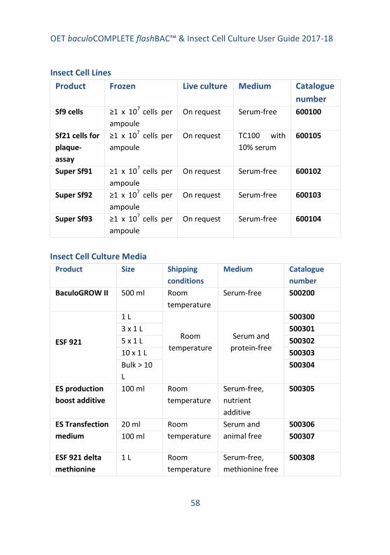
OET baculoCOMPLETE flashBAC™ & Insect Cell Culture User Guide 2017-18
58
Insect Cell Lines
Product Frozen Live culture Medium Catalogue
number
Sf9 cells ≥1 x 10⁷
cells per
ampoule
On request Serum-free 600100
Sf21 cells for
plaque-
assay
≥1 x 10⁷ cells per
ampoule
On request TC100 with
10% serum
600105
Super Sf91 ≥1 x 10⁷
cells per
ampoule
On request Serum-free 600102
Super Sf92 ≥1 x 10⁷ cells per
ampoule
On request Serum-free 600103
Super Sf93 ≥1 x 10⁷
cells per
ampoule
On request Serum-free 600104
Insect Cell Culture Media
Product Size Shipping
conditions
Medium Catalogue
number
BaculoGROW II 500 ml Room
temperature
Serum-free 500200
ESF 921
1 L
Room
temperature
Serum and
protein-free
500300
3 x 1 L 500301
5 x 1 L 500302
10 x 1 L 500303
Bulk > 10
L
500304
ES production
boost additive
100 ml Room
temperature
Serum-free,
nutrient
additive
500305
ES Transfection
medium
20 ml Room
temperature
Serum and
animal free
500306
100 ml 500307
ESF 921 delta
methionine
1 L Room
temperature
Serum-free,
methionine free
500308

OET baculoCOMPLETE flashBAC™ & Insect Cell Culture User Guide 2017-18
59
1. Introduction
1.1 Insect cell culture
This manual provides a guide to the growth and maintenance of insect cell
cultures. It is extremely important that the insect cells used for the
production and analysis of recombinant baculoviruses are of the highest
quality. Insect cells can also be transfected with suitable plasmids to
produce stable cell lines expressing a gene of choice. This manual provides
tried and trusted protocols used within the labs at OET Ltd and by scientists
with over thirty-years’ experience of working with insect cell cultures and
baculoviruses. We hope it is useful to both beginners and more experienced
researchers.
1.2 Choice of cell line
The insect cells most commonly used for the baculovirus expression system
are Sf21 cells, originally derived from the pupal ovarian cells of Spodoptera
frugiperda (fall army worm)1; Sf9 cells, which are a clonal isolate of Sf21
2; or
T. ni (TnHi5™) cells3, originally derived from the ovarian cells of Trichoplusia
ni (cabbage looper)4. Generally, Sf21 or Sf9 cells are used for co-
transfections, virus amplification and plaque assays. Whilst many labs use
Sf9 for all protocols, Sf21 cells are superior for plaque-assays and monitoring
virus cytopathic effects and are more tolerant to sub-optimal conditions;
and so ideal for those new to the system. Sf9 cells are usually better for
amplification of large stocks of virus and protein production and grow very
well in large-scale fermenters or shake flasks. TnHi5™ cells are often used to
achieve maximal protein production but should not be used to produce or
amplify virus because of the increased possibility of generating virus
mutants4. For reasons that remain unclear, some genes are expressed much
better in Sf cells than T. ni cells or vice versa, so testing expression in both
cell lines at an early stage is recommended.
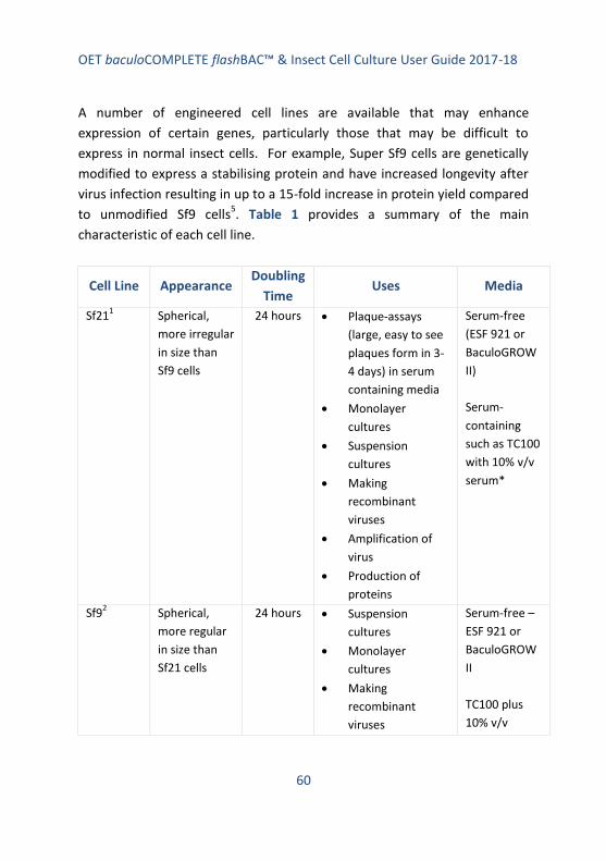
OET baculoCOMPLETE flashBAC™ & Insect Cell Culture User Guide 2017-18
60
A number of engineered cell lines are available that may enhance
expression of certain genes, particularly those that may be difficult to
express in normal insect cells. For example, Super Sf9 cells are genetically
modified to express a stabilising protein and have increased longevity after
virus infection resulting in up to a 15-fold increase in protein yield compared
to unmodified Sf9 cells5. Table 1 provides a summary of the main
characteristic of each cell line.
Cell Line Appearance Doubling
Time Uses Media
Sf211 Spherical,
more irregular
in size than
Sf9 cells
24 hours Plaque-assays
(large, easy to see
plaques form in 3-
4 days) in serum
containing media
Monolayer
cultures
Suspension
cultures
Making
recombinant
viruses
Amplification of
virus
Production of
proteins
Serum-free
(ESF 921 or
BaculoGROW
II)
Serum-
containing
such as TC100
with 10% v/v
serum*
Sf92 Spherical,
more regular
in size than
Sf21 cells
24 hours Suspension
cultures
Monolayer
cultures
Making
recombinant
viruses
Serum-free –
ESF 921 or
BaculoGROW
II
TC100 plus
10% v/v
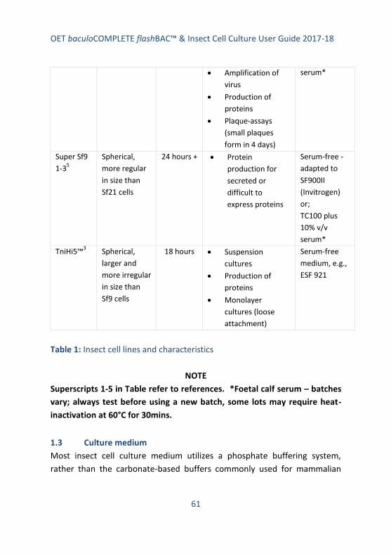
OET baculoCOMPLETE flashBAC™ & Insect Cell Culture User Guide 2017-18
61
Amplification of
virus
Production of
proteins
Plaque-assays
(small plaques
form in 4 days)
serum*
Super Sf9
1-35
Spherical,
more regular
in size than
Sf21 cells
24 hours + Protein
production for
secreted or
difficult to
express proteins
Serum-free -
adapted to
SF900II
(Invitrogen)
or;
TC100 plus
10% v/v
serum*
TniHi5™3 Spherical,
larger and
more irregular
in size than
Sf9 cells
18 hours Suspension
cultures
Production of
proteins
Monolayer
cultures (loose
attachment)
Serum-free
medium, e.g.,
ESF 921
Table 1: Insect cell lines and characteristics
NOTE
Superscripts 1-5 in Table refer to references. *Foetal calf serum – batches
vary; always test before using a new batch, some lots may require heat-
inactivation at 60°C for 30mins.
1.3 Culture medium
Most insect cell culture medium utilizes a phosphate buffering system,
rather than the carbonate-based buffers commonly used for mammalian

OET baculoCOMPLETE flashBAC™ & Insect Cell Culture User Guide 2017-18
62
cells. This means that CO2 incubators are not required. Serum is required for
the maintenance of certain cell lines, but many have now been adapted to
serum-free conditions. There is a large variety of insect cell culture media
available and it is beyond the scope of this manual to list them all; Table 1
lists the media currently in use in our labs. Sf9, Sf21 and TnHi5™ cells can all
be grown in medium with serum or serum-free media (Table 1). Always use
a different bottle of cell culture medium for each cell line. The addition of
antibiotics is optional (penicillin and streptomycin prepared with 5 units/ml-1
penicillin G sodium and 5 μl/ml-1
streptomycin sulphate in 0.85% saline can
be used) but generally it is not recommended for virus amplification or
protein production. Certainly it is best to maintain stock cultures without
antibiotics; otherwise you may be maintaining a low-level contaminant that
may later cause inefficient virus replication or protein production. Addition
of antibiotics to plaque-assay medium is recommended.
1.4 General requirements for insect cell culture
Insect cells have a relatively high dissolved oxygen content (DOC)
requirement, particularly when infected with virus. Maintaining the
appropriate DOC is important for cell growth and virus replication, and this
can be achieved in shake, spinner and tissue culture flasks by using vented
caps and not over-tightening lids. Most insect cells can be cultivated over a
temperature range from 25-30°C. The optimal temperature for cell growth
and infection for insect cells is considered to be 27-28°C. Insect cells can
also be cultured at room temperature (about 20-22°C) when a slower
growth rate is required. Virus infection is usually carried out at 27-28°C.
We recommend carrying out any cell culture work each day prior to
handling virus and only using one cell line at a time.

OET baculoCOMPLETE flashBAC™ & Insect Cell Culture User Guide 2017-18
63
1.5 Maintaining cell cultures
Insect cell lines can be maintained as either suspension cultures, in shake
flasks or in stirred vessels (Figure 1), or in monolayer adherent cultures in T
flasks or dishes. Generally, insect cells adapted to serum-free medium are
cultivated in suspension shake cultures whilst cells adapted to serum-
supplemented media are cultivated in monolayer cultures or stirred
suspension cultures (as growing these cells in shake culture generates
excessive foaming and subsequent cell damage). However, cells grown in
serum-free medium may also be cultured in monolayers.
Shake flasks may be recyclable glass or disposable. Stirred flasks are usually
glass and contain either a magnetic stirring bar or suspended magnetic
stirring rod (Figure 1). Both types are available from a range of suppliers. To
maintain optimum cell culture conditions in a suspension culture, cell
densities should be kept within certain ranges, i.e. within the log-phase of
growth (see Table 2). This is achieved by counting the number of cells, using
either a Neubauer counting chamber or an automatic cell counter. Sub-
culturing (or passaging) of cells allows them to be maintained within log
phase, preventing them from entering their stationary phase. Sub-culturing
of shaker or stirrer cultures requires the seeding density of each cell culture
to be determined before sub-culturing of cells can commence. We tend to
sub-culture cell lines continuously for approximately 30 passages before
returning to stocks stored in liquid nitrogen; but this is a matter of
preference for each lab.
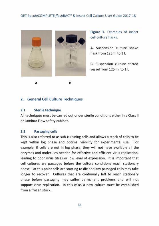
OET baculoCOMPLETE flashBAC™ & Insect Cell Culture User Guide 2017-18
64
A B
2. General Cell Culture Techniques
2.1 Sterile technique
All techniques must be carried out under sterile conditions either in a Class II
or Laminar Flow safety cabinet.
2.2 Passaging cells
This is also referred to as sub-culturing cells and allows a stock of cells to be
kept within log phase and optimal viability for experimental use. For
example, if cells are not in log phase, they will not have available all the
enzymes and molecules needed for effective and efficient virus replication,
leading to poor virus titres or low level of expression. It is important that
cell cultures are passaged before the culture conditions reach stationary
phase – at this point cells are starting to die and any passaged cells may take
longer to recover. Cultures that are continually left to reach stationary
phase before passaging may suffer permanent problems and will not
support virus replication. In this case, a new culture must be established
from a frozen stock.
Figure 1. Examples of insect
cell culture flasks.
A. Suspension culture shake
flask from 125ml to 3 L
B. Suspension culture stirred
vessel from 125 ml to 1 L

OET baculoCOMPLETE flashBAC™ & Insect Cell Culture User Guide 2017-18
65
At each passage, record the passage number on the culture flask. Generally,
insect cell cultures can be passaged for about 30 times before returning to
frozen stocks of cells to initiate a new culture.
When initiating a new culture from a frozen stock, we strongly recommend
starting the culture as an adherent culture and once the cells are growing
well (1-2 passages), transfer them into a suspension culture. It is also easier
to monitor cells visually under the microscope when growing in monolayer
culture. It is possible to go straight from a frozen vial to a suspension
culture but this requires a high density and high viability of the frozen cells.
See notes later on in this manual.
2.3 Adherent culture
Cells can be maintained in T25 or T75 flasks and grown at 27-28°C until the
cells just reach confluency. Cells can be maintained in serum-free media or
in media containing 10% serum (Table 1) Confluency means the cells have
just reached the point where they are touching each other, covering the
surface of the culture vessel (Figure 2). Cells that are passaged repeatedly
when the culture has past confluency will suffer from increased cell growth
time and the appearance of grainy and ‘sausage’ shaped cells that have
arrested mid cell division (Figure 2). Overgrown cells also have increasing
numbers of cells floating in the medium; healthy cultures have very few
floating cells. Attempting to passage cells before a culture reaches
confluency means it is harder to dislodge cells as they are firmly attached to
the flask surface and thus the chances of damaging the cells increase.
To sub culture adherent cells, they should be detached from the surface by
tapping the flask sharply on the bench top – this is the method we routinely
use. Cells can also be detached by repeatedly pipetting the culture media
over the monolayer to dislodge cells – use a Pasteur pipette.

OET baculoCOMPLETE flashBAC™ & Insect Cell Culture User Guide 2017-18
66
NOTE
Cell scrapers can be used but in our experience this method leads to an
excess of dead cells – home-made scrapers consisting of a bent glass rod
with an attached piece of soft silicon tubing, and sterilised by autoclaving,
are more gentle on cells than commercial scrapers.
Usually a 1 in 5 (Sf9) or 1 in 5 to 1 in 10 (Sf21/TniHi5™) with one part old
culture and 4 (or 9) parts fresh medium is sufficient to keep cells in a log-
phase culture. A typical 75 cm2 flask contains 10 ml of culture medium and
so 2 ml would be transferred into 8 ml of fresh medium for a 1 in 5 split (1
ml plus 9 ml for 1 in 10 split). These values are guides and will need to be
adapted to suit the actual growth parameters in each lab.

OET baculoCOMPLETE flashBAC™ & Insect Cell Culture User Guide 2017-18
67
A B
C D
E F

OET baculoCOMPLETE flashBAC™ & Insect Cell Culture User Guide 2017-18
68
It is also important not to make the density of cells too low when passing, as
cells need to be in reasonable proximity with other cells to promote growth.
Some cells are more tolerant of this than others: Sf21 cells are the most
tolerant, Sf9 cells are the least. Figure 2 shows an example of cell density
immediately after passaging and at confluent levels for Sf21, Sf9 and
TniHi5™ cells. If the cells in the newly seeded culture are too sparse, they
may not divide and the culture will not reach become confluent.
It is essential to monitor cells under an inverted microscope prior to and
after passing to check for confluency, detachment after tapping, or any signs
of poor health or over growth. Signs of poor health include: grainy cells,
sausage-shaped cells, floaters, longer doubling time, failure to reach
confluence. Grainy cells with refractive cuboidal-like structures in the
nucleus are a sign of wild-type baculovirus contamination.
Maintain a log book of passaging and record the passage number, date and
split ratio on the culture flask. After about 30 passages of being maintained
in log phase, cultures start to lose viability and virus replication can be
impaired. The old culture should be discarded and a new one established
from a frozen stock. The log book can also be used to record any
Figure 2 (page 67). Images to illustrate
insect cells at various stages of culture.
A. Sf21 cells - sub confluent
B. Sf21 cells - confluent
C. Sf9 cells – sub confluent
D. Sf9 cells – confluent
E. Tni cells – sub confluent
F. Tni cells - confluent

OET baculoCOMPLETE flashBAC™ & Insect Cell Culture User Guide 2017-18
69
observations about the culture and this is sometimes very helpful when
trouble shooting.
Whilst it is not essential, some labs prefer to count cells at each passage and
seed a certain number of cells per flask each time. Table 2 provides an
indication of seeding density for passaging cells grown in monolayer culture.
Cell line
Cells to
seed a T25
flask
Cells to
seed a T75
flask
Cells to seed a
T150 flask
Split ratio guide
(culture : fresh
medium)
Sf21
1 x 10
6 3 x 10
6 5-6 x 10
6 1:5 to 1:10
Sf9/Super
Sf9 1.5 x 10
6 5 x 10
6 1 x 10
7 1:5
TniHi5™
0.9 x 10
6 2-3 x 10
6 4-6 x 10
6 1:5 to 1:10
Culture
volume 5-7 ml 10-15 ml 30-40 ml
Table 2: Seeding density for passaging monolayer cultures
When harvesting cells for use in experimental work, always count the
number of cells and determine their viability so that the correct seeding
density can be achieved for transfections or virus amplification.
Key points when culturing cells:
Check cells each day under the microscope until a confluent
monolayer has formed
Passage cells when confluent or shortly after
Do not allow cells to become overgrown
Do not split at too high a ratio

OET baculoCOMPLETE flashBAC™ & Insect Cell Culture User Guide 2017-18
70
Keep a record of passage number, date and split ratio
Start a new culture from frozen stocks after about 30 passages
Do not use antibiotics in routine cultures
2.4 Suspension culture
Maintaining insect cells in suspension culture is very easy and provides a
ready source of cells for amplifying recombinant viruses and infecting cells
for protein production. There are two main methods for small-scale
suspension cultures – shake flasks or stirred flasks (Figure 1).
Shake flasks require the use of serum-free medium as otherwise serum
creates excess froth that results in cells bursting as their membranes fuse
with the bubbles. Most serum-free medium contain surfactants to reduce
frothing. A surfactant such as Pluronic®F-68 can be added to media to
reduce frothing but in our experience even adding surfactant to serum-
containing medium does not prevent damage to cells.
There are commercial disposable shake flasks in a range of sizes that permit
cultures from 10 ml to 1.5 L. As the culture volume increases attention must
be given to aeration since insect cells, particularly those infected with virus,
have a high oxygen requirement for metabolism. This can be achieved by
selectinging an appropriate rpm, not over filling the flask to maximise the
surface area for gas exchange and ensuring lids are vented.
For those on a restricted budget, it is cheaper to use reusable glass flasks
with cotton wool and loosely covered foil caps that have been sterilised by
autoclaving and then dried. However, insect cells are very susceptible to
contaminants in flasks and so any washing-up regime must be very
stringent.

OET baculoCOMPLETE flashBAC™ & Insect Cell Culture User Guide 2017-18
71
At OET, our washing up regime is as follows:
Disinfect flask with VirkonTM
Soak in hot water with mild detergent (washing up liquid) and scrub
internal surface with nylon bottle brush to remove adherent cell
debris
Rinse five times with hot water
Soak in hot water for 2 hours
Rinse in deionised water twice
Soak in deionised water overnight
Rinse in deionised water, dry and sterilise in an autoclave
Dry in warm cabinet prior to use
As insect cells do not require CO2, a shaking platform can be placed inside a
standard incubator maintained at 27-28°C or even a clean cupboard/room
maintained at this temperature.
Cells can also be grown in stirred cultures using commercial systems that
have vessels with either a vertical impeller or hanging stirring bar that sit on
a bespoke stirring device (Figure 1). There are often side ports to take
samples or add media/cells. Again, the caps should be vented or left loose
and the vessels must not be overfilled to ensure good aeration. Cost
effective home-made stirred flasks can be made using a flat-bottomed
round flask with a stirring bar sat on a conventional magnetic stirrer. Again
attention must be given to ensure the flasks are properly cleaned and
sterilised before use.
Cells detached from a healthy adherent culture should be counted and used
to set up a suspension culture according to the guidance in Table 3. It is
important that the cells have a high viability – at least 95%. When
establishing a suspension culture for the first time, set up a relatively

OET baculoCOMPLETE flashBAC™ & Insect Cell Culture User Guide 2017-18
72
modest scale flask (50-100 ml culture). Once the suspension culture is
established, a larger culture can be established if required.
Cells should be monitored daily by taking a small sample for counting and
determining cell viability. In this way a growth curve can be plotted and
monitored. Cells should be passaged before they reach stationary phase.
An example of a typical growth curve is shown in Figure 3. Whilst we
provide guidance on this matter, each lab should establish the optimal
conditions for their cells and media combination.
Passaging cells in suspension is very easy, after counting and determining
the viability, remove all the excess cells and, to the cells remaining in the
flask, add fresh medium to establish a new culture at the correct cell density
(Table 3). This can continue for up to about 30 passages (see notes under
adherent cultures). After this time, the culture needs to be set up from a
fresh adherent culture at low passage number.
The excess cells removed can be used to set up further suspension cultures
for virus infection – either to amplify stocks of recombinant virus or infect
cells for protein production. Cells can also be used to seed monolayer
cultures for experimental use e.g., 30mm dishes or multiwall plates for
plaque assay, co-transfections to make recombinant viruses or to test
expression levels.
Key points when setting up a new suspension culture:
Use healthy log phase cells from an adherent culture that is at least
95% viable
Count the cells and seed a culture no larger than 100 ml using the
guidelines in Table 4 using serum-free medium

OET baculoCOMPLETE flashBAC™ & Insect Cell Culture User Guide 2017-18
73
Ensure good aeration by not over filling flasks, maintaining optimal
surface area to volume ratio, using an appropriate rpm and vented
lids
Monitor cells daily and set up a growth curve
Passage cells by removing excess cells and adding fresh media to
achieve correct cell density (Table 4) before cells reach stationary
phase
Use excess cells to start cultures for virus amplification, protein
production or experimental use in monolayer cultures
Progress to larger volumes once cells established in culture
Always note passage number, date and cell count/viability data on
flask and in log book
After 30 passages (from retrieval of frozen stock – include passage
number data from adherent cultures), start a new adherent culture
from a frozen stock
Only use log phase cells for virus infections
Do not use TniHi5™ cells for making recombinant viruses or
amplifying viruses
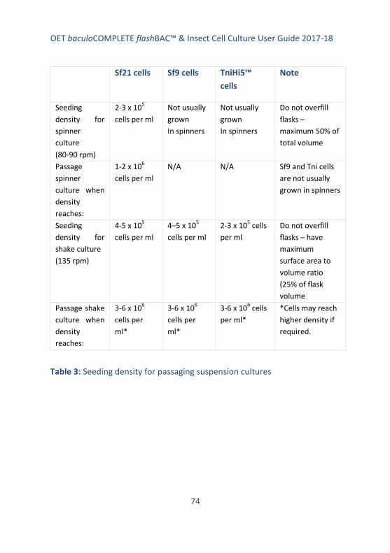
OET baculoCOMPLETE flashBAC™ & Insect Cell Culture User Guide 2017-18
74
Sf21 cells Sf9 cells TniHi5™
cells
Note
Seeding
density for
spinner
culture
(80-90 rpm)
2-3 x 10⁵
cells per ml
Not usually
grown
In spinners
Not usually
grown
In spinners
Do not overfill
flasks –
maximum 50% of
total volume
Passage
spinner
culture when
density
reaches:
1-2 x 10⁶
cells per ml
N/A N/A Sf9 and Tni cells
are not usually
grown in spinners
Seeding
density for
shake culture
(135 rpm)
4-5 x 10⁵
cells per ml
4–5 x 10⁵
cells per ml
2-3 x 10⁵ cells
per ml
Do not overfill
flasks – have
maximum
surface area to
volume ratio
(25% of flask
volume
Passage shake
culture when
density
reaches:
3-6 x 10⁶
cells per
ml*
3-6 x 10⁶
cells per
ml*
3-6 x 10⁶ cells
per ml*
*Cells may reach
higher density if
required.
Table 3: Seeding density for passaging suspension cultures

OET baculoCOMPLETE flashBAC™ & Insect Cell Culture User Guide 2017-18
75
Figure 3. Typical growth curve for Sf9 cells grown in suspension culture in
ESF 921 medium.
3. Freezing and Thawing Cells
3.1 Freezing cells
Once a cell line has been established in either adherent or suspension
culture, after two-three passages, stocks should be frozen in ampoules in
liquid nitrogen so that new cultures can be established when required, for

OET baculoCOMPLETE flashBAC™ & Insect Cell Culture User Guide 2017-18
76
example, after 30 passages or if the culture fails for some reason. It is
important to freeze cells at as lower passage number as possible.
There are many variations in the protocols published for the freezing down
and thawing of insect cells. The following is one that we find works well.
Protocol:
1. Set up a culture (suspension or adherent but suspension is better)
that will provide sufficient cells for freezing down (1 x 10⁷ cells per
vial). Freeze down several vials in one batch – at least 20.
2. Harvest cells from a log phase culture (mid log phase for
suspension or just prior to confluency for adherent cells). Count
cells and determine their viability. Cells need to be at least 95%
viable.
3. Place and label the required number of cryovials on ice.
4. Pellet required number of cells very gently at 500 rpm for 5 mins.
Remove and use the conditioned growth medium to prepare the
freezing mixture as follows:
Serum-free medium* Serum-containing medium
45% conditioned growth medium 40% conditioned medium
45% fresh medium 10% FBS (serum)
10% DMSO 10% DMSO
40% fresh medium
*use same freezing mix as serum-containing medium if preferred
5. Resuspend the required number of cells in freezing mixture, very
gently by pipetting up and down, to achieve a density of 1 x 10⁷
cells per ml. Place 1 ml aliquots into cryovials.
OET baculoCOMPLETE flashBAC™ & Insect Cell Culture User Guide 2017-18
77
6. Place the vials in a freezing chamber (e.g., we use Mr Frosty by
Nalgene) containing isopropanol and immediately place the
chamber at -80°C overnight before transferring to liquid nitrogen.
Or, place the vials at -20°C for one hour and then at -80°C overnight
before placing in liquid nitrogen.
7. After a few days, retrieve one vial to ensure that the freezing
process has been successful.
8. Keep a log book/e-record of where and when each cell line is
frozen, and when vials are recovered.
Key points when freezing cells:
Use healthy log phase cells with 95% or greater viability
Once DMSO has been added, cells must be cooled immediately to
avoid damage
Freeze slowly to avoid damage to cells
Check process has been successful after a few days of storage
Check liquid nitrogen levels regularly to ensure cells don’t start to
thaw during storage
Take all the normal H&S precautions when handling liquid nitrogen.
3.2 Thawing cells
It is important that cells are rapidly thawed and transferred into fresh
growth medium as soon as possible. DMSO is cytotoxic when cells are
thawed. We recommend recovering cells into an adherent culture for ease
of replacing the freezing mixture with fresh medium and monitoring the
cells under the microscope.

OET baculoCOMPLETE flashBAC™ & Insect Cell Culture User Guide 2017-18
78
Protocol:
1. Add 5 ml pre-warmed fresh growth medium to each of two T25
flasks.
2. Rapidly defrost the cells e.g., in a water bath at 37°C until just
thawed.
3. Sterilise the outside of the cryovial by misting with 70% ethanol
spray and transfer 0.5 ml of thawed cells to each flask. Mix to
generate an even distribution of cells and allow the live cells to
attach to the flask for about 1 hour at 28°C.
4. Monitor the cells under the microscope. Live cells should have
attached firmly within the hour incubation period; dead cells will
float. Remove the freezing medium and any floating cells (and
DMSO). Replace with fresh medium.
5. Continue to incubate the cells until a confluent monolayer is
formed and then passage/cub-culture as previously described,
setting up a suspension culture after 2-3 passages if required.
6. Alternatively, the whole 1 ml contents of the cryovial can be placed
in a 125 ml shake/stirred flask containing 25 ml pre-warmed fresh
medium and incubated as for shake/stirred cultures. Remove 5 ml
as a back-up into a T25 monolayer culture flask (and treat as above
steps 4-5). Continue the culture until the cell density is 2 x 10⁶
cells/ml and passage as normal.
It is recommended that suspension cultures set up in this way are
passaged two-three times before being used for experimental or
virus work.

OET baculoCOMPLETE flashBAC™ & Insect Cell Culture User Guide 2017-18
79
Key points when thawing cells:
Thaw quickly in a clean water bath
Sterilise the outside of the vial before opening
Set up a monolayer culture first so it is easy to remove dead cells
When cells are growing well, establish suspension cultures
4. Counting Cells and Determining Cell Viability
4.1 Counting cells
Before passaging cells or using cells for virus infection or transfections, cells
should be counted to establish an accurate count per ml. This can be
achieved using a commercial cell counter or by using a standard
haemocytometer. When establishing a new culture, new cell line or using a
new medium, it is well worth setting up a growth curve and monitoring cell
density every 24 hours to establish the growth pattern.
The following provides a protocol for counting cells using a Neubauer
haemocytometer.
Protocol:
1. Take a sample of cells from a shake or spinner culture or cells
harvested from a monolayer culture and using a Pasteur pipette,
load the prepared Neubauer chamber using capillary action (attach
cover slip firmly to form the counting chamber).
2. Count all the cells within the central 5 x 5 square grid (Figure 4) on
the counting chamber using a phase-contrast microscope (x10
objective). Count cells touching the etched triple line on the top
and left of squares. Do not count cells touching the triple lines on
the bottom or right of the squares.

OET baculoCOMPLETE flashBAC™ & Insect Cell Culture User Guide 2017-18
80
3. Count cells on both 5 x 5 grids and average the results. If the cells
are too dense to count accurately, dilute the sample an appropriate
amount to get a countable number of cells. If the cells are
clumped, they should be dispersed by gently pipetting up and
down to get single cells; otherwise the cell count will be inaccurate.
Ideally you need at least 30 cells and no more than 100.
4. The 5 x 5 square gives the number of cells present in 0.1 µl of
culture. To calculate the number of cells per ml, multiply by 10,000
(104). If the cells were diluted before counting, remember to
multiply the answer by the dilution factor.
Key points when counting cells:
Ensure cells are in a single cell suspension and not clumped
Count at least in duplicate to get an average
Don’t forget to multiply answer by any dilution factor
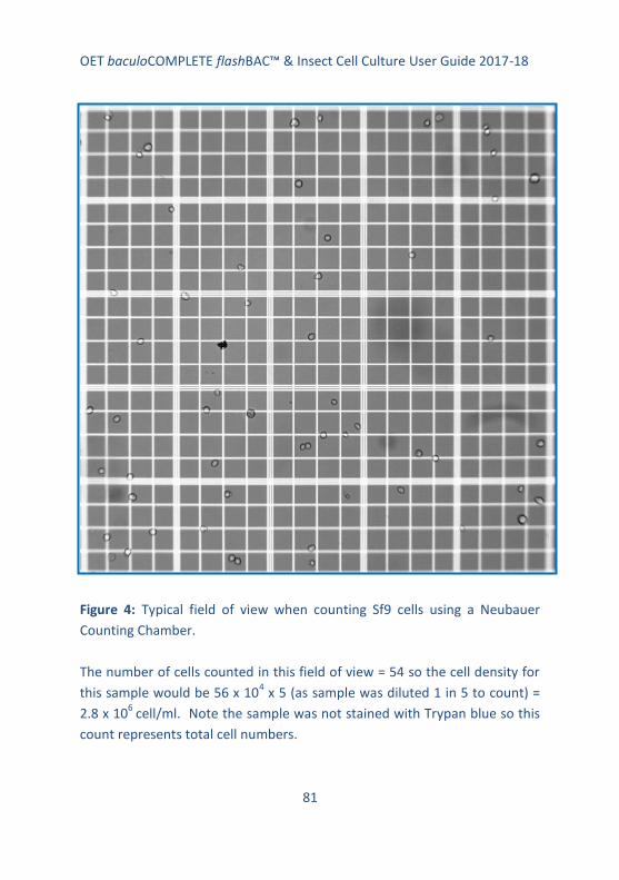
OET baculoCOMPLETE flashBAC™ & Insect Cell Culture User Guide 2017-18
81
Figure 4: Typical field of view when counting Sf9 cells using a Neubauer
Counting Chamber.
The number of cells counted in this field of view = 54 so the cell density for
this sample would be 56 x 104 x 5 (as sample was diluted 1 in 5 to count) =
2.8 x 10⁶
cell/ml. Note the sample was not stained with Trypan blue so this
count represents total cell numbers.

OET baculoCOMPLETE flashBAC™ & Insect Cell Culture User Guide 2017-18
82
4.2 Cell viability
Cell viability should be tested from time to time and every time cells are
used for virus work (transfections or virus infection) as poor viability is a
common reason for failure of virus to amplify to high titres or ensure high
yield of protein production.
The easiest method is Trypan blue exclusion and this can conveniently be
performed when counting cells. Trypan blue is a vital stain that is actively
excluded from live cells; therefore, dead cells take up and retain the stain
appearing blue under the microscope. Healthy cells appear refractile, bright
and clear. The percentage of dead cells can be calculated and used to
determine the overall viability of the culture. A good culture should be at
least 90% and preferably 95% viable.
Protocol:
1. Prepare a 2% (w/v) preparation of Trypan blue (e.g. Sigma) in PBS.
2. Dilute a sample of cells 1:1 with the stain (final concentration 1%)
and view cells under a phase contrast microscope immediately. It
is convenient to count the cells at this stage (see above protocol).
3. % viability = 100 - % dead cells
% dead cells = total blue cells counted/total cells counted x 100
Key points when determining viability:
Ensure cells are in a single cell suspension and not clumped
Count at least in duplicate to get an average
Don’t forget to multiply answer by two to take account of adding
stain
Don’t leave cells in stain for more than few minutes before
determining viability

OET baculoCOMPLETE flashBAC™ & Insect Cell Culture User Guide 2017-18
83
5. Establishing a New Culture from Living Cells
If cells are received from another lab or from a supplier (e.g., the
BaculoComplete kit from OET Ltd), you will need to establish a new culture
as soon as the cells arrive. You will need to ensure that you have a supply of
medium (see page 58), access to a Laminar Flow hood or Class II safety
cabinet, a supply of sterile flasks and pipettes, access to a cell counter or a
Neubauer haemocytometer to count cells, an incubator set at 27-28°C (no
CO2 required), access to a phase contrast inverted microscope and Trypan
blue stain (e.g. from Sigma)
Protocol:
1. View cells under an inverted light microscope and note if majority
of cells are floating or attached as a monolayer.
2. Decontaminate the outside of the flask by spraying with 70%
alcohol.
3. Place flask containing cells in a laminar flow hood or class 2 safety
cabinet and use aseptic technique through-out all procedures.
4. If many cells are still attached as a monolayer, decant the medium
and any floating cells into a discard container and replace with 5-6
ml fresh medium (e.g., baculoGROW II, ESF 921).
If majority of cells are floating, decant medium and cells into a
sterile centrifuge tube and gently pellet cells at 1000 rpm for 5-10
min in a bench top centrifuge.
Also add 5-6 ml fresh medium to the attached cells in the original
flask and incubate at 27-28°C until just about confluent (~80%).

OET baculoCOMPLETE flashBAC™ & Insect Cell Culture User Guide 2017-18
84
5. Decant the old medium from the pelleted cells (into discard) and
gently resuspend the cells in 5-6 ml of fresh medium to obtain a
single cell suspension. Place the cells into a fresh T-flask, ensure
they are evenly dispersed over the surface, and incubate over night
as described above. Any live cells will attach overnight. The
following day, remove floating dead cells and replace with 5-6 ml
fresh medium and incubate as above until cells are just about
confluent.
6. We recommend an initial 1 in 4 to 1 in 5 split of cells when just
confluent. For example, harvest the cells by tapping the flask
sharply on the bench to detach cells, and gently pipette up and
down to get a single cell suspension. For example, place 1.25 ml
cells with 4.75 ml fresh medium into a new T-flask.
Passage the cells through at least one further round of monolayer
culture before setting up shake cultures.
7. The cells can be maintained like this in monolayer culture,
expanding into larger T-flasks as required, or the cells can be grown
in suspension/shake culture if cultured in serum-free medium.
8. To establish a shake culture, harvest cells from 2 x T25 monolayer
cultures and transfer 8 ml into a 125ml shaker flask containing 12
ml fresh medium.
Ensure the lid is not on tightly to allow for aeration and shake at
100-110 rpm at 27-28°C for 3-4 days and then count the cells.
9. Set up a fresh shake culture at 1 x 10⁶ cells/ml with the appropriate

OET baculoCOMPLETE flashBAC™ & Insect Cell Culture User Guide 2017-18
85
amount of cell culture and fresh medium. Once the suspension
culture is established, seed the newly passaged cells at 0.8 x 10⁶
cells/ml or as required. Cells will grow to a density of 5-8 x 10⁶ cells
per ml.
10. Do not allow cells to overgrow in either monolayer or shake
culture. For optimal virus infections, cells should be used in log
growth phase. Undertaking a growth curve is useful to understand
the growth characteristics of the culture.
11. We recommend that as soon as possible a batch of expanded cells
are frozen down in liquid nitrogen to act as a source of low passage
number cells for long term use. We normally passage cells for 30
passages before retrieving fresh stocks from liquid nitrogen.
12. For general information about insect cell culture read pages 64 to
74.
6. Establishing a New Culture of Cells From a Frozen Ampoule
On receipt, it is essential that the ampoule of frozen cells is either
transferred to liquid nitrogen for storage or thawed to initiate a live cell
culture. Do not freeze insect cells at -80°C.
You must use aseptic technique through-out and work in a Class II Safety
Hood or Tissue Culture Laminar Flow Hood. Rinse or mist the vial of cells
with 70% alcohol before opening.
Read through the general information about insect cell culture on pages 64
to 74 before starting and have the following materials available: suitable
OET baculoCOMPLETE flashBAC™ & Insect Cell Culture User Guide 2017-18
86
growth medium (e.g., baculoGROW II, ESF 921 from OET Ltd, Table 2),
T25/T75 monolayer flasks, 1ml and 10 ml sterile pipettes, incubator at 27-
28°C, water bath at ~37°C (best to use a ‘temporary bath’ such as a clean
beaker with warm clean water rather than a dirty water bath).
Protocol:
1. On receipt, using aseptic technique, defrost the cells rapidly in a
clean water bath at 37°C until just thawed.
2. Rinse or mist the outside of the vial with 70% alcohol and then
transfer the contents of the ampoule between 2 T25/T75 flasks
containing 10-15 ml fresh culture medium (e.g., baculoGROW II,
ESF 921). Incubate the cells overnight at 27-28°C.
3. Check the cells under the microscope. Dead cells will be floating.
Live cells will have attached.
4. Decant medium containing floating cells into discard. Replace with
an appropriate amount of fresh media and continue to incubate
cells until they form a just confluent monolayer. Check daily.
5. When ready, passage cells and set up further monolayer or
suspension cultures as described
6. Cells can be used to prepare recombinant viruses as soon as they
have recovered from shipping and are doubling approximately
every 24 hours with a high viability (90% or more). This may take 2-
3 passages of cells.
It is important that cells are not used to make recombinant viruses
until they are growing well in a log phase culture.

OET baculoCOMPLETE flashBAC™ & Insect Cell Culture User Guide 2017-18
87
7. As soon as possible prepare a stock of cells for freezing down for
long term storage in liquid nitrogen (see page 75).
7. Adapting Cells to New Media
On occasion it is necessary to transfer cells into a new culture medium.
Sometimes this can be achieved by simply using the new medium in place of
the old. However, more often a period of adaption is required.
The accepted standard protocol is to culture the cells in 75% old medium: 25
% new for one to two passages, then 50% old: 50% new for one to two
passages followed by 75% new: 25% old before finally culturing the cells in
the new medium. This takes time but ensure the cells adapt well to a new
medium.
A quicker version of this method can often be successful. It simply involves
culturing the cells in 50:50, old: new medium for one passage (monolayer or
suspension culture) and then continuing to grow the cells in the new
medium for at least one passage before using them for experimental work
(e.g. amplifying virus or making recombinant viruses).
For this quicker protocol to be successful, it is important that the cells are in
log phase of growth (80% confluent for monolayer or 3-5 x 10⁶ cells/ml for
suspension cultures) and that you monitor the cells each day to ensure they
are still growing well.
The cells should be passaged as soon as they are confluent (monolayer
cultures) or when they reach a density of 4-5 x 10⁶ cells/ml (suspension
culture). If the cells take a very long time to reach confluency or a density of

OET baculoCOMPLETE flashBAC™ & Insect Cell Culture User Guide 2017-18
88
4-5 x 10⁶, or cell viability drops very low, the longer adaption process
described above will need to be followed.
7.1 Adapting cells to baculoGROW II or ESF 921
We have successfully adapted Sf9 cells to baculoGROW II and ESF 921 (from
a variety of media) using the quick adaption protocol described below.
Protocol:
1. Take a log phase culture of the cells to be adapted (80% confluent
monolayer culture) or cells at 3-5 x 10⁶ cells/ml for suspension
cultures. Ensure the culture is at least 90% viable.
2. For suspension cultures, dilute the culture with an equal amount of
the new medium and continue growing the cells until they reach 4-
5 x 10⁶ cells per ml.
For monolayer cultures, passage the cells using 50% old medium
saved from the previous culture and 50% new medium. Grow until
the cells are just confluent.
3. Passage the cells as normal using the new medium.
4. Use cells for experimental work after 1-2 passages in the new
medium.

OET baculoCOMPLETE flashBAC™ & Insect Cell Culture User Guide 2017-18
89
8. References
1. Vaughn, J. L., Goodwin, R. H., Tompkins, G. J. & McCawley, P.
(1977). In Vitro 13, 213-217.
2. Sf9 was cloned by G. E. Smith and C. L. Cherry from the parent line,
IPLB-SF 21 AE, by Vaughn et al., in 19771.
3. Wickham T.J., Davis T, Granados R.R., Shuler M.L. & Wood H.A.
(1992). Biotechnol Prog. 8, 391–396.
4. Hink, W. F. & Vail, P. V. (1973). J. Invertebr. Pathol. 22, 168-174.
Acknowledgements
This guide to insect cell culture was prepared by the OETeam of Robert
Possee, Adam Chambers, Olga Lissina, Aleksandra Kuczera, Linda King and
Laura Swan. We hope it is helpful.