Sertac Mustafaoglu - Urban / Retail / Street Furniture Design Selection
Chapter 3
Odontogenic Infections
Onur Gonul, Sertac Aktop, Tulin Satilmis,Hasan Garip and Kamil Goker
Additional information is available at the end of the chapter
http://dx.doi.org/10.5772/54645
1. Introduction
The incidence, severity, morbidity, and mortality of odontogenic infections have declineddramatically over the years. This reduction in mortality was not due to the first use of penicillinin the treatment of these infections. Rather, it was due to application of the principles of theinitial establishment of airway security, followed by early and aggressive surgical drainage ofall anatomical spaces affected by cellulitis or abscesses. Since then, with the use of antibioticsand advanced medical supportive care, mortality associated with Ludwig’s angina has beenfurther reduced, to 4% [1].
Determination of the severity of infection, evaluation of host defences, surgical management,medical support, administration of antibiotics, and frequent evaluations of the patient are themainstays of the management of odontogenic infections. Three major factors must be consid‐ered when determining the severity of an infection of the head and neck: anatomical location,rate of progression, and airway compromise.
The host response to a severe infection can place a severe physiological load on the body. Fevercan increase sensible and insensible fluid losses and caloric requirements. A prolonged fevermay cause dehydration, which can, in turn, decrease cardiovascular reserves and depleteglycogen stores, shifting the metabolism to a catabolic state [2].
The surgeon should also be aware that elderly individuals are not able to respond to highfevers, as is often seen in children. Thus, an elevated temperature in a patient of ad‐vanced age is not only a sign of a particularly severe infection, but also an omen of de‐creased cardiovascular and metabolic reserve, due to the demands placed on the elderlypatient’s physiology [3].
© 2013 Gonul et al.; licensee InTech. This is an open access article distributed under the terms of the CreativeCommons Attribution License (http://creativecommons.org/licenses/by/3.0), which permits unrestricted use,distribution, and reproduction in any medium, provided the original work is properly cited.
White blood cell count at admission has been reported to be a significant predictor of the lengthof hospital stay. Thus, evaluation of leukocytosis is important in determining the severity ofinfection, as well as in estimating the length of hospital stay.
The physiological stress of a serious infection can also disrupt previously well-establishedcontrol of systemic diseases, such as diabetes, hypertension, and renal disease. The increasedcardiac and respiratory demands of a severe infection may deplete scarce physiologicalreserves in a patient with chronic obstructive pulmonary disease or atherosclerotic heartdisease, for example. Thus, an otherwise mild or moderate infection may be a significant threatto a patient with pre-existing systemic disease, and the surgeon should be careful to evaluateand manage concurrent systemic diseases in conjunction with direct management of theinfection.
2. Microbiology of dental infections
Recent reports have confirmed that oral/dental infections are polymicrobial, includingfacultative anaerobes, such as viridans-group streptococci and the Streptococcus anginosusgroup, with predominantly strict anaerobes, such as anaerobic cocci, Prevotella and Fusobacte‐rium species. The use of sophisticated non-culture methods has identified a wider range oforganisms, such as Treponema species and anaerobic Gram-positive rods such as Bulleidiaextructa, Cryptobacterium curtum, and Mogibacterium timidum [4].
3. Anatomical Spread of Infection
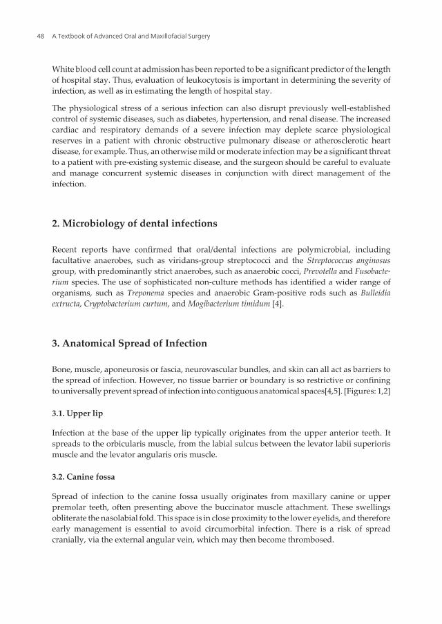
Bone, muscle, aponeurosis or fascia, neurovascular bundles, and skin can all act as barriers tothe spread of infection. However, no tissue barrier or boundary is so restrictive or confiningto universally prevent spread of infection into contiguous anatomical spaces[4,5]. [Figures: 1,2]
3.1. Upper lip
Infection at the base of the upper lip typically originates from the upper anterior teeth. Itspreads to the orbicularis muscle, from the labial sulcus between the levator labii superiorismuscle and the levator angularis oris muscle.
3.2. Canine fossa
Spread of infection to the canine fossa usually originates from maxillary canine or upperpremolar teeth, often presenting above the buccinator muscle attachment. These swellingsobliterate the nasolabial fold. This space is in close proximity to the lower eyelids, and thereforeearly management is essential to avoid circumorbital infection. There is a risk of spreadcranially, via the external angular vein, which may then become thrombosed.
A Textbook of Advanced Oral and Maxillofacial Surgery48
3.3. Buccal space
The attachment of the buccinator muscle to the base of the alveolar process can control thespread of infection in the region of the mandibular and maxillary molars. An infection spreadsintraorally, superficial to the buccinator muscle, in front of the anterior border of the massetermuscle. Thus, the clinical manifestations of infection in this space are characterized by swellingconfined to the cheek. However, infection may spread superiorly, towards the temporal space,inferiorly, to the submandibular space, or posteriorly, into the masseteric space. In some cases,infection may spread to the surface of the skin, leading to fistula formation
3.4. Palate
The palate is usually involved in infections originating from the maxillary lateral incisor or thepalatal roots of the posterior teeth. The infection spreads from the apices of these teeth,perforating the palatal alveolar bone, and pus accumulates below the palatal mucoperiosteum.
Figure 1. Severe infection of several fascial spaces.
Figure 2. Submental view.
Odontogenic Infectionshttp://dx.doi.org/10.5772/54645
49
It is important to be aware that although the lateral incisor is the most common source of palatalabscess, though most still present labially.
3.5. Pterygomandibular space
Infection in this space is manifested by trismus, due to the involvement of the pterygoidmuscles. This space is bounded medially by the medial pterygoid muscle and laterally by themedial surface of the mandible, anteriorly by the pterygomandibular raphe, and posteriorlyby the deep lobe of the parotid gland. The lateral pterygoid muscle forms the roof of this space.
3.6. Submasseteric space
The most common source of infection in this space is from lower third molar pericoronitis.This space is bound medially by the masseter muscle and laterally by the outer surface of theramus of the mandible. It is in direct communication with the lateral pharyngeal spaceposteriorly. The temporalis muscle divides the superior part of this space into two portions,the superficial temporal space, which is bounded by temporalis muscle medially, and the deeptemporal space, with the temporalis muscle laterally and the periosteum of the temporal bonemedially. Severe trismus due to spasm of the masseter muscle is a characteristic feature ofinvolvement of this fascial space.
3.7. Infratemporal space
Extension of infection from maxillary molars can pass into this space. Infection may also spreadfrom the pterygomandibular, parotid, or lateral pharyngeal region to the infratemporal space.The patient then complains of pain, particularly with mouth opening, some dysphagia, anddifficulty with lateral mandibular movements. This space is located behind the zygomatic boneposterior to the maxilla and medial to the insertion of the medial pterygoid muscle. Theinfratemporal space is bounded superiorly by the greater wing of the sphenoid and is in closeproximity to the inferior orbital fissure, with a possible risk of spread of infection to the orbit.
3.8. Parotid space
Involvement of this space may be an extension of infection in the middle ear or the mastoidregion. Infection in the masseteric or the lateral pharyngeal space may also spread to theparotid region. Thus, the most characteristic feature of involvement of this space is swellingof the parotid gland region, below the ear lobe. This space contains several important structuresthat may be affected by infections. These include the 7th cranial nerve, the auriculotemporalnerve, the facial vein, the parotid lymph node, and, more deeply, the external carotid with itsbranches.
3.9. Submandibular space
This space is located below the mylohyoid muscle, medial to the ramus and the body of themandible. It is bounded anteriorly by the attachments of the anterior belly of the digastricmuscle and posteriorly by the posterior belly of digastric muscle and the stylomandibular
A Textbook of Advanced Oral and Maxillofacial Surgery50
ligament. Infection from the posterior mandibular teeth may pass lingually, below theattachment of the mylohyoid muscle, into this space. Clinically, swelling of the submandibularregions tends to obliterate the angle of the mandible, causing pain and redness of the skinoverlying this region. Dysphagia is also usually a marked symptom.
3.10. Submental space
This space lies between the two anterior bellies of the digastric muscle. Anteriorly and laterallythis space is bounded by the body of the mandible. It is contained, superficially, by theplatysma muscle and, deeply and superiorly, by the mylohyoid muscle. Infection of this spaceusually arises from mandibular anterior teeth, where the infection perforates the lingual cortex;swelling of the submental region is a characteristic clinical feature. The skin over the swellingis stretched and hardened, and the patient experiences considerable pain and difficulty withswallowing. The infection may progress buccally, causing swelling in the labial sulcus andover the chin.
3.11. Sublingual space
Infection spreads into this space as the result of perforation of the lingual cortex, above theattachment of the mylohyoid muscle. This space is bounded superiorly by the mucousmembranes and inferiorly by the mylohyoid muscle. The genioglossus and geniohyoidmuscles form the medial boundary. Laterally, this space is bounded by the lingual surface ofthe mandible. Infection in this space will raise the floor of the mouth and displace the tongue,medially and posteriorly. Such tongue displacement may compromise the airway andimmediate intervention may be required. Dysphagia and difficulty with speech are alsocommon.
3.12. Pharyngeal space
This space is located on the lateral side of the neck, bounded medially by the superiorconstrictor muscle of the pharynx and posterolaterally by the parotid space. Infection in thisspace may originate from mandibular molars or third molar pericoronal suppuration. Thiscould also be a site of spread of infection from the parotid space or fascial space around thebody of the mandible. The lateral pharyngeal space contains the carotid sheath, glossophar‐yngeal nerve, accessory nerve, and the hypoglossal nerve, as well as the sympathetic trunk.Thus, spread of infection into this space carries a significant danger of spreading into adescending neck infection and involvement of the mediastinum. Clinically, stiffness of theneck, swelling of the lateral wall of the pharynx, medial displacement of the tonsils, dysphagia,and trismus are among the characteristic clinical features of involvement of this space.
3.13. Retropharyngeal space
This space is located between the posterior wall of the pharynx and the prevertebral fascia.This space is in direct communication with the base of the skull, superiorly, and the media‐
Odontogenic Infectionshttp://dx.doi.org/10.5772/54645
51
stinum, inferiorly. It has the same characteristic clinical features as infection of the lateralpharyngeal space and carries a significant complication risk of a descending neck infection.
4. Evaluation of patients with dentofacial infections
Patients with dentofacial infections may present with various signs and symptoms, rangingfrom less important to extremely serious. Quick assessment of the patient’s situation isessential as the first step of therapy. If the patient shows central nervous system changes,airway compromise, or toxification, then immediate hospitalization, aggressive medicaltreatment, and surgical intervention may be necessary. Basic principles of patient evaluationmust be followed. A complete patient history, physical examination, laboratory investigation,radiological investigation, and accurate and appropriate interpretation of findings must bemade. Following these basic principles provides the best chance of accurate diagnosis andtreatment [6,7].
4.1. History taking
History taking helps in obtaining information regarding the origin, extent, location, andpotential threat of the problem. History taking can be defined briefly as determining thepresent situation of the patient, previous hospitalization history of the patient, previoustrauma in the region, recurrent infections, and history of recent swelling and/or airwaycompromise.
4.2. Physical examination
Examination of the thorax, abdomen, extremities, cardiovascular system, recording of vitalsigns, and body temperature assessment are essential as part of the general patient evaluation.Next, the skin of the face, head, and neck, swellings, injuries, and areas of tenderness overmaxillary and frontal sinuses, sinus tracts, fistula formation, enlargement of underlying bonystructures, salivary glands, and lymph nodes must be examined. A comprehensive extraoralexamination includes inspection of the skin of the face, head, and neck, and of any swelling,injuries, fixation of skin, sinus, or fistula formation. Palpation of the size of any swelling,tenderness, local temperature, fluctuation, enlargement or tenderness over maxillary andfrontal sinuses, sinus tracts, fistula formation, enlargement and tenderness of underlying bonystructure, salivary glands, and lymph nodes is also important. A comprehensive intraoralexamination includes measurement of inter-incisal openings for the assessment of trismus,examination of the teeth, any localized fistula or swelling, sites of tooth extraction, percussionfindings, heat and cold testing, electrical pulp testing, visualization of opening ducts of salivaryglands, soft palate, tonsillar fossa, uvula, and oropharynx.
4.2.1. Clinical features
Clinical features must be definitively identified to evaluate the patient’s condition properly.Clinical features can be classified as follows.
A Textbook of Advanced Oral and Maxillofacial Surgery52
a. Signs of inflammation
Rubor: This symptom is usually present when the infection is close to an external tissue surface,due to vasodilation.
Tumor: This may be present at an infection site, due to accumulation of inflammatory exudateor pus.
Calor: This is due to warm blood from deeper tissues at the site of the infection, increasedvelocity of blood flow, and an increased rate of metabolism.
Dolor: This is due to pressure on sensory nerve endings, caused by distension of tissues, causedby the action of liberated or activated factors, such as kinins and histamine.
Loss of function: This is due to mechanical factors or reflex inhibition of muscle movements,associated with pain. This is reflected in difficulty in chewing and swallowing and respiratoryissues.
b. Fever
Fever is one of the most consistent signs of infection. However, other conditions that maymanifest fever should also be considered. Non-infectious inflammatory disorders, likerheumatoid arthritis, excess catabolism, as in thyrotoxicosis, neoplastic disease, like lympho‐ma, and post-operative release of endogenous pyrogens, which stimulate the hypothalamicthermoregulation centers, should be considered.
c. Repeated Chills
Generally seen in the presence of bacteraemia and pyogenic abscesses.
d. Lymphadenopathy
The condition of the lymph nodes depend on whether the situation is acute or chronic. In acuteinfections, lymph nodes are soft, tender, and enlarged. Surrounding tissues are edematous andthe overlying skin is erythematous. In chronic infections, lymph nodes are firm, non-tender,and enlarged. Edema of surrounding tissue may not be present. The location of affected lymphnodes may indicate the site of an infection.
e. Headache
This is usually associated with fever, and its thought to be due to stretching of sensitivestructures surrounding dilated intracranial arteries.
f. Other Clinical Features
Other clinical features include the presence of draining sinuses or fistulae, difficulty in openingthe mouth, difficulty in swallowing, increased salivation, changes in phonation, difficulty inbreathing.
• Clinical Symptoms of Possibly Life-Threatening Infections are as follows:
Odontogenic Infectionshttp://dx.doi.org/10.5772/54645
53
Respiratory impairment, difficulty in swallowing, impaired vision or eye movement or both,change in voice quality, lethargy, decreased level of consciousness, agitation, hypoxia.
• Clinical Symptoms of Toxicity are as follows:
Pallor, increased rate of respiration, fever, lethargy, diaphoresis.
• Central Nervous System Changes Associated with Infection are as follows:
Decreased level of consciousness, evidence of meningeal irritation, severe headache, stiff neck,vomiting, and oedema of the eyelids and other abnormal eye signs.
4.3. Radiological examination
A radiological examination may be helpful in locating the offending teeth or other underlyingcauses. Various radiographs can be useful, such as intraoral periapical radiographs, ortho‐pantomographs, and lateral oblique views of the mandible. A-P and lateral views of the neckcan be helpful in detecting retropharyngeal space infections. Other imaging techniques, suchas computed tomography, magnetic resonance imaging, and xeroradiography, are also usedfor detection of the localization of infection and infection-affected tissues. CT scanning is thegold standard in head and neck imaging. It is the advanced imaging modality most widelyused in the evaluation of facial infections. A CT scan can show the extent of soft tissueinvolvement, such as the extent of the inflammatory process, the epicenter of the inflammatoryprocess, differentiation between myositis–fasciitis and abscess formation, and can accuratelydemonstrate the airway status and lymph node involvement [8].
5. Antibiotic therapy in dentofacial infections
Choosing the appropriate antibiotic for treating an odontogenic infection must be done withcare. When all factors are considered, the clinician may decide that no antibiotic is necessaryat all, whereas in other situations, broad-spectrum or even combination antibiotic therapy maybe indicated. Various factors must be considered when choosing an antibiotic from the nearly70 that are currently available. Although appropriate use may result in dramatic resolutionand cure of patients with infections, inappropriate use of antibiotics provides little or no benefitto offset the risks and expense associated with antibiotic administration. Recent studies haveshown that even the administration of oral penicillin promotes the growth of penicillin-resistant organisms in the oropharyngeal flora of the patient. Thus, the following guidelinesshould be considered when choosing a specific antibiotic.
Determination of the need for antibiotic administration. A common misconception is thatall infections, by definition, require antibiotic administration. This is not necessarily the case.In some cases, antibiotics are not useful and may even be contraindicated. In making thisdetermination, three factors must be considered.
1. The seriousness of the infection.
A Textbook of Advanced Oral and Maxillofacial Surgery54
2. Whether adequate surgical treatment can be achieved.
3. The state of the patient’s host defenses.
When these three factors are balanced, several definite indications for antibiotic use becomeclear. These are:
• Swelling extending beyond the alveolar process
• Cellulitis
• Trismus
• Lymphadenopathy
• Fever
• Severe pericoronitis
• Osteomyelitis
Based on the same three criteria, antibiotic therapy is not indicated in other situations, suchas:
• Patient demand
• Toothache
• Periapical abscess
• Dry socket
• Multiple dental extractions in a non-compromised patient
• Mild pericoronitis
• Drained alveolar abscess
In summary, antibiotics should be used when clear evidence exists of bacterial invasion intodeeper tissues, which is greater than the host defenses can overcome. Patients who have animpaired ability to defend themselves against infection and patients who have infections thatare not immediately amenable to surgical treatment should also be considered for antibiotictherapy. Antibiotics should not be used when no evidence of bacterial invasion of deepertissues is found. It should be remembered that antibiotics do not improve wound healing anddo not benefit non-bacterial infections.
• Routine empirical antibiotic use. Because the microbiology and antibiotic sensitivity ofmany oral pathogens is well-known, it is reasonable to use one of the effective antibioticsempirically. This means to give the antibiotic with the assumption that an appropriate drugis being given. The drug of choice is usually penicillin. Alternative drugs for use in apenicillin-allergic patient are clindamycin and azithromycin. Metronidazole is usefulagainst anaerobic bacteria and should be reserved for a situation in which only anaerobicbacteria are suspected or used in combination with an antibiotic that has an anti-aerobic
Odontogenic Infectionshttp://dx.doi.org/10.5772/54645
55
bacteria effect, like penicillin. The most widely used, effective, orally administered antibi‐otics are:
• Penicillin
• Amoxicillin
• Clindamycin
• Azithromycin
• Metronidazole
• Moxifloxacin
• Narrowest spectrum antibiotic use. It is preferable to use an antibiotic with the narrowestspectrum that is effective against the organism(s) involved in the infection. The use of abroad-spectrum antibiotic should be avoided, because it increases the risk of the develop‐ment of resistant microbial strains and also increases the risk of superinfections, by dis‐rupting the normal bacterial flora in various body cavities and permitting ordinarily non-pathogenic bacteria to proliferate and cause disease.
• Antibiotic usage with the lowest incidence of toxicity and side effects. Most antibioticshave a variety of toxicities and side effects that limit their usefulness. These range from mildto so severe that the antibiotic cannot be used in routine clinical practice. The older antibi‐otics usually used for odontogenic infections have a surprisingly low incidence of toxicity-related problems. The more recent antibiotics, on the other hand, may have significanttoxicities and drug interactions. Thus, it is becoming increasingly important for the clinicianto understand the toxicities, side effects, and drug interactions of the drugs that areprescribed.
• Use of a bactericidal antibiotic, if possible. Antibiotics may either kill bacteria or interferewith their reproduction. Bactericidal antibiotics usually interfere with cell-wall productionin newly forming or growing bacteria. The antibiotic actually kills the bacteria, while thewhite blood cells, complement, and antibodies of the host play a less important role.Bacteriostatic antibiotics interfere with bacterial reproduction and growth. This slowing ofbacterial reproduction allows the host defenses to move into the area of infection, phago‐cytose existing bacteria, and kill them. Thus, bacteriostatic antibiotics require reasonablyintact host defences. Therefore, for patients with compromised host defences, bactericidalantibiotics should be the drug of choice [9,10,11].
6. Surgical management of odontogenic infections
The primary principle of the surgical management of odontogenic infections is to performsurgical drainage and to remove the cause of the infection. Surgical management may rangefrom something as simple as an endodontic extirpation of the necrotic tooth pulp to treatmentas complex as the wide incision of soft tissue in the submandibular and neck regions for a
A Textbook of Advanced Oral and Maxillofacial Surgery56
severe infection. The primary goal in surgical management of infection is to remove the causeof infection and to provide drainage of accumulated pus and necrotic debris. Surgical incisionand drainage helps to get rid of toxic purulent material, to decompress edematous tissues, toallow better perfusion of blood, which contains antibiotic and defense elements, and to increaseoxygenation of the infected area. When an abscess is drained surgically, appropriate dentaltreatment also should be instituted to achieve quick resolution. This may involve explorationof either the entire anatomical space or the abscess cavity. The abscess cavity is then irrigatedwith betadine and saline solution. A drain is inserted into the depth of the space. It may simplypass through a single incision and remain in the depth of the space, or it may be a through-and-through drain. The drain is typically secured to one of the margins of the incision with asuture. The method of opening an abscess ensures that no blood vessel or nerve in the regionis damaged, and can be defined in ten steps:
1. Topical anesthesia. Local anesthesia is achieved with the help of ethyl chloride spray;local anesthesia can then be achieved by subcutaneous ring blockage using a localanesthetic solution, such as articaine + epinephrine or lidocaine + epinephrine.
2. Incision. This is made over a point of fluctuation in the most dependent area along theskin crease, through undamaged skin and subcutaneous tissue.
3. If pus is not encountered, further deepening of the surgical site is achieved with sinusforceps.
4. Closed forceps are pushed through the deep fascia and advanced towards the puscollection.
5. The abscess cavity is entered and forceps opened in a direction parallel to vital structures.
6. Pus flows along the sides of the incision. [Figure 3]
7. Explore the entire cavity for additional loci.
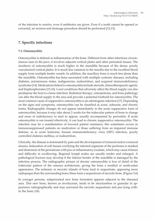
8. Placement of a drain. A soft corrugated rubber drain is inserted into the depth of theabscess cavity, and the external part is secured to the wound margin with the help of asuture. [Figure 4]
9. The drain is left in place for at least 24 h.
10. A dressing is applied over the site of the incision, without pressure.
The purpose of the drain is to allow the discharge of tissue fluids and pus from the wound bykeeping it patent. The drain also allows debridement of the abscess cavity by irrigation. Tissuefluids flow along the surface of drain. Thus, it is not always necessary to make perforations inthe drain, which could weaken and possibly cause fragmentation within the tissue. Drainsshould be removed when the drainage is nearly completed. Drains have been shown to allowingress of skin flora along their surfaces. Some forms of drains, such as latex drains in particular,can be irritating to the surrounding tissues and may themselves stimulate some exudateformation. Thus, drains are usually left in infected wounds for 2–7 days. Removal is achievedby simply cutting the suture and slipping the drain from the wound.
Odontogenic Infectionshttp://dx.doi.org/10.5772/54645
57
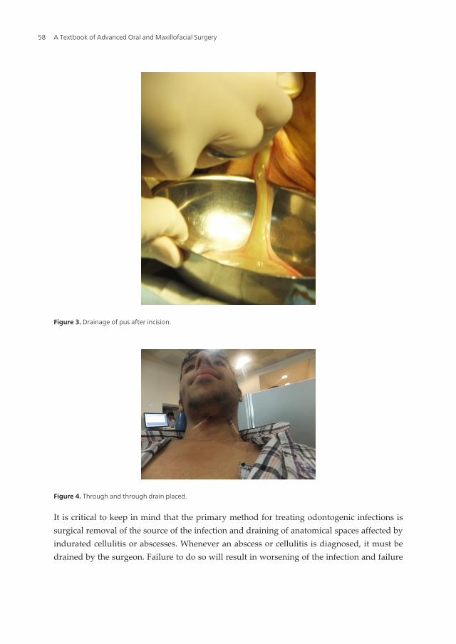
Figure 3. Drainage of pus after incision.
Figure 4. Through and through drain placed.
It is critical to keep in mind that the primary method for treating odontogenic infections issurgical removal of the source of the infection and draining of anatomical spaces affected byindurated cellulitis or abscesses. Whenever an abscess or cellulitis is diagnosed, it must bedrained by the surgeon. Failure to do so will result in worsening of the infection and failure
A Textbook of Advanced Oral and Maxillofacial Surgery58
of the infection to resolve, even if antibiotics are given. Even if a tooth cannot be opened orextracted, an incision and drainage procedure should be performed [12,13].
7. Specific infections
7.1. Osteomyelitis
Osteomyelitis is defined as inflammation of the bone. Different from other infectious circum‐stances seen in the jaws, it involves adjacent cortical plates and often periosteal tissues. Theincidence of osteomyelitis is much higher in the mandible because of the dense, poorlyvascularized cortical plates. It is much less common in the maxilla due to the excellent bloodsupply from multiple feeder vessels. In addition, the maxillary bone is much less dense thanthe mandible. Osteomyelitis has been associated with multiple systemic diseases, includingdiabetes, autoimmune states, malignancies, malnutrition, and acquired immunodeficiencysyndrome [14]. Medications linked to osteomyelitis include steroids, chemotherapeutic agents,and bisphosphonates [15,16]. Local conditions that adversely affect the blood supply can alsopredispose the host to a bone infection. Radiation therapy, osteopetrosis, and bone pathologycan alter the blood supply to the area and provide a potential foothold for osteomyelitis. Themost common cause of suppurative osteomyelitis is an odontogenic infection [17]. Dependingon the signs and symptoms, osteomyelitis can be classified as acute, subacute, and chronicforms. Radiographic changes do not appear immediately in the acute suppurative form ofosteomyelitis, because it may take about 2 weeks for the trabecular pattern of bone to changeand areas of radiolucency to start to appear, usually accompanied by periostitis. If acuteosteomyelitis is not treated effectively, it can lead to chronic suppurative osteomyelitis. Theinfection may be a manifestation of lowered patient resistance; this sometimes occurs inimmunosuppressed patients on medication or those suffering from an impaired immunedefense, as in acute leukemia, human immunodeficiency virus (HIV) infection, poorlycontrolled diabetes mellitus, or malnutrition.
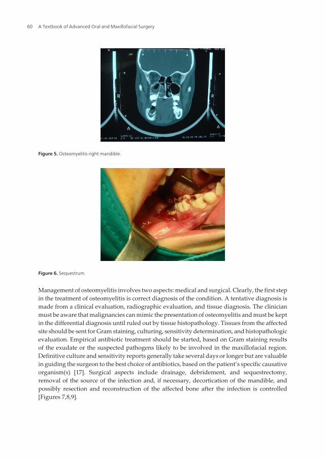
Clinically, the disease is dominated by pain and the development of intraoral and/or extraoralsinuses. Induration of soft tissues overlying the infected segments of the jawbones is markedand distension of the periosteum with pus or inflammatory exudate, which may cause trismusand difficulty in swallowing. Regional lymph nodes are usually tender and enlarged. Apathological fracture may develop if the inferior border of the mandible is damaged by theinfection process. The radiographic picture of chronic osteomyelitis is loss of detail of thetrabecular pattern of the osseous architecture, giving the bone a mottled or moth-eatenappearance. The ischemic or necrotic islands of bone tend to sequestrate, appearing moreradiopaque than the surrounding bone; these form a sequestrum of necrotic bone. [Figure 5,6]
In younger persons, subperiosteal new bone formation appears adjacent to the diseasedarea. This new bone, known as involucrum, tends to be structureless or granular in ap‐pearance radiographically and may surround the necrotic sequestrum and pus lying with‐in the bone (18).
Odontogenic Infectionshttp://dx.doi.org/10.5772/54645
59
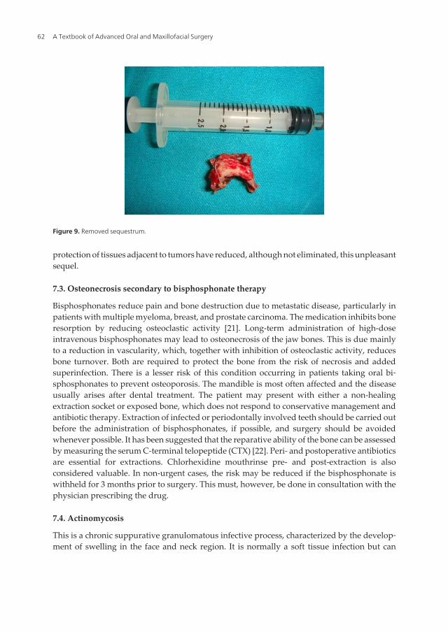
Management of osteomyelitis involves two aspects: medical and surgical. Clearly, the first stepin the treatment of osteomyelitis is correct diagnosis of the condition. A tentative diagnosis ismade from a clinical evaluation, radiographic evaluation, and tissue diagnosis. The clinicianmust be aware that malignancies can mimic the presentation of osteomyelitis and must be keptin the differential diagnosis until ruled out by tissue histopathology. Tissues from the affectedsite should be sent for Gram staining, culturing, sensitivity determination, and histopathologicevaluation. Empirical antibiotic treatment should be started, based on Gram staining resultsof the exudate or the suspected pathogens likely to be involved in the maxillofacial region.Definitive culture and sensitivity reports generally take several days or longer but are valuablein guiding the surgeon to the best choice of antibiotics, based on the patient’s specific causativeorganism(s) [17]. Surgical aspects include drainage, debridement, and sequestrectomy,removal of the source of the infection and, if necessary, decortication of the mandible, andpossibly resection and reconstruction of the affected bone after the infection is controlled[Figures 7,8,9].
Figure 5. Osteomyelitis right mandible.
Figure 6. Sequestrum.
A Textbook of Advanced Oral and Maxillofacial Surgery60
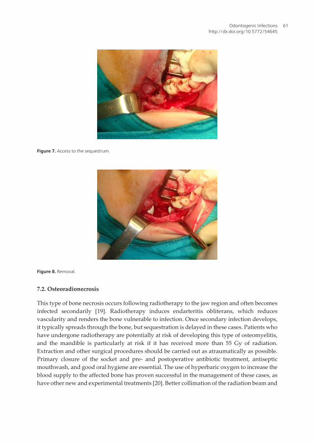
Figure 7. Access to the sequestrum.
Figure 8. Removal.
7.2. Osteoradionecrosis
This type of bone necrosis occurs following radiotherapy to the jaw region and often becomesinfected secondarily [19]. Radiotherapy induces endarteritis obliterans, which reducesvascularity and renders the bone vulnerable to infection. Once secondary infection develops,it typically spreads through the bone, but sequestration is delayed in these cases. Patients whohave undergone radiotherapy are potentially at risk of developing this type of osteomyelitis,and the mandible is particularly at risk if it has received more than 55 Gy of radiation.Extraction and other surgical procedures should be carried out as atraumatically as possible.Primary closure of the socket and pre- and postoperative antibiotic treatment, antisepticmouthwash, and good oral hygiene are essential. The use of hyperbaric oxygen to increase theblood supply to the affected bone has proven successful in the management of these cases, ashave other new and experimental treatments [20]. Better collimation of the radiation beam and
Odontogenic Infectionshttp://dx.doi.org/10.5772/54645
61
protection of tissues adjacent to tumors have reduced, although not eliminated, this unpleasantsequel.
7.3. Osteonecrosis secondary to bisphosphonate therapy
Bisphosphonates reduce pain and bone destruction due to metastatic disease, particularly inpatients with multiple myeloma, breast, and prostate carcinoma. The medication inhibits boneresorption by reducing osteoclastic activity [21]. Long-term administration of high-doseintravenous bisphosphonates may lead to osteonecrosis of the jaw bones. This is due mainlyto a reduction in vascularity, which, together with inhibition of osteoclastic activity, reducesbone turnover. Both are required to protect the bone from the risk of necrosis and addedsuperinfection. There is a lesser risk of this condition occurring in patients taking oral bi‐sphosphonates to prevent osteoporosis. The mandible is most often affected and the diseaseusually arises after dental treatment. The patient may present with either a non-healingextraction socket or exposed bone, which does not respond to conservative management andantibiotic therapy. Extraction of infected or periodontally involved teeth should be carried outbefore the administration of bisphosphonates, if possible, and surgery should be avoidedwhenever possible. It has been suggested that the reparative ability of the bone can be assessedby measuring the serum C-terminal telopeptide (CTX) [22]. Peri- and postoperative antibioticsare essential for extractions. Chlorhexidine mouthrinse pre- and post-extraction is alsoconsidered valuable. In non-urgent cases, the risk may be reduced if the bisphosphonate iswithheld for 3 months prior to surgery. This must, however, be done in consultation with thephysician prescribing the drug.
7.4. Actinomycosis
This is a chronic suppurative granulomatous infective process, characterized by the develop‐ment of swelling in the face and neck region. It is normally a soft tissue infection but can
Figure 9. Removed sequestrum.
A Textbook of Advanced Oral and Maxillofacial Surgery62
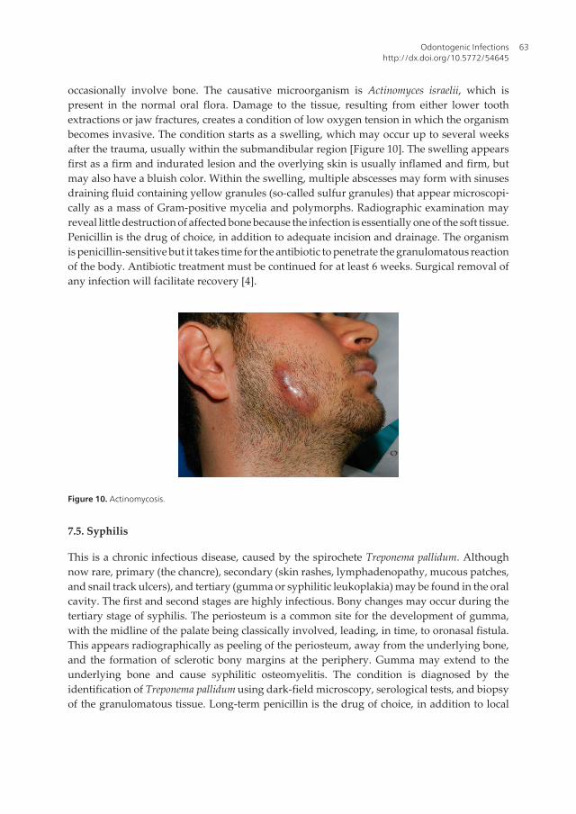
occasionally involve bone. The causative microorganism is Actinomyces israelii, which ispresent in the normal oral flora. Damage to the tissue, resulting from either lower toothextractions or jaw fractures, creates a condition of low oxygen tension in which the organismbecomes invasive. The condition starts as a swelling, which may occur up to several weeksafter the trauma, usually within the submandibular region [Figure 10]. The swelling appearsfirst as a firm and indurated lesion and the overlying skin is usually inflamed and firm, butmay also have a bluish color. Within the swelling, multiple abscesses may form with sinusesdraining fluid containing yellow granules (so-called sulfur granules) that appear microscopi‐cally as a mass of Gram-positive mycelia and polymorphs. Radiographic examination mayreveal little destruction of affected bone because the infection is essentially one of the soft tissue.Penicillin is the drug of choice, in addition to adequate incision and drainage. The organismis penicillin-sensitive but it takes time for the antibiotic to penetrate the granulomatous reactionof the body. Antibiotic treatment must be continued for at least 6 weeks. Surgical removal ofany infection will facilitate recovery [4].
Figure 10. Actinomycosis.
7.5. Syphilis
This is a chronic infectious disease, caused by the spirochete Treponema pallidum. Althoughnow rare, primary (the chancre), secondary (skin rashes, lymphadenopathy, mucous patches,and snail track ulcers), and tertiary (gumma or syphilitic leukoplakia) may be found in the oralcavity. The first and second stages are highly infectious. Bony changes may occur during thetertiary stage of syphilis. The periosteum is a common site for the development of gumma,with the midline of the palate being classically involved, leading, in time, to oronasal fistula.This appears radiographically as peeling of the periosteum, away from the underlying bone,and the formation of sclerotic bony margins at the periphery. Gumma may extend to theunderlying bone and cause syphilitic osteomyelitis. The condition is diagnosed by theidentification of Treponema pallidum using dark-field microscopy, serological tests, and biopsyof the granulomatous tissue. Long-term penicillin is the drug of choice, in addition to local
Odontogenic Infectionshttp://dx.doi.org/10.5772/54645
63
measures to deal with damaged soft tissue, sequestered bone, and involved teeth. The fourthstage of syphilis is rare; it affects the cardiovascular system, causing aortic aneurysms or aorticvalve incompetence. The central nervous system may also become involved, which may leadto dementia or spinal cord disease [4].
Author details
Onur Gonul*, Sertac Aktop, Tulin Satilmis, Hasan Garip and Kamil Goker
Department of Oral and Maxillofacial Surgery, Marmara University, Istanbul, Turkey
References
[1] Hought RT, Fitzgerald BE, Latta JE, Zallen, RD. Ludwig’s angina: report of two casesand review of the literature from 1945 to January 1979. J Oral Surg 1980;38:849–55.
[2] Miloro M. Peterson’s Principle of Oral and Maxillofacial Surgery. Second Edition.. InMiloro M.,Ghali G. E., Larsen P. E., Waite P. Editors. BC Decker Inc. 2004 p.277-79
[3] Flynn TR, Topazian RG. Infections of the oral cavity. In: Waite D, editor. Textbook ofpractical oral and maxillofacial surgery. 3rd Ed. Philadelphia (PA): Lea & Febiger;1987. p. 273–310.
[4] Andersson L. Oral and Maxillofacial Surgery. In Andersson L., Kahnberg K. E., Pog‐rel M.A.editors Wiley Blackwell 2010 p.280-314
[5] Flynn T. Anatomy and surgery of oral and maxillofacial infections. J Oral MaxillofacSurg 2006; 64: 100–5
[6] Malik N. A. Textbook of Oral and Maxillofacial Surgery. Secpnd Edition. JaypeBrothers Medical Publishers (P) Ltd. India 2008 p. 587-636
[7] Hupp J. R. Contemporary Oral and Maxillofacial Surgery. In Hupp J. R., Ellıs III E.,Tucker R. M. Editors.Fifth Edition. Mosby Elsevier 2008
[8] Miller WD, Furst IM, Sandor GKB, et al. A prospective blinded comparison of clinicalexamination and computed tomography in deep neck infections. Laryngoscope 1999;109:1873–9
[9] Flynn TR, Halpern LR. Antibiotic selection in head and neck infections. Oral Maxillo‐fac Surg Clin North Am 2003;15:17–38.
[10] Kuriyama T, Karasawa T, Nakagawa K, Nakamura S, Yamamoto E. Anatomical sus‐ceptibility of major pathogens of orofacial odontogenic infections to 11 β-lactam anti‐biotics. Oral Microbiol Immunol 2002; 17: 285–9
A Textbook of Advanced Oral and Maxillofacial Surgery64
[11] Fazakerley MW, McGowan P, Hardy P, et al. A comparative study of cephradine,amoxycillin and phenoxymethylpenicillin in the treatment of acute dentoalveolar in‐fection.Br Dent J 1993;174:359–63.
[12] Flynn T. Surgical management of oral infections. Atlas Oral Maxillofac Surg ClinNorth Am 2000; 8: 77–100.
[13] Flynn TR. The timing of incision and drainage. In: Piecuch JF, editor. Oral and maxil‐lofacial surgery knowledge update 2001. Rosemont (IL): American Association of Or‐al and Maxillofacial Surgeons; 2001. p. 75–84.
[14] Marx RE. Chronic osteomyelitis of the jaws. Oral Maxillofac Surg Clin North Am1991;3:367–81.
[15] Marx RE. Pamidronate and zoledronate induced avascular necrosis of the jaws. J Or‐al Maxillofac Surg 2003;61:1115–8.
[16] .Migliorati CA. Bisphosphonates and oral cavity avascular bone necrosis. J Clin On‐col2003;21:4253–4.
[17] Koorbush GF, Fotos P, Goll TK. Retrospective study of osteomylitis. Aetiology, dem‐ographics, risk factors and management in 35 cases. Oral Surg Oral Med Oral Path‐ol1992; 74: 149–54.
[18] Cierny G,Mader J, Pennick J. A clinical staging system for osteomyelitis. ContempOrthop 1985;10:17.
[19] Marx RJ. Osteonecrosis: a new concept of its pathology. J Oral Maxillofac Surg 1983;41: 283–8.
[20] Marx RE, Johnson RP, Kline SN. Prevention of osteoradionecrosis: a randomized pro‐spective clinical trial of hyperbaric oxygen versus penicillin. J Am Dent Assoc 1985;111:49–54.
[21] Mavrokokki T, Cheng A, Stein B, Goss A. Nature and frequency of bisphosphonateassociated osteonecrosis of the jaws in Australia. J Oral Maxillofac Surg 2009; 65: 415–23.
[22] Lyons A, Ghazali N. Oral bisphosphonate induced osteonecrosis, risk factors, predic‐tion of risk using CTX testing, prevention, and treatment. Br J Oral Maxillofac Surg2008; 46: 653–61.
Odontogenic Infectionshttp://dx.doi.org/10.5772/54645
65