
Racisim in Trinidad by Charleen Agostini - Process Work Institute
Upload
adele-toddCategory
view
216download
0
Ocular Pathology Case
Kari Eisley, M.D.
PGY3
Pathological images © Charleen T. Chu, 2010

Clinical History
• 66 yo white female
• Presents to ED with right eye redness, drainage and decreased vision x 2 weeks
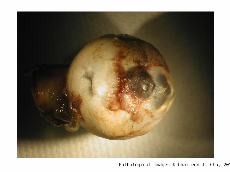
• Has a history of mental retardation• Eye enucleated (PHS10-11472)

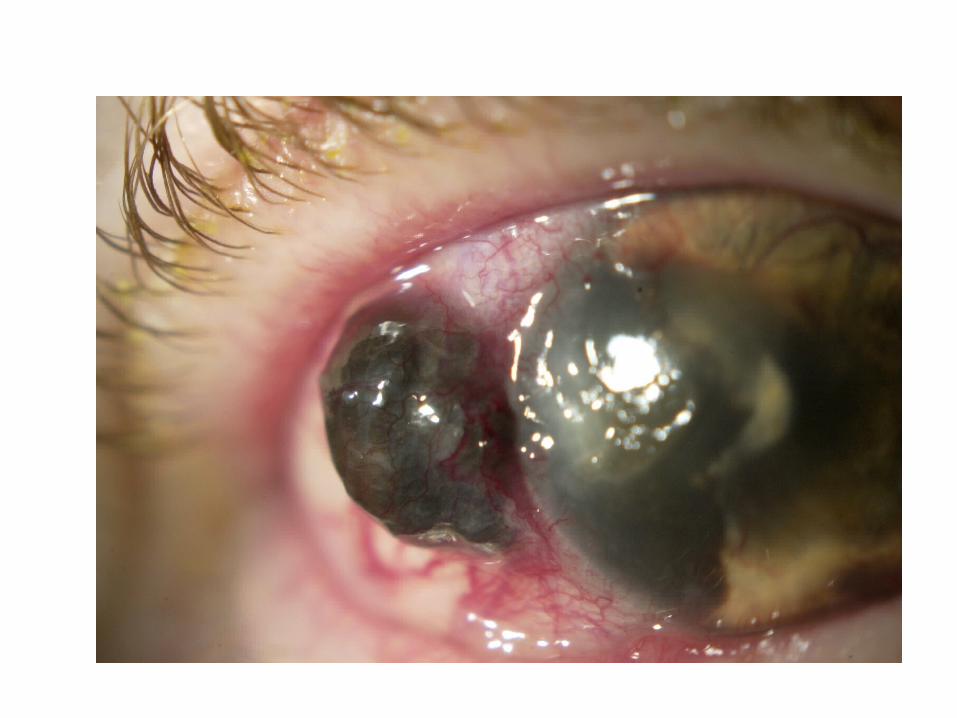

Extrascleral Mass
Cornea
Mass in Anterior Chamber
Pathological images © Charleen T. Chu, 2010
Pathological images © Charleen T. Chu, 2010
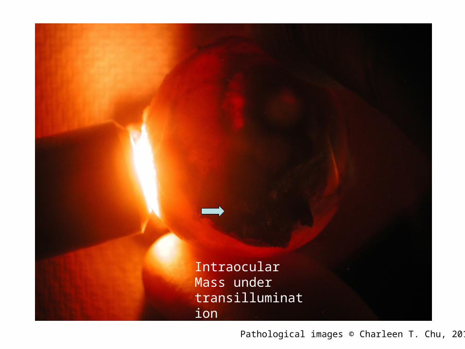
Intraocular Mass under transillumination
Pathological images © Charleen T. Chu, 2010
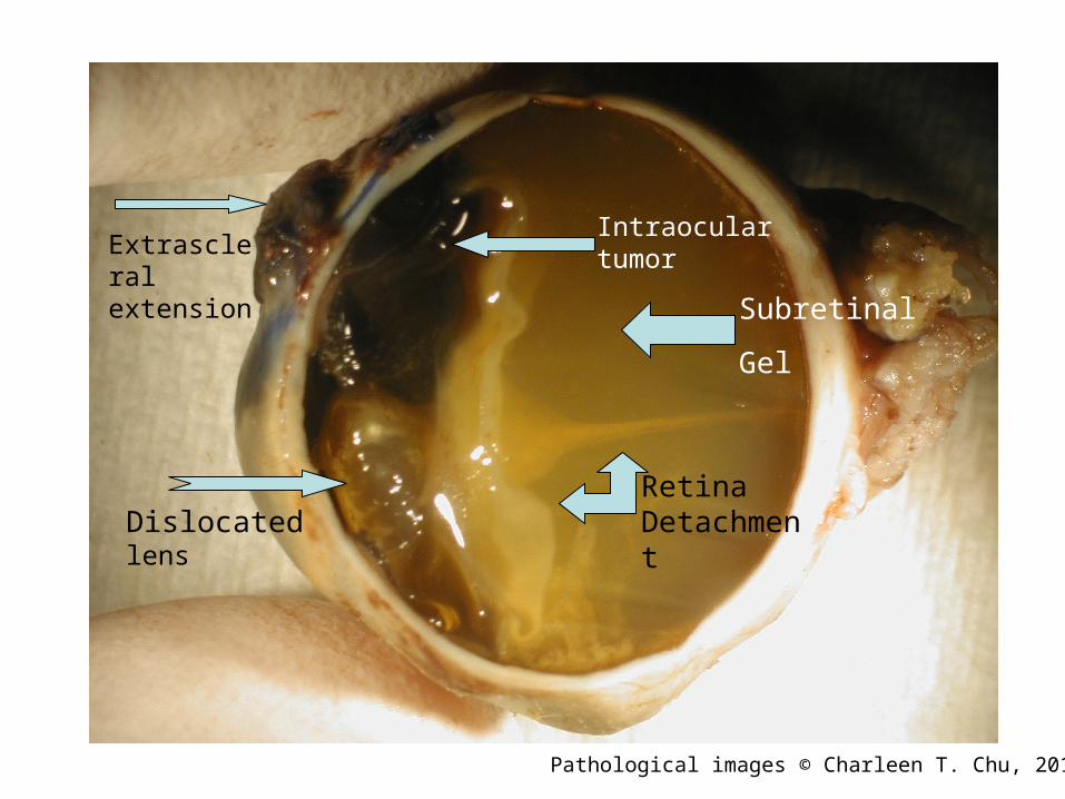
Retina DetachmentDislocated
lens
Extrascleral extension
Intraocular tumor
Subretinal
Gel
Pathological images © Charleen T. Chu, 2010
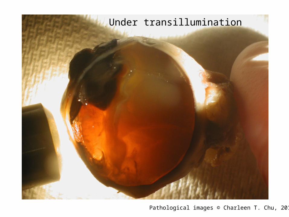
Under transillumination
Pathological images © Charleen T. Chu, 2010
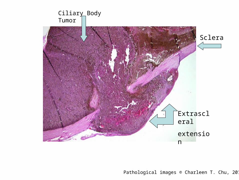
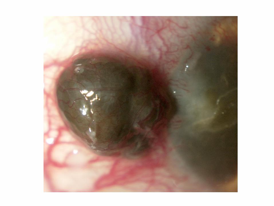
Sclera
Ciliary Body Tumor
Extrascleral
extension
Pathological images © Charleen T. Chu, 2010

Dislocated Lens Anterior Subcapsular Cataract
Posterior Polar Cataract
Tumor
Detached Retina with Subretinal Gel
Pathological images © Charleen T. Chu, 2010
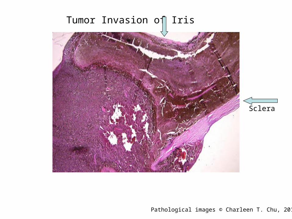
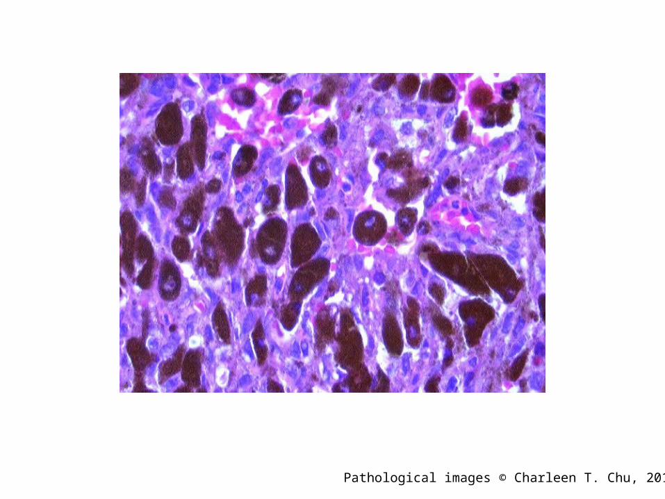
Tumor Invasion of Iris
Sclera
Pathological images © Charleen T. Chu, 2010
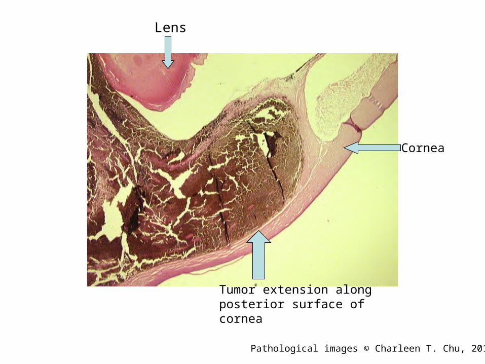
Lens
Cornea
Tumor extension along posterior surface of cornea
Pathological images © Charleen T. Chu, 2010
Pathological images © Charleen T. Chu, 2010
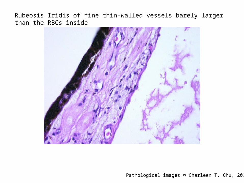
Rubeosis Iridis of fine thin-walled vessels barely larger than the RBCs inside
Pathological images © Charleen T. Chu, 2010
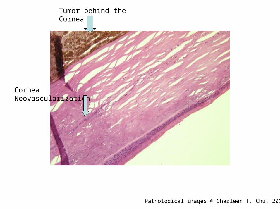
Tumor behind the Cornea
Cornea Neovascularization
Pathological images © Charleen T. Chu, 2010
Pathological images © Charleen T. Chu, 2010
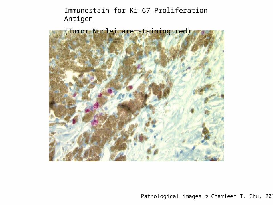
Immunostain for Ki-67 Proliferation Antigen
(Tumor Nuclei are staining red)
Pathological images © Charleen T. Chu, 2010
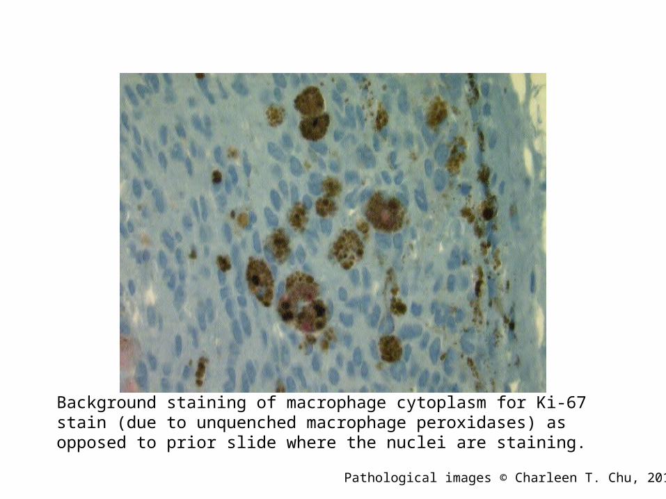
Background staining of macrophage cytoplasm for Ki-67 stain (due to unquenched macrophage peroxidases) as opposed to prior slide where the nuclei are staining.
Pathological images © Charleen T. Chu, 2010
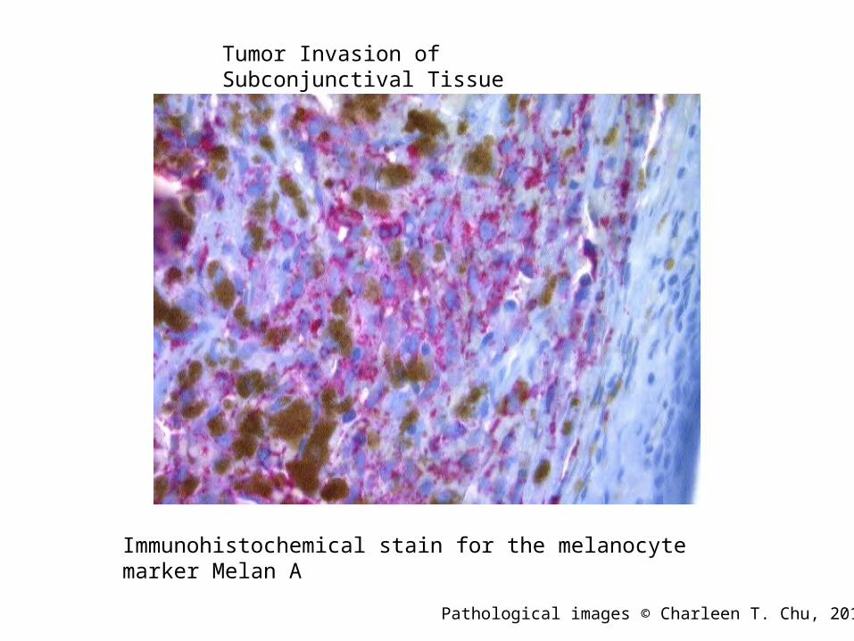
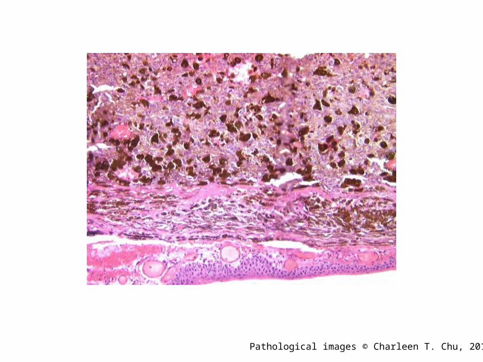
Immunohistochemical stain for the melanocyte marker Melan A
Tumor Invasion of Subconjunctival Tissue
Pathological images © Charleen T. Chu, 2010

Melan A Melanocyte Immunostain
Pathological images © Charleen T. Chu, 2010

Immunohistochemical Stain HMB-45
Melanophages
Tumor cells with positive HMB-45
Pathological images © Charleen T. Chu, 2010
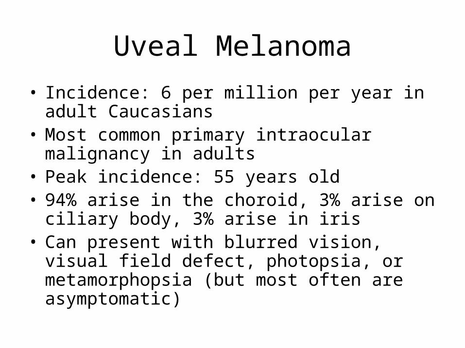
Uveal Melanoma
• Incidence: 6 per million per year in adult Caucasians
• Most common primary intraocular malignancy in adults
• Peak incidence: 55 years old• 94% arise in the choroid, 3% arise on ciliary
body, 3% arise in iris• Can present with blurred vision, visual field
defect, photopsia, or metamorphopsia (but most often are asymptomatic)
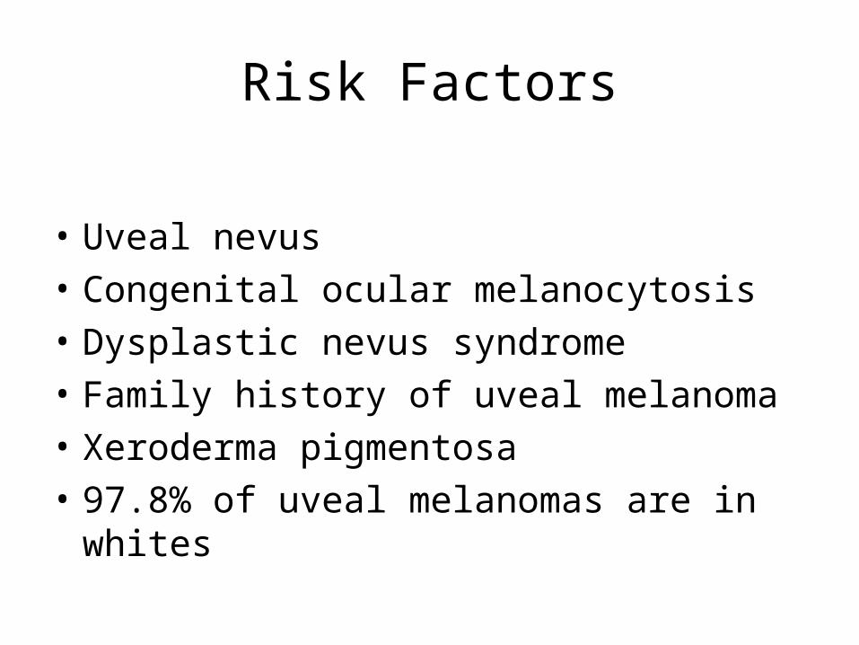
Risk Factors
• Uveal nevus
• Congenital ocular melanocytosis
• Dysplastic nevus syndrome
• Family history of uveal melanoma
• Xeroderma pigmentosa
• 97.8% of uveal melanomas are in whites
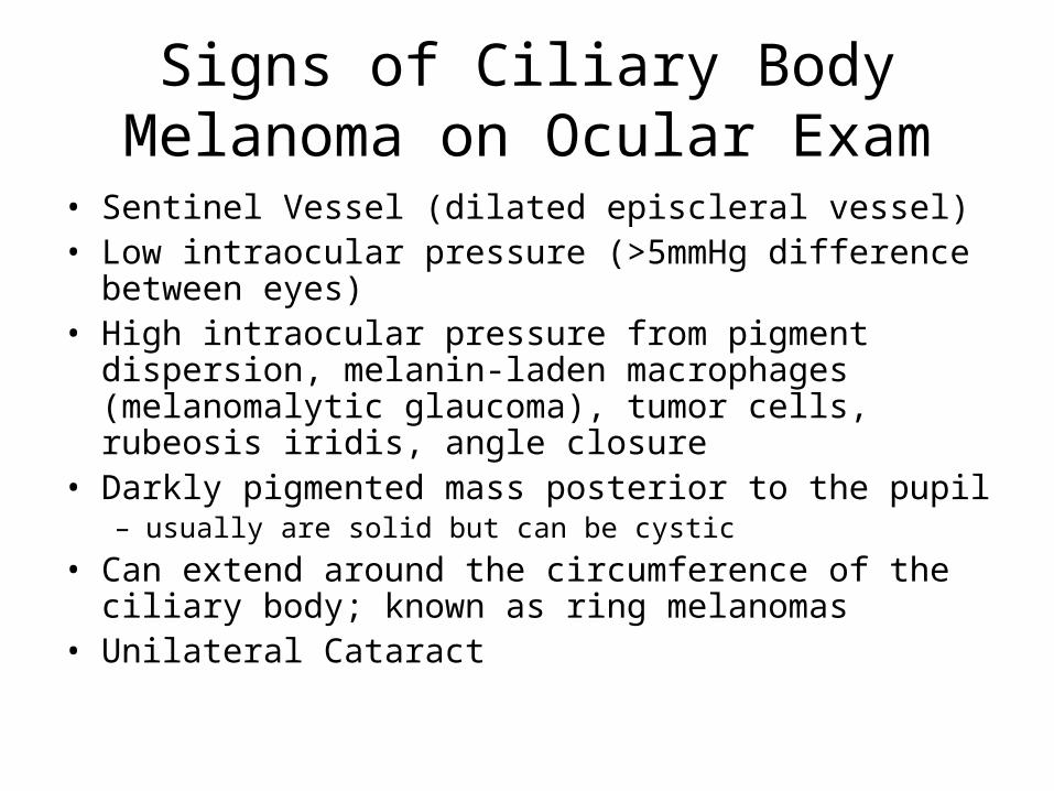
Signs of Ciliary Body Melanoma on Ocular Exam
• Sentinel Vessel (dilated episcleral vessel)• Low intraocular pressure (>5mmHg difference between
eyes)• High intraocular pressure from pigment dispersion,
melanin-laden macrophages (melanomalytic glaucoma), tumor cells, rubeosis iridis, angle closure
• Darkly pigmented mass posterior to the pupil– usually are solid but can be cystic
• Can extend around the circumference of the ciliary body; known as ring melanomas
• Unilateral Cataract
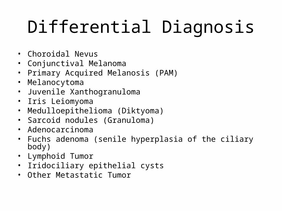
Differential Diagnosis• Choroidal Nevus• Conjunctival Melanoma• Primary Acquired Melanosis (PAM)• Melanocytoma• Juvenile Xanthogranuloma• Iris Leiomyoma• Medulloepithelioma (Diktyoma)• Sarcoid nodules (Granuloma)• Adenocarcinoma• Fuchs adenoma (senile hyperplasia of the ciliary body)• Lymphoid Tumor• Iridociliary epithelial cysts • Other Metastatic Tumor
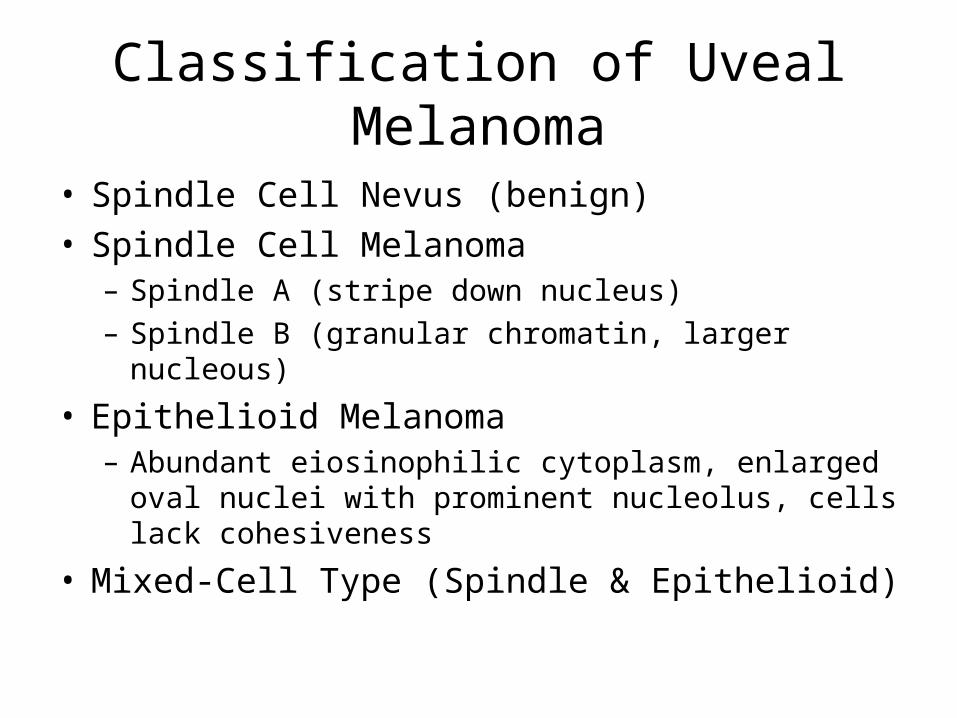
Classification of Uveal Melanoma
• Spindle Cell Nevus (benign)• Spindle Cell Melanoma
– Spindle A (stripe down nucleus)– Spindle B (granular chromatin, larger nucleous)
• Epithelioid Melanoma– Abundant eiosinophilic cytoplasm, enlarged oval
nuclei with prominent nucleolus, cells lack cohesiveness
• Mixed-Cell Type (Spindle & Epithelioid)
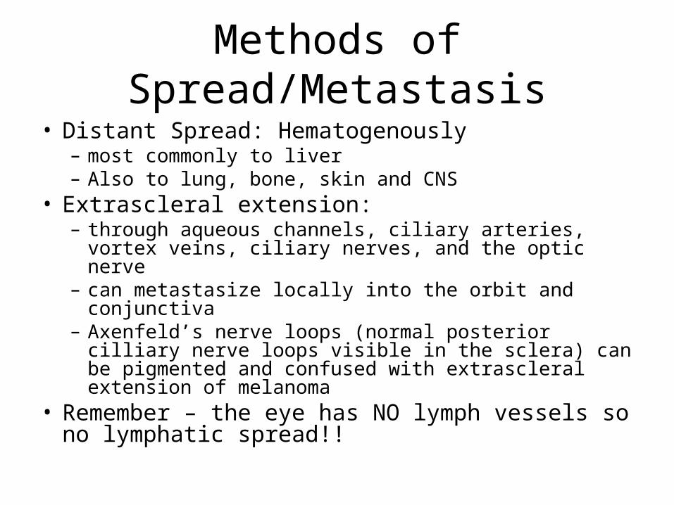
Methods of Spread/Metastasis
• Distant Spread: Hematogenously – most commonly to liver– Also to lung, bone, skin and CNS
• Extrascleral extension: – through aqueous channels, ciliary arteries, vortex
veins, ciliary nerves, and the optic nerve– can metastasize locally into the orbit and conjunctiva– Axenfeld’s nerve loops (normal posterior cilliary nerve
loops visible in the sclera) can be pigmented and confused with extrascleral extension of melanoma
• Remember – the eye has NO lymph vessels so no lymphatic spread!!

Prognosis
• Mortality of Ciliary Body Melanoma is 30-50% within 10 years from diagnosis
• Ciliary Body Melanoma has a worse prognosis than posterior choroidal melanomas, likely due to delayed diagnosis
• Spindle Cell – Best• Epithelioid – Worst• Mixed-cell Type – Intermediate• With Metastatic disease : < 6 months
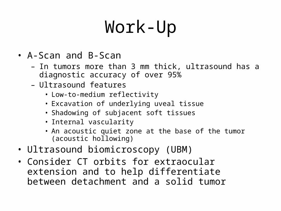
Work-Up
• A-Scan and B-Scan– In tumors more than 3 mm thick, ultrasound has a diagnostic
accuracy of over 95% – Ultrasound features
• Low-to-medium reflectivity • Excavation of underlying uveal tissue • Shadowing of subjacent soft tissues • Internal vascularity • An acoustic quiet zone at the base of the tumor (acoustic hollowing)
• Ultrasound biomicroscopy (UBM) • Consider CT orbits for extraocular extension and to help
differentiate between detachment and a solid tumor

Metastatic Work-Up
• Importance: – To determine medical risk of surgery– If metastatic disease is present, enucleation is
inappropriate unless the eye is painful
• Thorough physical, especially to hepatic abdominal region & skin/subcutaneous tissues
• LFTs, CT/Ultrasound of liver• Chest X-Ray
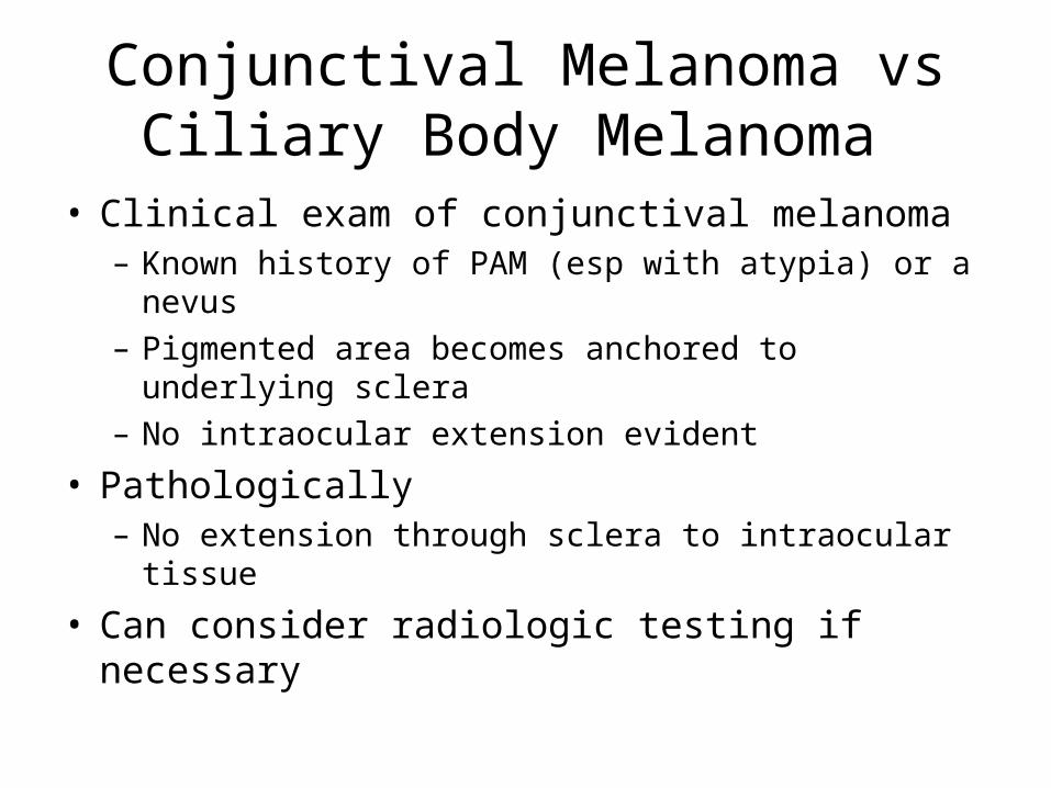
Conjunctival Melanoma vs Ciliary Body Melanoma
• Clinical exam of conjunctival melanoma– Known history of PAM (esp with atypia) or a nevus– Pigmented area becomes anchored to underlying
sclera– No intraocular extension evident
• Pathologically– No extension through sclera to intraocular tissue
• Can consider radiologic testing if necessary
Ciliary Body Melanoma Treatment
• Many factors considered in treatment approach:– Visual acuity of the affected eye– visual acuity of the contralateral eye – intraocular pressure– ocular structures involved – size of the tumor – age and general health of the patient– presence of metastases

Ciliary Body Melanoma Treatment
1. Enucleation has been the preferred treatment for advanced and complicated tumors, especially when other treatment has failed
2. External beam irradiation with either protons or helium ions is a frequently used alternative method to treat medium-size tumors (<10 mm in height and 15 mm in diameter).
– Complications include cataract, dry eyes, radiation retinopathy, and rubeosis iridis
3. Plaque brachytherapy is a widely accepted alternative to enucleation for medium-size posterior uveal melanomas
– Local recurrence rate of about 12%– Can still get radiation retinopathy but less frequently than external beam
4. Block excision, or sclerouvectomy, is an alternative treatment method for ciliary body melanomas covering less than 4 clock hours of the circumference.
5. If there is extensive orbital extension, consider orbital exenteration• Overall, remains controversial• adjuvant systemic treatment is not currently advocated without metastases
Metastatic Ciliary Body Melanoma Treatment
• Systemic chemotherapy is the primary treatment
• Enucleation is only offered as palliative treatment if the eye is painful


![Pulmonary subspeciality rounds Dr.Krock [pulmonology] Dr.Poddutoori [PGY3, I.M]](https://static.fdocuments.us/doc/165x107/56649c7d5503460f94931f73/pulmonary-subspeciality-rounds-drkrock-pulmonology-drpoddutoori-pgy3.jpg)