Ocular and Periocular Tumors in Cats - In Focus · PDF fileOcular and Periocular Tumors in...
72
Ocular and Periocular Tumors in Cats Richard R Dubielzig
Transcript of Ocular and Periocular Tumors in Cats - In Focus · PDF fileOcular and Periocular Tumors in...

Ocular and Periocular Tumorsin Cats
Richard R Dubielzig
Anatomic distribution of feline Anatomic distribution of feline primary ocular neoplasia (n = 2599)primary ocular neoplasia (n = 2599)
82%
12.3%
4.4%
1.15%


Melanoma: 1510 of 2766 tumors or 55%
– Diffuse Iris Melanoma ... 1340 (263 Early)– “Atypical” ….. 27– Limbal ……… 46– Conjunctival… 27– 70 mostly DIM improperly labeled
Kalishman JV, Chappell R, Flood LA, Dubielzig RR (1998).A matched observational study of survival in cats with enucleation due to diffuse iris melanoma. Vet. Ophthal. 1: 25-29.
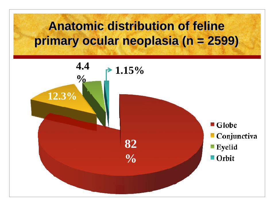
Typical Clinical Appearance of Feline Diffuse Iris Melanoma
Asymmetrical Darkening of the IrisThis process can occur rapidly
or it can take years

Typical Histopathologic Appearance of Feline Diffuse Iris Melanoma

Typical Gross Appearance of Feline Diffuse Iris Melanoma
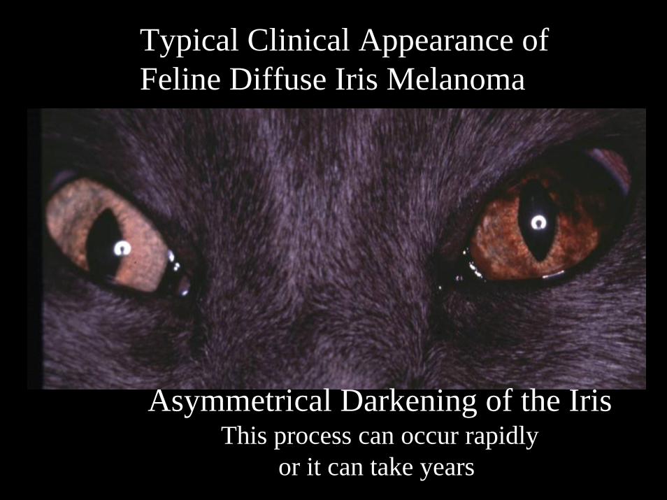
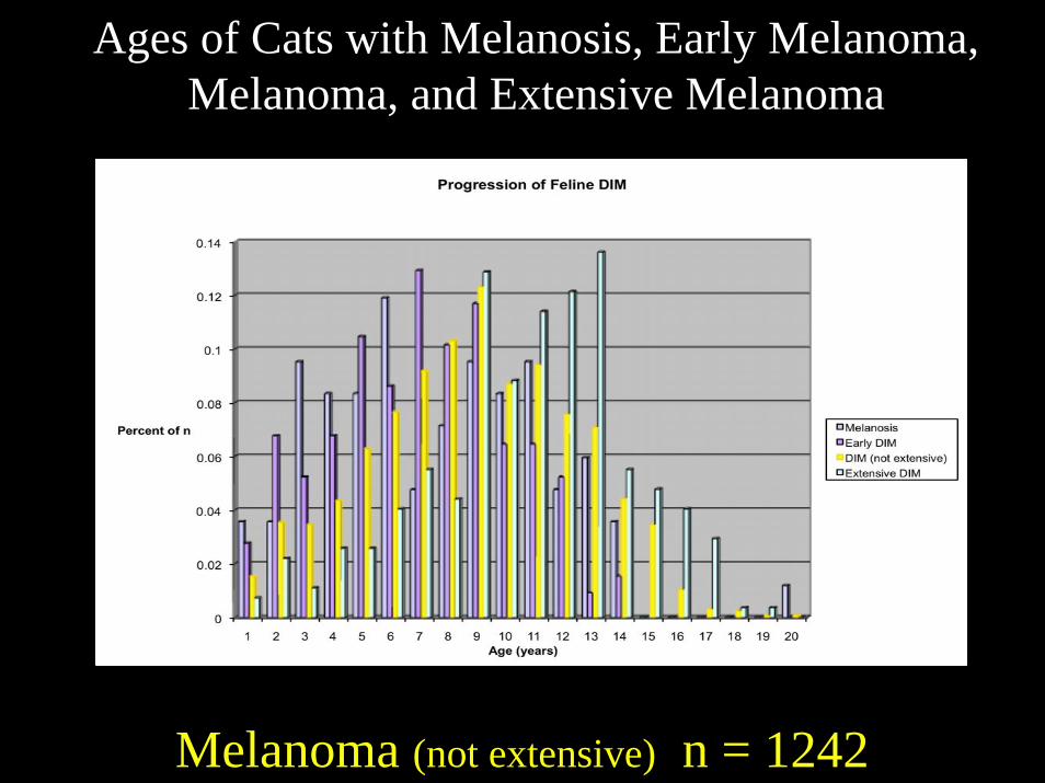
Ages of Cats with Melanosis, Early Melanoma, Melanoma, and Extensive Melanoma
0
0.02
0.04
0.06
0.08
0.1
0.12
0.14
Percent of n
1 2 3 4 5 6 7 8 9 10 11 12 13 14 15 16 17 18 19 20Age (years)
Progression of Feline DIM
MelanosisEarly DIMDIM (not extensive)Extensive DIM
Melanosis n = 84

Ages of Cats with Melanosis, Early Melanoma, Melanoma, and Extensive Melanoma
0
0.02
0.04
0.06
0.08
0.1
0.12
0.14
Percent of n
1 2 3 4 5 6 7 8 9 10 11 12 13 14 15 16 17 18 19 20Age (years)
Progression of Feline DIM
MelanosisEarly DIMDIM (not extensive)Extensive DIM
Early Melanoma n = 325
Ages of Cats with Melanosis, Early Melanoma, Melanoma, and Extensive Melanoma
Melanoma (not extensive) n = 1242

Ages of Cats with Melanosis, Early Melanoma, Melanoma, and Extensive Melanoma
0
0.02
0.04
0.06
0.08
0.1
0.12
0.14
Percent of n
1 2 3 4 5 6 7 8 9 10 11 12 13 14 15 16 17 18 19 20Age (years)
Progression of Feline DIM
MelanosisEarly DIMDIM (not extensive)Extensive DIM
Extensive Melanoma n = 272

Metastatic Potential of Feline Diffuse Iris Melanoma
All of the cases with metastasisin the COPLOW collection wereextensive in the original enucleation

Evisceration Followed by IntrascleralProsthesis is Not Recommended
in Cats with FDIM

Cats with Extensive FDIM atthe Time of Enucleation are MoreLikely to Have a Shortened Life
7 of 9 cats alive at follow-up
6 of 12 cats alive at follow-up
2 of 13 cats alive at follow-up

Early Stages of Feline Diffuse Iris Melanoma:
• Melanosis• Early Melanoma

Melanosis
Proliferating melanocytes are entirely limited to the anterior
surface of the iris
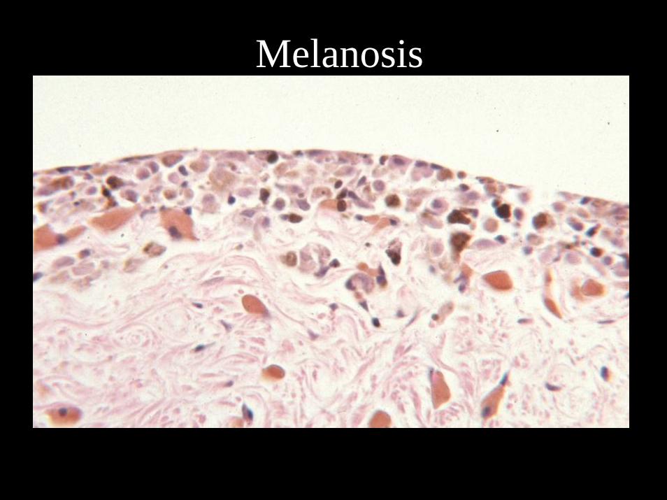
Melanosis

Early FDIMTumor confined to the iris
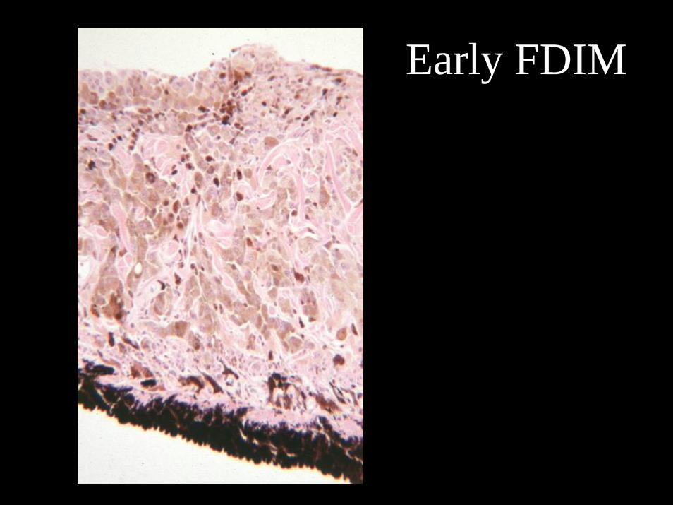
Early FDIM

Feline Ocular Post-traumatic Sarcoma:325 of 4035 tumors, or 5.5%
Male Bias• Spindle cell variant, 67%
– 217 cases, 43 “early”– Lens epithelial origin
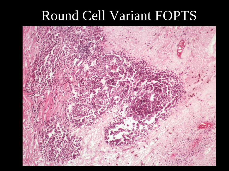
• Round cell variant, 21%– 69 cases, 5 “early”– Variant of lymphoma
• Osteosarcoma/Chondrosarcoma, 10%– 33 OSA cases– 6 Chondrosarcoma cases– Unknown cell of origin
Feline Ocular Post-traumatic Sarcoma
• Almost all cases have documented chronic eye disease– 81 cases have a documented traumatic event– Time between trauma and enucleation
• 60 cases have the dates recorded• Average time is 6.35 years• Range is 1 to 17 years

Reasons to believe FOPTS is related to trauma
• Lens capsule rupture• History of trauma or abnormal eye• Time between trauma and tumor
– Between 2 months and 15 years


Early Spindle Cell Variant FOPTS
Spindle Cell PTS with
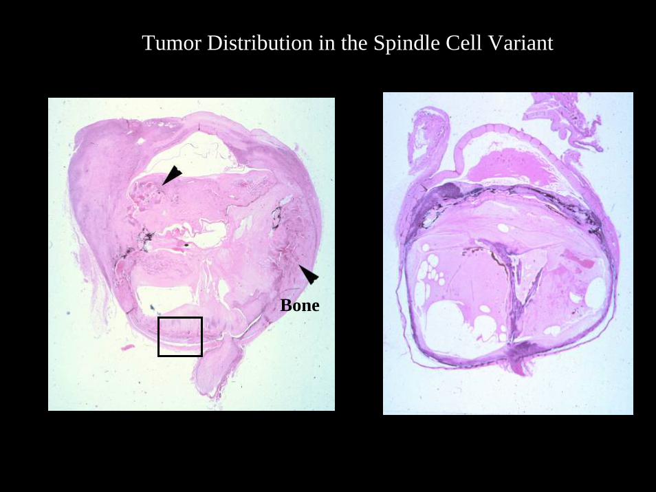
Tumor Distribution in the Spindle Cell Variant
Bone

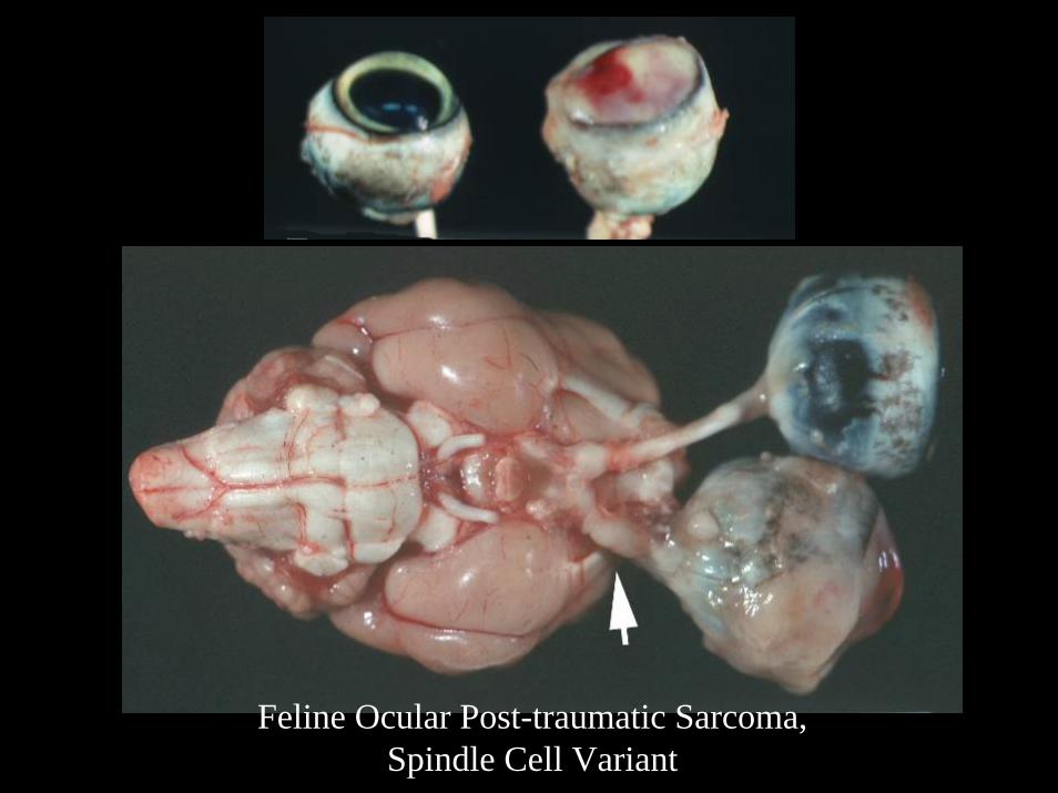
Feline Ocular Post-traumatic Sarcoma, Spindle Cell Variant
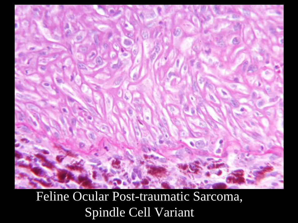
Feline Ocular Post-traumatic Sarcoma,Spindle Cell Variant

Cellular Features of Spindle Cell FOPTS
Collagen 4
Vimentin αA Crystallin
Feline Ocular Post-traumatic Sarcoma,Spindle Cell Variant

Follow-up Spindle Cell Variant
• Cases which have extended beyond the sclera have a bad prognosis– Local recurrence– Extension towards the brain
• Cases removed within the sclera have a good prognosis
• 8% of traumatized globes removed prophylactically have early FOPTS

Round Cell Variant FOPTS

Round Cell PTS
Round Cell Variant FOPTS
Round Cell Variant FOPTS


FOPTS Osteosarcoma
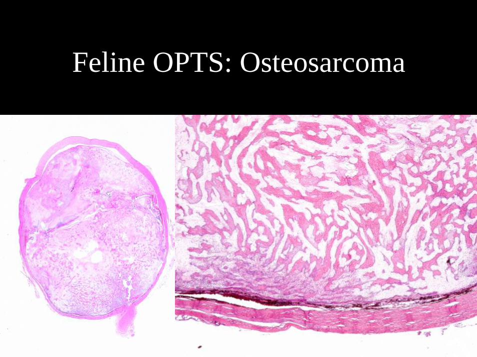
Feline OPTS: Osteosarcoma

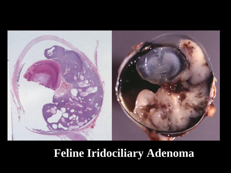
Feline Iridociliary Epithelial Tumors
• 102 of 2766 neoplastic cases, ~4%• Tend to be non-pigmented• Solid• Cavitated• About half have metaplastic
bone• Vimentin+, Cytokeratin-
Neglected Feline Iridociliary Adenoma

Feline Iridociliary Epithelial Tumors
Feline Iridociliary Adenoma

Feline Iridociliary Adenoma

Feline Iridociliary AdenomaSpindle Cells

Feline Iridociliary Epithelial TumorsImmunohistochemistry
NSE+ 100%Cytokeratin+ 20%Not related to tumor type
Vimentin+ 34%


Feline Conjunctival and Lid Squamous Cell Carcinoma
•Total SCC in the Database: 233•Multifocal (Bowenoid): 15•Orbital: 30•Associated with lymphocytic inflammation: 53•No age or breed associations

Feline Conjunctival and Lid Squamous Cell Carcinoma

Feline Conjunctival and Lid Squamous Cell Carcinoma
Multifocal or Bowenoid

Feline Orbital Squamous Cell CarcinomaDDx Feline Restrictive Orbital Myofibroblastic Sarcoma
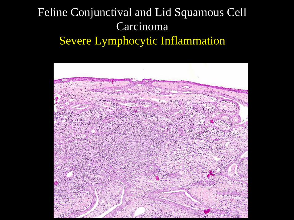
Feline Conjunctival and Lid Squamous Cell Carcinoma
Severe Lymphocytic Inflammation
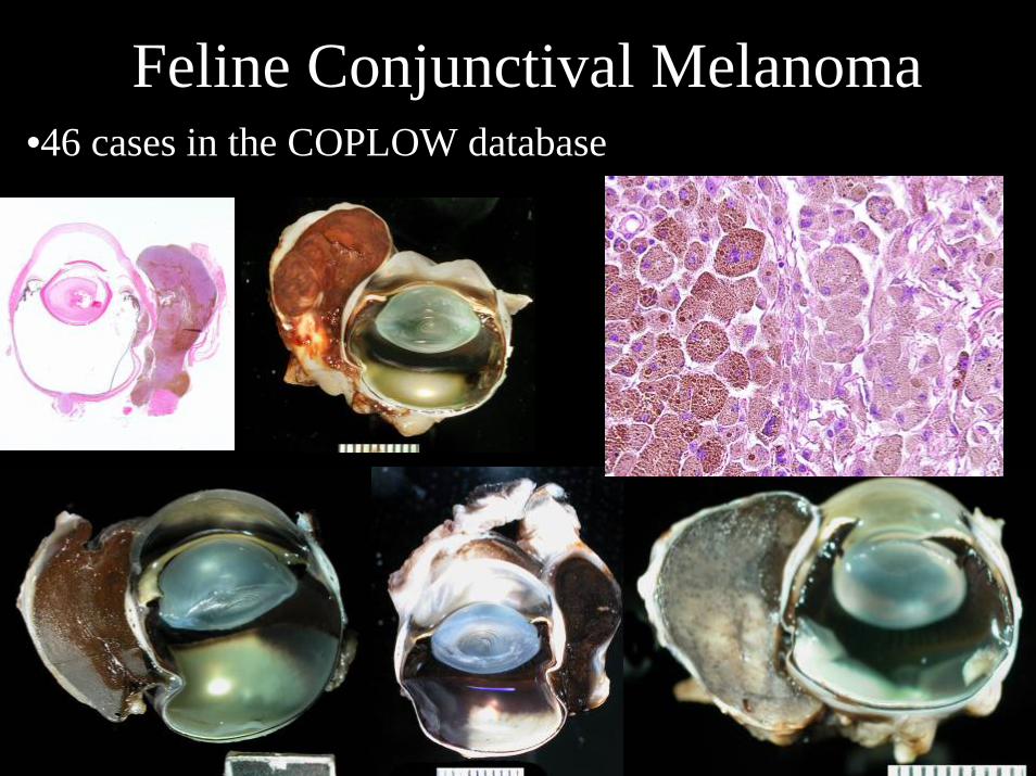
Feline Conjunctival Melanoma•46 cases in the COPLOW database

Feline Conjunctival Surface Adenocarcinoma
•Formerly Mucoepidermoid Carcinoma•18 cases in the COPLOW database•Malignant potential

Feline Conjunctival Surface Adenocarcinoma

Feline Conjunctival Surface AdenocarcinomaMetastatic Potential
L


Feline HidrocystomaApocrine gland originSiamese predilection

Feline Eyelid Peripheral Nerve Sheath Tumor
36 cases in the COPLOW Database
S100
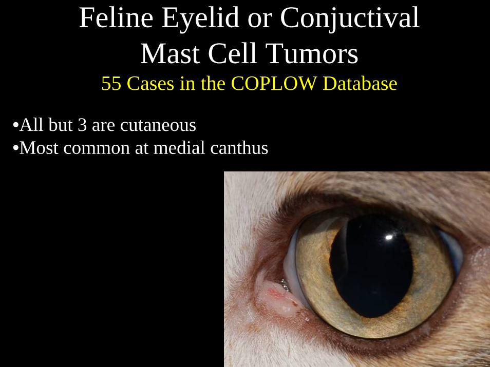
Feline Eyelid or ConjuctivalMast Cell Tumors
55 Cases in the COPLOW Database
•All but 3 are cutaneous•Most common at medial canthus

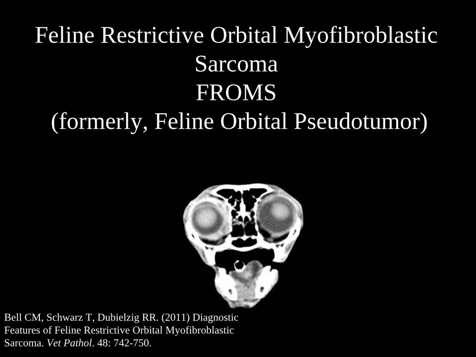
Feline Restrictive Orbital MyofibroblasticSarcomaFROMS
(formerly, Feline Orbital Pseudotumor)
Bell CM, Schwarz T, Dubielzig RR. (2011) Diagnostic Features of Feline Restrictive Orbital MyofibroblasticSarcoma. Vet Pathol. 48: 742-750.

14 cases of FROMS• Breed:
– 9 DSH– 3 DLH– 1 Maine Coon– 1 unknown
• Gender– MN = 5 FS = 9
• Age– Mean = 10.5 years, Median = 10 years– Range = 4 - 16 years
• Unilateral = 13 Bilateral = 1• Oral lesions = 1

Clinical Characteristics• Restricted mobility of globe and eyelids• Thickened and distorted eyelids • Profound corneal disease

FROMS Clinical Characteristics• Thickening +/- ulceration of lips• Proliferative gingival lesions (neoplastic?)
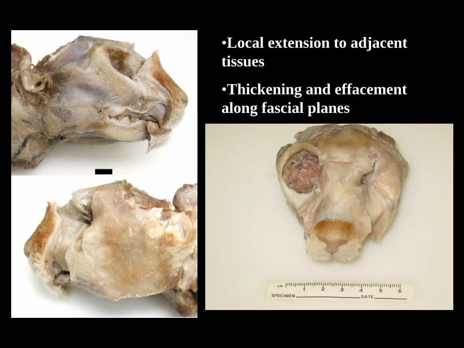
•Local extension to adjacent tissues
•Thickening and effacement along fascial planes
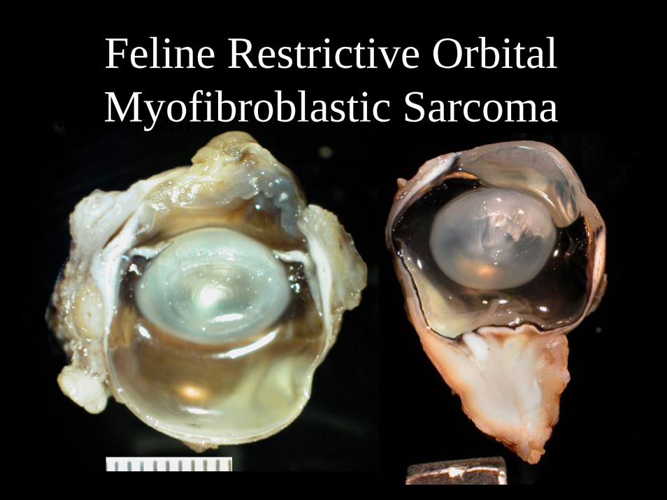
•Severe corneal disease
•Dense, fibrous orbital tissues
•Globe spared
Feline Restrictive Orbital Myofibroblastic Sarcoma

Feline Restrictive Orbital Myofibroblastic Sarcoma

Feline Restrictive Orbital Myofibroblastic Sarcoma

Subepithelial neoplastic cellsare not SMA+, unlike the remainder of the tumor
Smooth Muscle Actin (SMA)

Tooth
Orbit
Orbit from necropsy specimen
Orbit
Tumor in bone

FROMS Histopathology:
•Spindle cells in irregular short bundles with collagenous matrix
•Bland nuclear profile
•Mitotic activity virtually absent
Trichrome

Total + -Vimentin 8 8 0S-100 8 8 0SMA 8 8 0Melan A 2 0 2
FROMS Immunohistochemistry

Clinical Progression & Survival
• 9 of 10 cases with adequate follow-up had spread to the contralateral eye and/or oral cavity/lips
• All cats (5) that were confirmed deceased were euthanized due to progressive FROMS
• Of 3 cats currently living, 2 have signs of progressive FROMS
Feline Restrictive Orbital Myofibroblastic Sarcoma: Summary
• FROMS behavior is locally invasive and severely restricts the mobility of globe, eyelids and lips
• Morphology suggests an infiltrative myofibroblasticsarcoma, seldom forms a mass lesion, lacks cellular atypia
• Diagnosis requires histopathology plus clinical picture• Distribution and extent in the oral cavity and elsewhere
in the head is not obvious at the first diagnosis but becomes very apparent at necropsy
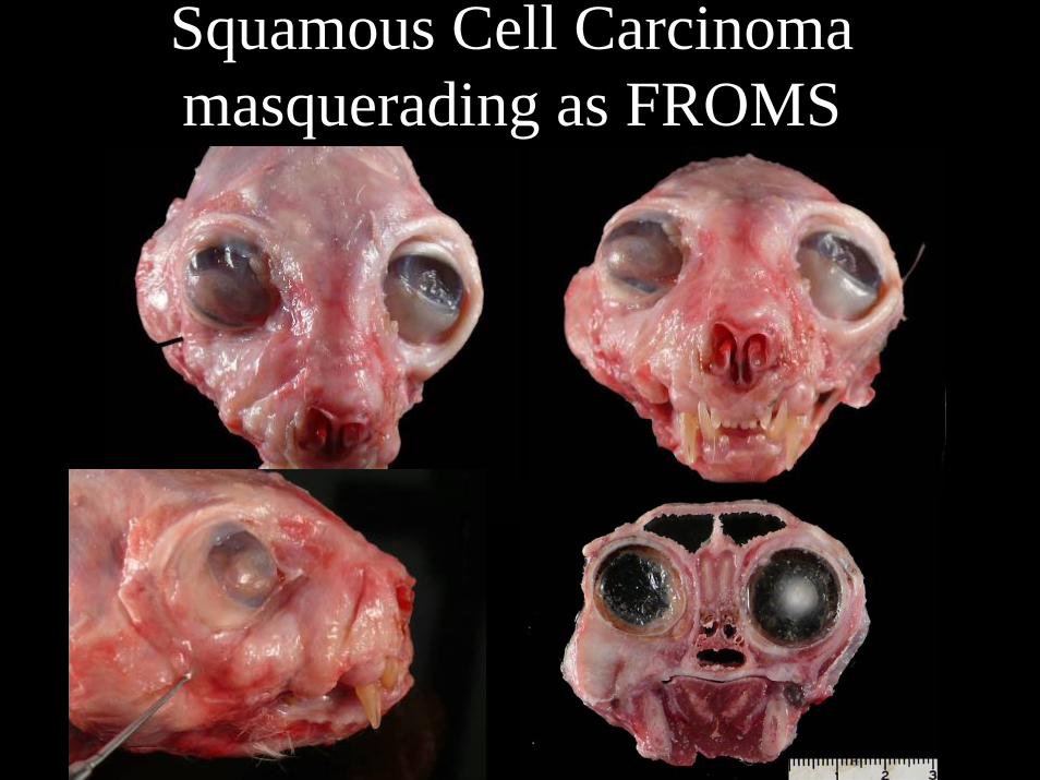
Squamous Cell Carcinomamasquerading as FROMS