Obturation an Overview - IJCE
10
IP Indian Journal of Conservative and Endodontics 2021;6(1):11–20 Content available at: https://www.ipinnovative.com/open-access-journals IP Indian Journal of Conservative and Endodontics Journal homepage: https://www.ipinnovative.com/journals/IJCE Review Article Obturation an Overview Surabhi Soumya 1 , Pratik Agarwal 1 , Gaurav Patri 1 , Sidhartha S P Behera 2, *, Madhurendra Kumar 2 1 Dept. of Conservative, Kalinga Institute of Dental Sciences, Bhubaneswar, Odisha, India 2 Dept. of Prosthodontics & Implantology, Smile Dental Pvt. Ltd, Hyderabad, Telangana, India ARTICLE INFO Article history: Received 14-01-2021 Accepted 14-02-2021 Available online 16-03-2021 Keywords: Guttapercha Obturation Obturation technique Obturating material ABSTRACT The triad of endodontics is incomplete without the vital step of obturation. The success of an endodontically treated tooth vastly relies upon achieving a “fluid tight seal” carrying out an obturation adequately and accurately. Choosing a particular technique among several obturation techniques available is based upon the anatomy of the root canal and achieving certain objectives specific to each case. The two basic obturation procedures are lateral condensation and warm vertical compaction. There has been an advent of newer devices and techniques, such as those which employ heat and vibration for condensation and compaction of the obturating material, dramatically transforming the practice of endodontics and making success of an obturation more predictable. This article briefly covers the obturating materials and the techniques used for obturation along with their present status in today’s endodontic practice. © This is an open access article distributed under the terms of the Creative Commons Attribution License (https://creativecommons.org/licenses/by/4.0/) which permits unrestricted use, distribution, and reproduction in any medium, provided the original author and source are credited. 1. Introduction The clinical goal of a root canal obturation is to fill the empty spaces, achieve hermetic sealing and preventing any bacterial activity to infiltrate into the periapical tissues. 1–3 Clinician should choose a path of treatment that will result in best possible cleaning & shaping of the root canal system, coupled with an obturation technique providing a three dimensional complete seal of the entire root canal space. 4 Achieving this goal shall primarily depend upon efficient bio mechanical preparation being carried out by a skilled clinician. Other factors that shall determine the clinical success of an obturation would include how and what are the materials used for obturation. Proper post-endodontic restoration pertaining to the coronal anatomy of the tooth being treated also plays an important role as studies provide reasonable evidence of coronal leakage and failure because of improper or fractured restorations or inadequate periodontal support present. 5 * Corresponding author. E-mail address: [email protected] (S. S. P. Behera). Hence, the objectives of this review paper are: 1. To understand the requirements for successful obturation of root canals 2. Clinical knowledge of the root canal obturating materials along with what constitutes an ideal root canal filling material. 3. Understanding of the obturation techniques and expertise over carrying out the steps skilfully 4. Substantial knowledge of newer available devices employing heat and vibration during root canal obturation and knowing the principles and purpose behind each of them. 2. Ideal Root Canal Filling Materials Obturation materials have been classified into: 6 1. Plastics: Gutta Percha, Resilon 2. Metal cores or solids: Silver points, coated cones 3. Titanium and Irridio Platinum https://doi.org/10.18231/j.ijce.2021.004 2581-9534/© 2021 Innovative Publication, All rights reserved. 11
Transcript of Obturation an Overview - IJCE

IP Indian Journal of Conservative and Endodontics 2021;6(1):11–20
Content available at: https://www.ipinnovative.com/open-access-journals
IP Indian Journal of Conservative and Endodontics
Journal homepage: https://www.ipinnovative.com/journals/IJCE
Review Article
Obturation an Overview
Surabhi Soumya1, Pratik Agarwal1, Gaurav Patri1, Sidhartha S P Behera2,*,Madhurendra Kumar2
1Dept. of Conservative, Kalinga Institute of Dental Sciences, Bhubaneswar, Odisha, India2Dept. of Prosthodontics & Implantology, Smile Dental Pvt. Ltd, Hyderabad, Telangana, India
A R T I C L E I N F O
Article history:Received 14-01-2021Accepted 14-02-2021Available online 16-03-2021
Keywords:GuttaperchaObturationObturation techniqueObturating material
A B S T R A C T
The triad of endodontics is incomplete without the vital step of obturation. The success of an endodonticallytreated tooth vastly relies upon achieving a “fluid tight seal” carrying out an obturation adequately andaccurately. Choosing a particular technique among several obturation techniques available is based upon theanatomy of the root canal and achieving certain objectives specific to each case. The two basic obturationprocedures are lateral condensation and warm vertical compaction. There has been an advent of newerdevices and techniques, such as those which employ heat and vibration for condensation and compactionof the obturating material, dramatically transforming the practice of endodontics and making success of anobturation more predictable. This article briefly covers the obturating materials and the techniques used forobturation along with their present status in today’s endodontic practice.
© This is an open access article distributed under the terms of the Creative Commons AttributionLicense (https://creativecommons.org/licenses/by/4.0/) which permits unrestricted use, distribution, andreproduction in any medium, provided the original author and source are credited.
1. Introduction
The clinical goal of a root canal obturation is to fill theempty spaces, achieve hermetic sealing and preventingany bacterial activity to infiltrate into the periapicaltissues.1–3Clinician should choose a path of treatment thatwill result in best possible cleaning & shaping of theroot canal system, coupled with an obturation techniqueproviding a three dimensional complete seal of the entireroot canal space.4Achieving this goal shall primarilydepend upon efficient bio mechanical preparation beingcarried out by a skilled clinician. Other factors that shalldetermine the clinical success of an obturation wouldinclude how and what are the materials used for obturation.Proper post-endodontic restoration pertaining to the coronalanatomy of the tooth being treated also plays an importantrole as studies provide reasonable evidence of coronalleakage and failure because of improper or fracturedrestorations or inadequate periodontal support present.5
* Corresponding author.E-mail address: [email protected] (S. S. P. Behera).
Hence, the objectives of this review paper are:
1. To understand the requirements for successfulobturation of root canals
2. Clinical knowledge of the root canal obturatingmaterials along with what constitutes an ideal rootcanal filling material.
3. Understanding of the obturation techniques andexpertise over carrying out the steps skilfully
4. Substantial knowledge of newer available devicesemploying heat and vibration during root canalobturation and knowing the principles and purposebehind each of them.
2. Ideal Root Canal Filling Materials
Obturation materials have been classified into:6
1. Plastics: Gutta Percha, Resilon2. Metal cores or solids: Silver points, coated cones3. Titanium and Irridio Platinum
https://doi.org/10.18231/j.ijce.2021.0042581-9534/© 2021 Innovative Publication, All rights reserved. 11
12 Soumya et al. / IP Indian Journal of Conservative and Endodontics 2021;6(1):11–20
4. Cements and Pastes: Hydron, MTA, CalciumPhosphate, Gutta Flow
Properties of an ideal obturation material have beendescribed by Grossman et al.1,7However, no singlematerial currently satisfies all these requirements. An idealcombination of a good sealer and a reliable core materialensures an optimal obturation.
Characteristics of an ideal root canal filling material1. Easily introduced in the canal.2. Seal canal laterally and apically3. Dimensionally stable after being inserted4. Impervious to moisture.5. Bacteriostatic or does not promote bacterial growth6. Radiopaque.7. Non staining to tooth structure.8. Non irritating.9. Sterile/easily sterilized.10. Removed easily from canal if required.
3. Types of Sealers
Sealers are an integral part of a successful obturationbecause they bridge the empty space between the dentinalwall and the obturating material along with any gapsbetween the core segments. They are also able to fillin irregularities within the canal system and to occludeaccessory canals as well as isthumi between canals.Grossman has described the properties of an ideal sealermaterial Table 1)
Again, no such material exists which fulfills all thecriteria of an ideal root canal sealer (Table 2)
Core MaterialsA plethora of core materials are availableto be used along with a sealer or cement (Table 3)
3.1. Gutta Percha
Gutta-Percha is a dried coagulated extract of plants ofPalaquium which are natural inhabitants of South EastAsia.6,8 It derives its meaning from two words, “GETAH”meaning gum and “PERTJA” for the name of the tree inMalay language. It supposedly has least toxicity, least tissueirritability and minimal allergic reaction when present fora long duration in the root canal6,9 So, Gutta-percha is thepreferred choice as a solid, core filling material for canalobturation.
Amorphous gutta-percha exists in molten state.Crystalline Gutta-percha exists in Alpha or Beta phase.α - Phase (lower viscosity): Pliable and tacky at 56-
64◦C,available in bars and pellets, used in thermoplasticizedtechniqueβ - Phase (higher viscosity): Rigid and solid at 42-44◦C,
used for manufacturing gutta-percha points and sticks.Different types of gutta percha available are:
1. Gutta-percha points: They have size and shape similarto ISO standardization (2% taper from sizes No. 15 to140)
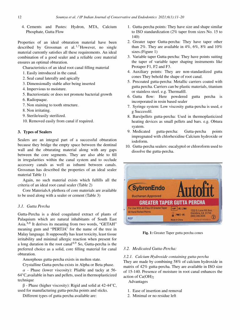
2. Greater taper Gutta-percha: They have taper otherthan 2%. They are available in 4%, 6%, 8% and 10%sizes.(Figure 1)
3. Variable taper Gutta-percha: They have points suitingthe taper of variable taper shaping instruments likeProtaper F1, F2 and F3.
4. Auxiliary points: They are non-standardized guttacones They behold the shape of root canal.
5. Precoated gutta-percha: Metallic carriers coated withgutta percha. Carriers can be plastic materials, titaniumor stainless steel. e.g. Thermafill.
6. Gutta flow: Here powdered gutta percha isincorporated in resin based sealer
7. Syringe system: Low viscosity gutta-percha is used, eg Successfil.
8. Bars/pellets gutta-percha: Used in thermoplasticizedheating devices as small pellets and bars. e.g. Obturasyatem.
9. Medicated gutta-percha: Gutta-percha pointsimpregnated with chlorhexidine Calcium hydroxide oriodoform.
10. Gutta-percha sealers: eucalyptol or chloroform used todissolve the gutta-percha.
Fig. 1: Greater Taper gutta percha cones
3.2. Medicated Gutta-Percha:
3.2.1. Calcium Hydroxide containing gutta-perchaThey are made by combining 58% of calcium hydroxide inmatrix of 42% gutta-percha. They are available in ISO sizeof 15-140. Presence of moisture in root canal enhances theaction of Ca(OH)2
Advantages
1. Ease of insertion and removal2. Minimal or no residue left
Soumya et al. / IP Indian Journal of Conservative and Endodontics 2021;6(1):11–20 13
Table 1:Should be tacky on mixing and adhere to the canal wall when setShould exist in fine powder form to mix with the liquid component easilyShould be radio-opaqueShould not shrink on settingShould not stain tooth structureShould establish a hermetic sealShould have bacteriostatic propertiesShould exhibit a slow setShould not be soluble in tissue fluidsShould not irritate periapical tissueShould be soluble in a common solvent if it is necessary to remove it
Table 2: Advantages and disadvantages of different commercially available sealers:
Type of sealer Example Advantages DisadvantagesZinc oxide eugenol Pulp Canal Sealer EWT Roth’s
Sealer Tubli-seal Wach’sSealer
Long history of use will absorb ifextruded Slow setting timeAntimicrobial effectRadio-opaque
Shrinkage on setting Soluble Canstain tooth structure May negativelyaffect bonding of core materials
Calcium hydroxide Seal Apex Apexit Apexit plus Antimicrobial effectRadio-opaque
Soluble May weaken dentine
Glass ionomer Activ GP Ketac Endo Dentine-bonding properties Hard to remove in retreatmentMinimal antimicrobial effect
Resin AH-26 AH Plus DiaketEndoREZ Epiphany RealSeal
Good adhesion to the wall andcore Do not contain eugenolSlow set
Some release formaldehyde whenset Bond strenght compromisedwith use of chlorhexidine Bondingcomparable to conventional sealers
Silicone GuttaFlow Roeko Seal Triturated Long working timeFills canal irregularities withconsistency Biocompatible
Expand slightly on setting Settingtime is inconsistent Setting timegets delayed by use of sodiumhypochlorite
Bioceramic SmartSeal SmartPaste Bio BioRoot RCS
Hydrophilic No shrinking onsetting BiocompatibleAntimicrobial properties
Minimal supporting clinical dataQuestions raised over ease ofremoval for retreatment
Table 3: Advantages and disadvantages of different core materials:
Core material Advantages DisadvantagesGutta Percha • Ease of manipulation
•Minimal toxicity• Radio-opacity• Ease of removal with heat or solvent
Improper adhesion to dentine When heatedshrinks on cooling
Coated cones • Similar to gutta-percha• Adhesion to canal wall
Incomplete adhesion with evidence ofleakage
Resilon • Similar to gutta-percha•May form a monoblock
Formation of monoblock is controversial
Pro-Points • Expand to fill voids and lateral canals• Used with bio-active sealer
Limited evidence on effectiveness
3. Firm for easy insertion
Disadvantages:
1. Short lived action2. Radiolucent3. Lack of sustained release
3.2.2. Iodoform containing gutta-perchaIodoform containing gutta-percha remains inert till it comesin contact with the tissue fluids. Free iodine gets released incontact with tissue fluids giving an antibacterial property.
3.2.3. Gutta percha with chlorhexidine diacetate5% chlorhexidine diacetate is embodied into the guttapercha matrix. This material is used as an intracanalmedicament.

14 Soumya et al. / IP Indian Journal of Conservative and Endodontics 2021;6(1):11–20
3.3. Coated cones
Available in two versions
1. Ultradent - have gutta percha surface coated witha resin, bond is formed when resin sealer comesin contact with resin coated gutta-percha cone. Thisinhibits leakage between the solid core and sealer. Useof EndoRez sealer is advocated.
2. Active GP Plus - with coating of glass ionomer ongutta-percha, designed for use with their glass ionomersealer.
3.4. Resilon
Resilon became popular as a material substitutingguttapercha to be used with a resin sealer like Epiphanyto achieve an adhesive bond at three interfaces, the corematerial, the canal wall and the sealer. This techniquecreates a monoblock with a bond to canal wall on oneside and a bond to the core material on the other. Resiloncontains polymers of polyesters, bioactive glass and fillerslike barium sulphate. It has similar handling properties asthat of guttapercha, can be softened with heat or dissolvedwith solvents like chloroform.4,10Resilon is a nontoxic,non-mutagenic, and biocompatible material.4,11,12
3.5. Pro points
Also known as “Smart sealing” system consisting of a nylonpolymer core and a hydrophilic polymer coating. Can beused with a hydrophilic MTA based sealer in contact whichthe points go through hygroscopic expansion within thecanal to fill voids.1
4. Obturation Techniques
Several obturation techniques exist with little difference intheir long term outcome results and there is no techniquethat prevents leakage.1,13,14There is some evidence tosuggest that warm vertical compaction is superior to lateralcompaction.1,15,16
Classification of Root Canal Obturation Techniques:According to the mode of practical use, the obturation
techniques are classified as.17,18
1. Single cone technique- Custom-made roll cone technique/tailor-made gutta-percha- Prefabricated stainless steel file method
2. Multiple-cone technique- Cold and warm lateral condensation- Warm vertical compaction- Continuous wave of condensation- Thermo-mechanical compaction
3. Chemo-plasticized gutta-percha- Chloropercha
- Eucapercha4. Thermo-plasticized injectable gutta-percha obturation
- Obtura II- Ultrafil 3D- Thermafil- Solid core carrier-based systems
5. Paste-only root fillings- Zinc oxide eugenol paste- Calcium hydroxide paste- Iodoform paste- Paraformaldehyde containing paste
6. SPAD/resorcinol formaldehyde- Diaket- AH-26- Lee-EndoFill- Hydron
4.1. Single cone technique
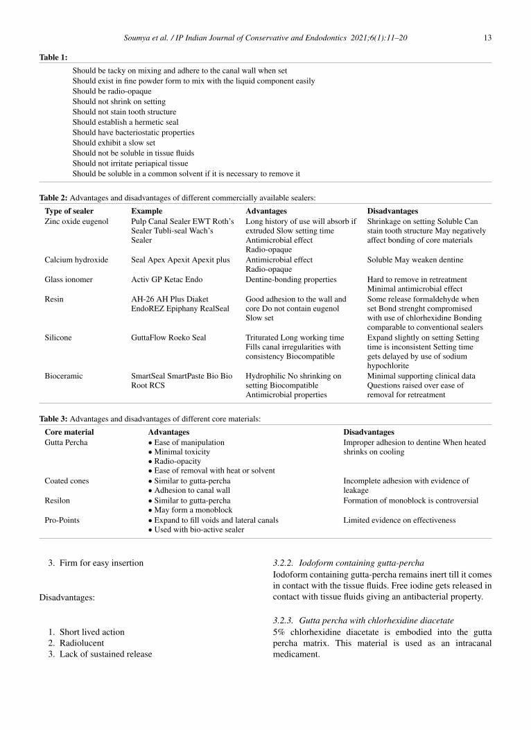
In this technique, a greater taper cone of specific size fitsexactly to the prepared canal. Such an approach is often usedin conjunction with specific filing systems. This techniquedepends on the sealer and a three dimensional seal might notbe achieved. Nonetheless, the apical portion should be wellfitting. The Active GP precision obturation system utilizesGlass ionomer technology, extending the working time ofthe Glass ionomer sealer by modifying its particle size to thenanoparticle level. The gutta-percha cones are coated withGlass ionomer particles at a thickness of 2µm. The accuracyof the size of canal preparation to the size of cone to be usedis very important to minimize the amount of sealer used andany chances of shrinkage as well.19 Up to 49% of dentistsfavour this technique and laboratory evidence suggests thatthis is comparable to lateral compaction1 (Figure 2)
4.2. Cold lateral compaction
Use of a master cone which corresponds to the size of finalcanal preparation, smeared with sealer and placed into thecanal. Additional accessory cones are compacted laterallyto the master cone with the help of spreaders.1Figure 5
4.2.1. Advantages
Overfilling can be avoided and can be easily done in allkinds of root canal morphology.
4.2.2. Disadvantages
Gaps can exist if not compacted well and may not producea homogeneous obturation.
Soumya et al. / IP Indian Journal of Conservative and Endodontics 2021;6(1):11–20 15
Fig. 2: Single cone obturation. Voids around the GP necessitate agreater dependency on sealer
Fig. 3: Master GP placed and lateral compaction done againstthe canal wall with a spreader. An additional GP point is placedinto the void left by the spreader. Accessory cones placed until noempty space is left.
5. Variations of Lateral Compaction Technique
5.1. Curved canals
NiTi spreaders are used. For severely curved canals,thermoplasticized gutta percha technique is preferred.
5.2. Blunderbuss canals
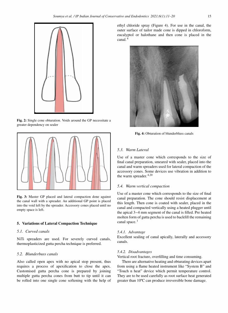
Also called open apex with no apical stop present, thusrequires a process of apexification to close the apex.Customised gutta percha cone is prepared by joiningmultiple gutta percha cones from butt to tip until it canbe rolled into one single cone softening with the help of
ethyl chloride spray (Figure 4). For use in the canal, theouter surface of tailor made cone is dipped in chloroform,eucalyptol or halothane and then cone is placed in thecanal.4
Fig. 4: Obturation of blunderbluss canals
5.3. Warm Lateral
Use of a master cone which corresponds to the size offinal canal preparation, smeared with sealer, placed into thecanal and warm spreaders used for lateral compaction of theaccessory cones. Some devices use vibration in addition tothe warm spreader.4,20
5.4. Warm vertical compaction
Use of a master cone which corresponds to the size of finalcanal preparation. The cone should resist displacement atthis length. Then cone is coated with sealer, placed in thecanal and compacted vertically using a heated plugger untilthe apical 3−4 mm segment of the canal is filled. Pre heatedmolten form of gutta percha is used to backfill the remainingcanal space.1
5.4.1. AdvantageExcellent sealing of canal apically, laterally and accessorycanals.
5.4.2. DisadvantagesVertical root fracture, overfilling and time consuming.
There are alternative heating and obturating devices apartfrom using a flame heated instrument like “System B” and“Touch n heat” device which permit temperature control.They are to be used carefully as root surface heat generatedgreater than 10ºC can produce irreversible bone damage.
16 Soumya et al. / IP Indian Journal of Conservative and Endodontics 2021;6(1):11–20
Fig. 5: The ‘continuous wave’ technique: following placementof master cone, a heated plugger is inserted into the canal tosever the coronal GP and warm the apical GP. Remaining GPcompacted with pluggers and remaining canal space back-filledwith plasticized GP.
5.5. System B/Continuous Wave
It was developed by Buchanan to help warm the guttapercha in the canal. It monitors the tip temperature of theheat carrier pluggers and thus delivers a specific amount ofheat. To achieve a good three dimensional obturation usingsystem B, the canal should have an adequate taper and theset temperature should be such that it does not burn the guttapercha.
5.5.1. AdvantagesIt ensures proper condensation into main and lateral canalswith excellent apical control. It creates single wave ofheating and compacting thereby compaction of fillingmaterial can be done at the same time when it is heatsoftened.
5.6. The Down Pak - 3D Obturation with heat andvibration
The Down Pak is an innovative device introduced to allowthree dimensional obturation with heat and vibration. It hasa vibrating spreader device that can be used for gutta percha,resilon & hybrid resin filling material by both warm verticaland lateral condensation techniques. Down Pak offers awide selection of tips in NiTi and ultrasoft stainless steel.It ensures a denser and more compact filling of root canalspace.
5.7. Lateral/Vertical compaction of warm gutta perchatechnique
Lateral compaction controls length of obturation whilevertical compaction gives a three dimensional obturation.Endotec II combines advantages of both these techniques.
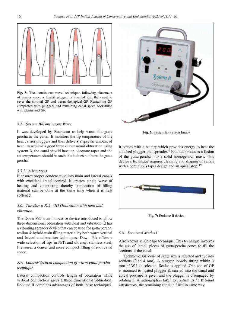
Fig. 6: System B (Sybron Endo)
It comes with a battery which provides energy to heat theattached plugger and spreader.4 Endotec produces a fusionof the gutta-percha into a solid homogenous mass. Thisdevice’s technique requires cleaning and shaping of canalswith a continuous taper design and an apical stop.19
Fig. 7: Endotec II device
5.8. Sectional Method
Also known as Chicago technique. This technique involvesthe use of small pieces of gutta-percha cones to fill thesections of the canal.
Technique: GP cone of same size is selected and cut intosections (3 to 4 mm). A plugger loosely fitting within 3mm of W.L is selected. Sealer is applied. One end of GPis mounted to heated plugger & carried into the canal andapical pressure is given and the plugger is disengaged byrotating it. A radiograph is taken to confirm its fit. If foundsatisfactory, the remaining canal in filled in same way.

Soumya et al. / IP Indian Journal of Conservative and Endodontics 2021;6(1):11–20 17
5.8.1. AdvantageIdeal in cases of post core where only apical portion of canalis to be filled.
5.8.2. DisadvantageTime consuming and difficult to remove the sections of guttapercha if there is overfilling.4
5.9. Thermo-mechanical compaction (McSpaddenCompactor)
Mac-spadden compactor is used which resembles a reverseH-file. A cone coated with sealer is placed in the root canal,engaged with a rotary instrument (running between 5,000and 10,000 rpm) that frictionally warms, plasticizes andcompacts it into the root canal.1 (Figure 8)
Fig. 8: Thermo mechanical compaction using Mc spaddencompactor
5.9.1. AdvantagesIt requires less chair side time, dense three dimensionalobturation is achieved.
5.9.2. DisadvantagesIts difficult to use in narrow and curved canals becauseof frequent breakage of compactor blades. Overfilling ofcanals is also seen with this technique.
5.10. Thermo Plasticized GP injection techniques
The heating of gutta percha outside the tooth and injectingthe material into the canal is the highlight of this technique.
The clinician should take care of any extrusions.(1)Obtura II& III, Calamus, Elements, Hotshot and Ultrafil 3D are basedon this concept.
A. Obtura II (Obtura Spartan, Earth City,Mo.): Introduced at Harvard Institute in 1977. It consistsof an electric control unit with pistol grip syringe andspecially designed gutta-percha pellets which are heated toapproximately 365 – 3900 F(185-2000◦C) for obturation.Root canal space has to be prepared well giving a smoothcontinuous taper. Obtura II is indicated in roots with straightor slightly curved canals and also used in cases of obturationof roots with internal resorption or perforations.4
Fig. 9: Obtura II device
B. The Calamus Flow Obturation Delivery System:(Dentsply-Tulsa Dental. Tulsa, OK): Has a handpiece andactivation cuff to enable control of the flow and temperatureof the gutta-percha in to the canal. The activation cuffis released to stop the flow. It uses disposable single usecatridges with a filling material indicator. The temperatureof the thermoplasticized gutta-percha as it is extrudedthrough the needle tip ranges from 38 o C to 44 o C. Thegutta-percha remains able to flow for 45 to 60 seconds,depending on the viscosity.19 (Figure 11)
Fig. 10: Calamus and Elements obturating unit

18 Soumya et al. / IP Indian Journal of Conservative and Endodontics 2021;6(1):11–20
C. The Elements Obturation Unit (Sybron Endo): Itcontains a system B device and a gutta-percha extruder ina motorized handpiece. The extruder tips are sized 20, 23& 25 gauge and are pre-bent. The disposable cartridgesof gutta-percha are heated quickly and the unit shuts offautomatically to prevent overheating of the material.19
D. The Ultrafill 3D System (Hygienic- Coltene-Whaledent): It is low heat (70◦C) system with sterilizableinjection syringe, three different types of disposable gutta-percha cannula with needles attached that can be pre-curvedand a portable heating unit. Moisture or temperature doesnot affect its setting or solubility. The material expandsslightly (0.2%) resulting in an excellent seal of the rootcanal.19 (Figure 11)
Fig. 11: Ultrafil 3D obturation unit
5.11. Apical Third Filling
Classification-
1. Carrier based: Simplifill obturator, Fiberfill obturator2. Paste based system: Dentin chip filling, Calcium
hydroxide, MTA
5.12. Carrier Based Gutta-Percha:19
5.12.1. Thermafil (Dentsply - Tulsa Dental)Alpha form of guttapercha is coated on a plastic core andtemperature is controlled by a heating device. A clinicianhas a time of approximately 10 seconds after heating toplace it into the canal without rotating or twisting it. Thecarrier is resected after leaving it for 2-4 minutes for the GPto cool off.
5.12.1.1. Advantage. Effective sealing of lateral andaccessory canals
5.12.1.2. Disadvantage. Occasional extrusion of materialsbeyond apex (Figure 12)
Fig. 12: Thermafil Obturation
5.12.2. Successfil (Hygienic-Coltene Whaledent)In this system guttapercha is available in syringe and carriers(Titanium or Radiopaque plastic) are inserted in to thesyringe to a measured length of the canal. The rate ofwithdrawl from the syringe determines the amount andshape of guttapercha. The gutta-percha can be compactedaround the carrier with various pluggers, depending on canalmorphology. This is followed by the severing of the carrierslightly above the orifice using a bur.(Figure 13)
Fig. 13: Successfil obturation system and simplifill carriers withapical plug of GP
5.12.3. SimpliFill (Discus Dental, Culver City, CA)It is employed following canal preparation using Lightspeedinstruments. Here there is 5mm of guttapercha as an apicalplug to the carrier. After canal preparation, the carrieris advanced within 1mm of the canal length prepared,pushed to the determined length using digital pressureand the carrier is then severed rotating the handle counterclockwise atleast four times. The coronal space can thenbe filled with gutta-percha. This sectional technique isefficient.(Figure 13)
5.13. Paste Based System
5.13.1. Dentin chip fillingIt is also known as BIOLOGICAL SEAL. Herecircumferential filing using H - file produces dentin
Soumya et al. / IP Indian Journal of Conservative and Endodontics 2021;6(1):11–20 19
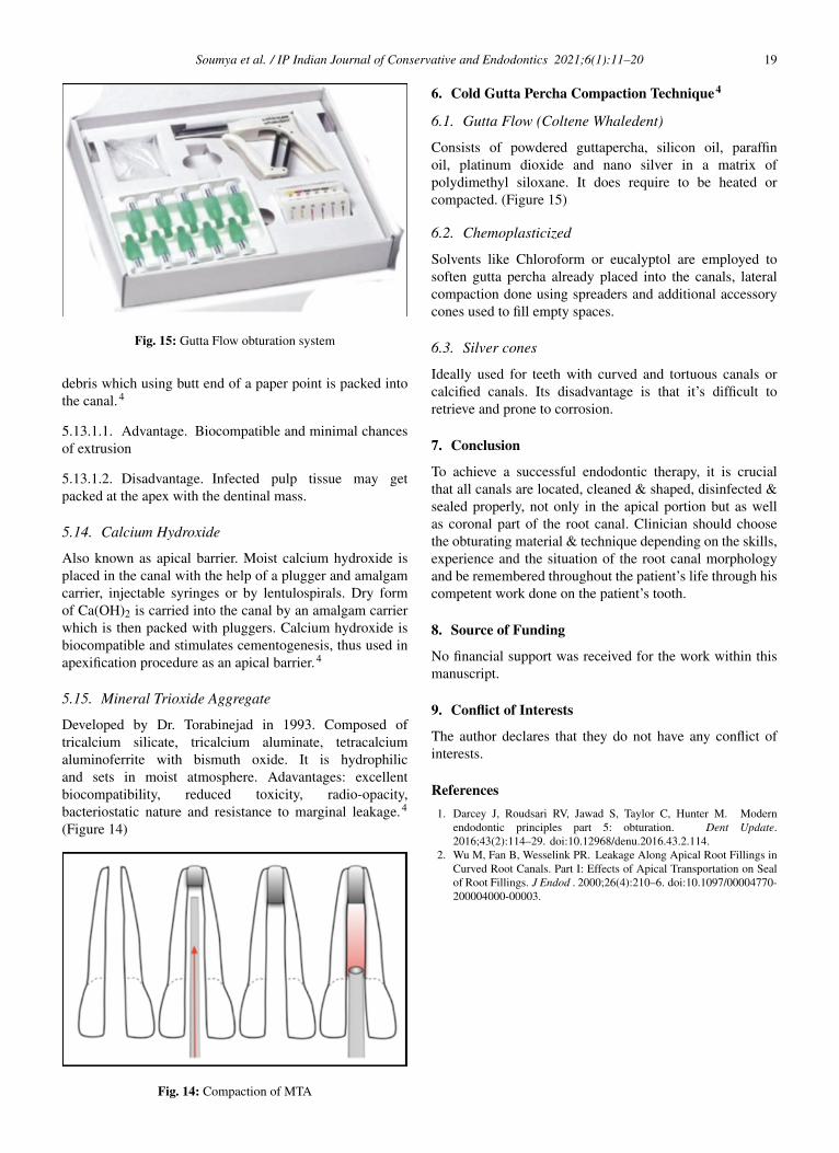
Fig. 15: Gutta Flow obturation system
debris which using butt end of a paper point is packed intothe canal.4
5.13.1.1. Advantage. Biocompatible and minimal chancesof extrusion
5.13.1.2. Disadvantage. Infected pulp tissue may getpacked at the apex with the dentinal mass.
5.14. Calcium Hydroxide
Also known as apical barrier. Moist calcium hydroxide isplaced in the canal with the help of a plugger and amalgamcarrier, injectable syringes or by lentulospirals. Dry formof Ca(OH)2 is carried into the canal by an amalgam carrierwhich is then packed with pluggers. Calcium hydroxide isbiocompatible and stimulates cementogenesis, thus used inapexification procedure as an apical barrier.4
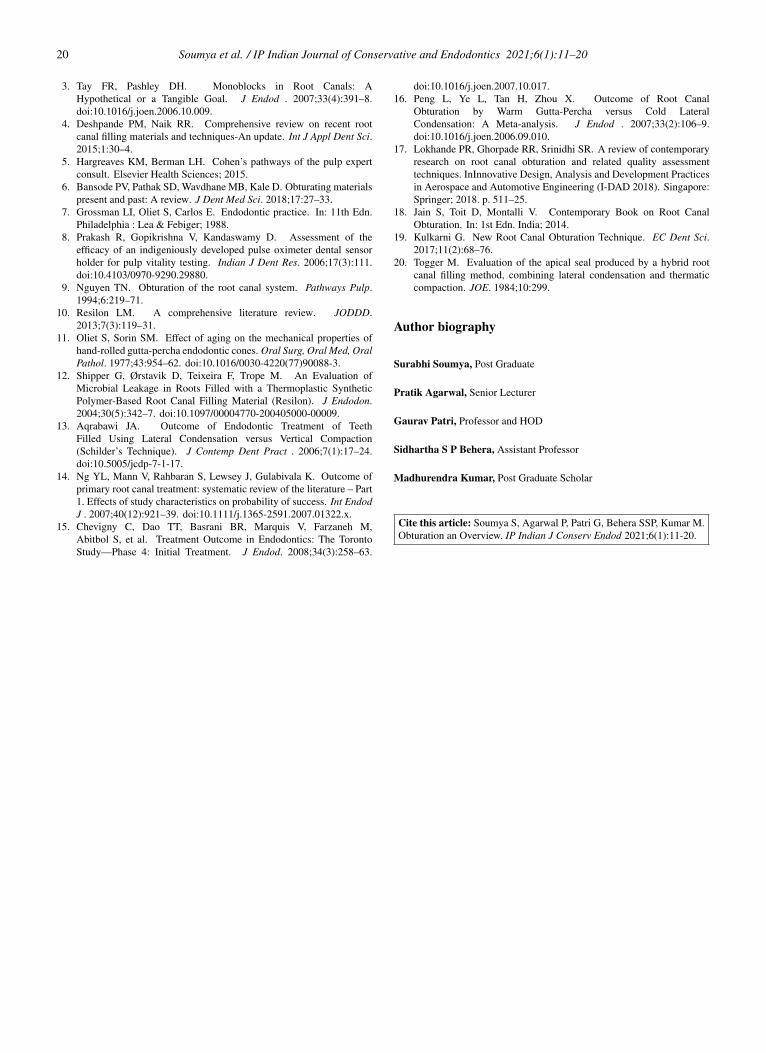
5.15. Mineral Trioxide Aggregate
Developed by Dr. Torabinejad in 1993. Composed oftricalcium silicate, tricalcium aluminate, tetracalciumaluminoferrite with bismuth oxide. It is hydrophilicand sets in moist atmosphere. Adavantages: excellentbiocompatibility, reduced toxicity, radio-opacity,bacteriostatic nature and resistance to marginal leakage.4
(Figure 14)
Fig. 14: Compaction of MTA
6. Cold Gutta Percha Compaction Technique4
6.1. Gutta Flow (Coltene Whaledent)
Consists of powdered guttapercha, silicon oil, paraffinoil, platinum dioxide and nano silver in a matrix ofpolydimethyl siloxane. It does require to be heated orcompacted. (Figure 15)
6.2. Chemoplasticized
Solvents like Chloroform or eucalyptol are employed tosoften gutta percha already placed into the canals, lateralcompaction done using spreaders and additional accessorycones used to fill empty spaces.
6.3. Silver cones
Ideally used for teeth with curved and tortuous canals orcalcified canals. Its disadvantage is that it’s difficult toretrieve and prone to corrosion.
7. Conclusion
To achieve a successful endodontic therapy, it is crucialthat all canals are located, cleaned & shaped, disinfected &sealed properly, not only in the apical portion but as wellas coronal part of the root canal. Clinician should choosethe obturating material & technique depending on the skills,experience and the situation of the root canal morphologyand be remembered throughout the patient’s life through hiscompetent work done on the patient’s tooth.
8. Source of Funding
No financial support was received for the work within thismanuscript.
9. Conflict of Interests
The author declares that they do not have any conflict ofinterests.
References1. Darcey J, Roudsari RV, Jawad S, Taylor C, Hunter M. Modern
endodontic principles part 5: obturation. Dent Update.2016;43(2):114–29. doi:10.12968/denu.2016.43.2.114.
2. Wu M, Fan B, Wesselink PR. Leakage Along Apical Root Fillings inCurved Root Canals. Part I: Effects of Apical Transportation on Sealof Root Fillings. J Endod . 2000;26(4):210–6. doi:10.1097/00004770-200004000-00003.
20 Soumya et al. / IP Indian Journal of Conservative and Endodontics 2021;6(1):11–20
3. Tay FR, Pashley DH. Monoblocks in Root Canals: AHypothetical or a Tangible Goal. J Endod . 2007;33(4):391–8.doi:10.1016/j.joen.2006.10.009.
4. Deshpande PM, Naik RR. Comprehensive review on recent rootcanal filling materials and techniques-An update. Int J Appl Dent Sci.2015;1:30–4.
5. Hargreaves KM, Berman LH. Cohen’s pathways of the pulp expertconsult. Elsevier Health Sciences; 2015.
6. Bansode PV, Pathak SD, Wavdhane MB, Kale D. Obturating materialspresent and past: A review. J Dent Med Sci. 2018;17:27–33.
7. Grossman LI, Oliet S, Carlos E. Endodontic practice. In: 11th Edn.Philadelphia : Lea & Febiger; 1988.
8. Prakash R, Gopikrishna V, Kandaswamy D. Assessment of theefficacy of an indigeniously developed pulse oximeter dental sensorholder for pulp vitality testing. Indian J Dent Res. 2006;17(3):111.doi:10.4103/0970-9290.29880.
9. Nguyen TN. Obturation of the root canal system. Pathways Pulp.1994;6:219–71.
10. Resilon LM. A comprehensive literature review. JODDD.2013;7(3):119–31.
11. Oliet S, Sorin SM. Effect of aging on the mechanical properties ofhand-rolled gutta-percha endodontic cones. Oral Surg, Oral Med, OralPathol. 1977;43:954–62. doi:10.1016/0030-4220(77)90088-3.
12. Shipper G, Ørstavik D, Teixeira F, Trope M. An Evaluation ofMicrobial Leakage in Roots Filled with a Thermoplastic SyntheticPolymer-Based Root Canal Filling Material (Resilon). J Endodon.2004;30(5):342–7. doi:10.1097/00004770-200405000-00009.
13. Aqrabawi JA. Outcome of Endodontic Treatment of TeethFilled Using Lateral Condensation versus Vertical Compaction(Schilder’s Technique). J Contemp Dent Pract . 2006;7(1):17–24.doi:10.5005/jcdp-7-1-17.
14. Ng YL, Mann V, Rahbaran S, Lewsey J, Gulabivala K. Outcome ofprimary root canal treatment: systematic review of the literature – Part1. Effects of study characteristics on probability of success. Int EndodJ . 2007;40(12):921–39. doi:10.1111/j.1365-2591.2007.01322.x.
15. Chevigny C, Dao TT, Basrani BR, Marquis V, Farzaneh M,Abitbol S, et al. Treatment Outcome in Endodontics: The TorontoStudy—Phase 4: Initial Treatment. J Endod. 2008;34(3):258–63.
doi:10.1016/j.joen.2007.10.017.16. Peng L, Ye L, Tan H, Zhou X. Outcome of Root Canal
Obturation by Warm Gutta-Percha versus Cold LateralCondensation: A Meta-analysis. J Endod . 2007;33(2):106–9.doi:10.1016/j.joen.2006.09.010.
17. Lokhande PR, Ghorpade RR, Srinidhi SR. A review of contemporaryresearch on root canal obturation and related quality assessmenttechniques. InInnovative Design, Analysis and Development Practicesin Aerospace and Automotive Engineering (I-DAD 2018). Singapore:Springer; 2018. p. 511–25.
18. Jain S, Toit D, Montalli V. Contemporary Book on Root CanalObturation. In: 1st Edn. India; 2014.
19. Kulkarni G. New Root Canal Obturation Technique. EC Dent Sci.2017;11(2):68–76.
20. Togger M. Evaluation of the apical seal produced by a hybrid rootcanal filling method, combining lateral condensation and thermaticcompaction. JOE. 1984;10:299.
Author biography
Surabhi Soumya, Post Graduate
Pratik Agarwal, Senior Lecturer
Gaurav Patri, Professor and HOD
Sidhartha S P Behera, Assistant Professor
Madhurendra Kumar, Post Graduate Scholar
Cite this article: Soumya S, Agarwal P, Patri G, Behera SSP, Kumar M.Obturation an Overview. IP Indian J Conserv Endod 2021;6(1):11-20.