OBSTETRICAL EMERGENCIES in the Intrapartal · PDF fileOBSTETRICAL EMERGENCIES in the...
49
OBSTETRICAL EMERGENCIES in the Intrapartal Period Everyone Is At Risk Developed by - Lisa Fikac, MSN, RNC-NIC Original Authors - Sherrie Reites, BSN, RN Stacey Cashwell, MSN, RN Expiration Date - 12/10/16
Transcript of OBSTETRICAL EMERGENCIES in the Intrapartal · PDF fileOBSTETRICAL EMERGENCIES in the...

OBSTETRICAL EMERGENCIES
in the
Intrapartal Period
Everyone Is At Risk Developed by - Lisa Fikac, MSN, RNC-NIC
Original Authors - Sherrie Reites, BSN, RN Stacey Cashwell, MSN, RN
Expiration Date - 12/10/16

This continuing education activity is provided by Cape Fear Valley Health System, Training and Development Department, which is an approved provider of Continuing Nursing Education by the North Carolina Nurses Association, an accredited approver by the American Nurses Credentialing Center’s Commission on Accreditation.
1.5 Contact hours will be awarded upon completion of the following criteria:
• Completion of the entire activity • Submission of a completed evaluation form • Completion a post-test with a grade of at least 85%.
The planning committee members and content experts have declared no financial relationships which would influence the planning of this activity.
Microsoft Office Clip Art is the source for all graphics unless otherwise noted.

• Describe the difference between immediate vs. imminent obstetrical emergencies • Describe maternal mortality and common risk factors for obstetrical emergencies • Discuss the management of common obstetrical emergencies

In the United States and other developed countries, the obstetrical patient has -
• Well-trained physicians and support staff available • Modern, well-equipped medical facilities • This translates into excellent outcomes for both mother and baby
However, an unexpected obstetrical emergency may interfere with the outcome. Therefore -
• The physician and/or Certified Nurse Midwife must be prepared to provide the best possible management for unexpected events, AND
• Support staff must be prepared to react and intervene appropriately and promptly

At any time, any pregnant patient may encounter an emergency--immediate or imminent
Immediate emergencies are those that -
• Allow for little time to review • Require quick reaction because seconds count!
Imminent emergencies are those that -
• May be just as life-threatening as immediate emergencies, but there is a little time to plan care • In this case, minutes count!
Always remember the ABCDEs of trauma!
Mothers may be able to have more babies, but babies cannot have more mothers!
Some examples of IMMEDIATE obstetrical emergencies include -
• Cardiorespiratory compromise • Bleeding
o Third trimester bleeding o Hemorrhage during or immediately post-delivery

o Hemorrhagic shock • Trauma
o Accidental injury o Birth injury to the genital tract
• Hypertensive crisis – eclampsia • Shoulder dystocia • Vasovagal syncope or CNS changes • Uterine rupture • Complications of anesthesia
Some examples of IMMINENT obstetrical emergencies include -
• Severe preeclampsia • Coagulopathy • Hemorrhage • Fetal distress • Preterm labor • Acute abdominal pain
o Ectopic pregnancy • Infection
o Septic shock o Ruptured pelvic abscess
• Sickle cell crisis • Acute renal failure • Diabetic ketoacidosis (DKA)

Maternal Mortality Rates - United States, 1999-2007
National Center for Health Statistics, Final Mortality Data. National Center for Health Statistics, Final Natality Data. Retrieved October 15, 2013, from www.marchofdimes.com/peristats
Causes of maternal mortality after the first trimester include -
• Pulmonary embolism and bleeding • Gestational hypertension • Postpartum bleeding • Cerebral vascular accident (CVA) • Anesthesia-related issues • Abortion-related issues • Cardiomyopathy • Infection • Indirect causes • Prior to the end of the first trimester, ectopic pregnancy must also be considered

Bleeding during pregnancy and/or during the intrapartal period may be an emergency that poses a threat to the mother or to both the mother and fetus.
Bleeding may be due to a variety of reasons. Some of the possible causes include -
• Abnormal Placental Implantation o Placenta previa o Placenta abruptio o Placenta accreta o Ectopic pregnancy
• Trauma during labor and delivery o Episiotomy o Complicated vaginal delivery o Low- or mid-forceps delivery o Cesarean birth o Hysterectomy o Uterine rupture
• Uterine atony due to - o Over-distention of the uterus
Fetal macrosomia Multiple gestation Polyhydramnios
o Anesthesia or analgesia o Exhausted myometrium as a result of -
Rapid or prolonged labor Oxytocin or prostaglandin stimulation Chorioamnionitis
• Impaired coagulation due to - o Prolonged retention of a dead fetus o Amniotic fluid embolism o Sepsis o Severe intravascular hemolysis o Multiple transfusions o Severe preeclampsia and/or eclampsia o Anticoagulant treatment
• Miscellaneous risks - o Obesity o Native American ethnicity o Previous history of hemorrhage

Clinical Presentation
Obvious bleeding
• If there is no obvious bleeding noted and symptoms cannot be explained by other changes in the mother's condition, consider occult or sequestered bleeding.
Pain
Changes in maternal -
• Vital signs (T, P, R, BP) • Oxygen saturations • Level of consciousness (LOC)
Changes in fetal heart rate and activity
Clinical Management
Treatment focuses on the underlying cause of the bleeding
Specific causes of bleeding are addressed in the following pages
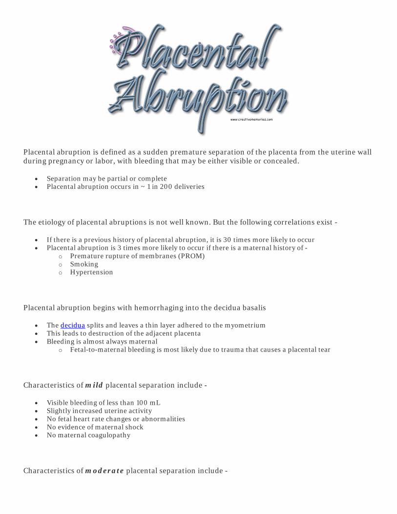
Placental abruption is defined as a sudden premature separation of the placenta from the uterine wall during pregnancy or labor, with bleeding that may be either visible or concealed.
• Separation may be partial or complete • Placental abruption occurs in ~ 1 in 200 deliveries
The etiology of placental abruptions is not well known. But the following correlations exist -
• If there is a previous history of placental abruption, it is 30 times more likely to occur • Placental abruption is 3 times more likely to occur if there is a maternal history of -
o Premature rupture of membranes (PROM) o Smoking o Hypertension
Placental abruption begins with hemorrhaging into the decidua basalis
• The decidua splits and leaves a thin layer adhered to the myometrium • This leads to destruction of the adjacent placenta • Bleeding is almost always maternal
o Fetal-to-maternal bleeding is most likely due to trauma that causes a placental tear
Characteristics of mild placental separation include -
• Visible bleeding of less than 100 mL • Slightly increased uterine activity • No fetal heart rate changes or abnormalities • No evidence of maternal shock • No maternal coagulopathy
Characteristics of moderate placental separation include -

• Visible bleeding of between 100-500 mL • Increased uterine tone • Fetal heart rate may show signs of distress, or it may be absent • Evidence of mild maternal shock
Characteristics of severe placental separation include -
• Bleeding of greater than 500 mL that may be visible or completely concealed • The uterus is firm and contracted • Evidence of moderate to severe maternal shock • Evidence of maternal coagulopathy • Fetal death
Placental abruption may also be classified by the location of the separation -
• Marginal - at the edge of implantation • Partial - at the center of placental implantation • Complete - full separation of placental implantation
Classifications of Placental Abruption

From Howard, PK and Steinmann, RA. (2009).Sheehy's Emergency Nursing, 6th Edition. Mosby Elsevier. Retrieved October 24, 2013, from Mosby's Nursing Consult.
Risk factors for placental abruption include -
• Increased maternal age • Multiparity - especially in women < 30 years of age • Hypertension - most common condition associated with placental abruption
o Preeclampsia o Chronic maternal hypertension
• Rupture of membranes (ROM) o There is a 5-10 times greater chance of placental abruption with premature and prolonged ROM
Risk increases with accompanying infection o Causes of sudden uterine decompression with ROM include -
Second twin Polyhydramnios Chorioamnionitis
• Multifetal gestation • Cigarette smoking - most preventable risk factor • Maternal thrombophilias such as factor V Leiden or prothrombin gene mutation • History of abortions - spontaneous and elective • Illicit drug use
o Cocaine o Methamphetamine
• Short fetal umbilical cord • Abdominal trauma • Uterine fibroids, or leiomyomas - especially if located behind the site of placental implantation • Placental abruption in previous pregnancies
Clinical Presentation
Maternal
• Bleeding with or without abdominal tenderness • Continuous dull back pain and abdominal pain • Intermittent abdominal cramping • Frequent, mild uterine contractions • With significant bleeding, the mother may exhibit -
o Tachycardia with hypotension This is a late finding
o Pale, clammy skin o Increasing uterine distention and tone o Nausea and vomiting o Shock o Decreased urine output o Psychosocial findings
Anxiety Fear of loss of pregnancy Fear for self Confusion Pain

Fetal
• Fetal heart rate (FHR) changes o Baseline FHR may be increased, initially, with decreased baseline variability o Over time, the fetal heart rate will trend to bradycardia and then to absence o Variability is decreased
• Late decelerations are evident on electronic fetal monitor tracings
Clinical Management
• Consider transferring to a tertiary care facility if - o Placental separation is mild o Both the maternal and fetal conditions are stable o Delivery is not imminent o Medical and nursing staff are available to accompany the mother on transport
• Monitor maternal vitals every 15 minutes or more frequently as indicated. • If the mother does not already have an IV, start one or more IVs with an 18-gauge or larger catheter.
o Give IV fluids rapidly - Crystalloid - normal saline, lactated ringers Colloid - PRBCs and/or fresh frozen plasma (FFP)
• Measure and estimate blood loss using the following methods - o Weight of blood saturated items - 1 gram = 1 mL o Mark abdominal fundal height, especially if the bleeding is concealed o Measure and document abdominal girth
• Begin oxygen via face mask at 8-10 L/min • Position in supine with a wedge under the hips
o If maternal condition worsens, position in Trendelenburg • Monitor labor progression
o Contractions may be stronger • Monitor electronic fetal monitor for -
o Fetal heart rate (FHR) o FHR pattern o FHR variability o Decelerations
• Avoid vaginal exams until placenta previa is ruled out • Although the uterus is often hypertonic, contractions are frequently not regular. Oxytocin may be used
to.
Diagnostic Procedures
Laboratory studies
• Type and cross-match for 2-4 units of PRBCs • CBC with differential and platelet count • Clotting factors - prothrombin time (PT), partial thromboplastin time (PTT), fibrinogen, fibrin split
products • D-dimer - blood test used to rule out active blood clot formation • Determine the presence of fetal erythrocytes
o APT or Loendersloot - rapid tests • Urine drug screen - if the mother is a recent admission or is there is poor or no prenatal care • Uterine ultrasound to -
o Determine the location of the placental separation

o Determine the severity of the placental separation o Identify any concealed bleeding
Additional preparation
• Notify the NICU and the pediatrician • Position the mother for comfort
o Analgesics may be contraindicated if maternal or fetal compromise is a concern • Be prepared for delivery
o In the event of a non-viable fetus and/or ripe cervix - The membranes may be artificially ruptured
This is thought to provide better compression of the spiral artery and decrease bleeding
Provide established evidence-based care for the laboring mother Provide compassionate care for the mother and fetus after delivery
o In the event of an emergency Cesarean birth, provide the following care in addition to the care mentioned above -
Prepare the abdomen for surgery Insert a urinary catheter Witness surgical consent Have the neonatal resuscitation team in the operating room
Potential Complications
Maternal
• Couvelaire uterus - blood is forced back into the muscle fibers of the uterus. • Hypovolemic shock • Coagulopathy • Maternal renal failure • Hypoxic damage to -
o Liver o Adrenal glands o Anterior pituitary gland
Sheehan Syndrome • Death
Fetal/newborn
• Fetal growth restriction • Asphyxia • Fetal exsanguination • Prematurity • Coagulopathy • Death


Postpartum hemorrhage is defined as estimated blood loss within the first 24 hours post delivery -
• Greater than 500 mL of blood loss after a vaginal delivery • Greater than 1000 mL of blood loss after a Cesarean delivery
Postpartum hemorrhage is classified as -
• Primary - when the bleeding occurs within the first 24 hours after birth • Secondary - if blood loss occurs more than 24 hours postpartum and prior to 6 weeks after delivery
Risk factors for postpartum hemorrhage include -
• Uterine atony - most common cause o Occurrence in prior pregnancies increases the risk in the current pregnancy
• Precipitous or prolonged first or second stage of labor or both • Induced or augmented labor • Pregnancy induced hypertension (PIH) • Over-distention of the uterus due to -
o Fetal macrosomia (> 4500 gram infant) o Polyhydramnios o Multiple gestation
• Use of certain drugs o General anesthesia - halogenated hydrocarbons o Magnesium sulfate o Prolonged use of oxytocin
• Trauma caused by - o The instruments used to assist delivery o Intravaginal manipulation
• Multiparity • Previous postpartum hemorrhage • Previous uterine rupture • Previous uterine surgery

• Abnormal placental implantation - o Placenta previa o Placenta accreta o Placenta increta o Placenta percreta
• Uterine malformations or fibroids • Maternal exhaustion, malnutrition, or anemia • Lacerations of the upper or lower genitourinary tract • Retained products of pregnancy • Inherited or acquired coagulopathy • Uterine infection - chorioamnionitis • Uterine rupture
Clinical Presentation
• See Bleeding Emergencies for general symptoms • If initial changes go unrecognized or untreated, postpartum hemorrhage can progress into shock.
Clinical Management
No matter what the underlying etiology is, the woman who is bleeding -
• Can very quickly loose 1/3 of her blood volume • SHOCK is the logical progression if clinical symptoms go unrecognized or untreated
The following acronym can be used to help with organization of care of the mother experiencing a postpartum hemorrhage
"ORDER"
O=Oxygenate
• Assure and secure a patent airway • Administer oxygen at 6-8 L/min • Oxygen delivery device - use whatever is necessary
o Mask o Nasal cannula o Endotracheal tube
R=Restore circulatory volume
• If not already in place, start one or more IV lines • Crystalloids or colloids may be administered initially • Blood should be replaced with blood when possible
o Packed red blood cells (PRBCs) - administer 200 mL This will increase the hematocrit 60-65%
• If blood replacement is not possible, remember to replace clotting factors o Concentrated platelets - administer 40-60 mL
This will increase the platelet count by 25,000-35,000

• Cryoprecipitate - administer 30-50 mL o This supplies fibrinogen, Factor VIII, Factor XIII
This is equivalent to 3-10 times the volume of plasma • Fresh frozen plasma (FFP) - administer 200 mL
o This supplies all the clotting factors except for platelets o This supplies 1 gram of fibrinogen
• Fibrinogen - administer 200 mL o This increases fibrinogen by 1-2 grams
D=Drug therapy
• In general, avoid vasopressors • If the woman is in cardiac failure, load with digoxin • Treat the underlying cause -
o Repair lacerations o Oxytocin or prostaglandin E2 may be given to stimulate the myometrium to contract in uterine
atony o Treat infections with antibiotics
E=Evaluate
• Evaluate the woman's response to therapy • Monitor vital signs every 15-30 minutes or as needed • Again, determine the underlying cause of the hemorrhaging and intervene appropriately • If undelivered, closely monitor the condition of the fetus
o External fetal monitoring o Reposition mom, as able
R=Remedy the basic problem
• Repair any lacerations • Massage the boggy uterus • Assess lochia for amount, odor, color, consistency, clots, pad count • Remove secundines • Ligate the ovarian and hypogastric artery • In life-threatening situations, hysterectomy may be necessary
Potential Complications
• Hypovolemic shock • Coagulopathy • Acute renal failure • Seizures • Coma • Death

Uterine rupture is a rare occurrence and is considered an obstetric emergency. It is usually associated with -
• Uterine scarring • Uterine anomalies • Uterine trauma • Prior invasive molar pregnancy • Excessive uterine stimulation during an attempted vaginal birth after Cesarean (VBAC) • History of placenta percreta or increta • Obstructed labor • Mid- to high-operative vaginal delivery • Fetal malpresentation
Risk factors for uterine rupture include -
• Previous Cesarean birth o More than one o Interval less than 2 years o History of no prior vaginal delivery
• Uterine incision • VBAC requiring induction of labor
o This is the most common risk factor • Labor induction or augmentation with oxytocin • Uterine anomalies • Abdominal trauma • Previous uterine trauma
Uterine rupture occurs most frequently after 32 weeks gestation
Clinical Presentation
• Fetal bradycardia or late/variable decelerations at a point during labor when they are usually unlikely to occur
• Decreased baseline uterine pressure • Loss of uterine contractility • Abdominal pain • Vaginal bleeding • Signs of shock

Clinical Management
Immediately notify the physician
• If the mother does not already have an IV, start one or more IVs with an 18-gauge or larger catheter • Prepare for emergency Cesarean birth • Notify the NICU and the pediatrician • Monitor FHR • Position the mother on her side to maximize uterine blood flow
The uterus may be repaired when -
• The rupture is minimal • Further pregnancies are anticipated • The mother's health is not in jeopardy
The uterus is removed when -
• There are no further pregnancies anticipated • The mother's health is in jeopardy
Potential Complications
Maternal
• Hysterectomy • Hypovolemic shock • Coagulopathy • Maternal renal failure • Hypoxic damage to -
o Liver o Adrenal glands o Anterior pituitary gland
• Death
Fetal/newborn
• Asphyxia • Coagulopathy • Death

Trauma is the -
• Most frequent cause of death in women 35 years of age or younger • Leading cause of death in women of childbearing age
About 1 in 12 pregnant women experiences a significant traumatic injury
• About 10-15% of injuries occur in the first trimester • About 32-40% of injuries occur in the second trimester • About 50-54% of injuries occur in the third trimester
Most traumatic injuries during pregnancy are fairly minor
Causes of injury include -
• Motor vehicle accidents (MVAs) ~55% • Falls ~22% • Assaults ~22%
o Including domestic violence • Burns ~1%
Injuries may potentially jeopardize BOTH the mother and fetus
Maternal injuries can include -
• Head injuries • Chest injuries • Abdominal and pelvic injuries • Spinal trauma • Burns and inhalation injuries • Fractures, sprains, dislocations • Soft tissue injuries

Fetal injuries can include -
• Skull fractures • Direct injury to fetal body parts • Fetal death
TRAUMA CONCEPTS
The maternal-fetal response to trauma is affected by -
• The mechanisms of maternal and fetal injury • Gestational age of the fetus • Any secondary complications
The normal changes that occur with pregnancy affect the mother's response to traumatic injury
• Changes may mask and affect the patterns of injury
Trauma in pregnancy involves not one, but two patients--the mother and the fetus
• Minor injuries to the mother may cause significant or even fatal injuries to the fetus • Maternal outcomes to trauma correspond to the severity of the injury. HOWEVER -
o Fetal outcomes depend on the injury as well as the mother's physiologic response to the injury
The initial GOAL in treating trauma is maternal stabilization
• Resuscitation during pregnancy is the same as with any trauma patient. Rapid initial assessment to identify and treat immediate emergencies, followed by a more thorough secondary assessment.
It is important to keep in mind -
• Initial contact is usually in the Emergency Department (ED) where staff are unfamiliar with obstetrics and the normal changes of the pregnant woman
• Obstetric staff are unfamiliar with the principles of trauma stabilization and management
• Collaboration is essential!

Clinical Presentation
Assessment
Trauma assessment begins with prehospital care and continues once the patient is transported to the ED.
• Obtain the patient's history - if available, history of this pregnancy as well as all past history
• Determine the mechanism of injury o Blunt force - MVA, assault, fall o Penetrating - stab wound, gunshot o Burns - causative agent
• Primary survey is conducted in the ED to identify any life-threatening conditions, initiate or continue resuscitation measures initiated in the field, and begin any other necessary treatments. (Based on ATLS Protocols)
o Airway assessment and maintenance, protection of the cervical spine o Bleeding assessment
This includes the amount and source of bleeding o Circulation
This includes - Hemorrhage control Treatment of hypovolemic shock
o Disability and displacement This includes -
Neurological assessment Displacement of the uterus to avoid -
Uterine compression of the vena cava and aorta Supine hypotension
o Exposure This includes -
Undressing the patient to assess for injury Avoiding hypothermia
• Secondary survey includes a detailed physical exam, management of non-life-threatening injuries, and continued reassessment of maternal and fetal condition.
o Vital signs Maternal heart rate, respiratory rate, BP, temperature Fetal heart rate
o History Allergies Medications Past illnesses/pregnancy Last meal Events
o Head-to-toe physical assessment including pregnancy specific evaluation for - Vaginal bleeding Rupture of membranes
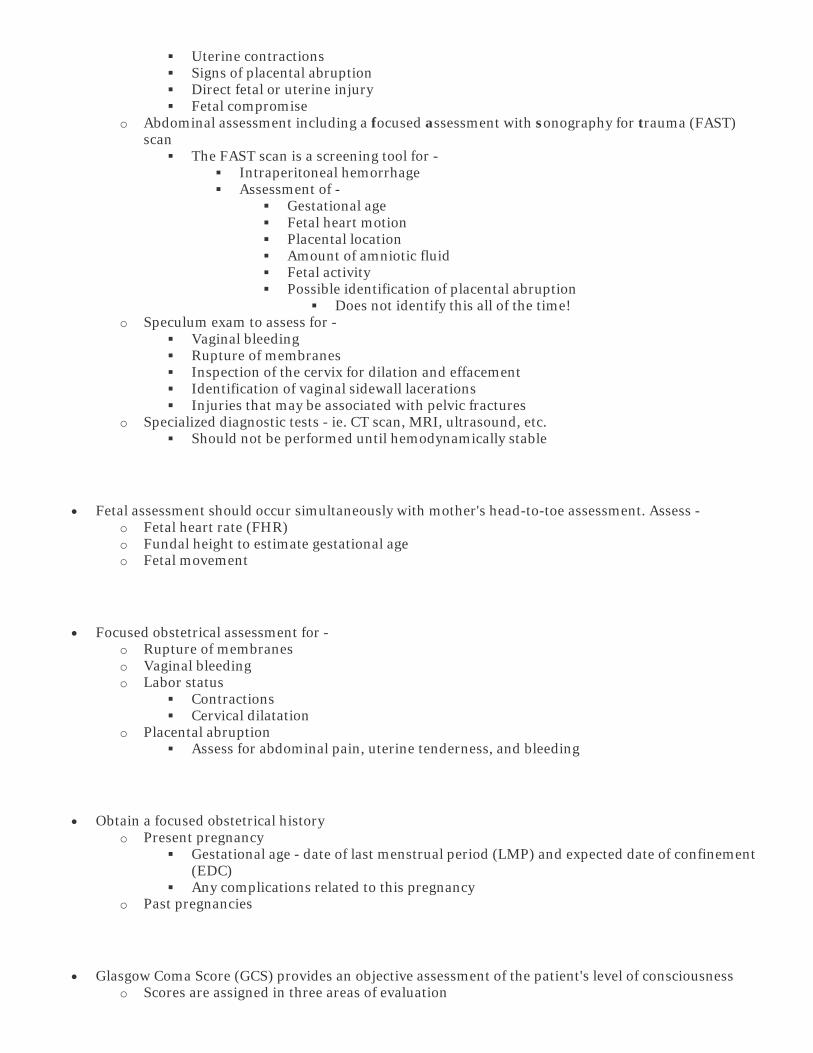
Uterine contractions Signs of placental abruption Direct fetal or uterine injury Fetal compromise
o Abdominal assessment including a focused assessment with sonography for trauma (FAST) scan
The FAST scan is a screening tool for - Intraperitoneal hemorrhage Assessment of -
Gestational age Fetal heart motion Placental location Amount of amniotic fluid Fetal activity Possible identification of placental abruption
Does not identify this all of the time! o Speculum exam to assess for -
Vaginal bleeding Rupture of membranes Inspection of the cervix for dilation and effacement Identification of vaginal sidewall lacerations Injuries that may be associated with pelvic fractures
o Specialized diagnostic tests - ie. CT scan, MRI, ultrasound, etc. Should not be performed until hemodynamically stable
• Fetal assessment should occur simultaneously with mother's head-to-toe assessment. Assess - o Fetal heart rate (FHR) o Fundal height to estimate gestational age o Fetal movement
• Focused obstetrical assessment for - o Rupture of membranes o Vaginal bleeding o Labor status
Contractions Cervical dilatation
o Placental abruption Assess for abdominal pain, uterine tenderness, and bleeding
• Obtain a focused obstetrical history o Present pregnancy
Gestational age - date of last menstrual period (LMP) and expected date of confinement (EDC)
Any complications related to this pregnancy o Past pregnancies
• Glasgow Coma Score (GCS) provides an objective assessment of the patient's level of consciousness o Scores are assigned in three areas of evaluation

o A GCS of 8 or less may indicate significant neurological pathology and the need for emergency life support
• Psychosocial findings o Anxiety, fear, guilt o Potential domestic abuse - injuries inconsistent with reported history
Clinical Management
Airway protection is a priority and may require intervention to maintain a patent airway and adequate oxygenation and ventilation.
• Aggressive maternal ventilatory support may be needed to avoid fetal hypoxemia • High flow oxygen via mask should be initiated even if there are no visible signs of respiratory distress
o Oxygen demands are increased during pregnancy o Traumatic injury increases oxygen demands even more
Interventions are based on maternal and fetal assessments and status, as well as, the particular injuries sustained
• Assessment includes - o Electronic fetal monitoring (EFM) for 24-48 hours if the pregnancy is 20 weeks gestation or
more o Maternal respirations for effectiveness and air exchange o Effectiveness of maternal cardiac output and circulation o Vaginal bleeding o Signs and symptoms of shock o Uterine contractions and/or other abdominal pains or complaints o Fetal heart rate, pattern, activity
• Anticipation of the possibility of surgical intervention is based on the type of trauma • Consistently communicate with the physician to provide updates on maternal and fetal status • Provide emotional support to the patient, family, and significant others
Diagnostic Procedures
• Laboratory tests o CBC with differential and platelet count o Blood type, Rh, cross match o Sodium, potassium, chloride o Glucose, blood urea nitrogen (BUN), creatinine o Clotting factors - prothrombin time (PT), partial thromboplastin time (PTT), fibrinogen, fibrin
split products o D-dimer - blood test used to rule out active blood clot formation o Amylase and liver function tests (LFTs) o Urinalysis

o Arterial blood gases, serum sodium bicarbonate o Blood alcohol and/or toxicology studies, as needed o Urine or serum beta-hCG o Determine the presence of fetal erythrocytes
Kleihauer-Betke or hemoglobin electrophoresis - takes 1 hour for test to be performed Ogita, APT or Loendersloot - rapid tests
• Radiologic tests - injury area-specific o Computed Tomography (CT) Scan o Magnetic Resonance Imaging (MRI) o Ultrasonography
This is a good tool in triage It is safe and non-invasive Helps to determine fetal well-being
• Electronic fetal and uterine monitoring o Note FHR and variability o Monitor uterine contractions
• Peritoneal lavage - a surgical technique used to diagnose intraperitoneal bleeding o This is indicated when the mother has -
Altered LOC Unexplained shock Major chest injuries Signs and symptoms of intraabdominal bleeding
Not all tests or studies may be needed on all patients, but all tests and studies should be considered and either obtained or ruled out
Potential Complications
• Preterm labor • Placental abruption • Uterine rupture • Maternal cardiopulmonary arrest with fetal delivery • Maternal and fetal death

Anaphylactoid syndrome of pregnancy, also known as amniotic fluid embolism, occurs after maternal intravascular exposure to fetal tissue.
• It is normal for maternal exposure to fetal tissue to occur during the intrapartal period • Exposure may also happen with -
o Placement of an intrauterine catheter o Cesarean delivery o Spontaneous or surgical abortion
• In 1995, the National Registry for Amniotic Fluid Embolus was started o Data from 69 cases that occurred between 1983-1993 were analyzed o Data indicated that exposure of the maternal circulation to small amounts of amniotic fluid
caused a syndrome similar to anaphylaxis and/or septic shock • Amniotic fluid is recognized by the mother's body as a foreign substance that is similar to a bacterial
endotoxin or specific antigen o This causes the release of the following into the maternal circulation -
Histamine Bradykinin Cytokines Prostaglandins Leukotrienes Thromboxane Arachidonic acid
Amniotic fluid is made up of -
• Maternal extracellular fluid • Fetal urine • Fetal squamous cells • Lanugo • Vernix caseosa • Mucin • Meconium • Arachidonic acid metabolites • Increased concentrations of prostaglandins, in late pregnancy
Anaphylactoid syndrome of pregnancy is a rare and extremely dangerous, often fatal, obstetrical complication
• It is unpredictable and unpreventable • In the United States, it's estimated incidence is between 1 in 8,000 and 1 in 30,000 deliveries
o The true incidence is unknown due to difficulty in diagnosis and confirmation of cases • The mortality rate is between 61-86%

• Of the women who do survive - o ~ 85% will have permanent neurological impairment o ~ 40% will develop -
Adult respiratory distress syndrome (ARDS) Left-sided heart failure Disseminated intravascular coagulation (DIC) Multisystem organ failure
Potential risk factors
There are no reliable risk factors that predict anaphylactoid syndrome of pregnancy
It is theorized that amniotic fluid enters the maternal circulation in one of the following ways -
• Through the endocervix after ROM o Tears in the endocervical vessels
• At the site of placental separation • At the site of uterine trauma
o Lacerations that occur during the normal course of labor o Small tears in the lower uterine segment
Once the membranes are ruptured, alteration in the pressure gradient may allow amniotic fluid to enter the uterine vessels and maternal circulation
Early reports suggested a potential link between hypertonic uterine contractions or oxytocin administration
• However, analysis of data from the National Registry found that the hypertonic contractions were due to the release of catecholamines into the circulation
o This showed that hypertonic uterine contractions are actually part of the initial hemodynamic response
• The National Registry also found that oxytocin was not used more often in patients who suffered anaphylactoid syndrome of pregnancy
Clinical Presentation
The pathophysiology of anaphylactoid syndrome of pregnancy is not well understood
• It is unclear why some women react so severely to this exposure
The onset of symptoms most often occurs during labor or immediately following delivery
• In the classic presentation, the woman experiences an acute onset of - o Hypoxia

Probably due to severe ventilation/perfusion mismatch caused by the embolic event o Hypotension
Probably due to - Left-ventricular heart failure Pulmonary hypertension Hypovolemia
o Cardiopulmonary arrest
The clinical course of anaphylactoid syndrome of pregnancy has three phases -
Phase Pathophysiology Signs and Symptoms
Phase 1 Amniotic fluid enters the maternal pulmonary vasculature
Respiratory distress
Hypoxemia with respiratory acidosis
Cyanosis
Dyspnea
Cough
Bronchospasm
Pulmonary edema
Phase 2 Inflammatory mediators are released
Myocardial depression occurs
Lung injury occurs
Neurologic compromise occurs
Acute renal failure
Shock and hypotension
Left ventricular heart failure
Arrhythmias
Altered level of consciousness
Seizures
Phase 3 Immunologic activation of coagulation pathway
Intravascular congestion
Bleeding
Disseminated intravascular coagulopathy
(DIC)
Multiple organ system failure

Non-reassuring fetal heart rate (FHR) findings commonly occur just prior to or at the onset of maternal symptoms
Clinical Management
Recognize that this is a life-threatening event!!!!!!
• Diagnosis can, and should, be made from the clinical picture • Fetal debris may be detected in blood aspirated from the mother via
a pulmonary artery catheter o Only present in ~50% of cases o Often found at autopsy in ~75% of cases
• Prompt recognition of clinical symptoms and communication of changes to the physician is key!
The overall goal of clinical management is to optimize -
• Hemodynamic function • Cardiac output • Oxygen transport
This goal will -
• Improve tissue oxygenation • Reduce the risk of end-organ failure • Reduce mortality and morbidity
Assess the patient and initiate CPR if needed
• Call for Rapid Response Team as needed
Administer oxygen at a high concentration
• If there are no spontaneous respirations, ventilate the patient and provide 100% oxygen • Insertion of a pulmonary artery catheter can assist with assessment of -
o Hemodynamic function o Oxygen transport status
Monitor vital signs every 5-10 minutes
• Maternal pulse and BP, and fetal heart rate • Observe for signs of shock and cardiovascular impairment

IV access
• If there is no IV access, start one immediately • Preferably more than one line • Anticipate the placement of a central line
Assess the fetal status
• In the presence of non-reassuring FHR findings, initiate intrauterine support • Depending on maternal status, consider delivery
o If the mother is hemodynamically unstable but has not gone into cardiac arrest, stabilization of the mother may also result in fetal improvement
o Once the mother has gone into cardiac arrest, delivery of the neonate via C-section should be performed immediately
Unfortunately, at this juncture, maternal outcomes are not good, and all efforts to deliver the baby should happen immediately
Optimize preload
• Consider inotropic support if left ventricular contractility is decreased • For hypotension that is unresponsive to volume and inotropic support, vasopressors may be necessary • Volume replacement
o Administer IV fluids rapidly Manage volume loss and hypotension Avoid fluid overload since this may result in the development of pulmonary edema and
ARDS o Administer colloids, like PRBCs, as needed
Treat DIC with blood products as needed
• Bleeding may occur during ante-, intra- and/or postpartum periods. So, assess for - o Oozing from puncture sites o Uncontrolled bleeding
Hydrocortisone therapy may be considered by the physician
Diagnostic Procedures
Laboratory studies -
• ABGs • CBC • Platelet count • Coagulation panel

o Fibrinogen o Fibrin split products o PT and PTT
Anticipated procedures include -
• Chest X-ray • 12-lead ECG
Potential Outcomes
• The mother and infant survive without sequelae • The mother survives, but the infant does not • The mother survives the following complications -
o DIC corrected and controlled CBC and coagulation panel within normal limits
o Respiratory distress is corrected and controlled Oxygen saturation is within normal limits ARDS is resolved
• The mother survives with severe neurological sequelae • The mother does not survive

Umbilical cord prolapse is a life threatening event for the fetus and requires immediate intervention
• Umbilical cord prolapse occurs when the cord - o Falls below the presenting part or o Is compressed between the presenting part and the pelvis or cervix
• Umbilical cord prolapse can occur prior to or during delivery of the fetus, but it often occurs when the membranes rupture (ROM)
Umbilical Cord Prolapse
• A - Complete occult prolapse • B - Cord presenting in front of the fetal head; may be seen in the vagina • C - Frank breech presentation with prolapsed cord
From Lowdermilk, DL, and others. (2012). Maternity and Women's Health Care, 10th Edition. St. Louis: Mosby. Retrieved October 31, 2013, from Mosby's Skills.
Cord prolapse is estimated to occur in approximately 1 in 265-600 births
Umbilical cord prolapse is a life-threatening event for the fetus
• Pressure on the cord causes oxygen deprivation to the fetus and can result in fetal death

Risk factors for umbilical cord prolapse include -
• Amniotomy with a high presenting part • Premature rupture of membranes • Long umbilical cord • Fetal malpresentation - breech, transverse lie • Preterm, low birthweight (LBW), or small for gestational age (SGA) fetuses • Multiple pregnancy • Polyhydramnios • Cephalopelvic disproportion (CPD) that prevents fetal engagement • Lack of engagement prior to the onset of labor • Multiparity
Interventions that may predispose the woman to developing an umbilical cord prolapse include -
• Artificial rupture of membranes • Internal scalp electrode application • Intrauterine pressure catheter insertion • Manual rotation of the fetal head • Amnioinfusion or amnioreduction • External cephalic version with ruptured membranes • Application of forceps or vacuum
Clinical Presentation
The first sign of umbilical cord prolapse is usually -
• A severe, prolonged, fetal bradycardia OR • A change from a normal fetal monitor tracing to severe variable decelerations
Overt prolapse occurs when the umbilical cord is protruding from the vagina or is palpated during a vaginal exam
• The station of the presenting part may be 0 to -4 cm
Occult prolapse occurs when the cord is not visible or palpable, but it is located between the presenting part and the pelvis or cervix.....look for changes to an abnormal fetal heart rate tracing
Clinical Management - Prevention
Admission assessment includes -
• Fetal presenting part and station • Cervical dilatation • Status of the membranes • Fetal heart rate, pattern, variability

• History for presence of the above listed risk factors
Consider discontinuing ambulation if -
• Risk factors are present • The membranes are ruptured • The presenting part is not engaged
At the time of ROM -
• Initiate electronic fetal monitoring if not already in use • Perform a vaginal exam
Clinical Management - Prolapse Occurs
The provider who identifies the prolapse keeps his/her hand in the vagina to push the presenting part away from the cord until the infant is delivered
• Continue to palpate for pulsation of the cord • If the cord has prolapsed out of the vagina, keep it moist
Sterile Vaginal Exam (SVE) to Relieve Pressure on the Umbilical Cord
• A - Vertex presentation • B - Breach presentation
From Lowdermilk, DL, and others. (2012). Maternity and Women's Health Care, 10th Edition. St. Louis: Mosby. Retrieved October 31, 2013, from Mosby's Skills.
Assist the patient into a position where her hips are elevated above her head. Positions include -
• Knee-chest position - patient on her knees with her head and arms resting on the bed • Trendelenburg position • Modified Sims position - patient in side-lying position with hips elevated on pillows

Administer 100% oxygen by face mask
Continuously monitor the fetal heart rate
Prepare for immediate delivery -
• Forcep vaginal delivery if the cervix is fully dilated • If the cervix is not fully dilated or the fetus in distress, the fetus is delivered by emergency Cesarean
Potential Complications
Maternal
• Trauma to the birth canal from a rapid forceps delivery • Uterine atony with subsequent postpartum bleeding from general anesthesia • Blood loss from Cesarean birth • The usual concerns following any type of surgery (e.g., infection, ileus)
Fetus/Newborn
• Fetal anoxia that results in long-term neurologic delays/deficits for the infant • Neonatal infection • Fetal/newborn death
o The longer the cord prolapse persists, the greater the risk of mortality for the fetus/newborn

Shoulder dystocia is the inability to deliver the infant's shoulders by the usual maneuvers used following the delivery of the infant's head
Dystocia is derived from the Greek work "dystokia"
• Root word "tokos" meaning childbirth • "Dys" meaning difficult • Therefore, the literal meaning of dystocia is "difficult childbirth" • Today, dystocia is commonly defined as the abnormal progress of labor
Shoulder dystocia occurs in 0.2-3% of all deliveries
• Since criteria used to diagnose shoulder dystocia varies, some sources consider this to be an under-reported complication.
• There is a greater incidence in infants > 4000 grams
Shoulder dystocia is a potentially serious complication of labor. It is a true obstetrical emergency and can result in increased maternal morbidity as well as increased neonatal morbidity and mortality.
Some studies conclude that shoulder dystocia can be anticipated but cannot be predicted or prevented
• There are no accurate methods to identify which fetus will experience shoulder dystocia • Ultrasound measurements for fetal macrosomia have limited accuracy
Shoulder dystocia may be recognized by the "turtle" sign
• This occurs when the presenting head extends and retracts, then, recoils against the mother's perineum with contractions and pushing
Shoulder dystocia is one of the more common reasons for a primary Cesarean delivery

Potential risk factors
• Preconceptual risks include - o Flat pelvis o Abnormal size and/or shape of the pelvis o Macrosomic maternal birthweight - > 4000 grams o Shoulder dystocia with a previous delivery
This increases the incidence to 10-14% in subsequent pregnancies o A prior macrosomic infant o Diabetes and/or gestational diabetes o Short stature o Maternal obesity o Multiparity o Advanced maternal age
• Antepartal risks include - o Glucose intolerance or gestational diabetes o Excessive maternal weight gain - > 20 Kg or ~ 45 lbs o Maternal obesity - > 81 Kg or ~ 180 lbs o Macrosomic infant - > 4000 grams o Abnormal size and/or shape of the pelvis o Shoulder dystocia with a previous delivery o Male fetus o Post-dates - > 42 weeks' gestation
• Intrapartal risks include - o Alteration in uterine myometrial function and labor contraction pattern
Uterine contractions may be insufficient or inappropriately coordinated to cause cervical dilatation and effacement
Contractions, when combined with pushing, may be insufficient to cause descent and expulsion of the fetus
o Malposition or abnormal development of the fetus can prevent entrance into or passage through the birth canal
o Interference in the descent of the fetus through the birth canal from either the mother's bony pelvis or soft tissues
Variation in the size and shape of the bony pelvis or other abnormalities of the reproductive tract can interfere with engagement, descent, or expulsion of the fetus
The fetus' anterior shoulder fails to rotate and becomes impacted behind the mother's symphysis pubis
The fetus' posterior shoulder is impacted behind the mother's sacral promontory (rare) Enlarging caput succedaneum Increased fetal weight appears to be a common thread through most reported cases of
shoulder dystocia o A prolonged second stage of labor with or without mid-pelvis forcep delivery o Epidural anesthesia
Clinical Management
Timely recognition and management are imperative to prevent birth trauma to both the mother and fetus
During labor -
• Recognize potential risk factors

o Keep physician/CNM updated on the patient's progress, status, trends, changes • Frequent vaginal exams
o Facilitates timely recognition and intervention o Perform vaginal exams approximately every 2 hours, unless otherwise ordered by the
physician/CNM • Standard nursing care includes -
o Vital signs o Fetal monitoring o IV fluids o Positioning o Labor support
• Anticipate physician/CNM activities o Artificial rupture of membranes (AROM) may stimulate effective contractions
Remember, this is a commitment to deliver the fetus within a reasonable time
o Oxytocin augmentation
During delivery -
• Standard delivery room set-up includes making sure - o Radiant warmer is turned on o Resuscitation equipment and medications are available
• Adequate analgesia/anesthesia o Anesthesia staff is available
• Anticipate the need for additional nursing staff to assist physician/CNM during delivery and to continue patient support
• Alert the nursery and/or NICU staff to the impending delivery • Be prepared for emergency Cesarean delivery • Be prepared to obtain cord blood gases to document acid-base status of the infant at birth • Anticipate physician/CNM activities to facilitate delivery
o Extend episiotomy o Moderate suprapubic pressure may be requested to assist with the delivery of the fetal head
Apply pressure over the suprapubic area, NEVER the fundus
• The physician/CNM should not request fundal pressure because it may - o Further impact the shoulder of the fetus o Fracture the infant's clavicle o Cause uterine rupture
• The Rubin maneuver uses two fingers, palm, or fist applied posteriorly to the anterior shoulder of the fetus, toward the fetus' face, to move the shoulder laterally
o 1st - Abdominally rock fetal shoulders from side-to-side to free-up a shoulder and accomplish delivery, if unsuccessful
o 2nd: Reach in the vagina and push most accessible fetal shoulder toward the fetus' anterior chest wall resulting in abduction of both shoulders
This reduces the shoulder-to-shoulder diameter releasing impacted the anterior shoulder from behind the maternal symphysis pubis, allowing delivery

From Rodis, JF. (2013). Intrapartum management and outcome of shoulder dystocia. In UpToDate Online 21.8-C21.151. Retrieved October 31, 2013, from UpToDate Online.
• The Massanti technique uses the palm applied posteriorly and laterally above the symphysis pubis to dislodge and adduct the fetus' anterior shoulder and rotate it underneath the symphysis pubis
• McRoberts maneuver o Remove the mother's legs from the stirrups and sharply flex them on the mother's abdomen o This requires two assistants--one assistant for each leg--if the mother is unable to perform on
her own

From Rodis, JF. (2013). Intrapartum management and outcome of shoulder dystocia. In UpToDate Online 21.8-C21.151. Retrieved October 31, 2013, from UpToDate Online.
o This maneuver results in straightening the sacrum, rotating the symphysis pubis toward the mother's head and a decreasing the pelvic angle
o This does not increase pelvic dimensions but the pelvic rotation may allow room and free-up the impacted anterior shoulder and expulsion of the fetal head
o Prolonged or excessive maternal hip flexion has been associated with transient joint dislocation, separation of the symphysis and/or femoral cutaneous neuropathy
• The Zavanelli maneuver is used to - o Return the fetal head into the pelvis prior to a Cesarean delivery o Return the fetal head to occiput anterior or occiput posterior position if the
head has rotated from either position o With direct pressure on the head, flex the head and push it back into
vagina as far as possible o Perform an emergency Cesarean delivery
Nursing documentation should include -
• Flow sheet and narrative notations summarizing what happened o Avoid using the term "shoulder dystocia," since this is a medical diagnosis o Assign one RN whose only job is to document, similar to a code situation o Avoid late entries
• Record of attendees present • Record of who did what at what time
o Correct names AND titles, not just titles o Use correct times - DON'T GUESS
Use the same clock/watch o The outcome
• List the maneuvers and/or techniques used, in the order used o Nursing interventions should reference that they were performed with an order from and under
the direction of the physician/CNM • Record calls for assistance
o Who was called o Why called o Time called

o Time assistance arrived • Delivery of the infant
o Time o APGAR score o Time of delivery of the head and the time of delivery of the infant's body
• Condition of the infant at delivery o Resuscitation efforts o Who participated in the infant's care with correct names and titles o The outcome
• Maternal condition at delivery and during the recovery period • Labwork obtained
o Include results in narrative note or lab slip in the chart • Be logical, clear, and concise
Potential Complications
Maternal
• Uterine atony or rupture • Hematoma • Bladder atony • Postpartum hemorrhage
o This is usually due to uterine atony o May also be from vaginal and/or cervical lacerations
• Postpartum infection • Separation of the symphysis pubis • Endometriosis
Fetal/Infant
• Infant bruising • Transient brachial plexus injury • Fractures of the clavicle or humerus • Meconium aspiration • Soft tissue laceration received during an emergency Cesarean delivery
o This may require surgical closure • Asphyxia • Intracranial hemorrhage • Fetal or neonatal death • Despite quick recognition and prompt, appropriate intervention, traumatic injuries may occur in 8-20%
of cases o Most injuries resolve between 6-24 months of age o If sequelae persist after this time, the injury (ies) may be permanent (rare) o Persistent injury is most likely to be brachial plexus injury

Ectopic pregnancy refers to any pregnancy implanted somewhere other than the endometrium
Frequent sites of implantation include -
• Fallopian tube (~97%) • Ovary (~0.5%) • Cervix (~0.3%) • Abdominal cavity (~1.5%)
Ectopic pregnancy occurs in ~ 0.2% of pregnancies
• Incidence has increased with the use of assisted reproductive technologies (ART)
Potential risk factors
• Most ectopic pregnancies result from anything that narrows, obstructs, constricts or delays passage of the ovum through the fallopian tube to the uterus
• Chronic or previous tubal infections can cause strictures and slowing of fallopian tube motility o Chlamydial and/or gonorrheal salpingitis o Postpartum endometritis o Post-abortion uterine infection
• Previous tubal or pelvic surgery o Tubal reversal o This may cause tubal adhesions
• Hormonal factors - interfere with normal tubal motility • Contraceptive failure
o Particularly intrauterine devices (IUD) • ART - use of ovulation-stimulating drugs alter estrogen and progesterone levels which can affect tubal
motility • Maternal smoking - thought to affect the ciliary action in the fallopian tubes • Transmigration of ovum - the ovum may migrate from one ovary to the opposite fallopian tube which
delays transportation to the uterus but not the development of the ovum
Clinical Presentation
The initial presentation of an ectopic pregnancy varies depending on the site implantation

From Howard, PK and Steinmann, RA. (2009).Sheehy's Emergency Nursing, 6th Edition. Mosby Elsevier. Retrieved October 31, 2013, from Mosby's Nursing Consult.
The classic symptoms of ectopic pregnancy include -
• Abdominal pain • Delayed menses • Abnormal vaginal bleeding
o 6-8 weeks after the last normal menstrual period
Abdominal pain occurs in almost all cases of ectopic pregnancy
• Pain usually begins as a dull lower quadrant pain caused by tubal stretching • The pain progresses to a sharp, colicky pain caused by further stretching of the tube and contractions • Finally, the pain progresses to a severe, generalized constant pain throughout the lower abdomen
Many women experience -
• A period that is delayed by 1-2 weeks
OR
• A lighter than usual or irregular period
Many women do not experience the common signs of pregnancy since pregnancy hormonal levels are decreased

Symptoms after tubal rupture include -
• Acute generalized lower quadrant abdominal pain o This is caused by blood irritating the peritoneum
• Referred shoulder pain o This is related to irritation of the diaphragm caused by blood in the peritoneal cavity
• Faintness and dizziness • Signs of shock • Afebrile state
Clinical Management
Early diagnosis is the optimal goal
Obtain a detailed health history to determine risk
Perform a thorough physical assessment
• A non-uterine, abdominal mass would be a positive finding
Standard nursing care for all types of ectopic pregnancies includes -
• Frequent assessment of vital signs • Pain assessment • Assessment for vaginal bleeding
o Compare to normal menstrual history • Assess for signs of syncope • Assess for signs of shock
Diagnostic Procedures
• Laboratory studies o Serial measurements of human chorionic gonadotropin (hCG) o Hemoglobin and hematocrit
• Radiologic studies o Transvaginal ultrasound to demonstrate the presence or absence of an intrauterine pregnancy
Treatment Prior to Tubal Rupture
Prior to rupture, ectopic pregnancy may be treated surgically with a laparoscopic procedure
• An incision is made over the site of pregnancy and the products of conception are carefully removed to control bleeding
For women who are hemodynamically stable, ectopic pregnancy may be treated with methotrexate. Criteria for its use include -

• Ectopic sac smaller than 3.5 cm in diameter • Fetus is not alive - confirmed by lack of cardiac activity • Serum hCG levels < 5000 mIU/mL • Normal liver and renal function tests • No evidence of peptic ulcer disease, ulcerative colitis, or pulmonary disease • No evidence of leukopenia or thrombocytopenia • No evidence of AIDS due to added immunosuppressive effects
Treatment After Tubal Rupture
After a ruptured tubal pregnancy, the affected fallopian tube is usually removed
• If perfusion to the adjacent ovary has been compromised, it may be removed...preservation of the ovary is recommended
Treatment of an Abdominal Ectopic Pregnancy
With an abdominal pregnancy, hemorrhage is a very real and serious possibility
• Once an abdominal pregnancy is diagnosed, surgery to remove the fetus is usually performed • The ligation and removal of the placenta is also performed to decrease the risk of -
o Infection o Hemorrhage o Intestinal obstruction
• If the placenta cannot be removed, methotrexate may be used to facilitate involution
Treatment of a Cervical Ectopic Pregnancy
Surgical removal of a cervical ectopic pregnancy is the last treatment option due to risks of -
• Uncontrollable bleeding • Urinary tract injury
Methotrexate is the treatment of choice and may be given systemically or injected directly into the gestational sac
Potential Complications
Ectopic pregnancy is the number one cause of maternal death in the first trimester
Fetal death approaches 100%
• About 5% of abdominal pregnancies reach viability o Continuation of this type of pregnancy is not encouraged due to the high risk of hemorrhage o There is also a high risk of fetal deformity caused by oligohydramnios


American Psychological Association. (2010). Publication Manual of the American Psychological Association, 6th Edition. Washington, DC: Author.
Ananth, C.V. & Kinzler, W.L. (2013). Clinical features and diagnosis of placental abruption. In UpToDate Online 21.8-C21.151.http://www.uptodate.com/contents/placental-abruption-clinical-features-and-diagnosis?detectedLanguage=en&source=search_result&search=placental+abruption&selectedTitle=1%7E135&provider=noProvider (Retrieved November 1, 2013).
Bush, M., Eddleman, K. & Belogolovkin, V. (2013). Umbilical cord prolapse. In UpToDate Online 21.8-C21.151.http://www.uptodate.com/contents/umbilical-cord-prolapse?detectedLanguage=en&source=search_result&search=umbilical+cord+prolapse&selectedTitle=1%7E16&provider=noProvider (Retrieved November 1, 2013).
Cunningham, F.G., Leveno, K.J., Bloom, S., Hauth, J.C., Rouse, D.J., & Spong, C.Y. (2009). Williams Obstetrics, 23rd Edition. New York: McGraw-Hill.
F.A. Davis Company. (2013). Taber’s Online. http://www.tabers.com/tabersonline (Retrieved October 25, 2013).
Howard, P.K. and Steinmann, R.A. (2009).Sheehy's Emergency Nursing, 6th Edition. Mosby Elsevier. (Retrieved October 24, 2013, from Mosby's Nursing Consult).
Lowdermilk, DL, and others. (2012). Maternity and Women's Health Care, 10th Edition. St. Louis: Mosby. Retrieved October 31, 2013, from Mosby's Skills.
Mattson, S. & Smith, J.E. (2011). Core Curriculum for Maternal-Newborn Nursing, 4th Edition. St. Louis: Saunders-Elsevier.
Rodis, J.F. (2013). Intrapartum management and outcome of shoulder dystocia. In UpToDate Online 21.8-C21.151.http://www.uptodate.com/contents/intrapartum-management-and-outcome-of-shoulder-dystocia?detectedLanguage=en&source=search_result&search=shoulder+dystocia&selectedTitle=1%7E40&provider=noProviderIn (Retrieved October 31, 2013).
Stepp Gilbert, E. (2011). Manual of High Risk Pregnancy and Delivery, 5th Edition. St. Louis: Elsevier
Troiano, N.H., Harvey, C.J., and Chez, B. F. (2013). AWHONN High-Risk & Critical Care Obstetrics, 3rd Edition. Philadelphia: Lippincott Williams & Wilkins.

Tulandi, T. (2013). Clinical manifestations, diagnosis, and management of ectopic pregnancy. In UpToDate Online 21.8-C21.151. http://www.uptodate.com/contents/clinical-manifestations-diagnosis-and-management-of-ectopic-pregnancy?detectedLanguage=en&source=search_result&search=ectopic+pregnancy&selectedTitle=1%7E150&provider=noProvider (Retrieved October 31, 2013).
UpToDate, Inc. (2013). McRoberts maneuver [Illustration]. In UpToDate Online 21.8-C21.151.http://www.uptodate.com/contents/5397?search=McRoberts%20maneuver&source=graphics_search&imageKey=OBGYN/74910#graphicRef74910 (Retrieved October 31, 2013).
UpToDate, Inc. (2013). Rubin maneuver [Illustration]. In UpToDate Online 21.8-C21.151.http://www.uptodate.com/contents/5397?search=Rubin%20maneuver&source=graphics_search&imageKey=OBGYN/62024#graphicRef62024 (Retrieved December 30, 2010).
Verklan, M.T. & Walden, M. (2010). Core Curriculum for Neonatal Intensive Care Nursing, 4th Edition. St. Louis: Saunders-Elsevier.