Objectives - NDAFP
130
Transcript of Objectives - NDAFP


Objectives
Recognize the impact of musculoskeletal referrals
Recognize indications for orthopedic referrals
Recognize evaluations, testing and nonoperativetreatments to consider
Recognize when a rheumatologist may become appropriate
Recognize indications for ultrasound imaging

In 2002, the Centers for Disease Control reported that
musculoskeletal problems are second only to upper
respiratory illness as reasons why people seek medical
attention in the emergency department.
McCaig LF, Burt CW. National Hospital Ambulatory
Medical Care Survey: 2001 emergency department
summary. Adv Data. 2003;335: 1-29.

Musculoskeletal symptoms are also the most common
reason for visits to outpatient departments
Ly N, McCaig LF. National Hospital Ambulatory
Medical Care Survey: 2000 outpatient department
summary. Adv Data. 2002;327: 1-27

Economic burden of injury
In 2000 are estimated to cost the U.S. health care system
$1.1 billion for fatal injuries
$33.7 billion for injury hospitalizations
$31.8 billion for injury emergency department visits
$13.6 billion for other outpatient visits
costs reflect treatment for physical injuries only
https://www.cdc.gov/nchs/data/misc/injury2007.pdf

Testing
Xray
CT
MRI
US
EMG/NCS
Vascular studies
Lab
Serum
Synovial fluid analysis

Nonoperative Treatments
Physical therapy
Eccentric exercise
Stretching
AROM/PROM
ASTYM
Modalities
Medications
NSAIDs
Oral
Topical
Opioids
Tylenol
Injections
Cortisone
PRP
Stem Cells
Hyaluranate
Immobilization
Bracing/Splints
Sling
Casting
Boots

Education in
musculoskeletal
medicine has been
shown to be inadequate
in some medical school
curricula
Seventy-nine percent of
the participants failed the
basic musculoskeletal
cognitive examination.
Freedman KB, Bernstein J. The adequacy of medical school education in
musculoskeletal medicine. J Bone Joint Surg Am. 1998;80(10):1421-1427.

One of the most frequent referrals from primary care
to specialist care is for a patient with a musculoskeletal
complaint
Professional society practice guidelines exist for many
musculoskeletal diseases
Referring Wisely: Orthopedic Referral Guidelines at an Academic Institution
Am J Manag Care. 2016;22(5):e185-e191

American Academy of
Orthopedic Surgeons


ACOEM guidelines
American College of Occupational and Environmental
Medicine
http://www.mdguidelines.com

Sideline Guidelines
Searchable database on over 250 conditions
Annotated x-ray, MRI, CT, and photographic examples
References to related scientific literature
Access to emergency guidelines

AO Surgery Reference
AO Foundation
Arbeitsgemeinschaft fur
Osteosynthesefragen/Ass
ociation for the study of
Internal Fixation

Consensus for Referral
University of California,
San Francisco (UCSF)
Health
36 clinical scenarios
214 questions
178 PCPs
24 orthopedists
~65,000 primary care
patients
5000 referrals to
orthopedics per year
Referring Wisely: Orthopedic Referral Guidelines at an Academic Institution,
The American Journal of Managed Care, VOL. 22, NO. 5, e185-e191

Consensus for Referral
What tests and treatments should be performed in
primary care prior to orthopedic consultation for
specific common musculoskeletal problems?
Which common musculoskeletal problems could be
managed by the PCP with an eConsult by an
orthopedist, in place of a face-to-face patient visit?
Referring Wisely: Orthopedic Referral Guidelines at an Academic Institution,
The American Journal of Managed Care, VOL. 22, NO. 5, e185-e191

Consensus for Referral
Referring Wisely: Orthopedic Referral Guidelines at an Academic Institution,
The American Journal of Managed Care, VOL. 22, NO. 5, e185-e191

Consensus for Referral
Referring Wisely: Orthopedic Referral Guidelines at an Academic Institution,
The American Journal of Managed Care, VOL. 22, NO. 5, e185-e191

Consensus for Referral
Acute
Chronic
> 3 months
Acute exacerbation of a
chronic condition
Principles of Ambulatory Medicine

Consensus for Referral
Little agreement exists regarding which orthopedic problems a primary care pediatrician should understand to care effectively for children.
no set of referral guidelines has been established
judgment has been primarily subjective
Previous attempts to create orthopedic referral guidelines have not been successful, even with primary care support.
It therefore is difficult to establish criteria for appropriate referral to a pediatric orthopedic surgeon.
Referral Patterns to a Pediatric Orthopedic Clinic: Implications for Education
and Practice. PEDIATRICS Vol. 113 No. 3 March 2004 e163

Referral Patterns to a Pediatric Orthopedic Clinic: Implications for Education
and Practice. PEDIATRICS Vol. 113 No. 3 March 2004 e163

Consensus for Referral
A large proportion of referrals indicated either a lack
of basic textbook knowledge or lack of examination
skills and appropriate diagnostic tools as
demonstrated by a high number of definitive
diagnosis indicating normal variants.
Referral Patterns to a Pediatric Orthopedic Clinic: Implications for Education
and Practice. PEDIATRICS Vol. 113 No. 3 March 2004 e163

Consensus for Referral
Referral Patterns to a Pediatric Orthopedic Clinic: Implications for Education
and Practice. PEDIATRICS Vol. 113 No. 3 March 2004 e163
Consensus for Referral
Referrals of patients by family physicians to consultants: a survery of the Israeli
Family Practice Research Network. Family Practice. 1998. Vol 15, N2. 158-164.

Consensus for Referral
Referrals of patients by family physicians to consultants: a survery of the Israeli
Family Practice Research Network. Family Practice. 1998. Vol 15, N2. 158-164.

Consensus for Referral
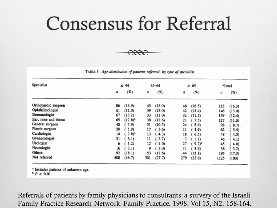
Referrals of patients by family physicians to consultants: a survery of the Israeli
Family Practice Research Network. Family Practice. 1998. Vol 15, N2. 158-164.

Orthopedic Referral
Expectations/Goals
Not met
Not set
Does the patient want to be referred?
Is the patient agreeable to surgery if recommended?

Orthopedic Referrals
Emergent Non emergent
Urgent
Elective

Emergent Referrals
Amputations
Open Fractures
Closed Fractures with Gross Deformity
Dislocations
Acute Compartment Syndrome
Neurovascular Compromise

Open Fractures
Wound cultures
Not recommended
29% of positive pre-debridement
60% of post-debridement cultures
Tetanus prophylaxis
Booster
10 years or more if vaccination history
contaminated wounds if more than 5 since the last tetanus vaccination history
IG 3000 – 5000 units IM
Highly contaminated wound
Antibiotic prophylaxis
Recommended within 3 hrs
except open finger fx
First-generation cephalosporin and an aminoglycoside
Duration debated > 24 hours
Surgical debridement
Critical step
Within 6 hours
Irrigation solution
Type and volume debated
Suspicion for compartment syndrome
Acute Management of Open Fractures: An Evidence-Based Review.
Orthopedics. 2015 Nov;38(11):e1025-33.

Distal Phalynx Fractures
Commonly missed open
fracture
AO Foundation

Closed Reductions
Fractures/Dislocations
Multiple techniques depending on site
If it won’t reduce
Fracture (complicated)
Tissue impeding
Muscle spasm
Maintaining reduction
Immobilization
Swelling
Adequate anesthesia

Anesthesia
Local Anesthetic
Hematoma block/Digital block/Intraarticular injection
No monitoring
Shorter time to discharge
Regional
Nerve block/Bier block
Pain relief
Neurovascular injury, time, equipment
General
Muscular relaxation
Pain relief
Expense, time, monitoring, adverse effects to meds

Dislocations
Milch techniqueExternal rotation

Dislocations
Cunningham techniqueStimson technique

Dislocations
Traction/Counter Traction

Dislocations
Sometimes overlooked
Perilunate
High suspicion
Nurse maid’s elbow

Crush injuries
leg
forearm
Compartment syndrome
Rapid Set-up
simple, sterile, disposable
Accurate
built-in microchip technology
Convenient
pre-filled syringe, hand held and easy to transport
Versatile
quick and continuous pressure monitoring

Neurovascular compromise
Warmth of extremity
Color of extremity
Pulses
Pain
Capillary refill
Sensation
Active movement

Nonemergent Referrals
Urgent/Elective
Structural
Congenital
Physeal/Apophyseal
Bone
Fracture
Tumor/Metastasis
Avascular necrosis
Joint
Cartilage
Ligament
Capsule
Soft Tissue
Muscle
Tendon
Bursa
Hardware
Retained ORIF
TJA
Foreign body/penetrating trauma
Synovitis/Tenosynovitis
Rheumatologic
Crystal induced
Infectious
Reactive
Neuromuscular
Entrapments
Dystrophies
Arthralgias/Myalgias
Hemochromotosis
Vitamin D deficiency
Thyroid disorders
Medication induced
Statins
Accutane

Congenital
Developmental dysplasia of the hip
Femoral acetabular impingement
Femoral torsion
Genu varum and genu valgum
Tibial torsion
Metatarsus adductus
Club foot
Tarsal coalition
Congenital trigger thumb
Syndactyly
Webbed digits
Polydactylyl
Osteogenesis imperfecta
Ehlers Danlos
Marfans
Trisomy 21

Apophyseal Injuries
Apophysitis
Iliac Crest
ASIS
Ischial Tuberosity
Greater Trochanter
Inferior patellar pole
Sindig-Larsen-Johansson
Proximal tibial tubercle
Osgood-Schlatter
Calcaneal
Severs
Imaging Findings of Lower Limb Apophysitis. AJR:196, March 2011

Avulsion of Apophysis

Fracture
Location, location,
location

High-Risk Stress Fractures: Diagnosis and Management. PM R. 2016 Mar;8(3
Suppl):S113-24.

High-Risk Stress Fractures: Diagnosis and Management. PM R. 2016 Mar;8(3
Suppl):S113-24.

Fracture Classification
Integrity of Skin
Open (compound)
Closed

Fracture Blisters
Delayed treatment
Leave intact
Alter treatment
difficult to splint or cast
surgical incision sites
Fracture blisters. West J Emerg Med. 2011 Feb;12(1):131-3.

Fracture Classification
Stable vs unstable
Displacement &
Angulation
deforming forces

Fracture Classification
Orientation of
Fracture

Fracture Classification
Physis
contributes to length
Apophysis
is a secondary center of ossification
contributes to contour
Weakest parts of a developing skeleton
Vulnerable to injury

SCFE
More likely or common
during a growth spurt
boys than girls
10-16 years of age
Risk factors
excessive weight or obesity
95th percentile
family history of SCFE
hyperthyroidism
AAOS

http://www.orthopaedicsone.com/display/Main/Osteogenesis+and+exercise

High Risk Stress Fractures
High-Risk Stress Fractures: Diagnosis and Management. PM R. 2016 Mar;8(3
Suppl):S113-24.

High-Risk Stress Fractures: Diagnosis and Management. PM R. 2016 Mar;8(3
Suppl):S113-24.

Bony mallet fracture
> 50% articular surface
Subluxation of joint
K wire fixation
The "Fish Hook" Technique for Bony
Mallet Finger. Orthopedics. 2016 Sep
1;39(5):295-8.

Clavicle Fractures
Compromise skin
integrity
extremely rare of the
skin to be perforated
from within
Is Skin Tenting Secondary to Displaced Clavicle Fracture More Than a Theoretical
Risk? A Report of 2 Adolescent Cases. Am J Orthop (Belle Mead NJ). 2015
Oct;44(10):E414-6

Clavicle Fractures
Midshaft
Distal
Unstable - ligamentous
Controversial
Proximal
Uncommon
Ligamentous support –
rarely displace
Fracture of distal end clavicle: A review. J Clin Orthop Trauma. 2014 Jun;5(2):65-73.

Clavicle Fractures
Displacement
Nonunion of displaced
midshaft clavicular
fractures was 15.1%
after nonoperative care
compared with 2.2%
after plate fixation
Nonoperative treatment compared with plate fixation of displaced midshaft clavicular
fractures. Surgical technique. J Bone Joint Surg Am. 2008 Mar;90 Suppl 2 Pt 1:1-8.

Clavicle Fractures
Shortening
> 2 cm
predispose to nonunion
and weakness
Wick, Orthop Trauma
Surg. 2001
A comparative study of non-operative and operative management in fracture clavicle.
J Indian Med Assoc. 2013 Dec;111(12):806, 808-9.

Fracture Nonunion
According to American Food and Drug
Administration
A non-union is established when a minimum of 9
months has elapsed since injury and the fracture shows
no visible progressive signs of healing for three months.
Fracture non-union epidemiology and treatment. Trauma 2016, Vol. 18(1) 3–11

Fracture Nonunion
Incidence and prevalence
vary significantly based
on anatomic region and
the criteria used to
define non-union
It has been estimated
that 100,000 fractures go
on to non-union each
year in the USA
Fracture non-union epidemiology and treatment. Trauma 2016, Vol. 18(1) 3–11

Fracture Nonunion
Risk factors
location of the fracture site
surgical treatment
bone displacement
type of fixation
treatment delay
comminution
inadequate treatment
wound infection
Biological causes
patient age
smoking
diabetes
obesity
NSAID use
Biological Risk Factors for Nonunion of Bone Fracture.Zura R, Mehta S, Della
Rocca GJ, Steen RG.JBJS Rev. 2016 Jan 5;4(1).

Fracture Nonunion
Locations prone to
nonunion
Scaphoid
Femur
Tibia
Humerus
Clavicle
5th MT

Bone Stimulators
Bone stimulators
Exogen
86% healed in an average
treatment time of 22
weeks
Low-intensity pulsed ultrasound in the treatment of nonunions. J Trauma.
2001 Oct;51(4):693-702

Bone Tumors
Detected
Painful
Associated with a
palpable mass
Associated with a
pathologic fracture
Discovered incidentally
on an imaging study
Lytic bone lesions
often not detectable on
standard radiographs
until the tumor has
resulted in 30–50% loss
of mineralization
Staging of Bone Tumors: A Review with Illustrative Examples. AJR:186, April 2006

Bone Tumors
Staging of Bone Tumors: A Review with Illustrative Examples. AJR:186, April 2006


6-year old girl with
Ewing’s sarcoma

16-year-old boy with
osteosarcoma

17-year-old boy with soft
tissue mass and
osteosarcoma

11-year-old girl with
osteosarcoma

Giant cell tumor Chondroblastoma

Benign Bone Tumors
Benign tumours of the bone: A review. J Bone Oncol. 2015 Jun; 4(2): 37–41.

Metastatic Bone Disease
Presentation
Pain
Pathologic Fracture
Impending Fracture
Treatment
Radiation
Bisphosphonates
Chemo
Hormones
Management of skeletal metastases: An orthopaedic surgeon's guide. Indian J Orthop.
2015 Jan-Feb; 49(1): 83–100.

Metastatic Bone Disease
Management of skeletal metastases: An orthopaedic surgeon's guide. Indian J Orthop.
2015 Jan-Feb; 49(1): 83–100.

Avascular Necrosis
Lunate
Humeral head
Femoral head
Knee
Tarsal Navicular
2nd MT head
UpToDate

Legg-Calve Perthes
Male to female ratio 3:1
Short stature
Younger age 4-8
? Coagulopathies
higher incidence of factor V Leiden mutation, protein S deficiency, elevated factor VIII, and prothrombinG20210A mutation in LCP patients, especially males
Vosmaer JBJS 2010
Legg-Calve ́-Perthes Disease An Overview with Recent Literature. Bulletin of the
Hospital for Joint Diseases 2014;72(1):18-27

Labral tears
Shoulder Hip

Osteochondral Lesions
Surgical techniques
Palliation
chondroplasty and debridement
Repair
drilling and microfracture
Restoration
autologous chondrocyteimplantation [ACI]
osteochondral autograft transfer [OAT]
osteochondral allograft [OCA]
Knee Articular Cartilage Repair and Restoration Techniques: A Review of
the Literature.Richter DL, Schenck RC Jr, Wascher DC, Treme G.Sports
Health. 2016 Mar-Apr;8(2):153-60.

Arthritis
Primary
Degenerative
Secondary
Disease driven
Post Traumatic
Injury driven
Erosive
Auto immune
Infectious
Neuropathic
Injections
Cortisone
Hyaluronte
PRP
Stem Cells
Palpation vs guided
Bracing
Surgical
Fusion
TJA

Ligament Injuries
Scapholunate ligament
Thumb UCL
Stener lesion
Elbow UCL
HAGL lesion
SC Separation
posterior
AC Separation
Grade 3-6
Lisfranc injury
Syndesmotic injury
Persistent instability after Grade 3 sprain of ankle and knee
Cruciate ligament injury

Soft Tissue
Gas Injected Material

Muscle
Strains
Contusion
Myositis ossificans

Tendon
Ruptures
Achilles
Peroneals
PTT
Quad
Patellar
Hamstring
RTC
Distal biceps
Long head biceps
FDP
Calcific tendinitis
Tenosynovitis
Tendinopathies
Tendinitis
Tendinosis
Trigger fingers
Tendon subluxation
Snapping hip
Snapping triceps

Bursitis
Ankle
Medial malleolar
Knee
Pes anserine
Hip
Greater trochanter
Iliopsoas bursa
Elbow
Olecranon
Distal biceps (cubital)
Shoulder
Subacromial
Subcorocoid

Nonemergent Referrals
Urgent/Elective
Structural
Congenital
Physeal/Apophyseal
Bone
Fracture
Tumor/Metastasis
Avascular necrosis
Joint
Cartilage
Ligament
Capsule
Soft Tissue
Muscle
Tendon
Bursa
Hardware
Retained ORIF
TJA
Foreign body/penetrating trauma
Synovitis/Tenosynovitis
Rheumatologic
Crystal induced
Infectious
Reactive
Neuromuscular
Entrapments
Dystrophies
Arthralgias/Myalgias
Hemochromotosis
Vitamin D deficiency
Thyroid disorders
Medication induced
Statins
Accutane

Limping Child
Initial workup
Labs
CBC, ESR, and CRP
X-ray
Pelvis and frog leg
lateral of affected
side
Transient synovitis, septic hip, and Legg-Calvé-Perthes disease: an approach to the
correct diagnosis. Pediatr Clin North Am. 2014 Dec;61(6):1109-18.

Rheumatologic
ESR, CRP
CBC
CMP
RF, anti-CCP
fANA, anti-DS DNA ab
Complement
Ro ab
ssA, ssB
HLA B27
Anti-centromereab, Anti-smooth muscle ab
Thyroid ab
25 hydroxy vitamin D
CK
UA
Synovial fluid analysis
Cell count
Crystals
Cultures

Hardware
TJA
Loosening
Osteolysis
Wear
Dislocated poly
Periprosthetic fracture
Retained
Migrating
Failure
Penetrating foreign body

Accessory Ossicles

Musculoskeletal Ultrasound
Advantages Ready accessibility
Portability
Quick scan time
Dynamic
Better patient tolerability No Radiation, No Side Effects,
No Contraindications
Cost
Guided procedure(s)
Personal interaction with the patient directed examination –
extension of physical exam
specific for each individual
Scanning technique is easily modified, as needed, to optimize the diagnostic effectiveness of the study contralateral comparison
Disadvantages
Training
highly operator-dependent
Equipment
Time

Musculoskeletal Ultrasound
Physics
Transducer
Emits sounds waves
(1%) and detects
returning echoes (99%)
Linear array (large
footprint & obvious
orientation)
Superficial > 10 MHz
Deep 5-7 MHz

Physics of Sound Waves
Transducers
Linear, (M/S) Curvilinear
(OB/ABD)

Musculoskeletal Ultrasound
Technological advances

Musculoskeletal Ultrasound

Musculoskeletal Ultrasound


MSK US Applications
Diagnosis
RTC
Carpal tunnel
Achilles tendon
Synovitis
Guided injections
Joint
Tendon
Cyst
Percutataneous lavage
Calcific tendinitis
Percutaneous release
TF
CT

Supraspinatous
Longitudinal

Supraspinatous
Transverse

Small Full Thickness
Supraspinatous Tear
Longitudinal

Small Full Thickness
Supraspinatous Tear
Transverse

MSK US vs MRI
Ultrasound detection of rotator cuff and biceps tendon
integrity is comparable to MRI and should be preferred
in revision cases.
Ultrasound vs. MRI in the assessment of rotator cuff structure prior to shoulder
arthroplasty. J Orthop. 2015 Jan 28;12(1):23-30.


MSK US Carpal Tunnel
Better specificity and
equal sensitivity as
compared with those of
electrodiagnostic testing
Comparison of ultrasound and electrodiagnostic testing for diagnosis of carpal
tunnel syndrome: study using a validated clinical tool as the reference standard.
Fowler JR, Munsch M, Tosti R, Hagberg WC, Imbriglia JE.J Bone Joint Surg Am.
2014 Sep 3;96(17):e148.

MSK US Carpal Tunnel
Screening for Carpal Tunnel Syndrome Using Sonography. J Ultrasound Med
2011; 30:1657–1667

Ultrasound Guided
Injection
AMSSM 2014 Position Statement
Ultrasound guided injections vs Landmark guided
injections
717 % increase in outpatient diagnostic MSK US from
2000-2009

Ultrasound Guided
Injection

Large joint injection

Large joint injection

Small Joint Injections

Small Joint Injection

Soft Tissue Injections

Baker’s Cyst

Soft Tissue Injection

Soft Tissue Injection

Soft Tissue Injection

Evaluating for disability
Disability Injury

Before and After

Teleconsultation
Assessment of imaging
Smart phone camera/display
Triage
Initial treatment/reduce waiting times
HIPPA compliance

Dr. Google


Public Expectations

Clinically Correlate


![[ OBJECTIVES ]](https://static.fdocuments.us/doc/165x107/61d432ab7761d92b9c077f35/-objectives-.jpg)