Obesogens in the aquatic environment an evolutionary and ...
17
Contents lists available at ScienceDirect Environment International journal homepage: www.elsevier.com/locate/envint Review article Obesogens in the aquatic environment: an evolutionary and toxicological perspective Ana Capitão a,b,⁎ , Angeliki Lyssimachou a , Luís Filipe Costa Castro a,b,⁎ , Miguel M. Santos a,b,⁎ a CIMAR/CIIMAR- Interdisciplinary Centre of Marine and Environmental Research, University of Porto, Terminal de Cruzeiros do Porto de Leixões, Avenida General Norton de Matos, S/N, 4450-208 Matosinhos, Portugal b FCUP – Department of Biology, Faculty of Sciences, University of Porto, Rua do Campo Alegre, 4169-007 Porto, Portugal. ARTICLE INFO Keywords: Obesogens Endocrine disruption Evolution Nuclear receptor Lipid metabolism Aquatic animals ABSTRACT The rise of obesity in humans is a major health concern of our times, affecting an increasing proportion of the population worldwide. It is now evident that this phenomenon is not only associated with the lack of exercise and a balanced diet, but also due to environmental factors, such as exposure to environmental chemicals that interfere with lipid homeostasis. These chemicals, also known as obesogens, are present in a wide range of products of our daily life, such as cosmetics, paints, plastics, food cans and pesticide-treated food, among others. A growing body of evidences indicates that their action is not limited to mammals. Obesogens also end up in the aquatic environment, potentially affecting its ecosystems. In fact, reports show that some environmental che- micals are able to alter lipid homeostasis, impacting weight, lipid profile, signaling pathways and/or protein activity, of several taxa of aquatic animals. Such perturbations may give rise to physiological disorders and disease. Although largely unexplored from a comparative perspective, the key molecular components implicated in lipid homeostasis have likely appeared early in animal evolution. Therefore, it is not surprising that the obesogen effects are found in other animal groups beyond mammals. Collectively, data indicates that suspected obesogens impact lipid metabolism across phyla that have diverged over 600 million years ago. Thus, a consistent link between environmental chemical exposure and the obesity epidemic has emerged. This review aims to summarize the available information on the effects of putative obesogens in aquatic organisms, considering the similarities and differences of lipid homeostasis pathways among metazoans, thus contributing to a better un- derstanding of the etiology of obesity in human populations. Finally, we identify the knowledge gaps in this field and we set future research priorities. 1. Introduction 1.1. Endocrine disruption The steep increase of chemical production and use since the 1940s coincides with the rise of several endocrine-related disorders in humans and wildlife populations, suggesting a relationship between both events (Bergman et al., 2013; Diamanti-Kandarakis et al., 2009; Grün and Blumberg, 2006; Kabir et al., 2015). The number of studies supporting this hypothesis is growing and it is now established that several of these chemicals have endocrine disrupting properties (Bergman et al., 2013). Endocrine disrupting chemicals (EDC) interfere with the normal http://dx.doi.org/10.1016/j.envint.2017.06.003 Received 31 March 2017; Received in revised form 30 May 2017; Accepted 3 June 2017 ⁎ Corresponding authors at: CIMAR/CIIMAR- Interdisciplinary Centre of Marine and Environmental Research, University of Porto, Terminal de Cruzeiros do Porto de Leixões, Avenida General Norton de Matos, S/N, 4450-208 Matosinhos, Portugal. E-mail addresses: [email protected] (A. Capitão), fi[email protected] (L.F.C. Castro), [email protected] (M.M. Santos). Abbreviations: ACA, Acetyl-CoA Acyltransferase; ACACa, acetyl-CoA carboxylase 1; ACACb, acetyl-CoA carboxylase 2; ACC, Acetyl CoA Carboxylase; ACOX, Acyl-CoA Oxidase; ACS, Acetyl-CoA synthetase; APOA-1, apolipoprotein A-I; BBzP, Butyl benzyl phthalate; BPA, Bisphenol A; BZF, bezafibrate; CA, clofibric acid; C/EBPs, CCAAT/enhancer binding proteins; CPT, carnitine palmitoyltransferase; CYP27a, sterol 27-hydroxylase; CYP4, cytochrome P450 4; DBD, DNA binding domain; DDT, dichlorodiphenyltrichloroethane; DEHP, di-2-ethyl- hexylphthalate; DiDP, diisodecyl phthalate; ECR, Ecdysone Receptor; EDC, Endocrine disrupting chemicals; ER, estrogen receptor; FA, fatty acid; FABP, fatty acid binding protein; FADS, fatty acid desaturase; FASN, Fatty Acid Synthase; FXR, farnesoid X receptor; GPAT1, glycerol-3-phosphate acyltransferase 1; HL, hepatic lipase; HNF4A, Hepatocyte Nuclear Factor 4 A; HSL, hormone sensitive lipase; IPA, Ingenuity pathway analysis; KSI, kidney somatic index; LBD, ligand biding domain; LPL, Lipoprotein Lipase; LSI, liver somatic index; LXR, liver X receptor; MEHP, Mono-ethyl-hexyl phthalate; NP, nonylphenol; NRs, nuclear receptors; PA, phthalic acid; PAHs, Polycyclic aromatic hydrocarbons; PBBs, polybrominated biphenyls; PCBs, polychlorinated biphenyls; PFOA, perfluorooctanoic acid; PPARs, peroxisome proliferator-activated receptors; PXR, pregnane X receptor; Rosi, Rosiglitazone; RXR, retinoid X receptor; SCD1, stearoyl-CoA desaturase 1; sod1, superoxide dismutase; SREBPs, sterol regulatory element-binding proteins; STA, Steroidogenic Acute Regulatory Protein; t-OP, octyl- phenol; TAG, Triacylglycerol; TBBPA, Tetrabromobisphenol A; TBT, Tributyltin; TCBPA, tetrachlorobisphenol A; TPT, Triphenyltin; WAT, White Adipose Tissue; ZOT, Zebrafish obe- sogenic test Environment International 106 (2017) 153–169 Available online 26 June 2017 0160-4120/ © 2017 Elsevier Ltd. All rights reserved. MARK
Transcript of Obesogens in the aquatic environment an evolutionary and ...
Contents lists available at ScienceDirect
Environment International
journal homepage: www.elsevier.com/locate/envint
Review article
Obesogens in the aquatic environment: an evolutionary and toxicologicalperspective
Ana Capitãoa,b,⁎, Angeliki Lyssimachoua, Luís Filipe Costa Castroa,b,⁎, Miguel M. Santosa,b,⁎
a CIMAR/CIIMAR- Interdisciplinary Centre of Marine and Environmental Research, University of Porto, Terminal de Cruzeiros do Porto de Leixões, Avenida GeneralNorton de Matos, S/N, 4450-208 Matosinhos, Portugalb FCUP – Department of Biology, Faculty of Sciences, University of Porto, Rua do Campo Alegre, 4169-007 Porto, Portugal.
A R T I C L E I N F O
Keywords:ObesogensEndocrine disruptionEvolutionNuclear receptorLipid metabolismAquatic animals
A B S T R A C T
The rise of obesity in humans is a major health concern of our times, affecting an increasing proportion of thepopulation worldwide. It is now evident that this phenomenon is not only associated with the lack of exerciseand a balanced diet, but also due to environmental factors, such as exposure to environmental chemicals thatinterfere with lipid homeostasis. These chemicals, also known as obesogens, are present in a wide range ofproducts of our daily life, such as cosmetics, paints, plastics, food cans and pesticide-treated food, among others.A growing body of evidences indicates that their action is not limited to mammals. Obesogens also end up in theaquatic environment, potentially affecting its ecosystems. In fact, reports show that some environmental che-micals are able to alter lipid homeostasis, impacting weight, lipid profile, signaling pathways and/or proteinactivity, of several taxa of aquatic animals. Such perturbations may give rise to physiological disorders anddisease. Although largely unexplored from a comparative perspective, the key molecular components implicatedin lipid homeostasis have likely appeared early in animal evolution. Therefore, it is not surprising that theobesogen effects are found in other animal groups beyond mammals. Collectively, data indicates that suspectedobesogens impact lipid metabolism across phyla that have diverged over 600 million years ago. Thus, a consistentlink between environmental chemical exposure and the obesity epidemic has emerged. This review aims tosummarize the available information on the effects of putative obesogens in aquatic organisms, considering thesimilarities and differences of lipid homeostasis pathways among metazoans, thus contributing to a better un-derstanding of the etiology of obesity in human populations. Finally, we identify the knowledge gaps in this fieldand we set future research priorities.
1. Introduction
1.1. Endocrine disruption
The steep increase of chemical production and use since the 1940scoincides with the rise of several endocrine-related disorders in humans
and wildlife populations, suggesting a relationship between both events(Bergman et al., 2013; Diamanti-Kandarakis et al., 2009; Grün andBlumberg, 2006; Kabir et al., 2015). The number of studies supportingthis hypothesis is growing and it is now established that several of thesechemicals have endocrine disrupting properties (Bergman et al., 2013).Endocrine disrupting chemicals (EDC) interfere with the normal
http://dx.doi.org/10.1016/j.envint.2017.06.003Received 31 March 2017; Received in revised form 30 May 2017; Accepted 3 June 2017
⁎ Corresponding authors at: CIMAR/CIIMAR- Interdisciplinary Centre of Marine and Environmental Research, University of Porto, Terminal de Cruzeiros do Porto de Leixões, AvenidaGeneral Norton de Matos, S/N, 4450-208 Matosinhos, Portugal.
E-mail addresses: [email protected] (A. Capitão), [email protected] (L.F.C. Castro), [email protected] (M.M. Santos).
Abbreviations: ACA, Acetyl-CoA Acyltransferase; ACACa, acetyl-CoA carboxylase 1; ACACb, acetyl-CoA carboxylase 2; ACC, Acetyl CoA Carboxylase; ACOX, Acyl-CoA Oxidase; ACS,Acetyl-CoA synthetase; APOA-1, apolipoprotein A-I; BBzP, Butyl benzyl phthalate; BPA, Bisphenol A; BZF, bezafibrate; CA, clofibric acid; C/EBPs, CCAAT/enhancer binding proteins;CPT, carnitine palmitoyltransferase; CYP27a, sterol 27-hydroxylase; CYP4, cytochrome P450 4; DBD, DNA binding domain; DDT, dichlorodiphenyltrichloroethane; DEHP, di-2-ethyl-hexylphthalate; DiDP, diisodecyl phthalate; ECR, Ecdysone Receptor; EDC, Endocrine disrupting chemicals; ER, estrogen receptor; FA, fatty acid; FABP, fatty acid binding protein; FADS,fatty acid desaturase; FASN, Fatty Acid Synthase; FXR, farnesoid X receptor; GPAT1, glycerol-3-phosphate acyltransferase 1; HL, hepatic lipase; HNF4A, Hepatocyte Nuclear Factor 4 A;HSL, hormone sensitive lipase; IPA, Ingenuity pathway analysis; KSI, kidney somatic index; LBD, ligand biding domain; LPL, Lipoprotein Lipase; LSI, liver somatic index; LXR, liver Xreceptor; MEHP, Mono-ethyl-hexyl phthalate; NP, nonylphenol; NRs, nuclear receptors; PA, phthalic acid; PAHs, Polycyclic aromatic hydrocarbons; PBBs, polybrominated biphenyls;PCBs, polychlorinated biphenyls; PFOA, perfluorooctanoic acid; PPARs, peroxisome proliferator-activated receptors; PXR, pregnane X receptor; Rosi, Rosiglitazone; RXR, retinoid Xreceptor; SCD1, stearoyl-CoA desaturase 1; sod1, superoxide dismutase; SREBPs, sterol regulatory element-binding proteins; STA, Steroidogenic Acute Regulatory Protein; t-OP, octyl-phenol; TAG, Triacylglycerol; TBBPA, Tetrabromobisphenol A; TBT, Tributyltin; TCBPA, tetrachlorobisphenol A; TPT, Triphenyltin; WAT, White Adipose Tissue; ZOT, Zebrafish obe-sogenic test
Environment International 106 (2017) 153–169
Available online 26 June 20170160-4120/ © 2017 Elsevier Ltd. All rights reserved.
MARK

function of the endocrine system by mimicking, blocking and/or al-tering hormone roles and metabolism (Diamanti-Kandarakis et al.,2009; Kabir et al., 2015; Schug et al., 2011). Although more than 1300chemicals have been identified to potentially interfere with hormonalmetabolism, very few have been screened for their capacity to causeendocrine effects in vivo (Bergman et al., 2013; “TEDX The EndocrineDisrupting Exchange,” 2017).
This vast number of compounds identified as EDCs have distinctchemical structures, proprieties and applications. Some of these com-pounds are used as synthetic hormones (e.g. ethynilestradiol), plastics (e.g.bisphenol A (BPA), phthalates), pesticides and fungicides (e.g.: organotins,methoxychlor, chlorpyrifos, dichlorodiphenyltrichloroethane (DDT), vin-clozolin), solvents (e.g.: polychlorinated biphenyls (PCBs), poly-brominated biphenyls (PBBs), dioxins), pharmaceutical agents (e.g. thia-zolidinediones, atypical anti-psychotics, antihistamines, antidepressants)and personal care products (e.g. triclosan). Besides synthetic chemicals,some natural compounds are also known EDCs (e.g. phytoestrogens, in-cluding genistein and coumestrol) (Castro and Santos, 2014; Kabir et al.,2015; Schug et al., 2016). Several of these compounds can undergobioaccumulation and biomagnification through the food-chain, beingpersistent in the environment. In contrast, others are easily degraded buttheir continuous release into the environment still makes them a cause ofconcern (Bergman et al., 2013; Diamanti-Kandarakis et al., 2009; Kabiret al., 2015) (See Fig. 1).
The effects of different EDCs in non-target organisms have been welldocumented (Ferreira et al., 2009; Liu et al., 2014; Melvin, 2016;Rodrigues et al., 2006; Schug et al., 2016). Two well-known examplestargeting the reproductive system are organotins that cause imposex ingastropods mollusks (a condition characterized by the development ofmale secondary sexual characteristics in females) (Abidli et al., 2009;Lima et al., 2011; Pascoal et al., 2013), and ethinylestradiol that altersthe fecundity and sex ratio of fish (Runnalls et al., 2015; Soares et al.,2009). Alterations in gene and protein expression, as well as physio-logical and behavioral changes are also observed frequently as a con-sequence of EDC exposure (Brander, 2013; Sárria et al., 2013). Morerecently, evidences emerged regarding disruption in lipid homeostasisby EDCs. Since lipid metabolism dysregulation is related with severalimportant diseases in the human population, the mode of action ofthese compounds - also known as obesogens- is now under strong scru-tiny (Castro and Santos, 2014; De Cock and Van de Bor, 2014;Diamanti-Kandarakis et al., 2009; Grün and Blumberg, 2006; Ouadah-Boussouf and Babin, 2016; Santos et al., 2012). These compounds canincrease the number of fat cells and/or the amount of fat stored in eachcell by altering the pathways of energy metabolism and food intake
(Holtcamp, 2012; Janesick and Blumberg, 2011). Several mechanismsof action have been suggested, including epigenetic changes that will beinherited by the future generations (Holtcamp, 2012). However, only asmall portion of chemicals has been tested so far for their potential todisrupt lipid homeostasis and a considerable amount of those fall in theobesogens category, such as organotins, BPA, perfluorooctanoic acid(PFOA), phthalates and some pharmaceuticals (Bašić et al., 2012;Bergman et al., 2013; Legler et al., 2015).
1.2. Lipid homeostasis
Lipid homeostasis is vital for the normal development, maintenanceand reproduction of metazoans, given their transversal involvement ina great variety of metabolic processes, such as energy storage, mem-brane composition, as intracellular signaling molecules, enzyme co-factors and several others (Birsoy et al., 2013). In vertebrates, lipidmetabolism is tightly regulated so that the organism can meet itsphysiological needs (Castro et al., 2016; Mello, 2010; Santos et al.,2012) (see Fig. 2). This metabolic regulation involves three major forms(Desvergne et al., 2006):
a. Allosteric control of enzyme activity along a metabolic pathwaythrough the binding of an activator (for example, the enzyme-sub-strate);
b. Post-translational modifications, which activate/deactivate the en-zyme;
One example is the phosphorylation/dephosphorylation of AcetylCoA Carboxylase (ACC). Low glucose levels cause the depho-sphorylation of ACC down-regulating the fatty acid synthesis, whilehigh glucose levels cause the opposite response. This regulation is es-sential in the balance between β-oxidation and fatty acid (FA) synthesis.(Berg et al., 2002; Nelson et al., 2005).
c. Transcriptional regulation;
This regulation of lipid metabolism occurs through the action ofseveral transcription factors, including nuclear receptors (NRs), sterolregulatory element-binding proteins (SREBPs) and CCAAT/enhancerbinding proteins (C/EBPs) (Desvergne et al., 2006). The transcriptionfactors can up or down-regulate the transcription of specific genes andprotein synthesis (Lempradl et al., 2015; Lyssimachou et al., 2015). NRsinclude peroxisome proliferator-activated receptors (PPARs), pregnaneX receptor (PXR), liver X receptor (LXR), farnesoid X receptor (FXR), all
Fig. 1. Schematic illustration of EDCs input in theaquatic environment (lakes, rivers and sea)(modified from Pait and Nelson, 2002; Sumpter,2005).
A. Capitão et al. Environment International 106 (2017) 153–169
154

heterodimeric partners of retinoid X receptor (RXR), among others. ThePPARs are crucial in the regulation of fat storage and FA β-oxidation.Mammals display three different PPAR genes: PPARα regulates en-zymes involved in the up-take of fatty acids, fatty acid esterification andβ-oxidation; PPARβ regulates FA oxidation in the muscles and PPARγregulates and is essential for adipogenesis. In vertebrates, in addition tofunction as an xenobiotic sensor, PXR is also involved in the regulationof lipid homeostasis in the liver and in the regulation of PPARγ geneexpression (Carazo et al., 2017; Mello, 2010). LXR, FXR and SREBP-2play a key role in cholesterol homeostasis; LXR regulates the expressionof SREBP-1c (Eberlé et al., 2004), while FXR activates the expression ofPPARα. C/EBPs are involved in adipogenesis and can activate the ex-pression of PPARγ (Nerlov, 2007) and SREBP-1c apart from cholesterolis also involved in the fatty acid synthesis (Reviewed by Alaynick, 2008;Berkenstam and Gustafsson, 2005; Desvergne et al., 2006; Mello, 2010).
1.3. Obesogens
Although this research area is relatively recent, several in vitro andin vivo studies have already shown the effect of obesogens in vertebrates(Chamorro-García et al., 2013; Grün and Blumberg, 2009a, 2009b). Thedifferentiation of 3T3-L1 cells in adipocytes is stimulated in the
presence of several putative obesogenic compounds such as organotins(Tributyltin (TBT) and Triphenyltin (TPT)) (Pereira-Fernandes et al.,2013), phthalates (Mono-ethyl-hexyl phthalate (MEHP), mono-benzylphthalate and mono-sec-butyl phthalate) (Hurst and Waxman, 2003)and BPA (Masuno et al., 2005). Several in vivo observations supportthese in vitro results. Chamorro-Garcia and co-workers studied the ef-fect of TBT and Rosiglitazone (Rosi) (a therapeutic drug and PPARγagonist) throughout 3 generations of C57/6J mice. The parental gen-eration (F0) was exposed to TBT (5.42, 54.2 and 542 nM) or Rosi(500 nM), the offsprings (F1) were exposed in utero, F2 was exposed asgerm cells and F3 was not exposed at all. All 3 generations (F1, F2 andF3) from the TBT parental exposure presented, to some extent, an in-crease in the white adipocyte tissue (WAT), liver lipids and alteration inthe expression profile of several genes involved in lipid metabolism(Pparα, Pparγ, Lipoprotein Lipase (Lpl), Srebp1c, Acyl-CoA Oxidase(Acox) and Fatty Acid Synthase (Fasn)). Similar results, although lesspronounced, were observed with Rosi for epidydimal and perirenalWAT. The adipocyte number in F3 remained unchanged while in F1 andF2 it decreased particularly in epidydimal and perirenal WAT. It isimportant to highlight that the observations in F3 animals revealtransgenerational effects (Chamorro-García et al., 2013).
An increase in the offspring weight were also observed after
Fig. 2. Schematic representation of three key pathways involved in human lipid metabolism highlighting the involvement of Nuclear Receptors (PPARα ;PPARβ ; LXR ). Fatty acidsynthesis – acetyl CoA carboxylase (ACC) catalyses the synthesis of malonyl-CoA from acetyl CoA; fatty acid synthetase (FAS) uses malonyl-CoA, acetyl CoA and NADPH to synthetizesaturated fatty acids. Fatty acid catabolism – acyl-CoA synthetase (ACS) catalyse the formation of fatty acyl-CoA from fatty acids and coenzyme A (CoA); carnitine palmitoyltransferase 1and 2 (CPTI and CPT II) transport the fatty acyl-CoA across the mitochondrial membrane; the fatty acyl-CoA is degraded in the β-oxidation pathway producing acyl-CoA; acyl-CoA is theused in the citric acid cycle (TCA) to produce ATP. Cholesterol synthesis -Hydroxymethylglutaryl-Coenzyme A synthase (HMGCS) and Hydroxymethylglutaryl-Coenzyme A reductase(HMGCR) uses acetyl-CoA and acetoacetyl-CoA to produce Mevalonate; Mevalonate is transformed in isopentenyl pyrophosphate (IPP) through the enzyme diphosphomevalonatedecarboxylase (MVD); Farnesyl diphosphate synthase (FDPS) transforms IPP in squalene; squalene is transformed in Cholestrol by squalene epoxidase (SE) and lanosterol synthase (LSS)(Based on (Alaynick, 2008; Berkenstam and Gustafsson, 2005; Birsoy et al., 2013; Desvergne et al., 2006; Mello, 2010; Shi and Burn, 2004).
A. Capitão et al. Environment International 106 (2017) 153–169
155

exposure to phthalates (diethyl-hexyl-phthalate) (0.25 mg/kg b.w.)(Hao et al., 2013) and to BPA (70 μg BPA/kg/day) (Somm et al., 2009)in C57BL/6 mice and Sprague-Dawley rats, respectively. Importantly,in the last decades an increase in the medium weight has been observedin several other mammalian species living in the vicinity of humanpopulations, e.g. macaques, chimpanzees, vervet monkeys, marmosets,mice, rats, dogs and cats (Klimentidis et al., 2011). This gives furthersupport to the hypothesis that environmental factors of anthropogenicorigin are likely to contribute to human obesity.
1.4. Interaction with NR
Several EDCs have been reported to interfere with the metabolismthrough interaction with NRs and the lipid metabolism is no exception(Santos et al., 2012). The NRs present two very conserved domains, theDNA binding domain (DBD) and the ligand biding domain (LBD). TheDBD is responsible for the ligation of the NR to the DNA responsiveelement of target genes, while the LBD accommodate the ligand (Evansand Mangelsdorf, 2014; Mello, 2010; Thornton, 2003). The NRs ligandsare very diverse and normally are small hydrophobic molecules. Al-though the NRs present specific natural ligands, their relation with therecent boom of chemicals seems to be promiscuous (Thornton, 2003).Human and/or mouse PPARs, for example, were reported to interactwith several groups of chemicals that include organotins (Harada et al.,2015; Hiromori et al., 2009; Kanayama et al., 2005), several phthalates(Bility et al., 2004; Cocci et al., 2015; Feige et al., 2007; Hurst andWaxman, 2003; Kanayama et al., 2005; Schlezinger et al., 2004) andsome pesticides (Kanayama et al., 2005; Takeuchi et al., 2006). Otherimportant NRs implicated in lipid homeostasis have also been reportedto be modulated by EDCs, e.g., human FXR by alkylphenols, BPA and aphthalate (Butyl benzyl phthalate (BBzP)) (Kanayama et al., 2005), andLXRα was modulated by alkylphenols and phthalates (Kanayama et al.,2005; Mozzicafreddo et al., 2015).
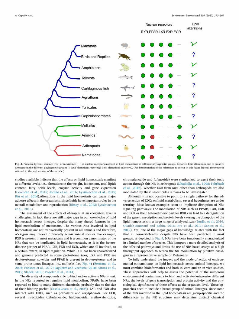
Metazoans, commonly known as animals, include a wide variety oforganisms that can be organized into several groups on the basis of theirevolutionary history (see Fig.3) (Jenner, 2007). The key molecularcomponents implicated in lipid metabolism have likely appeared in theancestor of animals, although for the vast majority of animal lineagesfunctional studies are still missing (Castro et al., 2016; Castro andSantos, 2014). This includes several transcription factors such SREBPand C/EBP, genes coding for metabolic enzymes, for example Acetyl-CoA Acyltransferase (acaa1) and carnitine palmitoyltransferse (cpt),and the storage of fat as Triacylglycerol (TAG) is also a common fea-ture. When looking into fat storage we find an increasing complexity.Several early branching lineages do not possess a specific organ for fataccumulation, but for example arthropods present an organ with si-milar liver and adipose tissue functions, called fat body (Birsoy et al.,2013; Lavarías et al., 2009). In vertebrates, liver and adipose tissue aredistinct and while in fishes, amphibians and reptiles the adipose tissueis concentrated in intra-abdominal regions, in mammals it is distributedwidely in the body. In mammals the hepatic lipid stores are less relevantthan in early-branching vertebrates (reviewed by Birsoy et al., 2013).The genomic and physiological evolution occurred in parallel (Brown,2002). During the course of animal evolution, several duplicationswaves occurred (e.g. genome duplications in vertebrates), and thesehave likely contributed to shape the gene repertoire participating inlipid cascades (Brown, 2002; Crow and Wagner, 2006; Lundin, 1999).Within NRs, RXR is present in most metazoans with exception ofsponges and some cnidarian species (Sladek, 2011). In contrast, LXRand FXR seem to be deuterostome novelties (Bertrand et al., 2004;Fonseca et al., 2017). Mammals have three PPARs and up to five areobserved in teleost genomes (Bertrand et al., 2004; Santos et al., 2012).Outside deuterostomes, PPAR has only been reported in mollusks(Vogeler et al., 2014), though without any functional characterization.The Ecdysone Receptor (ECR), a potential LXR/FXR orthologue(Ollikainen et al., 2006) was thought to be unique of arthropods but has
recently been reported in nematodes, mollusks and annelids (Laguerreand Veenstra, 2010). The existence of this diversity of NRs seems to belinked with genome evolution. It appears that although several differ-ences exist in lipid accumulation processes throughout metazoans,several important features are conserved. Therefore, it is not surprisingthat the obesogen effects are observed in other animal groups beyondmammals (Birsoy et al., 2013; Jordão et al., 2015; Lyssimachou et al.,2015).
This review aims to summarize the available information on theeffects of putative obesogens in aquatic organisms. This objective isapproached from an evolutionary standpoint, considering the simila-rities and differences of lipid homeostasis pathways among metazoans.Although aquatic animals are likely to be exposed to several obesogensin their habitat (Lyssimachou et al., 2015), the main focus of previousstudies, including recent reviews, has been the modulation of lipidhomeostasis by obesogens in mammalian models. However, a growingbody of literature indicates that many more metazoan groups in addi-tion to mammals are likely to be affected by this group of chemicals.Here, we review the current knowledge on the field, identifying gapsand highlighting research priorities.
2. Amphibians
2.1. Organotins
Organotins are persistent organic pollutants, introduced in the en-vironment in 1960s (Antizar-Ladislao, 2008; Maynard, 2002), and wereproduced for diverse uses from antifouling agents in paints to the textileindustry (Antizar-Ladislao, 2008; Bašić et al., 2012; García-Mayor et al.,2012; Maynard, 2002). Although TBT has been banned since 2008 bythe European Union, this compound is still widely distributed in marineand freshwater environments (Antizar-Ladislao, 2008; Gipperth, 2009).A recent study in one of the largest harbors in china revealed biologicalsamples with concentrations of organotins ranging from non-detectableup to 826.7 μg/kg. Triphenyltin was detected at concentrations of92.4 μg/kg (Chen et al., 2017).
Organotins have been reported to alter amphibian lipid metabolism.Xenopus laevis when exposed (from stage 48 to metamorphosis) to dif-ferent concentrations of TBT (0.33–3.3 μg/L) revealed a dose-depen-dent increase in the formation of ectopic adipocytes in and around thegonads of both sexes. In males exposed to 3.3 μg/L of TBT, the testiculartissue was replaced by adipocytes along the anterior-posterior axis(Grün et al., 2006). Exposure, under the same conditions, to the RXR-specific ligands (10–100 nM of LG100268 and AGN195203) and PPARγagonist troglitazone (0.1–1 μM) resulted in similar alterations, pointingto RXR/PPARγ as the target pathway, which is consistent with theobservations in mammalian models (Grün et al., 2006). Despite the lackof alterations in the whole-body weight, the exposure of the wood frogLithobates sylvaticus from 0.1–5 μg/L TPT from day 3 post-hatching upto 100 days, altered the expression of key lipid metabolic genes of theRXR/PPAR signaling pathway. A decrease in the expression of rxrα andpparα was observed in the animals exposed to both concentrations ofTPT after 7 days and a decrease in lpl was observed in the animals ex-posed just to the lowest concentration. After 100 days, an increase inthe expression of genes such as pparα and cyp4b1 was observed in theanimals from both concentrations and an increase of pparγ and lpl wasobserved in the animals from the highest concentration (Higley et al.,2013). These findings suggest a similar mechanism of action to thatdescribed in mammalians, where both TPT and TBT were reported toactivate RXRα and PPARγ (Grün et al., 2006; Kanayama et al., 2005).
2.2. Atrazine
The herbicide atrazine ((2-chloro)-4-(ethylamino)-6-(isopropylamino)-S-triazine), a photosynthesis inhibitor commonly detected in the watersurface (McCarthy and Fuiman, 2008) also interferes with the lipid
A. Capitão et al. Environment International 106 (2017) 153–169
156

metabolism in amphibians. In Texas, near agriculture areas, concentra-tions of 62.5 μg/L were reported (Pennington et al., 2001), but im-mediately after application it is commonly detected in puddles at con-centrations of 400 μg/L (Zaya et al., 2011a).
Atrazine caused a decrease in the fat body size and in the weight ofXenopus laevis exposed to concentrations close to environmental re-levance (200 μg/L and 400 μg/L) from stage 47 until stage 66 (or 43 to62) without observing any alteration in the feeding behaviour(Langerveld et al., 2009; Zaya et al., 2011a, 2011b). The animals ex-posed to 400 μg/L presented an increase in the transcription of genesinvolved in lipid catabolism and carbohydrates metabolism and a de-crease in the genes involved in lipid storage (Langerveld et al., 2009;Zaya et al., 2011a). These pathways have been reported to be involvedwith the decrease in fat storage in mammalian. One possible me-chanism proposed for the response to atrazine was the down-regulationof glucocorticoid signaling, and an increase in the expression of pparβinvolved in the utilization of fat (Zaya et al., 2011a). The results ob-served are in accordance with previous studies in mammalians; low-doses of atrazine can lead to weight gain through mild mitochondrialdamage, while high doses can be toxic and prevent weight gain (Limet al., 2009).
2.3. Other compounds
Different classes of pharmaceuticals are found worldwide in the
aquatic ecosystems, with concentrations ranging from the ng/L or μg/Lin urban environments up to the mg/L range near input sites (Brodinet al., 2014). A mixture of pharmaceuticals (naproxen, diclofenac,atenolol and gemfibrozil) (100 and 1000 μg/L) caused a decrease in thehepatic triglycerides levels and an increase in the condition factor(body weight/snout-vent length3 × 100) of Limnodynastes peronei fol-lowing 30-days exposure after reaching Gosner developmental stage 26.The triglycerides decrease is probably due to gemfibrozil since thiscompound is used to regulate cholesterol levels in humans and is knownto interact with lipid metabolism (Melvin, 2016).
BPA is a component of polycarbonated plastics widely used in dif-ferent products like baby bottles, beverage containers and food cans(Bašić et al., 2012). The aquatic contamination with BPA is very vari-able, the concentrations can vary from non-detected to 17 mg/L inlandfill leachate to 370 μg/L in paper-mill effluents (Fukazawa et al.,2002; Yamamoto et al., 2001; Canesi and Fabbri, 2015; Flint et al.,2012). Although BPA has an obesogenic effect in several species (Guanet al., 2016; Somm et al., 2009), the exposure of Xenopus tropicalisembryos to BPA (3.4 mg/L), during 11 weeks, did not alter the pparαand pparγ gene expression neither the weight of the animals (Mathieu-Denoncourt et al., 2015). However, it would be important to evaluate alarger range of concentration since it cannot be discarded the possibilitythat the lack of effects can be associated with the high concentrationtested (see Table 1).
Fig. 3. Phylogenetic tree highlighting the groups addressed in this review. The most basal metazoans don't show bilateral symmetry. Within those presenting a bilateral symmetry, inprotostomes during the embryonic development the blastopore originate the mouth whereas in deuterostomes the blastopore originate the anus. The protostomes include the Ecdysozoa(such as arthropods and nematodes), animals with periodical molting process, and Lophotrochozoa (such as mollusks and annelids), most of the animals included in this group present alophophore during some stage of their life cycle. The deuterostomes include Echinodermata, Hemichordata and Chordata (Tunicata, Cephalochordata, Vertebrata) (Jenner, 2007).
A. Capitão et al. Environment International 106 (2017) 153–169
157

3. Teleosts
3.1. Organotins
Alterations in lipid storage are among the effects reported in fishesexposed to TBT. Several species (Oncorhynchus tshawytscha, Danio rerio,Carassius auratus and Sebastiscus marmoratus) when exposed to TBTthrough water (1 ng/L–16.5 μg/L) or diet (0.4–150 ng/g/fish/day) ex-hibit an increase in lipid and/or TAG content, adiposity (adipocyte size)and/or altered body weight. The alterations reported were observed inshort-term (16.5 ng/L–16.5 μg/L for 1 day) and chronic experiments(1 ng/L–135 ng/L during 48 days −9 months) (Lyssimachou et al.,2015; Meador et al., 2011; Ouadah-Boussouf and Babin, 2016; Riuet al., 2014; Tingaud-Sequeira et al., 2011; Zhang et al., 2016; Zhanget al., 2012; Zhang et al., 2013). The effects in the expression pattern ofseveral genes involved in the lipid metabolism, in particular the al-terations observed in the nuclear receptors pparγ and rxr and theirdownstream pathways is in line with the hypothesis that TBT alter thelipid metabolism through the interaction with the heterodimer PPARγ/RXR (Lyssimachou et al., 2015; Zhang et al., 2009; Dimastrogiovanniet al., 2015; Li et al., 2014). The impact of TBT in the metabolism of fishseems to be gender and tissue-specific. A different expression pattern ofkey genes involved in lipid metabolism was observed in liver and brainand in females and males of Danio rerio when exposed to TBT(Lyssimachou et al., 2015).
The alterations in the expression of the NRs rxr and ppar, and theirdown-stream pathways are in line with the response of mammalianmodels (Harada et al., 2015; Kanayama et al., 2005; le Maire et al.,2009; Santos et al., 2012). More recently, new evidences suggests that
TBT may impact lipid homeostasis not only through PPARγ/RXR butalso through the LXR/RXR heterodimer (Ouadah-Boussouf and Babin,2016). To clarify this hypothesis the authors measured the TBT-inducedadipocity using a fluorescent dye (Nile Red), that allowed to quantifythe lipid droplets in zebrafish; this test is known as the zebrafish obe-sogenic test (ZOT) (Tingaud-Sequeira et al., 2011). This study showedthat only the agonists (or antagonists) of PPARβ/δ (GW501516/GSK3787), PPARγ (rosiglitazone/T0070907), RXR(DHA/UVI3003) andLXR (GW3965/GSK1440233A) could increase (or decrease) the adip-osity, indicating that these NRs are the potential targets of TBT. Theantagonists of those NRs where then used to rescue the obesogenicresponse of TBT; the RXR antagonist was fully successful, while the LXRantagonist reverted only partially the TBT effect. The other antagonistsfailed to rescue the TBT effects. The use of an antagonist of RXRhomodimer, that is also an agonist of the heterodimer RXR/PPARγ,combined with LXR antagonist and TBT revealed that RXR/PPARγ isalso involved in the action of TBT since this mix could not rescue theeffect of TBT, but the addition of PPARγ antagonist could decrease theadiposity. Taken together the effects of TBT seem to be mediatedthrough RXR/LXR and/or RXR/PPARγ, both permissive heterodimers,which suggests that TBT appears to act through RXR, modulating thedownstream pathways to increase the adipocyte number (Ouadah-Boussouf and Babin, 2016). However, Riu and co-workers did not ob-serve any increase in lipid accumulation in the zebrafish embryos (from3dpf to 11dpf) exposed to 100 nM and 1 μM of Rosi probably because inthis early life stage WAT in fish is hardly developed (Flynn et al., 2009;Imrie and Sadler, 2010; Riu et al., 2014). Moreover, 1 μM of Rosi failedto transactivate zebrafish PPARγ, supporting the hypotheses that theeffect of TBT in the heterodimer RXR/PPARγ occurs through RXR (Riu
Table 1Summary of effects of environmental chemicals in amphibian lipid homeostasis (green: up-regulated; red: down-regulated; n.d – not determined; bold-minimum effect concentration).
Compound Exposure Species Endpoints Ref.Weight/Lipidsalterations
Alteration in gene expression Biochemicalalterations
TBT 0.33 – 3.3µg/L(water); stage 48 tometamorphosis;
Xenopus laevis Formation ofectopic adipocytes;
n.d n.d (Grün et al.,2006)
TPT 0.1 – 5.0 µg/L(water);7 and 100 days;
Lithobatessylvaticus
7 days: weight; dsnout–ventral–length;
7 days: rxrα; pparα; scd1 (5 µg/L); fas(5 µg/L); lpl (0.1 and 5 µg/L); 100 days:pparα; cyp4b; pparγ (1 µg/L);fas (1 µg/L); lpl (1 µg/L);
n.d (Higley et al.,2013)
Atrazine
200µg/L and400µg/L (water);Stage 47 – 66;
Xenopus laevis Fat body size;Weight;
n.d n.d (Zaya et al.,2011b)
400µg/L (water);Stage 47 – 66;
Xenopus laevis n.d Microarray analysis:Genes involved in the lipid catabolism;Genes involved with lipid storage;
n.d (Zaya et al.,2011a)
400ppb (water);Stage 43 – 62;
Xenopus laevis Fat body size; Microarray analysis:Genes involved with proteolysis,digestion and carbohydrates metabolism;
n.d (Langerveldet al., 2009)
Mixture:naproxen,diclofenac,atenolol andgemfibrozil
0.1, 1.0, 10, 100 and1000 µg/L (water);30 days;
Limnodynastesperonii
Condition factor;HSI;
n.d Hepatic triglyceridescontent (100 and1000 µg/L);
(Melvin, 2016)
Compound Exposure invitro
System Species Endpoints Ref.
TBT 2µg/L; Cells–Cos 7;Luciferase assay;
Xenopus laevis Transactivation RXRα and RXRγ; (Grün et al.,2006)
A. Capitão et al. Environment International 106 (2017) 153–169
158

et al., 2011). Moreover, it was observed that the cysteine present in theposition 285 is essential for the activation of human PPARγ by TBT(Harada et al., 2015) and Danio rerio PPARγ has a tyrosine in the cor-respondent position (Den Broeder et al., 2015). This substitution inPPARγ of Danio rerio is expected to affect the affinity of TBT to PPARγ.Interestingly, this mutation of cysteine 285 is not common to allmembers of Actinopterygii, since Sparus aurata and Pleuronectes pla-tessa, for example, have a cysteine molecule in the homologous position(Leaver et al., 2005).
3.2. BPA and BPA analogues
BPA and BPA analogues are ubiquitous chemicals detected in theenvironment. (Fukazawa et al., 2002; Yamamoto et al., 2001). More-over, recent data suggests that this group of chemicals is able to impactlipid homeostasis (Birceanu et al., 2015; Riu et al., 2014, 2011). Thereported effects of BPA and BPA analogues include increase in lipidaccumulation and body mass in Danio rerio when exposed to Tetra-bromobisphenol A (TBBPA) (5.5 and 55 μg/L) or tetrachlorobisphenolA (TCBPA) (37 μg/L) during 19 days, and in Sparus aurata when ex-posed to BPA (50 mg/kg bw) during 21 days, (Maradonna et al., 2015;Riu et al., 2014, 2011). However, in Gobiocypris rarus females a de-crease in serum TGA content was observed under BPA (15 μg/L) ex-posure (Guan et al., 2016). Although the transcription patterns of sev-eral genes involved in lipid metabolism were altered in Sparus aurata(exposed to 50 mg/kg bw of BPA) and in Gobiocypris rarus (exposed to15 μg/L BPA), the pattern of expression is not the same; the fasn geneappeared up-regulated in Sparus aurata and down-regulated in Gobio-cypris rarus males (Guan et al., 2016; Maradonna et al., 2015). The NRsreported to be involved in the lipid homeostasis (PPARα, β, γ and RXR)and lpl genes were up-regulated while hormone sensitive lipase (hsl)gene was down-regulated in Sparus aurata which is consistent with thelipid accumulation observed (Maradonna et al., 2015). In Gobiocyprisrarus the decrease of fasn, acetyl-CoA carboxylase 1 (acaca) and acetyl-CoA carboxylase 2 (acacb) expression is not consistent with the re-spective enzymatic activity, but carnitine palmitoyltransferase 1A(cptIa) expression and enzymatic activity is increased in males anddecreased in females (Guan et al., 2016). The disparity between geneexpression and enzymatic activity can be associated with several reg-ulatory processes (Vogel and Marcotte, 2012). The effect of BPA in lipidhomeostasis of Gobiocypris rarus suggests a gender-specific effect sincethe activity of CPT1 is increased in males, but decreased in femalessimilar to cptIa gene expression. The observed increase in the TAGcontent in females serum and the tendency for decrease in males isconsistent with the previous observations (Maradonna et al., 2015).One possible explanation for the differences in Sparus aurata and Go-biocypris rarus studies described above can be gender related given thatin the Sparus aurata study genders of the specimens were not dis-criminated (Guan et al., 2016; Maradonna et al., 2015).
One possible mechanism for the action of BPA analogues in the lipidmetabolism is the modulation of PPARγ. Riu et al. (2011) observed thatBPA analogues, 10 μM TBBPA and 10 μM TCBPA, are able of in vitrotransactivating zebrafish PPARγ with a similar affinity to that observedin human PPARγ (Riu et al., 2011). An alternative hypothesis involvesestrogenic pathways (García-Mayor et al., 2012), as BPA has the po-tential to bind and activate the estrogen receptor (ER), and interactwith a variety of other targets in mammalian cells, including thyroidhormone receptors (Bašić et al., 2012; Bonefeld-Jørgensen et al., 2007).Further research is needed to fully understand the underlying me-chanisms.
3.3. Alkylphenols
Alkylphenols are commonly used in industrial and consumer pro-ducts (Pereira-Fernandes et al., 2013). Concentrations between 1.1 and1347 μg/kg of nonylphenol (NP) and 0.73–54.4 μg/kg of octylphenol (t-OP) were detected in mussels and 5–60.5 μg/kg of NP and 0.2–31.4 μg/kg in fish tissues (David et al., 2009). The Exposure of Sparus aurata tohigh levels of NP (50 mg/kg bw) or t-OP (5 mg/kg bw and 50 mg/kgbw) caused an increase in the number of specimens with severe lipidaccumulation in the liver, more pronounced in the t-OP groups. Thetranscription patterns of several genes involved in lipid metabolism (i.e.pparα, pparβ, pparγ, rxr, fasn, lpl and hsl) were also increased, empha-sizing the effect of NP and t-OP in the lipid metabolism (Maradonnaet al., 2015). It would be interesting in future studies to test the effectsof lower NP and t-OP levels.
Alkylphenols, such as NP and t-OP, have been reported to activatethe human Estrogen Receptor (ER) and several evidences point to thepossible involvement of ER in the action of certain obesogens, thusexplaining the results observed (Bonefeld-Jørgensen et al., 2007;Kramarova et al., 2009; Yang et al., 2015).
3.4. Phthalates
Phthalates are synthetic organic compounds derived from phthalicacid. These compounds are used since the 1930s as plasticizing agentsin cosmetics, paints and medicines and were found in the aquatic en-vironment in concentrations as high as 98 μg/L (Fromme et al., 2002).Recent evidences shows that these compounds also target the lipidmetabolism of fish (García-Mayor et al., 2012; Grün and Blumberg,2009b, 2007; Pereira-Fernandes et al., 2013). Phthalates were reportedto increase the transcription of NR involved in lipid metabolism(ppaarα, pparβ and pparγ) in zebrafish exposed to 39 μg/L DEHP and1.7 μg/L–1.7 mg/L phthalic acid (PA) (Maradonna et al., 2013). InSparus aurata, exposure to diisodecyl phthalate (DiDP) (45.1–450. 7 μg/L) not only up regulated the expression of ppars and rxr, but also theexpression of some downstream genes, cptIa, cptIb, fatty acid desaturase2 (fads2), stearoyl-CoA desaturase 1A (scd1a), scd1, lpl, hepatic lipase(hl), fatty acid binding protein (fabp), apolipoprotein A-I (apoa-1) andsrebp) (Cocci et al., 2015). In zebrafish exposed to 5000 mg/kg di-2-ethylhexylphthalate (DEHP), during 10 days, no changes were observedin the expression of the downstream genes of the PPARs signalingpathways (acox and lpl) in the liver, although the hepatosomatic indexwas increased. The observed alteration in the hepatosomatic indexcould be associated with increase fat accumulation or as a toxic re-sponse to DEHP (Fabbrini et al., 2010; Milić et al., 2014; Sadekarpawarand Parikh, 2013; Uren-Webster et al., 2010). Taken together, the datasuggest that the interaction of phthalates with the lipid metabolism mayoccur through PPARs modulation. DiDPis can bind to Sparus aurataPPARα and γ with similar affinity as to the human PPARs, MEHP cantransactivate zebrafish PPARγ and the relation between the interactionof phthalates with PPARs and their obesogenic effect was previouslyestablished for mammalians (Cocci et al., 2015; Desvergne et al., 2006;Maradonna et al., 2013; Riu et al., 2014).
3.5. Organophosphates
Some organophosphates, such as Fenitrothion and Trichlorfon, arepesticides that have been used extensively in agriculture and can enter thefreshwater environments (McCarthy and Fuiman, 2008; Sancho et al., 2009;Xu et al., 2012). Xu and co-workers described an increase in the liver TAGcontent in the Carassius auratus gibelio exposed to trichlorfon (1.0–4.0 mg/L)for 30 days (Xu et al., 2012). However, Sancho and co-workers reported a
A. Capitão et al. Environment International 106 (2017) 153–169
159

decrease in liver total lipid content in Anguilla anguilla after a short-termexposure (2 and 96 h) to fenitrothion (0.4 mg/L) (Sancho et al., 1998). Themechanism of action of these compounds in lipid metabolism remains to beelucidated, but similar effects were already reported in mammalians (Meggsand Brewer, 2007).
3.6. Fibrates
Fibrates are commonly used to control hypercholesterolemia inhumans. Given their widespread use, they are frequently detected in thefreshwater environment (Velasco-Santamaría et al., 2011). One of thefirst fibrates to be reported in water samples was clofibric acid (CA), anactive and persistent metabolite of clofibrate, a blood lipid loweringagent (Emblidge and DeLorenzo, 2006; Runnalls et al., 2007). Clofi-brate was detected in surface water at concentrations of 6–7 μg/L, fe-nofibrate and gemfibrozil were detected in much lower concentrations(≈0.05 μg/L) (Corcoran et al., 2010). Several studies demonstrated thecapacity of these compounds to interact with the lipid metabolism innon-mammalian animal models. A decrease in the weight of mosquitofish (Gambusia holbrooki) males when exposed to clofibrate (18.4 to295 mg/L) and an increase in females exposed to CA (4.03 mg/L)during 28 days have been reported (Nunes et al., 2004). In contrast, anexposure to CA at 10 μg/L for 10 days did not alter the levels of lipidsand cholesterol in the fish Fundulus heteroclitus (Emblidge andDeLorenzo, 2006).
A multigenerational study using zebrafish revealed very informativeresults on the effect of CA. F0 generation was exposed to 1 mg/g and10 mg/g CA through the diet and the offspring (F1) were raised withcontrol diet. A decrease in the weight and muscle triglyceride levels wasobserved in F0, but F1 presented an increase in weight in the offspringsof the highest exposure group. The gene expression pattern for bothgenerations was also different. In male livers of the F0, the expression ofpparα and acox1 increased in the animals exposed to the highest con-centration, and in F1 although the expression of pparγ increased, theexpression of apoa-1 and pparβ1 decreased. This study reveals an op-posite pattern of response in the descendants when compared with theparent generation (Coimbra et al., 2015). pparα expression also in-creased in Cyprinus carpio exposed to 20 mg/L of CA during 4 days andacox1 expression increased not only in the animals exposed to 20 mg/Lbut also in the animals exposed at a lower concentration (4 μg/L). Theseresponses are in line with the ones observed in zebrafish. The expres-sion of other genes, such as Acetyl-CoA Acyltransferase 1 (acaa1), cy-tochrome P450 4 (cyp4), sterol 27-hydroxylase (cyp27a), apoA1, lpl andsuperoxide dismutase (sod1) also increased in one or both CA ex-posures. These observations support the evidence that these compoundsinterfere with the lipid metabolism not only in mammals but also in theActinopterygii group (Corcoran et al., 2015).
The effects of other fibrates, besides CA and clofibrate, have alsobeen reported. A decrease in plasma cholesterol levels was found inzebrafish exposed to 1.7, 33 and 70 mg bezafibrate (BZF)/g food after 2and 7 days of exposure, along with a decrease in the expression of pparβand pparγ after 2 days of exposure and an increase in the expression ofpparβ and Steroidogenic Acute Regulatory Protein (star) after 21 days ofexposure (Velasco-Santamaría et al., 2011). The exposure of rainbowtrout (Oncorhychus mykiss) to 100 mg of Gemfibrozil/kg during 15 daysincreased the relative cholesterol levels and caused a decrease in therelative TAG levels and the phospholipids/triacylglycerol ratio. Lpl geneexpression was increased, but no alteration was observed in the pparsgenes (Prindiville et al., 2011). These results seem to be in line withdata from mammalian models, as fibrates are known PPARα agonistsand this NR is associated with fatty acid uptake and oxidation (Coimbraet al., 2015). In contrast to most studies reported in this review, fibrates
mostly led to the depletion of lipid tissue levels.
3.7. Other compounds
Besides the classes of compounds highlighted above, other com-pounds have also been reported to interfere with fish lipid metabolism(Zhu et al., 2014). One example is the herbicide atrazine (0.17 mg/L)that caused an increase in hepatic lipid levels in the grey mullet (Lizaramada) after an exposure of 21 days (Biagianti-Risbourg and Bastide,1995). Another example is DDT, a pesticide and well documentedpersistent organic pollutant (Lyche et al., 2013). Zebrafish exposed to0.1 μg/L and 1 μg/L of DDT during 60 days displayed an increase in thesaturated long chain fatty acids C16:0 and C18:0 along with mono-unsaturated C18:1n9 accompanied with a decrease of the levels ofpolyunsaturated fatty acids C20:3n3, C20:4n6, and C22:6n3 in a DDT-concentration-dependent manner. A decrease in the weight of the fe-males was also observed for the concentration of 0.1 μg/L DDT (Zhonget al., 2012).
3.8. Environmental mixtures
In the environment compounds are not present alone but in complexmixtures. In a study aiming to evaluate the effects of these complexmixtures, zebrafish was exposed through the diet from day 6 until5 months post-fertilization to two different mixtures of organic pollu-tants extracted from fish liver of Lota lota, originated from two differentlakes in Norway (Lake Mjøsa and Lake Losna). An increase in weightand alterations in the expression of genes related to lipid metabolismwere observed. An Ingenuity Pathway Analysis (IPA) established a re-lationship between the affected genes and PPARγ, ERα and HepatocyteNuclear Factor 4 A (HNF4A), suggesting that these NRs are the keyregulators of the genes affected (Lyche et al., 2011; Nourizadeh-Lillabadi et al., 2009). The decedents of F1 were also exposed to themixtures and presented a decrease in weight and length (Berg et al.,2011). The 3rd generation embryos (4, 7.5, 12 and 24 hpf) also pre-sented alterations in the transcription pattern of the genes related withlipid metabolism (Lyche et al., 2013).
Several additional studies show evidences of changes in lipidhomeostasis associated with chemical exposure in the field.Accumulation of lipid droplets and an increase in the pparγ and rxrαtranscription levels was observed in the liver of Atlantic Bluefin tuna(Thunnus thynnus) associated with levels of dioxin-like PCBs above thesafe limits established by the European commission regulation (EU n.1259/2011 of 02.12.2011) (Maisano et al., 2015). In Tilapia guineensis,Sarotherodon galileaus and Oreochromis niloticus a correlation betweenthe concentrations of Polycyclic aromatic hydrocarbons (PAHs) andPCBs and the increase of gene transcription of ppars (α, β and γ) wasalso observed (Adeogun et al., 2016b). Sarotherodon melanotheron fromtwo sites in Nigeria with different contamination levels showed al-terations in the liver somatic index (LSI), kidney somatic index (KSI)and expression of the genes pparα, β and γ. The increase in LSI and ingene transcription was observed in a concentration dependent manner(Adeogun et al., 2016a).
Taking these studies together, it becomes evident that some EDCscan affect fish lipid homeostasis, even though only the sub-class teleostwas analyzed here. An important remark that should be considered infuture studies concerns gender differences; in some studies, males andfemales show different responses, highlighting the importance of ana-lysing the response in both genders (See Table 2).
A. Capitão et al. Environment International 106 (2017) 153–169
160

Table 2Summary of effects of environmental chemicals in fish lipid homeostasis (green: up-regulated; red: down-regulated; n.d – not determined; bold-minimum effect concentration).
Compound Exposure in vivo Species Endpoints Ref.
Weight/Lipidsalterations
Alteration in geneexpression
Biochemicalalterations
TBT
2.8 ng/g and 150ng/g (diet);62 days;
Oncorhynchustshawytscha
Total lipid content; n.d n.d (Meador et al., 2011)
16.3µg/L TBT(diet);1 day;
Danio rerio Adiposity; n.d n.d (Tingaud–Sequeira etal., 2011)
27.4 – 137.10ng/L(water);9 months;
Danio rerio Male weight.Male CF (27.4 ng/L);Females CF;Female LSI;
Tissue: liverMale: rxrα/a;pparγ; c/ebpβ; srbp1; dgat2; fasn; 11β–hsd2; igf–IIα;Female: pparγ; ifg–IIα;Tissue: brainMale: rxrα/a;c/ebpα; srbp1; chrebp; dgat2; fasn;igf–IIα;Female: rxrα/a;c/ebpβ; dgat2; accα; acoxI; 11β–hsd2; 11β–hsd3α;
Male hepatic triglyceride; (Lyssimachou et al.,2015)
2.44 ng/L and 24.4ng/L (water);54 days;
Carassius auratus Body weight;Food intake (only for2.44 ng/L);
n.d Metabolic rate indicators(only for 2.44 ng/L);
(Zhang et al., 2016)
0,75µg/L and 7,5µg/L (water);60 days;
Cyprinus carpio n.d n.d Enzymes activity: trypsin,lipase and amylase;
(Li et al., 2014)
0.1 ng/L; 10 ng/L;100ng/L;48 days;
Sebastiscusmarmoratus(females)
Tissue: Ovaries;Total lipid (100ng/L);
n.d n.d (Zhang et al., 2013)
2.7, 27.4, 274 ng/L(water);48 days;
Sebastiscusmarmoratus
Total lipid content intestes (274ng/L);
Tissue: Testesrxrβ (27.4 and 274 ngSn/L), rxrγ, pparγ;
n.d (Zhang et al., 2009)
3.25, 16.3, 163ng/L,1.63and 16.3µg/L(water); 1 day;
Danio rerio Adiposity instarvation (163ng/L,1.63 and 16.3µg/L).
n.d n.d (Ouadah–Boussoufand Babin, 2016)
325.5ng/L(water);3 dpf–11 dpf,followed by 19 dayswithout TBT;
Danio rerio Body mass index; n.d n.d (Riu et al., 2014)
TBBPA 5.5–55µg/L (water);3 dpf–11 dpf,followed by 19 dayswithout TBT;
Danio rerio Body mass index; n.d n.d (Riu et al., 2014)
TCBPA 37µg/L (water);3 dpf–11 dpf,followed by 19 dayswithout TBT;
Danio rerio Body mass index; n.d n.d
BPA
15 µg/L (water);28 days;
Gobiocypris rarus n.d Females: acaca; acacb;gpat1;Males: fasn; gpat1; cptIα;
Serum triglyceride content(females);Enzyme activity: FASN,ACC, CPTI (female); CPTI(males); GPAT (males);
(Guan et al., 2016)
5mg/Kg (1) bw and50mg/Kg (2) bw(diet);21 days;
Sparus aurata Liver: Lipid (1, t–OP1 and 2, BPA1);Food intake;
lpl; hsl; pparα; pparβ;pparγ; rxr (except t–OP);fas;
n.d (Maradonna et al.,2015)NP
t–OP
DEHP 0.5 – 5000 mg/Kg(injection);10 days;
Danio rerio (males) HIS (5000 mg/Kg); Tissue: testisacox1;
n.d (Uren–Webster et al.,2010)
trichlorfon 1.0 – 4.0 mg/L(water);30 days;
Carassius auratusgibelio
n.d n.d Triglyceride content in theliver;
(Xu et al., 2012)
fenitrothion 0.4mg/L (water);2 – 96 hours;
Anguilla Anguilla Lipid content inliver;
n.d n.d (Sancho et al., 1998)
clofibrate 18.4, 37.9, 73.8,147.5 and 295µg l−1
(water); 28 days;
Gambusiaholbrooki
Male weight; n.d n.d (Nunes et al., 2004)
A. Capitão et al. Environment International 106 (2017) 153–169
161

4. Non-vertebrates
In comparison with vertebrates, studies addressing the effects of pu-tative obesogens in non-vertebrate taxa are scarce. Only a few studies haveexamined the chemical-induced disruption of lipid metabolism, mostlyinvolving exposure to organotin compounds or complex mixtures.
4.1. Tunicata
4.1.1. OrganotinsCiona intestinalis ovaries exposed in vitro during 5 h to 0.33 and
32.6 μg/L TBT presented alterations in the lipid content. The increase inphospholipid levels and the decrease in TGA was proposed as a possible
Clofibric acid
F0 – 1 mg/gand 10mg/g(diet);F1 – control diet;
Danio rerio Weight (F0);Triglyceride levels(muscle ofmalefish);Weight (F1)(10mg/g);
F0: pparα; acox1;
F1: pparγ; apoa1; pparβ1;
n.d (Coimbra et al., 2015)
4µg/L (water);4 and 10 days;
Cyprinus carpio n.d Tissue: liveracox1; cyp4; lpl;acaa1; apoA1; cyp27a;(only 10 days)
ACOXactivity; (only 10 days)
(Corcoran et al., 2015)
20mg/L (water);4 and 10 days;
Tissue: liveracox1; cyp27a;pparα; (only 4 days)apoa1; sod1;
ACOX activity;
BZF 1.7, 33 and 70 mg/g(diet);2, 7 and 21 days;
Danio rerio (male) n.d 2days: pparβ; pparγ;21days: pparβ; star;cyp17a1;
Plasma cholesterol levels(7 and 21 days);
(Velasco–Santamaríaet al., 2011)
Gemfibrozil 100 mg/Kg(injection);15 days;
Oncorhychusmykiss (female)
n.d lpl; Relative cholesterol levels;Relative triacylglycerollevels; Ratio ofphospholipids/triacylglycerol;
(Prindiville et al.,2011)
Atrazine 0.17 mg/L (water);21 days;
Liza ramada Lipid accumulationin the liver.
n.d n.d (Biagianti–Risbourgand Bastide, 1995)
DDT 0.1µg/L and 1µg/L(water);60 days;
Danio rerio Weight of thefemales (0.1µg/L);
n.d Saturate long chain fattyacids (C16:0 and C18:0), inmonounsaturated long chainfatty acid (C18:1n9);Polyunsaturated fatty acids)C20:3n3, C20:4n6, andC22:6n3);
(Zhong et al., 2012)
PFNA 0.01, 0.1, and 1.0mg/L(water);180days;
Danio rerio Weight;Length;HIS (0.01 and 0.1mg/L);
Males:fabps; pparαa; pparαb;pparγ; pparβa; pparβb;c/ebps;Females:fabps; pparαa; pparαb;pparγ; pparβa; pparβb;c/ebps;
Tissue: liverTotal cholesterol level (0.1and 1.0 mg/L);Triglyceride content (male);Triglyceride content(female);
(Zhang et al., 2012)
Compound Exposure in vitro System Species Endpoints Ref.
TBT 32.6µg/L; Liver cell line(RTL–W1)
Rainbow trout Genes: abca1; lpl; fas (Dimastrogiovanniet al., 2015)
TPT 36.8µg/L; Genes: abca1; fatp1; fas; lxr;Lipids: TAG;
4–NP 4.4mg/L; Genes: abca1; cd36; lpl; fas; pparβ;Lipids: TAG;
DEHP 1.9mg/L; Genes: cd36; lpl; fas;Lipids: TAG;
BPA 2.28mg/L Genes: abca1; cd36; fatp1; lpl; fas; lxr; pparα; pparβ;Lipids: TAG;
TBBPA 5.44mg/L HGELN human cellline;(transfected withPPARγ)
Danio rerio Transactivation; (Riu et al., 2011)TCBPA 3.66mg/L
DEHP 19.53ng/L – 39µg/L;4 days
Hepatocytes Danio rerio Males genes (390ng/L – 39µg/L): pparα, pparβ, pparγ(39µg/L);Females genes (39µg/L): pparα; pparβ; pparγ;
(Maradonna et al.,2013)
PA 1.66µg/L – 1.66mg/L;4 days;
Males genes: pparα; pparβ;Females genes: pparα; pparβ;
Cu 635.5µg/L–6.35mg/L;24h; 48h; 96h;
Hepatocytes Ctenopharyngodonidellus
Genes: srebp–1c; acc; fas; hsl; pparα (24h); pparα (48h,96h); cptI (24h); cptI (48h; 96h);Lipids: TAG; Enzyme activity: CPTI;
(Zhu et al., 2014)
DiDP 44.7, 446.7µg/L and4.5mg/L;
Hepatocytes Sparus aurata Genes: pparα, pparβ, pparγ, rxrα, cptIa, cptIb (446.7µg/L),fads2, scd1a, scd1b, lpl, hl, fabp, apo–1a and srebp (44.7,446.7µg/L);
(Cocci et al., 2015)
A. Capitão et al. Environment International 106 (2017) 153–169
162

adaptive mechanism of resistance to the pollutant. An increase of thelong chain fatty acids was also observed and related to an intensifica-tion of the membrane fluidity, which is in accordance with the cap-ability of organotins to interact with membranes permeability (Pucciaet al., 2005).
4.2. Mollusca
4.2.1. OrganotinsOrganotins also alter the lipid homeostasis in mollusks. TBT
(1.4 μg/L) caused an increase in total lipids of female gastropod Marisacornuarietis and a shift was observed in the proportion of the fatty acids(an increase in the monounsaturated fatty acids and a decrease in thepolyunsaturated ones) after 100 days exposure (Janer et al., 2007). Incontrast to TBT, a short exposure to TPT caused a decrease in femaletotal lipids (1.5 μg/L) and in the total fatty acid levels (93.1 ng/L–1.5 μg/L) in Marisa cornuarietis after 7 days (Lyssimachou et al.,2009). In another gastropod species, Nucella lapillus, TBT exposure(100 ng/L–200 ng/L) caused alterations in the gene expression of keyenzymes of fatty acid metabolism, indicated by the up-regulation ofAcetyl—CoA synthetase (acs) and cptI (Pascoal et al., 2013). In verte-brates, both these genes are regulated by the heterodimer PPAR/RXR(Alaynick, 2008; Desvergne et al., 2006; Mello, 2010). The mechanismof action of these compounds in mollusks is still not well understood,although hypotheses involving modulation of NRs have been put for-ward. PPAR, RXR and ECR are the most likely candidates, althoughunlike RXR, that is present throughout metazoans, PPAR and ECR arenot present in all non-vertebrate lineages (Jordão et al., 2016; Laguerreand Veenstra, 2010; Santos et al., 2012). Additional functional studieswith mollusks PPAR and ECR have yet to be done which limits furtherinterpretations.
4.2.2. Other compoundsThe bivalve Dreissena polymorpha was exposed to clofibrate (200 ng/
L–2 mg/L) for 7 days and exhibited a decrease in the total triacylgly-cerol levels and an increase in the fatty acid concentration in a dose-dependent manner. The increase of fatty acids can be associated withthe hydrolysis and consequent decrease of TAGs (Lazzara et al., 2012).
4.2.3. Environmental mixturesIn the environment, animals are not exposed solely to single com-
pounds, but to complex mixtures, so it is essential to determine theeffect of these complex mixtures in the metabolism (Lyche et al., 2013).A few studies have already linked oil pollution to lipid metabolic dis-orders. The mussel Mytilus edulis exposed to oil (0.05–2.5 ml/L) pre-sented an increase in the gills total lipids (after 10 days) and TG levels(Fokina et al., 2014). An important point-source of xenobiotics to theaquatic ecosystems are municipal wastewaters. In the mussel Elliptiocomplanata exposure to 20% v/v of municipal wastewaters for 2 weeksinduced an increase in gonads lipid content (Gagné et al., 2011). Arelationship between environmental contamination and lipid metabo-lism disruption was also established in the field. The clam Scrobiculariaplana was collected from three locations with different levels of con-tamination (Bay of St Brieuc (reference site), Goyen and Blavet estu-aries), the Goyen and Blavet estuaries presented higher level of com-pounds with estrogenic activity and PAH. Total lipids and glycolipidswere increased in the contaminated sites and the ratio TAG/Phospho-lipids was also altered, being higher in the Goyan estuary and lower inBlavet estuary (Perrat et al., 2013). The oyster Strombus gigas was col-lected from two sites with a high level of TBT (Road Harbour and TrellisBay) and two reference sites (Guana Island and Anegada). A microarrayanalysis showed an alteration in the expression of lipid metabolism-related genes for the Road Harbour and Trellis Bay sites when com-pared with the reference sites (Titley-O'Neal et al., 2013).
4.3. Arthropoda
4.3.1. OrganotinsDaphnia magna (class:Branchiopoda) cultured from 4 to 8 h old at a
high food rate was exposed to 0.1 μg/L and 1 μg/L of TBT during theadolescent instar and produced a progeny with less polyunsaturatedfatty acids (PUFA) being the progeny less fit and smaller (Jordão et al.,2015). Additionally, levels of TBT (0.3–4.6 nM) that do not interferewith the feeding rate or the molt process lead to an increase in the lipiddroplets of the D. magna individuals and an alteration in the expressionpattern of several genes, including RXR and ECR. Interestingly, theincrease in the lipid droplets was more pronounced in the animals witha higher food input. On the contrary, exposure to TPT (2.6–5.2 nM)decreased lipid accumulation in the animals exposed during the ado-lescent instar (Jordão et al., 2016, 2015).
It has been demonstrated that Arthropoda, Mollusca and AnnelidaRXR can be activated by TBT and TPT (Nishikawa et al., 2004; Wanget al., 2011; Wang and LeBlanc, 2009; André et al., 2017). The knock-down of Drosophila ECR caused an increase in lipid accumulation, in-dicating that this NR may control the lipid accumulation in the fat bodyof this species (Kamoshida et al., 2012). In addition, in Daphnia magnathe ECR agonist (20-hydroxyecdysone) caused an increase in lipid ac-cumulation and the antagonist (fenarimol) caused a decrease (Jordãoet al., 2016). Furthermore TBT, in addition to the induction of lipidaccumulation in Daphnia magna, also caused a change in the expressionof some ECR downstream genes (Jordão et al., 2015). This suggests thatRXR/ECR is involved in the TBT-induced alterations of lipid metabo-lism in arthropods, possibly through RXR, since a number of studiescould not find any transactivation of ECR in the presence of TBT(Verhaegen et al., 2011; Wang et al., 2011; Wang and LeBlanc, 2009).Whether TBT has a synergistic or antagonistic effect in the transacti-vation of the heterodimer, appears to be species-specific. Wang andcolleagues observed that TBT increased the transactivation of theDaphnia magna heterodimer RXR/ECR in the presence of the ECRagonist (20-hydroxyecdysone) (Wang et al., 2011), but in the arthropodCrangon cangron TBT decreased the transactivation observed whenRXR/ECR was exposed to an ECR agonist (Ponasterone A) (Verhaegenet al., 2011).
4.3.2. Other compoundsThree studies used the arthropod Daphnia magna to run a screening
of the total body lipid content for a wide variety of compounds thatwere previously identified as obesogens in vertebrates: endogenouscompounds, pesticides, NR agonists and pharmaceuticals (Jordão et al.,2016; Sancho et al., 2009; Villarroel et al., 2013). Methylfarnesoate,bisphenol A, pyriproxyfen and 20-hydroxyecdysone in concentrationlevels that do not affect feeding caused an increase in lipid levels, whilefenarimol, fluoxetine, emamectin benzoate, nonylphenol, methopreneand di-2-ethylexyl phthalate, also in concentrations that do not affectfeeding, caused a decrease in the lipid content (Jordão et al., 2016).Tebuconazol and propanil decreased lipid accumulation and thefeeding rate in Daphnia magna (Sancho et al., 2009; Villarroel et al.,2013, 2003). In Metapenaeus monoceros (class: Malacostraca), exposureto Endosulfan (40 and 60 ng/L), an insecticide and acaricide, during23 days also led to a decrease in the total lipid content (Suryavanshiet al., 2009).
4.3.3. Environmental mixturesExposure of the arthropod Macrobrachium borellii to 0.6 mg/L of
water-soluble fraction of crude oil for 7 days caused an increase in theactivity of palmitoyl-CoA synthetase, TAG-lipase and β-oxidation, aswell as in the TAG stores. The ratio Phospholipids/Triacylglycerol wasalso altered, being higher in eggs and lower in adults. The resultssuggest an increase in the energy production through an increment inthe fatty acid oxidation (Lavarías et al., 2007, 2006) (See Table 3).
A. Capitão et al. Environment International 106 (2017) 153–169
163

5. Conclusions and research priorities
Today it is well established that the adverse effects of EDCs on livingorganisms go beyond the interaction with the reproductive system andare not limited to mammals. Considering the crucial role of lipids inbiological processes across metazoans, if we aim to protect biodiversityat an ecosystem scale, it is important to understand how chemicals
affect metabolic pathways across different lineages, the interplay withthe genetic repertoire and evolutionary histories and the significance ofthese alterations at the population level (Castro and Santos, 2014;Thornton, 2003; Adeogun et al., 2016a). Although our knowledge onthe full spectra of taxa that can experience disruption of lipid home-ostasis following chemical exposure is still limited, it is now recognizedthat a great variety of metazoans are impacted. The limited number of
Table 3Summary of effects of environmental chemicals in non-vertebrate lipid homeostasis (green: up-regulated; red: down-regulated; n.d – not determined; bold-minimum effect con-centration).
Compound Exposure Species Endpoints Ref.
Weight/Lipids alterations Alteration in geneexpression
Biochemicalalterations
TBT
82ng/L – 1.4µg/L(water);100 days;
Marisa cornuarietis Lipids;Fatty acids levels;
n.d n.d (Janer et al.,2007)
100 ng/L – 200 ng/L(water);3 months;
Nucella lapillus n.d acs; cptI; n.d (Pascoal et al.,2013)
TPT 93.1 ng/L – 1.5µg/L(water);7 days;
Marisa cornuaietis Lipids;Fatty acids levels;
n.d n.d (Lyssimachouet al., 2009)
Clofibrate 200ng/L – 2mg/L(water);7 days;
Dreissena polymorpha Triacylglycerol;Fatty acids levels;
n.d n.d (Lazzara et al.,2012)
Tebuconazole 0.41 – 1.14 mg/L(water);5 days;
Daphnia magna Lipids;Feeding rate;
n.d n.d (Sancho et al.,2009)
Propanil 0.07 – 0.55 mg/L(water);5 days;
Daphnia magna Lipids; n.d n.d (Villarroel etal., 2013)
Endosulfan 40 and 60 ng/L (water);23 days;
Metapenaeusmonoceros
Lipids; n.d n.d (Suryavanshiet al., 2009)
Oil pollution 0.05 – 2.5 ml/L;10 days;
Mytilus edulis Lipid (gills);Triacylglycerol;Phospholipids;Cholesterol (0.05 ml/L);Cholesterol (2.5 ml/L);
n.d n.d (Fokina et al.,2014)
Crude oil 0.6mg/L;7 days;
Macrobrachiumborellii
Lipid;TAG;Phospholipids/Triacylglycerol(eggs);Phospholipids/Triacylglycerol(adults);
n.d PCS activity;LPS activity;β–oxidation;
(Lavarías etal., 2007,2006)
TBT 0.1 and 1 µg/l(water);Adolescent instar;
Daphnia magna Lipid droplets; hr3; ecr b; neverland;met; scr; hb2; rxr; n.d
(Jordão et al.,2015)
TBT 0.98ng/L–1.5µg/L;
Daphnia magna Lipid droplets; n.d n.d (Jordão et al.,2016)
Methyl farnesoate 10 – 250.3µg/L; Lipid droplets; n.d n.d
BPA 299µg/L –10mg/L;
Lipid droplets; n.d n.d
Pyriproxyfen 51ng/L3µg/L; Lipid droplets; n.d n.d
20–hydroxyecdysone
9 – 480.64µg/L;
Lipid droplets; n.d n.d
Fenarimol 49.68 – 298µg/L;
Lipid droplets; n.d n.d
Fluoxetine 15.5 – 278.4µg/L;
Lipid droplets; n.d n.d
Emamectinbenzoate
30.25 – 201.65ng/L;
Lipid droplets; n.d n.d
Nonylphenol 11.7 – 103.35µg/L;
Lipid droplets; n.d n.d
Methoprene 3 – 100µg/L; Lipid droplets; n.d n.d
Triphenyltin 221ng/L –1.9µg/L;
Lipid droplets; n.d n.d
DEHP 9.76 – 199µg/L; Lipid droplets; n.d n.d
Compound Exposure in vitro System Species Endpoints Ref.
TBT 0.33 and 32.6 µg/L; Ovaries Ciona intestinalis Phospholipid levels;Triglycerides;
(Puccia et al.,2005)
Exp
osur
e du
ring
ado
lesc
ent i
nsta
r (w
ater
)
A. Capitão et al. Environment International 106 (2017) 153–169
164
studies available indicate that the effects on lipid homeostasis manifestat different levels, i.e., alterations in the weight, fat content, total lipidscontent, fatty acids levels, enzyme activity and gene expression(Corcoran et al., 2015; Jordão et al., 2016; Lyssimachou et al., 2015;Riu et al., 2014).Alterations in the lipid homeostasis can cause majoradverse effects in the organisms, since lipids have important roles in theoverall metabolism and reproduction (Birsoy et al., 2013; Lyssimachouet al., 2015).
The assessment of the effects of obesogens at an ecosystem level ischallenging. In fact, there are still major gaps in our knowledge of lipidhomeostasis across lineages, despite the many shared features in thelipid metabolism of metazoans. The various NRs involved in lipidhomeostasis are not transversally present in all animals and therefore,obesogens may interact differently across animal species. For example,RXR is present in most metazoans and is a common denominator of theNRs that can be implicated in lipid homeostasis, as it is the hetero-dimeric partner of PPAR, LXR, FXR and ECR, which are all involved, toa certain extent, in lipid regulation. While ECR has been characterizedand genome predicted in some protostome taxa, LXR and FXR aredeuterostomes novelties and PPAR is present in deuterostomes and insome protostomes lineages, i.e., mollusks (see Fig. 4) (Bertrand et al.,2004; Fonseca et al., 2017; Laguerre and Veenstra, 2010; Santos et al.,2012; Sladek, 2011; Vogeler et al., 2014).
The diversity of compounds able to bind and/or activate NRs is vast.In the NRs reported to regulate lipid metabolism, PPARs have beenreported to bind to many different chemicals, probably due to the sizeof their binding pocket (Casals-Casas et al., 2008). LXR and FXR alsointeract with EDCs, such as phthalates and alkylphenols. For ECR,several insecticides (tebufenozide, halofenozide, methoxyfenozide,
chromafenozide and fufenozide) were synthesized to exert their toxicaction through this NR in arthropods (Dhadialla et al., 1998; Fahrbachet al., 2012). Whether ECR from taxa other than arthropods are alsomodulated by these insecticides remains to be investigated.
Although it is not possible to point to a single pathway for the ad-verse action of EDCs on lipid metabolism, several hypotheses are underscrutiny. Most known examples seem to implicate disruption of NRssignaling pathways. The modulation of NRs such as PPARs, LXR, FXRand ECR or their heterodimeric partner RXR can lead to a deregulationof the gene transcription and protein levels causing the disruption of thelipid homeostasis in a large range of analyzed taxa (Jordão et al., 2016;Ouadah-Boussouf and Babin, 2016; Riu et al., 2011; Santos et al.,2012). Yet, one of the major gaps of knowledge relates with the factthat in non-vertebrates, despite NRs have been predicted in mostgroups, as depicted in Fig. 4, NRs have been functionally characterizedin a limited number of species. This hampers a more detailed analysis ofthe affected pathways and limits the use of NRs based-assays as a highthroughput approach to screen the NR modulation by putative obeso-gens in a representative sample of Metazoans.
To fully understand the impact and the mode of action of environ-mental contaminants on lipid homeostasis across animal lineages, wemust combine bioinformatics and both in vitro and an in vivo studies.These approaches will help to assess the potential of the numerousenvironmental contaminants to bind and activate/antagonize differentNRs, the levels of gene transcription and protein activity and the phy-siological significance of these effects at the organism level. These ap-proaches need to include a broad group of animal lineages, since someof the NRs involved in the lipid homeostasis are group-specific and thedifferences in the NR structure may determine distinct chemical
Fig. 4. Presence (green), absence (red) or inexistence (−) of nuclear receptors involved in lipid metabolism in different phylogenetic groups. Reported lipid alterations due to putativeobesogens in the different phylogenetic groups (√-lipid alterations reported;?-lipid alterations unknown). (For interpretation of the references to colour in this figure legend, the reader isreferred to the web version of this article.)
A. Capitão et al. Environment International 106 (2017) 153–169
165

binding and activation outcomes. Overall, a combination of approachesis fundamental to understand the involvement of NRs in the action ofobesogens across the different animal phyla and to estimate the ecolo-gical consequences. This first requires functional characterization ofNRs in representative taxa, then it will be important to disentangle thedownstream signaling pathways which is today possible due to omicsadvances (e.g. various Next Generation Sequencing platforms, the ad-vances in mass spectrometry in proteomics and metabolomics), thusidentifying the genes and proteins regulated by each NR to expand ourknowledge in the lipid homeostasis outside mammals. In addition, bothlaboratory and field studies are needed to evaluate the effects of sus-pected obesogens in different taxa given that for several lineages we stilllack information (Fig. 4). This will establish the foundations to explorethe obesogenic potential of suspected chemicals across differentlineages. Many key questions still persist and should be the focus offuture research; What is the taxonomic scope of lipid homeostasisperturbation by environmental chemicals? Are the observed effectsreversible in the absence of the chemical insult? Are the observed ef-fects transgenerational as has been suggested in mammalian models? Ifso, what is the role of epigenetic modifications? Are the affectedpathways evolutionary conserved? To what extent obesogens impactecologically relevant endpoints in exposed marine animal populations?
An evolutionary perspective is vital to understand the impact ofobesogens at an ecosystem scale and address the major challenges raisedabove, and therefore contribute also for a better understanding of theetiology of obesity in human populations.
Funding
This work was supported by Norte2020 and FEDER (Coral—SustainableOcean Exploitation—Norte-01-0145-FEDER-000036). Ana Capitão wassupported by the Fundação para a Ciência e a Tecnologia [SFRH/BD/90664/2012].
Acknowledgements
Animal images are a courtesy of the Integration and ApplicationNetwork, University of Maryland Center for Environmental Science(ian.umces.edu/symbols/).
References
Liu, S. Ying, Jin, Q., Huang, X. Hui, Zhu, G. Nian, 2014. Disruption of zebrafish (Daniorerio) sexual development after full life-cycle exposure to environmental levels oftriadimefon. Environ. Toxicol. Pharmacol. 37, 468–475. http://dx.doi.org/10.1016/j.etap.2013.11.007.
Abidli, S., Lahbib, Y., Menif, N. El, 2009. Effects of TBT on the imposex development,reproduction and mortality in Hexaplex trunculus (Gastropoda: Muricidae). J. Mar.Biol. Assoc. UK 89, 139–146. http://dx.doi.org/10.1017/S0025315408002282.
Adeogun, A.O., Ibor, O.R., Onoja, A.B., Arukwe, A., 2016a. Fish condition factor, per-oxisome proliferator activated receptors and biotransformation responses inSarotherodon melanotheron from a contaminated freshwater dam (Awba Dam) inIbadann Nigeria. Mar. Environ. Res. 121, 1–13. http://dx.doi.org/10.1016/j.marenvres.2016.02.002.
Adeogun, A.O., Ibor, O.R., Regoli, F., Arukwe, A., 2016b. Peroxisome proliferator-acti-vated receptors and biotransformation responses in relation to condition factor andcontaminant burden in tilapia species from Ogun River, Nigeria. Comp. Biochem.Physiol., Part C: Toxicol. Pharmacol. 183–184, 7–19. http://dx.doi.org/10.1016/j.cbpc.2015.12.006.
Alaynick, W.A., 2008. Nuclear receptors, mitochondria and lipid metabolism.Mitochondrion 8, 329–337. http://dx.doi.org/10.1016/j.mito.2008.02.001.
André, A., Ruivo, R., Capitão, A., Froufe, E., Páscoa, I., Costa Castro, L.F., Santos, M.M.,2017. Cloning and functional characterization of a retinoid X receptor orthologue inPlatynereis dumerilii: An evolutionary and toxicological perspective. Chemosphere182, 753–761. http://dx.doi.org/10.1016/j.chemosphere.2017.05.064.
Antizar-Ladislao, B., 2008. Environmental levels, toxicity and human exposure to tribu-tyltin (TBT)-contaminated marine environment. A review. Environ. Int. 34, 292–308.http://dx.doi.org/10.1016/j.envint.2007.09.005.
Bašić, M., Butorac, A., Landeka Jurčević, I., Bačun-Družina, V., 2012. Obesity: genomeand environment interactions. Arh. Hig. Rada Toksikol. 63, 395–405. http://dx.doi.org/10.2478/10004-1254-63-2012-2244.
Berg, J., Tymoczko, J., Stryer, L., 2002. Chapter 22: fatty acid metabolism. In:Biochemistry. W. H. Freeman, New York, pp. 897–942.
Berg, V., Lyche, J.L., Karlsson, C., Stavik, B., Nourizadeh-Lillabadi, R., Hårdnes, N.,Skaare, J.U., Alestrøm, P., Lie, E., Ropstad, E., 2011. Accumulation and effects ofnatural mixtures of persistent organic pollutants (POP) in Zebrafish after two gen-erations of exposure. J. Toxicol. Environ. Health A 74, 407–423. http://dx.doi.org/10.1080/15287394.2011.550455.
Bergman, Å., Heindel, J.J., Kasten, T., Kidd, K.A., Jobling, S., Neira, M., Zoeller, R.T.,Becher, G., Bjerregaard, P., Bornman, R., Brandt, L., Kortenkamp, A., Muir, D., Drisse,M.B., Ochieng, R., Skakkebaek, N.E., Byléhn, A.S., Iguchi, T., 2013.Perspectives|editorial the impact of endocrine disruption: a consensus statement onthe state of the science. Environ. Health Perspect. 121, 104–106. http://dx.doi.org/10.1289/ehp.1205448.
Berkenstam, A., Gustafsson, J.Å., 2005. Nuclear receptors and their relevance to diseasesrelated to lipid metabolism. Curr. Opin. Pharmacol. 5, 171–176. http://dx.doi.org/10.1016/j.coph.2005.01.003.
Bertrand, S., Brunet, F.G., Escriva, H., Parmentier, G., Laudet, V., Robinson-Rechavi, M.,2004. Evolutionary genomics of nuclear receptors: from twenty-five ancestral genesto derived endocrine systems. Mol. Biol. Evol. 21, 1923–1937. http://dx.doi.org/10.1093/molbev/msh200.
Biagianti-Risbourg, S., Bastide, J., 1995. Hepatic perturbations induced by a herbicide(atrazine) in juvenile grey mullet Liza ramada (Mugilidae, Teleostei): an ultra-structural study. Aquat. Toxicol. 31, 217–229. http://dx.doi.org/10.1016/0166-445X(94)00065-X.
Bility, M.T., Thompson, J.T., McKee, R.H., David, R.M., Butala, J.H., Vanden Heuvel, J.P.,Peters, J.M., 2004. Activation of mouse and human peroxisome proliferator-activatedreceptors (PPARs) by phthalate monoesters. Toxicol. Sci. 82, 170–182. http://dx.doi.org/10.1093/toxsci/kfh253.
Birceanu, O., Servos, M.R., Vijayan, M.M., 2015. Bisphenol A accumulation in eggs dis-rupts the endocrine regulation of growth in rainbow trout larvae. Aquat. Toxicol.161, 51–60. http://dx.doi.org/10.1016/j.aquatox.2015.01.028.
Birsoy, K., Festuccia, W.T., Laplante, M., 2013. A comparative perspective on lipid storagein animals. J. Cell Sci. 126, 1541–1552. http://dx.doi.org/10.1242/jcs.104992.
Bonefeld-Jørgensen, E.C., Long, M., Hofmeister, M.V., Vinggaard, A.M., 2007. Endocrine-disrupting potential of Bisphenol A, bisphenol A dimethacrylate, 4-n-nonylphenol,and 4-n-octylphenol in vitro: new data and a brief review. Environ. Health Perspect.115, 69–76. http://dx.doi.org/10.1289/ehp.9368.
Brander, S.M., 2013. Endocrine Disruption in Aquatic Life. Elsevier B.V.http://dx.doi.org/10.1016/B978-0-444-59395-5.00005-4.
Brodin, T., Piovano, S., Fick, J., Klaminder, J., Heynen, M., Heynen, M., Jonsson, M.,2014. Ecological effects of pharmaceuticals in aquatic systems — impacts throughbehavioural alterations. Philos. Trans. R. Soc. B 369, 20130580. http://dx.doi.org/10.1098/rstb.2013.0580.
Brown, T., 2002. How genomes evolve. In: Genomes. Wiley-Liss, Oxford (Online).Canesi, L., Fabbri, E., 2015. Environmental effects of BPA: focus on aquatic species. Dose-
Response 13. http://dx.doi.org/10.1177/1559325815598304.Carazo, A., Hyrsova, L., Dusek, J., Chodounska, H., Horvatova, A., Berka, K., Bazgier, V.,
Gan-schreier, H., Chamulitrat, W., Kudova, E., Pavek, P., 2017. Acetylated deoxy-cholic (DCA) and cholic (CA) acids are potent ligands of pregnane X (PXR) receptor.Toxicol. Lett. 265, 86–96. http://dx.doi.org/10.1016/j.toxlet.2016.11.013.
Casals-Casas, C., Feige, J.N., Desvergne, B., 2008. Interference of pollutants with PPARs:endocrine disruption meets metabolism. Int. J. Obes. 32 (Suppl. 6), S53–S61. http://dx.doi.org/10.1038/ijo.2008.207.
Castro, L.F.C., Santos, M.M., 2014. To bind or not to bind: the taxonomic scope of nuclearreceptor mediated endocrine disruption in invertebrate phyla. Environ. Sci. Technol.48, 5361–5363. http://dx.doi.org/10.1021/es501697b.
Castro, L.F.C., Tocher, D.R., Monroig, O., 2016. Long-chain polyunsaturated fatty acidbiosynthesis in chordates: insights into the evolution of Fads and Elovl gene re-pertoire. Prog. Lipid Res. 62, 25–40. http://dx.doi.org/10.1016/j.plipres.2016.01.001.
Chamorro-García, R., Sahu, M., Abbey, R.J., Laude, J., Pham, N., Blumberg, B., 2013.Transgenerational inheritance of increased fat depot size, stem cell reprogramming,and hepatic steatosis elicited by prenatal exposure to the obesogen tributyltin inmice. Environ. Health Perspect. 121, 359–366. http://dx.doi.org/10.1289/ehp.1205701.
Chen, Z., Chen, L., Chen, C., Huang, Q., Wu, L., Zhang, W., 2017. Organotin con-tamination in sediments and aquatic organisms from the Yangtze estuary and ad-jacent marine environments. Environ. Eng. Sci. 34, 227–235. http://dx.doi.org/10.1089/ees.2016.0370.
Cocci, P., Mosconi, G., Arukwe, A., Mozzicafreddo, M., Angeletti, M., Aretusi, G.,Palermo, F.A., 2015. Effects of diisodecyl phthalate on PPAR:RXR-dependent geneexpression pathways in sea bream hepatocytes. Chem. Res. Toxicol. 28, 935–947.http://dx.doi.org/10.1021/tx500529x.
Coimbra, A.M., Peixoto, M.J., Coelho, I., Lacerda, R., Carvalho, A.P., Gesto, M.,Lyssimachou, A., Lima, D., Soares, J., André, A., Capitão, A., Castro, L.F.C., Santos,M.M., 2015. Chronic effects of clofibric acid in zebrafish (Danio rerio): a multi-generational study. Aquat. Toxicol. 160, 76–86. http://dx.doi.org/10.1016/j.aquatox.2015.01.013.
Corcoran, J., Winter, M.J., Tyler, C.R., 2010. Pharmaceuticals in the aquatic environment:a critical review of the evidence for health effects in fish. Crit. Rev. Toxicol. 40,287–304. http://dx.doi.org/10.3109/10408440903373590.
Corcoran, J., Winter, M.J., Lange, A., Cumming, R., Owen, S.F., Tyler, C.R., 2015. Effectsof the lipid regulating drug clofibric acid on PPARα-regulated gene transcript levelsin common carp (Cyprinus carpio) at pharmacological and environmental exposurelevels. Aquat. Toxicol. 161, 127–137. http://dx.doi.org/10.1016/j.aquatox.2015.01.033.
Crow, K.D., Wagner, G.P., 2006. What is the role of genome duplication in the evolutionof complexity and diversity? Mol. Biol. Evol. 23, 887–892. http://dx.doi.org/10.
A. Capitão et al. Environment International 106 (2017) 153–169
166

1093/molbev/msj083.David, A., Fenet, H., Gomez, E., 2009. Alkylphenols in marine environments: distribution
monitoring strategies and detection considerations. Mar. Pollut. Bull. 58, 953–960.http://dx.doi.org/10.1016/j.marpolbul.2009.04.021.
De Cock, M., Van de Bor, M., 2014. Obesogenic effects of endocrine disruptors, what dowe know from animal and human studies? Environ. Int. 70, 15–24. http://dx.doi.org/10.1016/j.envint.2014.04.022.
Den Broeder, M.J., Kopylova, V.A., Kamminga, L.M., Legler, J., 2015. Zebrafish as amodel to study the role of peroxisome proliferating-activated receptors in adipo-genesis and obesity. PPAR Res. 2015. http://dx.doi.org/10.1155/2015/358029.
Desvergne, B., Michalik, L., Wahli, W., 2006. Transcriptional regulation of metabolism.Physiol. Rev. 86, 465–514. http://dx.doi.org/10.1152/physrev.00025.2005.
Dhadialla, T.S., Carlson, G.R., Le, D.P., 1998. New insecticides with ecdysteroidal andjuvenile hormone activity. Annu. Rev. Entomol. 43, 545–569. http://dx.doi.org/10.1146/annurev.ento.43.1.545.
Diamanti-Kandarakis, E., Bourguignon, J.-P., Giudice, L.C., Hauser, R., Prins, G.S., Soto,A.M., Zoeller, R.T., Gore, A.C., 2009. Endocrine-disrupting chemicals: an endocrinesociety scientific statement. Endocr. Rev. 30, 293–342. http://dx.doi.org/10.1210/er.2009-0002.
Dimastrogiovanni, G., Córdoba, M., Navarro, I., Jáuregui, O., Porte, C., 2015. Alterationof cellular lipids and lipid metabolism markers in RTL-W1 cells exposed to modelendocrine disrupters. Aquat. Toxicol. 165, 277–285. http://dx.doi.org/10.1016/j.aquatox.2015.06.005.
Eberlé, D., Hegarty, B., Bossard, P., Ferré, P., Foufelle, F., 2004. SREBP transcriptionfactors: master regulators of lipid homeostasis. Biochimie 86, 839–848. http://dx.doi.org/10.1016/j.biochi.2004.09.018.
Emblidge, J.P., DeLorenzo, M.E., 2006. Preliminary risk assessment of the lipid-regulatingpharmaceutical clofibric acid, for three estuarine species. Environ. Res. 100,216–226. http://dx.doi.org/10.1016/j.envres.2005.03.014.
Evans, R.M., Mangelsdorf, D.J., 2014. Nuclear receptors, RXR, and the big bang. Cell 157,255–266. http://dx.doi.org/10.1016/j.cell.2014.03.012.
Fabbrini, E., Sullivan, S., Klein, S., 2010. Obesity and nonalcoholic fatty liver disease:biochemical, metabolic and clinical Implications. Hepatology 51, 679–689. http://dx.doi.org/10.1002/hep.23280.Obesity.
Fahrbach, S.E., Smagghe, G., Velarde, R.A., 2012. Insect nuclear receptors. Annu. Rev.Entomol. 57, 83–106. http://dx.doi.org/10.1146/annurev-ento-120710-100607.
Feige, J.N., Gelman, L., Rossi, D., Zoete, V., Métivier, R., Tudor, C., Anghel, S.I.,Grosdidier, A., Lathion, C., Engelborghs, Y., Michielin, O., Wahli, W., Desvergne, B.,2007. The endocrine disruptor monoethyl-hexyl-phthalate is a selective peroxisomeproliferator-activated receptor γ modulator that promotes adipogenesis. J. Biol.Chem. 282, 19152–19166. http://dx.doi.org/10.1074/jbc.M702724200.
Ferreira, F., Santos, M.M., Castro, L.F.C., Reis-Henriques, M.A., Lima, D., Vieira, M.N.,Monteiro, N.M., 2009. Vitellogenin gene expression in the intertidal blenny Lipophryspholis: a new sentinel species for estrogenic chemical pollution monitoring in theEuropean Atlantic coast? Comp. Biochem. Physiol., Part C: Toxicol. Pharmacol. 149,58–64. http://dx.doi.org/10.1016/j.cbpc.2008.07.002.
Flint, S., Markle, T., Thompson, S., Wallace, E., 2012. Bisphenol A exposure, effects, andpolicy: a wildlife perspective. J. Environ. Manag. 104, 19–34. http://dx.doi.org/10.1016/j.jenvman.2012.03.021.
Flynn, E.J., Trent, C.M., Rawls, J.F., 2009. Ontogeny and nutritional control of adipo-genesis in zebrafish (Danio rerio). J. Lipid Res. 50, 1641–1652. http://dx.doi.org/10.1194/jlr.M800590-JLR200.
Fokina, N.N., Bakhmet, I.N., Shklyarevich, G.A., Nemova, N.N., 2014. Effect of seawaterdesalination and oil pollution on the lipid composition of blue mussels Mytilus edulisL. from the White Sea. Ecotoxicol. Environ. Saf. 110, 103–109. http://dx.doi.org/10.1016/j.ecoenv.2014.08.010.
Fonseca, E., Ruivo, R., Lopes-Marques, M., Zhang, H., Santos, M.M., Venkatesh, B., Castro,L.F.C., 2017. LXRα and LXRβ nuclear receptors evolved in the common ancestor ofgnathostomes. Genome Biol. Evol, evw305. http://dx.doi.org/10.1093/gbe/evw305.
Fromme, H., Küchler, T., Otto, T., Pilz, K., Müller, J., Wenzel, A., 2002. Occurrence ofphthalates and bisphenol A and F in the environment. Water Res. 36, 1429–1438.http://dx.doi.org/10.1016/S0043-1354(01)00367-0.
Fukazawa, H., Watanabe, M., Shiraishi, F., Shiraishi, H., Shiozawa, T., Matsushita, H.,Terao, Y., 2002. Formation of chlorinated derivatives of bisphenol A in waste paperrecycling plants and their estrogenic activities. J. Health Sci. 48, 242–249. http://dx.doi.org/10.1248/jhs.48.242.
Gagné, F., André, C., Cejka, P., Hausler, R., Fournier, M., 2011. Evidence of neuroendo-crine disruption in freshwater mussels exposed to municipal wastewaters. Sci. TotalEnviron. 409, 3711–3718. http://dx.doi.org/10.1016/j.scitotenv.2011.04.037.
García-Mayor, R.V., Larrañaga Vidal, A., Docet Caamaño, M.F., Lafuente Giménez, A.,2012. Endocrine disruptors and obesity: obesogens. Endocrinol. Nutr. 59, 261–267.(English Ed.). http://dx.doi.org/10.1016/j.endoen.2012.05.001.
Gipperth, L., 2009. The legal design of the international and European Union ban ontributyltin antifouling paint: direct and indirect effects. J. Environ. Manag. 90, 86–95.http://dx.doi.org/10.1016/j.jenvman.2008.08.013.
Grün, F., Blumberg, B., 2006. Environmental obesogens: organotins and endocrine dis-ruption via nuclear receptor signaling. Endocrinology 147, 50–55. http://dx.doi.org/10.1210/en.2005-1129.
Grün, F., Blumberg, B., 2007. Perturbed nuclear receptor signaling by environmentalobesogens as emerging factors in the obesity crisis. Rev. Endocr. Metab. Disord. 8,161–171. http://dx.doi.org/10.1007/s11154-007-9049-x.
Grün, F., Blumberg, B., 2009a. Minireview: the case for obesogens. Mol. Endocrinol. 23,1127–1134. http://dx.doi.org/10.1210/me.2008-0485.
Grün, F., Blumberg, B., 2009b. Endocrine disrupters as obesogens. Mol. Cell. Endocrinol.304, 19–29. http://dx.doi.org/10.1016/j.mce.2009.02.018.
Grün, F., Watanabe, H., Zamanian, Z., Maeda, L., Arima, K., Cubacha, R., Gardiner, D.M.,
Kanno, J., Iguchi, T., Blumberg, B., 2006. Endocrine-disrupting organotin compoundsare potent inducers of adipogenesis in vertebrates. Mol. Endocrinol. 20, 2141–2155.http://dx.doi.org/10.1210/me.2005-0367.
Guan, Y., Gao, J., Zhang, Y., Chen, S., Yuan, C., Wang, Z., 2016. Effects of bisphenol A onlipid metabolism in rare minnow Gobiocypris rarus. Comp. Biochem. Physiol., Part C:Toxicol. Pharmacol. 179, 144–149. http://dx.doi.org/10.1016/j.cbpc.2015.10.006.
Hao, C., Cheng, X., Guo, J., Xia, H., Ma, X., 2013. Perinatal exposure to diethyl-hexyl-phthalate induces obesity in mice. Front. Biosci. 5, 725–733.
Harada, S., Hiromori, Y., Nakamura, S., Kawahara, K., Fukakusa, S., Maruno, T., Noda,M., Uchiyama, S., Fukui, K., Nishikawa, J., Nagase, H., Kobayashi, Y., Yoshida, T.,Ohkubo, T., Nakanishi, T., 2015. Structural basis for PPARγ transactivation by en-docrine-disrupting organotin compounds. Sci Rep 5, 8520. http://dx.doi.org/10.1038/srep08520.
Higley, E., Tompsett, A.R., Giesy, J.P., Hecker, M., Wiseman, S., 2013. Effects of triphe-nyltin on growth and development of the wood frog (Lithobates sylvaticus). Aquat.Toxicol. 144–145, 155–161. http://dx.doi.org/10.1016/j.aquatox.2013.09.029.
Hiromori, Y., Nishikawa, J., Yoshida, I., Nagase, H., Nakanishi, T., 2009. Structure-de-pendent activation of peroxisome proliferator-activated receptor (PPAR) gamma byorganotin compounds. Chem. Biol. Interact. 180, 238–244. http://dx.doi.org/10.1016/j.cbi.2009.03.006.
Holtcamp, W., 2012. Obesogens: an environmental link to obesity. Environ. HealthPerspect. 120. http://dx.doi.org/10.1289/ehp.120-a62.
Hurst, C.H., Waxman, D.J., 2003. Activation of PPARα and PPARγ by environmentalphthalate monoesters. Toxicol. Sci. 74, 297–308. http://dx.doi.org/10.1093/toxsci/kfg145.
Imrie, D., Sadler, K.C., 2010. White adipose tissue development in zebrafish is regulatedby both developmental time and fish size. Dev. Dyn. 239, 3013–3023. http://dx.doi.org/10.1002/dvdy.22443.
Janer, G., Navarro, J.C., Porte, C., 2007. Exposure to TBT increases accumulation of lipidsand alters fatty acid homeostasis in the ramshorn snail Marisa cornuarietis. Comp.Biochem. Physiol., Part C: Toxicol. Pharmacol. 146, 368–374. http://dx.doi.org/10.1016/j.cbpc.2007.04.009.
Janesick, A., Blumberg, B., 2011. Minireview: PPARγ as the target of obesogens. J. SteroidBiochem. Mol. Biol. 127, 4–8. http://dx.doi.org/10.1016/j.jsbmb.2011.01.005.
Jenner, R., 2007. Metazoan phylogeny. In: Evolution of Nervous Systems. Elsevier Inc.,Bath, pp. 17–40. http://dx.doi.org/10.1016/B0-12-370878-8/00106-3.
Jordão, R., Casas, J., Fabrias, G., Campos, B., Piña, B., Lemos, M.F.L., Soares, A.M.V.M.,Tauler, R., Barata, C., 2015. Obesogens beyond vertebrates: lipid perturbation bytributyltin in the crustacean Daphnia magna. Environ. Health Perspect. 123, 813–819.http://dx.doi.org/10.1289/ehp.1409163.
Jordão, R., Garreta, E., Campos, B., Lemos, M.F.L., Soares, A.M.V.M., Tauler, R., Barata,C., 2016. Compounds altering fat storage in Daphnia magna. Sci. Total Environ.545–546, 127–136. http://dx.doi.org/10.1016/j.scitotenv.2015.12.097.
Kabir, E.R., Rahman, M.S., Rahman, I., 2015. A review on endocrine disruptors and theirpossible impacts on human health. Environ. Toxicol. Pharmacol. 40, 241–258. http://dx.doi.org/10.1016/j.etap.2015.06.009.
Kamoshida, Y., Fujiyama-Nakamura, S., Kimura, S., Suzuki, E., Lim, J., Shiozaki-Sato, Y.,Kato, S., Takeyama, K. Ichi, 2012. Ecdysone receptor (EcR) suppresses lipid accu-mulation in the drosophila fat body via transcription control. Biochem. Biophys. Res.Commun. 421, 203–207. http://dx.doi.org/10.1016/j.bbrc.2012.03.135.
Kanayama, T., Kobayashi, N., Mamiya, S., Nakanishi, T., Nishikawa, J., 2005. Organotincompounds promote adipocyte differentiation as agonists of the peroxisome pro-liferator-activated receptor γ/retinoid X receptor pathway. Mol. Pharmacol. 67,766–774. http://dx.doi.org/10.1124/mol.104.008409.family.
Klimentidis, Y.C., Beasley, T.M., Lin, H., Murati, G., Glass, G.E., Guyton, M., Newton, W.,Jorgensen, M., Heymsfield, S.B., Kemnitz, J., Fairbanks, L., Allison, D.B., 2011.Canaries in the coal mine: a cross-species analysis of the plurality of obesity epi-demics 1626–1632. http://dx.doi.org/10.1098/rspb.2010.1890.
Kramarova, T.V., Dahlman Wright, K., Pongratz, I., 2009. The role of the estrogen re-ceptors in obesity. Drug Discov. Today Dis. Mech. 6, e49–e54. http://dx.doi.org/10.1016/j.ddmec.2009.11.001.
Laguerre, M., Veenstra, J.A., 2010. Ecdysone receptor homologs from mollusks, leechesand a polychaete worm. FEBS Lett. 584, 4458–4462. http://dx.doi.org/10.1016/j.febslet.2010.10.004.
Langerveld, A.J., Celestine, R., Zaya, R., Mihalko, D., Ide, C.F., 2009. Chronic exposure tohigh levels of atrazine alters expression of genes that regulate immune and growth-related functions in developing Xenopus laevis tadpoles. Environ. Res. 109, 379–389.http://dx.doi.org/10.1016/j.envres.2009.01.006.
Lavarías, S., Pollero, R.J., Heras, H., 2006. Activation of lipid catabolism by the water-soluble fraction of petroleum in the crustacean Macrobrachium borellii. Aquat.Toxicol. 77, 190–196. http://dx.doi.org/10.1016/j.aquatox.2005.12.002.
Lavarías, S., García, F., Pollero, R.J., Heras, H., 2007. Effect of the water-soluble fractionof petroleum on microsomal lipid metabolism of Macrobrachium borellii (Arthropoda:Crustacea). Aquat. Toxicol. 82, 265–271. http://dx.doi.org/10.1016/j.aquatox.2007.02.017.
Lavarías, S., Pasquevich, M.Y., Dreon, M.S., Heras, H., 2009. Partial characterization of amalonyl-CoA-sensitive carnitine O-palmitoyltransferase I from Macrobrachium borellii(Crustacea: Palaemonidae). Comp. Biochem. Physiol. B Biochem. Mol. Biol. 152,364–369. http://dx.doi.org/10.1016/j.cbpb.2009.01.004.
Lazzara, R., Fernandes, D., Faria, M., López, J.F., Tauler, R., Porte, C., 2012. Changes inlipid content and fatty acid composition along the reproductive cycle of the fresh-water mussel Dreissena polymorpha: its modulation by clofibrate exposure. Sci. TotalEnviron. 432, 195–201. http://dx.doi.org/10.1016/j.scitotenv.2012.05.094.
Leaver, M.J., Boukouvala, E., Antonopoulou, E., Diez, A., Favre-Krey, L., Tariq Ezaz, M.,Bautista, J.M., Tocher, D.R., Krey, G., 2005. Three peroxisome proliferator-activatedreceptor isotypes from each of two species of marine fish. Endocrinology 146,
A. Capitão et al. Environment International 106 (2017) 153–169
167

3150–3162. http://dx.doi.org/10.1210/en.2004-1638.Legler, J., Fletcher, T., Govarts, E., Porta, M., Blumberg, B., Heindel, J.J., Trasande, L.,
2015. Obesity, diabetes, and associated costs of exposure to endocrine-disruptingchemicals in the European Union. J. Clin. Endocrinol. Metab. 100, 1278–1288.http://dx.doi.org/10.1210/jc.2014-4326.
Lempradl, A., Pospisilik, J.A., Penninger, J.M., 2015. Exploring the emerging complexityin transcriptional regulation of energy homeostasis. Nat. Rev. Genet. 16, 665–681.http://dx.doi.org/10.1038/nrg3941.
Li, Z.H., Li, P., Shi, Z.C., 2014. Molecular responses in digestive tract of juvenile commoncarp after chronic exposure to sublethal tributyltin. Ecotoxicol. Environ. Saf. 109,10–14. http://dx.doi.org/10.1016/j.ecoenv.2014.07.031.
Lim, S., Ahn, S.Y., Song, I.C., Chung, M.H., Jang, H.C., Park, K.S., Pak, Y.K., Lee, H.K.,2009. Chronic exposure to the herbicide, atrazine, causes mitochondrial dysfunctionand insulin resistance. PLoS One 4. http://dx.doi.org/10.1371/journal.pone.0005186.
Lima, D., Reis-Henriques, M.A., Silva, R., Santos, A.I., Castro, Filipe C.L., Santos, M.M.,2011. Tributyltin-induced imposex in marine gastropods involves tissue-specificmodulation of the retinoid X receptor. Aquat. Toxicol. 101, 221–227. http://dx.doi.org/10.1016/j.aquatox.2010.09.022.
Lundin, L.G., 1999. Gene duplications in early metazoan evolution. Semin. Cell Dev. Biol.10, 523–530. http://dx.doi.org/10.1006/scdb.1999.0333.
Lyche, J.L., Nourizadeh-Lillabadi, R., Karlsson, C., Stavik, B., Berg, V., Skåre, J.U.,Alestrøm, P., Ropstad, E., 2011. Natural mixtures of POPs affected body weight gainand induced transcription of genes involved in weight regulation and insulin sig-naling. Aquat. Toxicol. 102, 197–204. http://dx.doi.org/10.1016/j.aquatox.2011.01.017.
Lyche, J.L., Grześ, I.M., Karlsson, C., Nourizadeh-Lillabadi, R., Berg, V., Kristoffersen,A.B., Skåre, J.U., Alestrøm, P., Ropstad, E., 2013. Parental exposure to natural mix-tures of POPs reduced embryo production and altered gene transcription in zebrafishembryos. Aquat. Toxicol. 126, 424–434. http://dx.doi.org/10.1016/j.aquatox.2012.08.019.
Lyssimachou, A., Navarro, J.C., Bachmann, J., Porte, C., 2009. Triphenyltin alters lipidhomeostasis in females of the ramshorn snailMarisa cornuarietis. Environ. Pollut. 157,1714–1720. http://dx.doi.org/10.1016/j.envpol.2008.12.013.
Lyssimachou, A., Santos, J.G., André, A., Soares, J., Lima, D., Guimarães, L., Almeida,C.M.R., Teixeira, C., Castro, L.F.C., Santos, M.M., 2015. The mammalian “obesogen”tributyltin targets hepatic triglyceride accumulation and the transcriptional regula-tion of lipid metabolism in the liver and brain of zebrafish. PLoS One 10, 1–22.http://dx.doi.org/10.1371/journal.pone.0143911.
le Maire, A., Grimaldi, M., Roecklin, D., Dagnino, S., Vivat-Hannah, V., Balaguer, P.,Bourguet, W., 2009. Activation of RXR-PPAR heterodimers by organotin environ-mental endocrine disruptors. EMBO Rep. 10, 367–373. http://dx.doi.org/10.1038/embor.2009.8.
Maisano, M., Cappello, T., Oliva, S., Natalotto, A., Giannetto, A., Parrino, V., Battaglia, P.,Romeo, T., Salvo, A., Spanò, N., Mauceri, A., 2015. PCB and OCP accumulation andevidence of hepatic alteration in the Atlantic bluefin tuna, T. thynnus, from theMediterranean Sea. Mar. Environ. Res. 121, 40–48. http://dx.doi.org/10.1016/j.marenvres.2016.03.003.
Maradonna, F., Evangelisti, M., Gioacchini, G., Migliarini, B., Olivotto, I., Carnevali, O.,2013. Assay of vtg, ERs and PPARs as endpoint for the rapid in vitro screening of theharmful effect of Di-(2-ethylhexyl)-phthalate (DEHP) and phthalic acid (PA) in zeb-rafish primary hepatocyte cultures. Toxicol. in Vitro 27, 84–91. http://dx.doi.org/10.1016/j.tiv.2012.09.018.
Maradonna, F., Nozzi, V., Santangeli, S., Traversi, I., Gallo, P., Fattore, E., Mita, D.G.,Mandich, A., Carnevali, O., 2015. Xenobiotic-contaminated diets affect hepatic lipidmetabolism: Implications for liver steatosis in Sparus aurata juveniles. Aquat. Toxicol.167, 257–264. http://dx.doi.org/10.1016/j.aquatox.2015.08.006.
Masuno, H., Iwanami, J., Kidani, T., Sakayama, K., Honda, K., 2005. Bisphenol A accel-erates terminal differentiation of 3T3-L1 cells into adipocytes through the phospha-tidylinositol 3-kinase pathway. Toxicol. Sci. 84, 319–327. http://dx.doi.org/10.1093/toxsci/kfi088.
Mathieu-Denoncourt, J., de Solla, S.R., Langlois, V.S., 2015. Chronic exposures tomonomethyl phthalate in western clawed frogs. Gen. Comp. Endocrinol. 219, 53–63.http://dx.doi.org/10.1016/j.ygcen.2015.01.019.
Maynard, R., 2002. Late lessons from early warnings: the precautionary principle1896–2000. Occup. Environ. Med. http://dx.doi.org/10.1136/oem.59.11.789-a.
McCarthy, I.D., Fuiman, L.A., 2008. Growth and protein metabolism in red drum(Sciaenops ocellatus) larvae exposed to environmental levels of atrazine and ma-lathion. Aquat. Toxicol. 88, 220–229. http://dx.doi.org/10.1016/j.aquatox.2008.05.001.
Meador, J.P., Sommers, F.C., Cooper, K.A., Yanagida, G., 2011. Tributyltin and theobesogen metabolic syndrome in a salmonid. Environ. Res. 111, 50–56. http://dx.doi.org/10.1016/j.envres.2010.11.012.
Meggs, W.J., Brewer, K.L., 2007. Weight Gain Associated with Chronic Exposure toChlorpyrifos in Rats. pp. 3.
Mello, T., 2010. Nuclear receptors in the control of lipid metabolism. Curr. Cardiovasc.Risk Rep. 4, 142–149. http://dx.doi.org/10.1007/s12170-010-0080-1.
Melvin, S.D., 2016. Oxidative stress, energy storage, and swimming performance ofLimnodynastes peronii tadpoles exposed to a sub-lethal pharmaceutical mixturethroughout development. Chemosphere 150, 790–797. http://dx.doi.org/10.1016/j.chemosphere.2015.09.034.
Milić, S., Lulić, D., Štimac, D., 2014. Non-alcoholic fatty liver disease and obesity: bio-chemical, metabolic and clinical presentations. World J. Gastroenterol. 20,9330–9337. http://dx.doi.org/10.3748/wjg.v20.i28.9330.
Mozzicafreddo, M., Cuccioloni, M., Bonfili, L., Cecarini, V., Palermo, F.A., Cocci, P.,Mosconi, G., Capone, A., Ricci, I., Eleuteri, A.M., Angeletti, M., 2015. Environmental
pollutants directly affect the liver X receptor alpha activity: kinetic and thermo-dynamic characterization of binding. J. Steroid Biochem. Mol. Biol. 152, 1–7. http://dx.doi.org/10.1016/j.jsbmb.2015.04.011.
Nelson, D., Cox, M., Lehninger, A., 2005. Chapter 10: Lipids. In: Lehninger Principles ofBiochemistry. W. H. Freeman, pp. 343–368. http://dx.doi.org/10.1002/bmb.2005.494033010419.
Nerlov, C., 2007. The C/EBP family of transcription factors: a paradigm for interactionbetween gene expression and proliferation control. Trends Cell Biol. 17, 318–324.http://dx.doi.org/10.1016/j.tcb.2007.07.004.
Nishikawa, J.I., Mamiya, S., Kanayama, T., Nishikawa, T., Shiraishi, F., Horiguchi, T.,2004. Involvement of the retinoid X receptor in the development of imposex causedby organotins in gastropods. Environ. Sci. Technol. 38, 6271–6276. http://dx.doi.org/10.1021/es049593u.
Nourizadeh-Lillabadi, R., Lyche, J.L., Almaas, C., Stavik, B., Moe, S.J., Aleksandersen, M.,Berg, V., Jakobsen, K.S., Stenseth, N.C., Skåre, J.U., Alestrøm, P., Ropstad, E., 2009.Transcriptional regulation in liver and testis associated with developmental and re-productive effects in male zebrafish exposed to natural mixtures of persistent organicpollutants (POP). J. Toxic. Environ. Health A 72, 112–130. http://dx.doi.org/10.1080/15287390802537255.
Nunes, B., Carvalho, F., Guilhermino, L., 2004. Acute and chronic effects of clofibrate andclofibric acid on the enzymes acetylcholinesterase, lactate dehydrogenase and cata-lase of the mosquitofish, Gambusia holbrooki. Chemosphere 57, 1581–1589. http://dx.doi.org/10.1016/j.chemosphere.2004.09.018.
Ollikainen, N., Chandsawangbhuwana, C., Baker, M.E., 2006. Evolution of the thyroidhormone, retinoic acid, ecdysone and liver X receptors. Integr. Comp. Biol. 46,815–826. http://dx.doi.org/10.1093/icb/icl035.
Ouadah-Boussouf, N., Babin, P.J., 2016. Pharmacological evaluation of the mechanismsinvolved in increased adiposity in zebrafish triggered by the environmental con-taminant tributyltin. Toxicol. Appl. Pharmacol. 294, 32–42. http://dx.doi.org/10.1016/j.taap.2016.01.014.
Pait, A.S., Nelson, J.O., 2002. Endocrine disruption in fish, an assessment of recent re-search and results. NOAA Tech. Memo. NOS NCCOS CCMA 149, 1–55.
Pascoal, S., Carvalho, G., Vasieva, O., Hughes, R., Cossins, A., Fang, Y., Ashelford, K.,Olohan, L., Barroso, C., Mendo, S., Creer, S., 2013. Transcriptomics and in vivo testsreveal novel mechanisms underlying endocrine disruption in an ecological sentinel,Nucella lapillus. Mol. Ecol. 22, 1589–1608. http://dx.doi.org/10.1111/mec.12137.
Pennington, P.L., Daugomah, J.W., Colbert, A.C., Fulton, M.H., Key, P.B., Thompson, B.C.,Erich, D., Scott, G.I., 2001. Mid-Texas estuaries and risk assessment implications formarine phytoplankton. J. Environ. Sci. Health B 36, 1–14. http://dx.doi.org/10.1081/PFC-100000912.
Pereira-Fernandes, A., Vanparys, C., Hectors, T.L.M., Vergauwen, L., Knapen, D., Jorens,P.G., Blust, R., 2013. Unraveling the mode of action of an obesogen: mechanisticanalysis of the model obesogen tributyltin in the 3T3-L1 cell line. Mol. Cell.Endocrinol. 370, 52–64. http://dx.doi.org/10.1016/j.mce.2013.02.011.
Perrat, E., Couzinet-Mossion, A., Fossi Tankoua, O., Amiard-Triquet, C., Wielgosz-Collin,G., 2013. Variation of content of lipid classes, sterols and fatty acids in gonads anddigestive glands of Scrobicularia plana in relation to environment pollution levels.Ecotoxicol. Environ. Saf. 90, 112–120. http://dx.doi.org/10.1016/j.ecoenv.2012.12.019.
Prindiville, J.S., Mennigen, J.A., Zamora, J.M., Moon, T.W., Weber, J.M., 2011. The fi-brate drug gemfibrozil disrupts lipoprotein metabolism in rainbow trout. Toxicol.Appl. Pharmacol. 251, 201–208. http://dx.doi.org/10.1016/j.taap.2010.12.013.
Puccia, E., Messina, C.M., Cangialosi, M.V., D'Agati, P., Mansueto, C., Pellerito, C., Nagy,L., Mansueto, V., Scopelliti, M., Fiore, T., Pellerito, L., 2005. Lipid and fatty acidvariations in Ciona intestinalis ovary after tri-n-butyltin (IV) chloride exposure. Appl.Organomet. Chem. 19, 23–29. http://dx.doi.org/10.1002/aoc.765.
Riu, A., le Maire, A., Grimaldi, M., Audebert, M., Hillenweck, A., Bourguet, W., Balaguer,P., Zalko, D., 2011. Characterization of novel ligands of ERα, Er β, and PPARγ: thecase of halogenated bisphenol A and their conjugated metabolites. Toxicol. Sci. 122,372–382. http://dx.doi.org/10.1093/toxsci/kfr132.
Riu, A., Mccollum, C.W., Pinto, C.L., Grimaldi, M., Hillenweck, A., Perdu, E., Zalko, D.,Bernard, L., Laudet, V., Balaguer, P., Bondesson, M., Gustafsson, J.A., 2014.Halogenated bisphenol-A analogs act as obesogens in zebrafish larvae (Danio rerio).Toxicol. Sci. 139, 48–58. http://dx.doi.org/10.1093/toxsci/kfu036.
Rodrigues, P., Reis-Henriques, M.A., Campos, J., Santos, M.M., 2006. Urogenital papillafeminization in male Pomatoschistus minutus from two estuaries in northwesternIberian Peninsula. Mar. Environ. Res. 62, 258–262. http://dx.doi.org/10.1016/j.marenvres.2006.04.032.
Runnalls, T.J., Hala, D.N., Sumpter, J.P., 2007. Preliminary studies into the effects of thehuman pharmaceutical Clofibric acid on sperm parameters in adult fathead minnow.Aquat. Toxicol. 84, 111–118. http://dx.doi.org/10.1016/j.aquatox.2007.06.005.
Runnalls, T.J., Beresford, N., Kugathas, S., Margiotta-Casaluci, L., Scholze, M., Scott, A.P.,Sumpter, J.P., 2015. From single chemicals to mixtures-reproductive effects of le-vonorgestrel and ethinylestradiol on the fathead minnow. Aquat. Toxicol. 169,152–167. http://dx.doi.org/10.1016/j.aquatox.2015.10.009.
Sadekarpawar, S., Parikh, P., 2013. Gonadosomatic and Hepatosomatic Indices ofFreshwater Fish Oreochromis mossambicus in Response to a Plant Nutrient 8, 110–118.http://dx.doi.org/10.5829/idosi.wjz.2013.8.1.7268.
Sancho, E., Ferrando, M.D., Andreu, E., 1998. Effects of sublethal exposure to a pesticideon levels of energetic compounds in Anguilla anguilla. J. Environ. Sci. Health B 33,411–424. http://dx.doi.org/10.1080/03601239809373154.
Sancho, E., Villarroel, M.J., Andreu, E., Ferrando, M.D., 2009. Disturbances in energymetabolism of Daphnia magna after exposure to tebuconazole. Chemosphere 74,1171–1178. http://dx.doi.org/10.1016/j.chemosphere.2008.11.076.
Santos, M., Reis-Henriques, M., Castro, L., 2012. Chapter 7. Lipid homeostasis pertur-bation by organotins: effects on vertebrates and invertebrates. In: Biochemical and
A. Capitão et al. Environment International 106 (2017) 153–169
168
Physiological Effects of Organotins, pp. 83–96. http://dx.doi.org/10.2174/978160805265311201010083.
Sárria, M.P., Santos, M.M., Castro, L.F.C., Vieira, N.M., Monteiro, N.M., 2013. Estrogenicchemical effects are independent from the degree of sex role reversal in pipefish. J.Hazard. Mater. 263, 746–753. http://dx.doi.org/10.1016/j.jhazmat.2013.10.043.
Schlezinger, J.J., Howard, G.J., Hurst, C.H., Emberley, J.K., Waxman, D.J., Webster, T.,Sherr, D.H., 2004. Environmental and endogenous peroxisome proliferator-activatedreceptor g agonists induce bone marrow B cell growth arrest and apoptosis: inter-actions between mono (2-ethylhexyl)phthalate, 9-cis-retinoic acid, and 15-deoxy-Δ12,14-prostaglandin J21. J. Immunol. 173, 3165–3177. http://dx.doi.org/10.4049/jimmunol.173.5.3165.
Schug, T.T., Janesick, A., Blumberg, B., Heindel, J.J., 2011. Endocrine disrupting che-micals and disease susceptibility. J. Steroid Biochem. Mol. Biol. 127, 204–215.http://dx.doi.org/10.1016/j.jsbmb.2011.08.007.
Schug, T.T., Johnson, A.F., Birnbaum, L.S., Colborn, T., Guillette, L.J., Crews, D.P.,Collins, T., Soto, A.M., vom Saal, F.S., McLachlan, J.A., Sonnenschein, C., Heindel,J.J., 2016. Minireview: endocrine disruptors: past lessons and future directions. Mol.Endocrinol. 30, 833–847. http://dx.doi.org/10.1210/me.2016-1096.
Shi, Y., Burn, P., 2004. Lipid metabolic enzymes: emerging drug targets for the treatmentof obesity. Nat. Rev. Drug Discov. 3, 695–710. http://dx.doi.org/10.1038/nrd1469.
Sladek, F.M., 2011. What are nuclear receptor ligands? Mol. Cell. Endocrinol. 334, 3–13.http://dx.doi.org/10.1016/j.mce.2010.06.018.
Soares, J., Coimbra, A.M., Reis-Henriques, M.A., Monteiro, N.M., Vieira, M.N., Oliveira,J.M.A., Guedes-Dias, P., Fontaínhas-Fernandes, A., Parra, S.S., Carvalho, A.P., Castro,L.F.C., Santos, M.M., 2009. Disruption of zebrafish (Danio rerio) embryonic devel-opment after full life-cycle parental exposure to low levels of ethinylestradiol. Aquat.Toxicol. 95, 330–338. http://dx.doi.org/10.1016/j.aquatox.2009.07.021.
Somm, E., Schwitzgebel, V.M., Toulotte, A., Cederroth, C.R., Combescure, C., Nef, S.,Aubert, M.L., Hüppi, P.S., 2009. Perinatal exposure to bisphenol a alters early adi-pogenesis in the rat. Environ. Health Perspect. 117, 1549–1555. http://dx.doi.org/10.1289/ehp.11342.
Sumpter, J.P., 2005. Endocrine disrupters in the aquatic environment: an overview. ActaHydrochim. Hydrobiol. 33, 9–16. http://dx.doi.org/10.1002/aheh.200400555.
Suryavanshi, U., Sreepada, R.A., Ansari, Z.A., Nigam, S., Badesab, S., 2009. A study onbiochemical changes in the penaeid shrimp, Metapenaeus monoceros (Fabricius) fol-lowing exposure to sublethal doses of organochlorine pesticide (endosulfan).Chemosphere 77, 1540–1550. http://dx.doi.org/10.1016/j.chemosphere.2009.09.051.
Takeuchi, S., Matsuda, T., Kobayashi, S., Takahashi, T., Kojima, H., 2006. In vitroscreening of 200 pesticides for agonistic activity via mouse peroxisome proliferator-activated receptor (PPAR)α and PPARγ and quantitative analysis of in vivo inductionpathway. Toxicol. Appl. Pharmacol. 217, 235–244. http://dx.doi.org/10.1016/j.taap.2006.08.011.
TEDX, 2017. The endocrine disrupting exchange [WWW document]. URL. http://www.endocrinedisruption.org/endocrine-disruption/tedx-list-of-potential-endocrine-disruptors/chemicalsearch?action=search&sall=1 (accessed 2.10.17).
Thornton, J.W., 2003. Nonmammalian nuclear receptors: evolution and endocrine dis-ruption. Pure Appl. Chem. 75, 1827–1839. http://dx.doi.org/10.1351/pac200375111827.
Tingaud-Sequeira, A., Ouadah, N., Babin, P.J., 2011. Zebrafish obesogenic test: a tool forscreening molecules that target adiposity. J. Lipid Res. 52, 1765–1772. http://dx.doi.org/10.1194/jlr.D017012.
Titley-O'Neal, C.P., Spade, D.J., Zhang, Y., Kan, R., Martyniuk, C.J., Denslow, N.D.,MacDonald, B.A., 2013. Gene expression profiling in the ovary of queen conch(Strombus gigas) exposed to environments with high tributyltin in the British VirginIslands. Sci. Total Environ. 449, 52–62. http://dx.doi.org/10.1016/j.scitotenv.2013.01.028.
Uren-Webster, T.M., Lewis, C., Filby, A.L., Paull, G.C., Santos, E.M., 2010. Mechanisms oftoxicity of di(2-ethylhexyl) phthalate on the reproductive health of male zebrafish.Aquat. Toxicol. 99, 360–369. http://dx.doi.org/10.1016/j.aquatox.2010.05.015.
Velasco-Santamaría, Y.M., Korsgaard, B., Madsen, S.S., Bjerregaard, P., 2011. Bezafibrate,a lipid-lowering pharmaceutical, as a potential endocrine disruptor in male zebrafish(Danio rerio). Aquat. Toxicol. 105, 107–118. http://dx.doi.org/10.1016/j.aquatox.
2011.05.018.Verhaegen, Y., Parmentier, K., Swevers, L., Renders, E., Rougé, P., De Coen, W.,
Cooreman, K., Smagghe, G., 2011. The heterodimeric ecdysteroid receptor complexin the brown shrimp Crangon crangon: EcR and RXR isoform characteristics andsensitivity towards the marine pollutant tributyltin. Gen. Comp. Endocrinol. 172,158–169. http://dx.doi.org/10.1016/j.ygcen.2011.02.019.
Villarroel, M.J., Sancho, E., Ferrando, M.D., Andreu, E., 2003. Acute, chronic and sub-lethal effects of the herbicide propanil on Daphnia magna. Chemosphere 53, 857–864.http://dx.doi.org/10.1016/S0045-6535(03)00546-0.
Villarroel, M.J., Sancho, E., Andreu-Moliner, E., Ferrando, M.D., 2013. Caloric content ofDaphnia magna as reflect of propanil stress during a short-term exposure and its re-lationship to long-term responses. Environ. Toxicol. Pharmacol. 35, 465–472. http://dx.doi.org/10.1016/j.etap.2013.02.012.
Vogel, C., Marcotte, E.M., 2012. Insights into the regulation of protein abundance fromproteomic and transcriptomic analyses. Nat. Publ. Group 13, 227–232. http://dx.doi.org/10.1038/nrg3185.
Vogeler, S., Galloway, T.S., Lyons, B.P., Bean, T.P., 2014. The nuclear receptor gene fa-mily in the Pacific oyster, Crassostrea gigas, contains a novel subfamily group. BMCGenomics 15, 369. http://dx.doi.org/10.1186/1471-2164-15-369.
Wang, Y.H., LeBlanc, G.A., 2009. Interactions of methyl farnesoate and related com-pounds with a crustacean retinoid X receptor. Mol. Cell. Endocrinol. 309, 109–116.http://dx.doi.org/10.1016/j.mce.2009.05.016.
Wang, Y.H., Kwon, G., Li, H., LeBlanc, G.A., 2011. Tributyltin synergizes with 20-hy-droxyecdysone to produce endocrine toxicity. Toxicol. Sci. 123, 71–79. http://dx.doi.org/10.1093/toxsci/kfr154.
Xu, W., Liu, W., Shao, X., Jiang, G., Li, X., 2012. Effect of trichlorfon on hepatic lipidaccumulation in crucian carp Carassius auratus gibelio. J. Aquat. Anim. Health 24,185–194. http://dx.doi.org/10.1080/08997659.2012.675937.
Yamamoto, T., Yasuhara, A., Shiraishi, H., Nakasugi, O., 2001. Bisphenol A in hazardouswaste landfill leachates. Chemosphere 42, 1999–2002. http://dx.doi.org/10.1016/S0045-6535(00)00079-5.
Yang, O., Kim, H.L., Weon, J.-I., Seo, Y.R., 2015. Endocrine-disrupting chemicals: reviewof toxicological mechanisms using molecular pathway analysis. J. Cancer Prev. 20,12–24. http://dx.doi.org/10.15430/JCP.2015.20.1.12.
Zaya, R.M., Amini, Z., Whitaker, A.S., Ide, C.F., 2011a. Exposure to atrazine affects theexpression of key genes in metabolic pathways integral to energy homeostasis inXenopus laevis tadpoles. Aquat. Toxicol. 104, 254–262. http://dx.doi.org/10.1016/j.aquatox.2011.04.022.
Zaya, R.M., Amini, Z., Whitaker, A.S., Kohler, S.L., Ide, C.F., 2011b. Atrazine exposureaffects growth, body condition and liver health in Xenopus laevis tadpoles. Aquat.Toxicol. 104, 243–253. http://dx.doi.org/10.1016/j.aquatox.2011.04.021.
Zhang, J., Zuo, Z., He, C., Cai, J., Wang, Y., Chen, Y., Wang, C., 2009. Effect of tributyltinon testicular development in Sebastiscus marmoratus and the mechanism involved.Environ. Toxicol. Chem. 28, 1528–1535. http://dx.doi.org/10.1897/08-347.1.
Zhang, W., Zhang, Y., Zhang, H., Wang, J., Cui, R., Dai, J., 2012. Sex Differences inTranscriptional Expression of FABPs in Zebra Fish Liver After ChronicPerfluorononanoic Acid Exposure. http://dx.doi.org/10.1021/es300147w.
Zhang, J., Zuo, Z., Xiong, J., Sun, P., Chen, Y., Wang, C., 2013. Tributyltin exposurecauses lipotoxicity responses in the ovaries of rockfish, Sebastiscus marmoratus.Chemosphere 90, 1294–1299. http://dx.doi.org/10.1016/j.chemosphere.2012.10.078.
Zhang, J., Sun, P., Yang, F., Kong, T., Zhang, R., 2016. Tributyltin disrupts feeding andenergy metabolism in the goldfish (Carassius auratus). Chemosphere 152, 221–228.http://dx.doi.org/10.1016/j.chemosphere.2016.02.127.
Zhong, H., Dong, L., Dong, Q., Ke, C., Fu, J., Wang, X., Liu, C., Dai, L., 2012. Quantitativeanalysis of aberrant fatty acid composition of zebrafish hepatic lipids induced byorganochlorine pesticide using stable isotope-coded transmethylation and gas chro-matography-mass spectrometry. Anal. Bioanal. Chem. 404, 207–216. http://dx.doi.org/10.1007/s00216-012-6089-1.
Zhu, Q.L., Luo, Z., Zhuo, M.Q., Tan, X.Y., Sun, L.D., Zheng, J.L., Chen, Q.L., 2014. In vitroexposure to copper influences lipid metabolism in hepatocytes from grass carp(Ctenopharyngodon idellus). Fish Physiol. Biochem. 40, 595–605. http://dx.doi.org/10.1007/s10695-013-9869-4.
A. Capitão et al. Environment International 106 (2017) 153–169
169