OASIS-C Integument Assessment: Not for Wimps! Part I ... Training Subscription... · OASIS-C...
20
OASIS-C Integument Assessment: Not for Wimps! Part I: Pressure Ulcers Presented by: Rhonda Will, RN, BS, COS-C, HCS-D Assistant Director, OASIS Competency Institute 243 King Street, Suite 246 Northampton, MA 01060 413-584-5300 Fax: 413-584-0220 www.fazzi.com
Transcript of OASIS-C Integument Assessment: Not for Wimps! Part I ... Training Subscription... · OASIS-C...
OASIS-C Integument Assessment: Not for Wimps! Part I: Pressure Ulcers
Presented by: Rhonda Will, RN, BS, COS-C, HCS-D
Assistant Director, OASIS Competency Institute
243 King Street, Suite 246 Northampton, MA 01060
413-584-5300 Fax: 413-584-0220
www.fazzi.com

4/30/2012
1
©2010
©2012
©2012
Part I ‐ Pressure Ulcers
May 1, 2012
Rhonda Will, RN, BS, COS‐C, HCS‐D
Assistant Director
OASIS Competency Institute
OASIS‐C Integument Assessment:Not for Wimps!
©2010
©2012
Purpose
To master wound healing principles and terminology of pressure ulcers.
To utilize these definitions when documenting pressure ulcers in comprehensive assessments and OASIS data items.
©2010
©2012
Objectives
Define the principles of wound healing applied to pressure ulcers;
Define healing status of pressure ulcers; and
Identify and document the correct stage and any increase in pressure ulcers during the episode of home care
4/30/2012
2
©2010
©2012
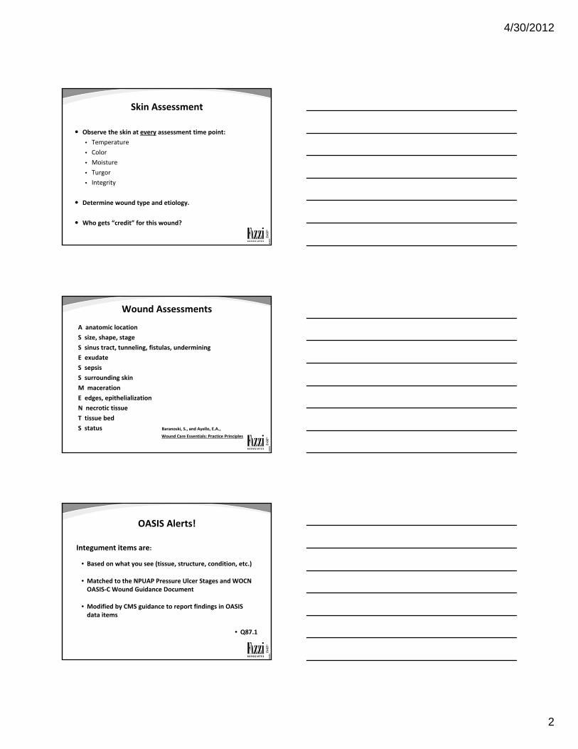
Skin Assessment
Observe the skin at every assessment time point:
• Temperature
• Color
• Moisture
• Turgor
• Integrity
Determine wound type and etiology.
Who gets “credit” for this wound?
©2010
©2012
Wound Assessments
A anatomic location
S size, shape, stage
S sinus tract, tunneling, fistulas, undermining
E exudate
S sepsis
S surrounding skin
M maceration
E edges, epithelialization
N necrotic tissue
T tissue bed
S status Baranoski, S., and Ayello, E.A., Wound Care Essentials: Practice Principles
©2010
©2012
OASIS Alerts!
Integument items are:
• Based on what you see (tissue, structure, condition, etc.)
• Matched to the NPUAP Pressure Ulcer Stages and WOCN OASIS‐C Wound Guidance Document
• Modified by CMS guidance to report findings in OASIS data items
• Q87.1

4/30/2012
3
©2010
©2012
©2010
©2012
Pressure Ulcer Definition
Localized injury to the skin and/or underlying tissue usually over a bony prominence, as a result of pressure or pressure in combination with shear and/or friction.
4 Stages + Suspected Deep Tissue Injury + Unstageable
(NPUAP 2007)
©2010
©2012
Unavoidable Pressure UlcerNPUAP March 2010
Unavoidable ‐means that the individual developed a pressure ulcer even though the provider had:
• Evaluated the individual's clinical condition and pressure ulcer risk factors;
• Defined and implemented interventions that are consistent with individual needs goals and recognized standards of practice;
• Monitored and evaluated the impact of the interventions; and
• Revised the approaches as appropriate.
4/30/2012
4
©2010
©2012
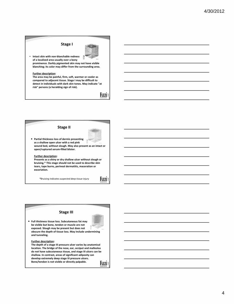
Stage I
• Intact skin with non‐blanchable redness of a localized area usually over a bony prominence. Darkly pigmented skin may not have visible blanching; its color may differ from the surrounding area.
Further description:The area may be painful, firm, soft, warmer or cooler as compared to adjacent tissue. Stage I may be difficult to detect in individuals with dark skin tones. May indicate "at risk" persons (a heralding sign of risk).
©2010
©2012
Stage II
Partial thickness loss of dermis presenting as a shallow open ulcer with a red pink wound bed, without slough. May also present as an intact or open/ruptured serum‐filled blister.
Further description:Presents as a shiny or dry shallow ulcer without slough or bruising.* This stage should not be used to describe skin tears, tape burns, perineal dermatitis, maceration or excoriation.
*Bruising indicates suspected deep tissue injury
©2010
©2012
Stage III
Full thickness tissue loss. Subcutaneous fat maybe visible but bone, tendon or muscle are not exposed. Slough may be present but does not obscure the depth of tissue loss. May include undermining and tunneling.
Further description: The depth of a stage III pressure ulcer varies by anatomical location. The bridge of the nose, ear, occiput and malleolus do not have subcutaneous tissue, and stage III ulcers can be shallow. In contrast, areas of significant adiposity can develop extremely deep stage III pressure ulcers. Bone/tendon is not visible or directly palpable.

4/30/2012
5
©2010
©2012
Stage IV
Full thickness tissue loss with exposed bone, tendon or muscle. Slough or eschar may be present on some parts of the wound bed. Often include undermining and tunneling.
Further description: The depth of a stage IV pressure ulcer varies by anatomical location. The bridge of the nose, ear, occiput and malleolus do not have subcutaneous tissue and these ulcers can be shallow. Stage IV ulcers can extend into muscle and/or supporting structures (e.g., fascia, tendon or joint capsule) making osteomyelitis possible. Exposed bone/tendon is visible or directly palpable.
©2010
©2012
OASIS Alert!
CMS modifies the NPUAP reference to “directly palpable” in a stage IV pressure description. For OASIS purposes, to classify a pressure ulcer as a stage IV, exposed bone/tendon must be “visible” and not “directly palpable”.
Q89.3
©2010
©2012
Suspected Deep Tissue Injury (SDTI)
• Purple or maroon localized area of discolored intact skin OR a blood filled blister due to damage of underlying soft tissue from pressure and/or shear.
• The area may be preceded by tissue that is painful, firm, mushy, boggy, warmer or cooler as compared to adjacent tissue.
(NPUAP 2007)
4/30/2012
6
©2010
©2012
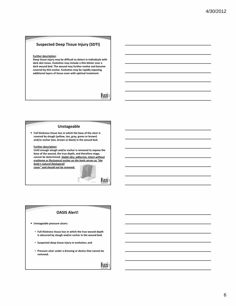
Suspected Deep Tissue Injury (SDTI)
Further description:Deep tissue injury may be difficult to detect in individuals with dark skin tones. Evolution may include a thin blister over a dark wound bed. The wound may further evolve and become covered by thin eschar. Evolution may be rapidly exposing additional layers of tissue even with optimal treatment.
©2010
©2012
Unstageable
Full thickness tissue loss in which the base of the ulcer is covered by slough (yellow, tan, gray, green or brown) and/or eschar (tan, brown or black) in the wound bed.
Further description:Until enough slough and/or eschar is removed to expose the base of the wound, the true depth, and therefore stage, cannot be determined. Stable (dry, adherent, intact without erythema or fluctuance) eschar on the heels serves as "the body's natural (biological) cover" and should not be removed.
©2010
©2012
OASIS Alert!
Unstageable pressure ulcers:
• Full thickness tissue loss in which the true wound depth is obscured by slough and/or eschar in the wound bed;
• Suspected deep tissue injury in evolution; and
• Pressure ulcer under a dressing or device that cannot be removed.

4/30/2012
7
©2010
©2012
OASIS Alert!
A previously stageable pressure ulcer now covered by eschar is considered “unstageable.”
Q98.1
Do not reverse stage pressure ulcers.
OASIS‐C Guidance Manual
©2010
©2012
Pressure Ulcers
Partial thickness tissue loss:
• Involves epidermis and into but not through the dermis
• Superficial; presents as shallow crater, abrasion or blister
• Heals by epithelialization
• Regeneration of epidermis across a wound surface
• Includes stage I and II pressure ulcers
©2010
©2012
Pressure Ulcers
Full thickness tissue loss:
• Penetrates through the fat (subcutaneous tissue) and may involve muscle, tendon, or bone
• Deep crater; may tunnel
• Heals by granulation, contraction and epithelialization
• Never considered fully healed
• Closed when fully granulated and covered with new epithelial tissue
• Includes stage III and IV pressure ulcers

4/30/2012
8
©2010
©2012
Degree/Status of HealingPressure Ulcer
Wound Status:
• Not healing
• Early/partial granulation
• Fully granulating
• Newly epithelialized
• When epithelial tissue has completely covered the wound surface, regardless of how long the pressure ulcer has been re‐epithelialized.
• www.wocn.org Wound Guidance Document
©2010
©2012
OASIS Alerts!
An ulcer diagnosed by a physician as a diabetic ulcer is not a pressure ulcer or stasis ulcer.
Q89
A pressure ulcer covered with a skin graft or debrided remains a pressure ulcer.
Q89.1 and 95
A pressure ulcer closed with a muscle flap becomes a surgical wound.
Q94
©2010
©2012
OASIS Alerts!
A previously closed stage III or IV pressure ulcer that is currently open again is reported at its worst stage.
OASIS‐C Guidance Manual
A pressure ulcer closed with a muscle flap that heals and then breaks down due to pressure becomes a new pressure ulcer.
Q94
4/30/2012
9
©2010
©2012
OASIS Alerts!
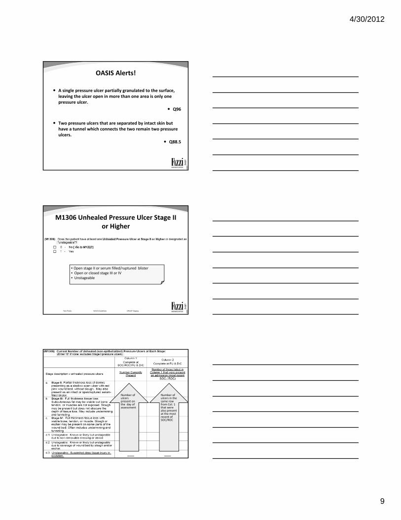
A single pressure ulcer partially granulated to the surface, leaving the ulcer open in more than one area is only one pressure ulcer.
Q96
Two pressure ulcers that are separated by intact skin but have a tunnel which connects the two remain two pressure ulcers.
Q88.5
©2010
©2012
M1306 Unhealed Pressure Ulcer Stage II or Higher
Time Points WOCN Guidelines NPUAP Staging
• Open stage II or serum filled/ruptured blister• Open or closed stage III or IV• Unstageable
©2010
©2012
M1308 Current Number of Unhealed Pressure Ulcers/Stage
Time Points WOCN Guidelines NPUAP Staging
Number of ulcers present on the day of assessment
Number of ulcers in the same row from Col. 1 that were also present at the most recent of SOC/ROC

4/30/2012
10
©2010
©2012
Time Points WOCN Guidelines NPUAP Staging
Patient 1 at SOC does not have an unhealed stage II pressure ulcer. There are no pressure ulcers.
©2010
©2012
Time Points WOCN Guidelines NPUAP Staging
Patient 1 at Follow Up has 1 unhealed stage II pressure ulcer.
1
0
0
0
0
0
0
0
0
0
0
0
©2010
©2012
Time Points WOCN Guidelines NPUAP Staging
Patient 2 at SOC has 1 unhealed stage III pressure ulcer.
0
1
0
0
0
0
4/30/2012
11
©2010
©2012
Time Points WOCN Guidelines NPUAP Staging
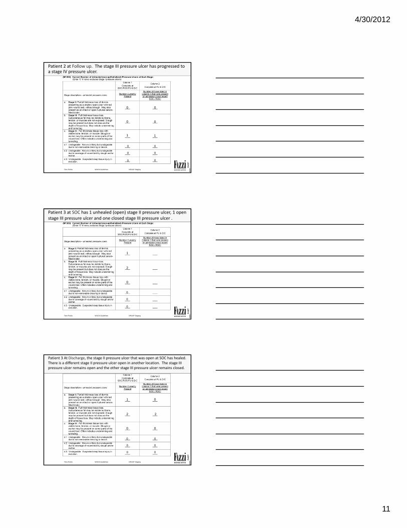
Patient 2 at Follow up. The stage III pressure ulcer has progressed to a stage IV pressure ulcer.
0 0
0 0
1 1
0 0
0 0
0 0
©2010
©2012
Time Points WOCN Guidelines NPUAP Staging
Patient 3 at SOC has 1 unhealed (open) stage II pressure ulcer, 1 open stage III pressure ulcer and one closed stage III pressure ulcer .
1
2
0
0
0
0
©2010
©2012
Time Points WOCN Guidelines NPUAP Staging
Patient 3 At Discharge, the stage II pressure ulcer that was open at SOC has healed. There is a different stage II pressure ulcer open in another location. The stage III pressure ulcer remains open and the other stage III pressure ulcer remains closed.
1 0
2 2
0 0
0 0
0 0
0 0

4/30/2012
12
©2010
©2012
Time Points WOCN Guidelines NPUAP Staging
Patient 4 At SOC there is 1 unhealed stage II pressure ulcer, 1 unhealed stage III pressure ulcer and 1 closed stage III pressure ulcer.
1
2
0
0
0
0
©2010
©2012
Time Points WOCN Guidelines NPUAP Staging
Patient 4 At Recertification, the stage II pressure ulcer that was open at SOC has fully re‐epithelialized. Another stage II PU is open in a different location. The stage III PU now has bone exposed and the other stage III PU remains closed.
1 0
1 1
1 1
0 0
0 0
0 0
©2010
©2012
Time Points WOCN Guidelines NPUAP Staging
Patient 5 At SOC, there is 1 pressure ulcer on the left heel covered with eschar and 1 blood filled blister on the right heel from pressure after many days of bed rest. There is a stage III pressure ulcer which closed in the hospital and remains closed.
0
1
0
0
1
1
4/30/2012
13
©2010
©2012
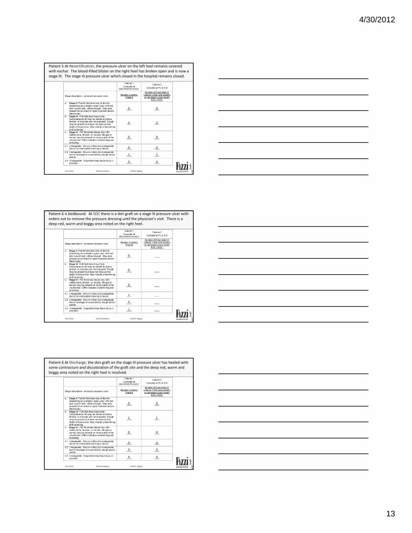
Time Points WOCN Guidelines NPUAP Staging
Patient 5 At Recertification, the pressure ulcer on the left heel remains covered with eschar. The blood‐filled blister on the right heel has broken open and is now a stage III. The stage III pressure ulcer which closed in the hospital remains closed.
0 0
2 2
0 0
0 0
1 1
0 0
©2010
©2012
Time Points WOCN Guidelines NPUAP Staging
Patient 6 is bedbound. At SOC there is a skin graft on a stage III pressure ulcer with orders not to remove the pressure dressing until the physician's visit. There is a deep red, warm and boggy area noted on the right heel.
0
0
0
1
0
1
©2010
©2012
Time Points WOCN Guidelines NPUAP Staging
Patient 6 At Discharge, the skin graft on the stage III pressure ulcer has healed with some contracture and discoloration of the graft site and the deep red, warm and boggy area noted on the right heel is resolved.
0 0
1 1
0 0
0 0
0 0
0 0

4/30/2012
14
©2010
©2012
Time Points WOCN Guidelines NPUAP Staging
Patient 7 at SOC has a stage I pressure ulcer on each heel.
©2010
©2012
Time Points WOCN Guidelines NPUAP Staging
Patient 7 At Recertification, there is no evidence of a stage I pressure ulcer on the right heel. The stage I pressure ulcer on the left heel is now a stage II.
1 1
0 0
0 0
0 0
0 0
0 0
©2010
©2012
Time Points WOCN Guidelines NPUAP Staging
Patient 8 at SOC has a stage III pressure ulcer.
0
1
0
0
0
0

4/30/2012
15
©2010
©2012
Time Points WOCN Guidelines NPUAP Staging
Patient 8 At Recertification, the stage III pressure ulcer from SOC is now covered with eschar.
0 0
0 0
0 0
0 0
1 1
0 0
©2010
©2012
M1310, M1312, M1314 ‐ Unhealed Stage III or IV Pressure Ulcer with Largest Surface Dimension
SOC/ROC/DC
Time Points WOCN Guidelines NPUAP Staging
Consider all stage III and IV pressure ulcers from M1308 Col.1 row b (stage III), row c (stage IV), and row d.2 (unstageablecovered w/ slough or eschar).
Depth does not include the depth of any tunneling.
©2010
©2012
OASIS Alert!
Status of the pressure ulcer needs to correspond to the visual assessment of the clinician on the day of the assessment.
Q98

4/30/2012
16
©2010
©2012
WOCN Definitions Degree of Healing
• Not healing
• Wound with ≥ 25% avascular tissue (eschar and/or slough) OR
• Signs/symptoms of infection OR
• Clean but non‐granulating wound bed OR
• Closed/hyperkeratotic wound edges OR
• Persistent failure to improve despite appropriate comprehensive wound management
• Early/partial granulation
• ≥ 25% of the wound bed is covered with granulation tissue
• < 25% of the wound bed is covered with avascular tissue (eschar and/or slough)
• No signs or symptoms of infection
• Wound edges open
©2010
©2012
WOCN DefinitionsDegree of Healing
• Fully granulating
• Wound bed filled with granulation tissue to the level of the surrounding skin
• No dead space
• No avascular tissue (eschar and/or slough)
• No signs or symptoms of infection
• Wound edges are open
• Newly epithelialized
• Wound bed completely covered with new epithelium
• No exudate
• No avascular tissue (eschar and/or slough)
• No signs or symptoms of infection
©2010
©2012
OASIS Alerts!
Stage II pressure ulcers that close/heal/fully epithelialize are not reportable on OASIS and therefore will not be “newly epithelialized” for data collection.
Stage II pressure ulcers do not granulate.
Stage II pressure ulcers (includes serum filled blister) can only be “not healing” for data collection.
4/30/2012
17
©2010
©2012
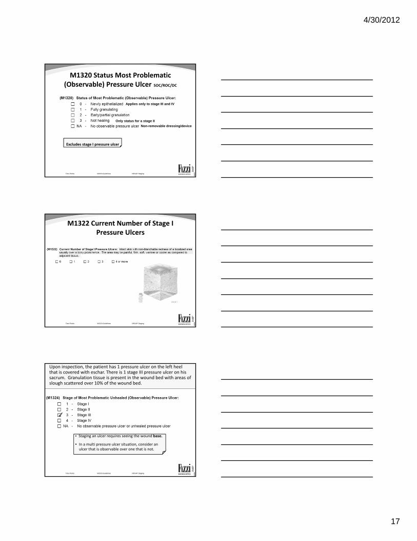
M1320 Status Most Problematic (Observable) Pressure Ulcer SOC/ROC/DC
Time Points WOCN Guidelines NPUAP Staging
Excludes stage I pressure ulcer
Applies only to stage III and IV
Only status for a stage II
Non-removable dressing/device
©2010
©2012
M1322 Current Number of Stage I Pressure Ulcers
Time Points WOCN Guidelines NPUAP Staging
©2010
©2012
M1324 Stage Most Problematic (Observable) Pressure Ulcer SOC/ROC/FU/DC
Time Points WOCN Guidelines NPUAP Staging
Upon inspection, the patient has 1 pressure ulcer on the left heel that is covered with eschar. There is 1 stage III pressure ulcer on his sacrum. Granulation tissue is present in the wound bed with areas of slough scattered over 10% of the wound bed.
• Staging an ulcer requires seeing the wound base.
• In a multi pressure ulcer situation, consider an ulcer that is observable over one that is not.
4/30/2012
18
©2010
©2012
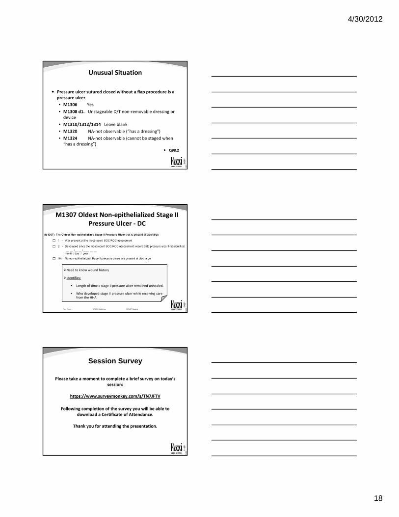
Unusual Situation
Pressure ulcer sutured closed without a flap procedure is a pressure ulcer
• M1306 Yes
• M1308 d1. Unstageable D/T non‐removable dressing or device
• M1310/1312/1314 Leave blank
• M1320 NA‐not observable (“has a dressing”)
• M1324 NA‐not observable (cannot be staged when “has a dressing”)
Q98.2
©2010
©2012
M1307 Oldest Non‐epithelialized Stage II Pressure Ulcer ‐ DC
Time Points WOCN Guidelines NPUAP Staging
Need to know wound history
Identifies:
• Length of time a stage II pressure ulcer remained unhealed.
• Who developed stage II pressure ulcer while receiving carefrom the HHA.
©2010
©2012
Session Survey
Please take a moment to complete a brief survey on today’s session:
https://www.surveymonkey.com/s/TN7JFTV
Following completion of the survey you will be able to download a Certificate of Attendance.
Thank you for attending the presentation.
4/30/2012
19
©2010
©2012
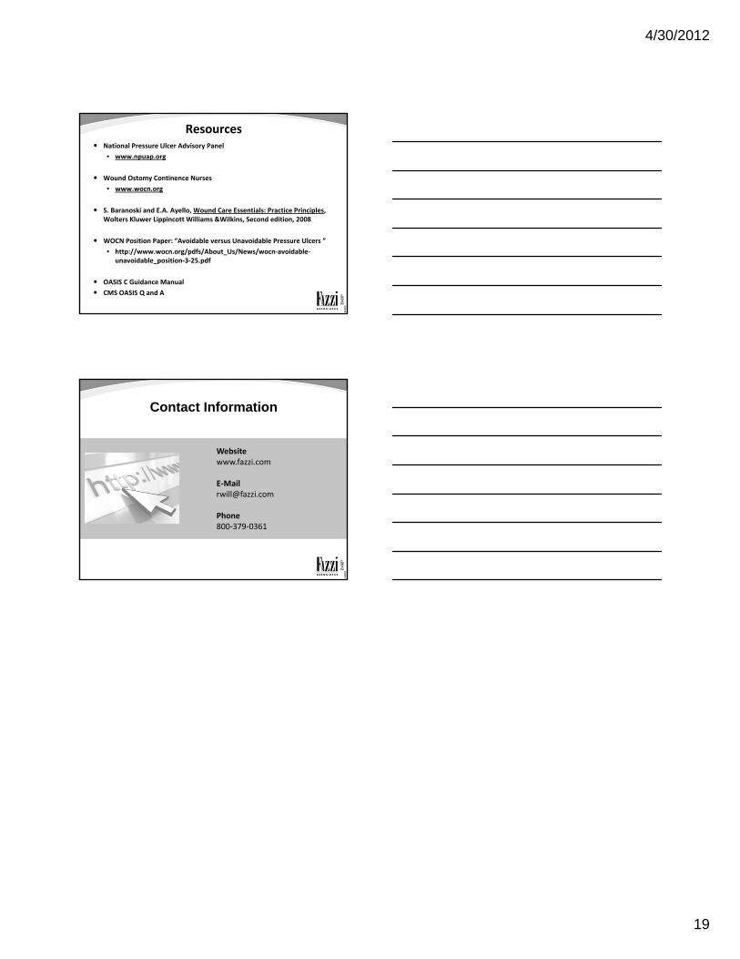
Resources National Pressure Ulcer Advisory Panel
• www.npuap.org
Wound Ostomy Continence Nurses
• www.wocn.org
S. Baranoski and E.A. Ayello, Wound Care Essentials: Practice Principles, Wolters Kluwer Lippincott Williams &Wilkins, Second edition, 2008
WOCN Position Paper: “Avoidable versus Unavoidable Pressure Ulcers ”
• http://www.wocn.org/pdfs/About_Us/News/wocn‐avoidable‐unavoidable_position‐3‐25.pdf
OASIS C Guidance Manual
CMS OASIS Q and A
©2010
©2012
Contact Information
Websitewww.fazzi.com
Phone800‐379‐0361