o Receptive Relaxation of the Fundus aka “Accommodation ... · o Feeding State (NB gastrocolic...
27
Stomach Stomach Anatomy o Arterial supply from Celiac Artery branches, Venous drainage into Portal Vein, Lymphatic Drainage into Celiac LNs Motility o Fasting State (MMC, refer above) o Feeding State (NB gastrocolic reflex occurs at this time) (1) Receptive Relaxation of the Fundus aka “Accommodation”: as food enters the stomach there is vagally mediated receptive relaxation of the fundus with storage of food for ~30min (2) Peristalsis of the Body aka “Titration”: vagally mediated segmental contractions 3x/min in body mixing food w/ gastric juice and saliva creating chyme (3) Peristalsis of the Antrum aka “Antroduodenal Coordination”: first wave closes pylorus, second wave pushes chyme against pylorus and chyme is propelled back allowing for continued mixing but a little (<3mL of chyme <2mm in size) is pushed thru, this occurs at 3cpm NB Pacemaker (juncture of the fundus and body of the greater curvature) NB duodenal pH and osmolarity receptors delay gastric emptying if too acidic or hypertonic Histology o similar to intestine except there are three muscularis propria layers (inner oblique, middle circular, outer longitudinal) and the epithelium creates gastric pits or foveolae (instead of villi) each w/ two glands Antral/Pyloric Glands (shorter glands b/c they don’t secrete much just mucus, loosely packed, purple) vs Oxyntic Glands (longer glands b/c they secrete a lot, packed tightly, pink) vs NB in general gastric lamina propria should never have inflammatory cells and if present then gastritis is present hence why pathologist almost always call mild gastritis on biopsy NB “foveolar hyperplasia” (surface mucin cells darken and proliferate creating a papillary appearance to the surface and corkscrewing profile deeper down, results from gastric irritation) NB “atrophy” (loss of glands, seen w/ chronic irritation, usually accompanied by intestinal metaplasia) Antral/Pyloric Glands Oxyntic Glands
Transcript of o Receptive Relaxation of the Fundus aka “Accommodation ... · o Feeding State (NB gastrocolic...

Stomach Stomach
Anatomy o Arterial supply from Celiac Artery branches, Venous drainage into Portal Vein, Lymphatic Drainage into Celiac LNs
Motility o Fasting State (MMC, refer above) o Feeding State (NB gastrocolic reflex occurs at this time)
(1) Receptive Relaxation of the Fundus aka “Accommodation”: as food enters the stomach there is vagally mediated receptive relaxation of the fundus with storage of food for ~30min
(2) Peristalsis of the Body aka “Titration”: vagally mediated segmental contractions 3x/min in body mixing food w/ gastric juice and saliva creating chyme
(3) Peristalsis of the Antrum aka “Antroduodenal Coordination”: first wave closes pylorus, second wave pushes chyme against pylorus and chyme is propelled back allowing for continued mixing but a little (<3mL of chyme <2mm in size) is pushed thru, this occurs at 3cpm
NB Pacemaker (juncture of the fundus and body of the greater curvature)
NB duodenal pH and osmolarity receptors delay gastric emptying if too acidic or hypertonic
Histology o similar to intestine except there are three muscularis propria layers (inner oblique, middle circular, outer longitudinal)
and the epithelium creates gastric pits or foveolae (instead of villi) each w/ two glands Antral/Pyloric Glands (shorter glands b/c they don’t secrete much just mucus, loosely packed, purple) vs
Oxyntic Glands (longer glands b/c they secrete a lot, packed tightly, pink) vs NB in general gastric lamina propria should never have inflammatory cells and if present then gastritis is
present hence why pathologist almost always call mild gastritis on biopsy NB “foveolar hyperplasia” (surface mucin cells darken and proliferate creating a papillary appearance to the
surface and corkscrewing profile deeper down, results from gastric irritation) NB “atrophy” (loss of glands, seen w/ chronic irritation, usually accompanied by intestinal metaplasia)
Antral/Pyloric Glands Oxyntic Glands


Secretory Function (2.5L/d) o General
Goals: decontaminate w/ acid, VitB12 absorption, some breakdown of protein/lipids more for hormone stimulation than for true digestion
Three Phases of Acid Secretion
Cephalic Phase (5 senses stimulate Vagus)
Gastric Phase (distension of food and amino acids stimulate G‐cells)
Intestinal Phase (amino acids in duodenum stimulate G‐cells) NB Cardia (looks more like antral mucosa than oxyntic mucosa!!!, controversial whether it is a pathologic area
2/2 GERD or normal area of the stomach) o 80% Oxyntic Glands (Fundus (mucus) + Corpus aka Body (HCl + Pepsin)) = active secretory portion (NB isthmus has surface
cells, neck has neck cells and parietal cells, base has chief cells, endocrine cells are scattered throughout) Columnar Epithelial Surface Cells aka Surface Cells (bicarb “alkaline tide” and insoluble mucus creating a
protective lining on stomach mucosa w/ pH gradient of 2 vs 7) vs Neck Cells (soluble mucus creating a smooth food bolus, neck cells also serve as stem cells for the cells below)
NB PGE2/F2/I2 stimulates mucus, bicarb, blood flow and epithelial regeneration which are all protective in addition to the tight junctions and intracellular buffers
o COX‐1 (constitutively expressed in the stomach to constantly maintain defense mechanisms) vs COX‐2 (inducibly expressed in the stomach in response to cytokines in order to create an inflammatory response)
+: Secretin
‐: Somatostatin Chief Cells (zymogen pepsinogen converted to active enzyme pepsin by acidic pH, pepsin can convert further
pepsinogen, pepsins are active at a pH of 1.8‐3.5, pepsin serves to breakdown proteins with the goal of releasing AAs which in turn trigger the release of important GI hormones such as gastrin/cholecystokinin thus pepsin is not really there for digestion, NB leptin and lipase is also secreted, gastric lipase is very different from pancreatic lipase in that it works at a lower pH and does not need colipase, the lipase secreted is a low amount capable of digesting 20% of dietary triglycerides, mainly used to breakdown TG from breast milk therefore important for babies)
+: M1, Other (CCK, Gastrin, GRP, secretin, VIP, histamine)
‐: Somatostatin, Other (Neuropeptide Y, PYY, IL‐1beta) Parietal Cells (secrete HCl & IF)
+: Gastrin‐CCK2 (from G Cells in Pylorus = endocrine, don’t stimulate acid rather are trophic and potentiate other secretagogues), M3 (from Vagus Nerve = neurocrine, don’t stimulate acid rather are trophic, NB alcohol stimulate M3 also), H2 (from near‐by ECL Cells = paracrine, most important!!!)
‐: PGE2, Secretin (from S Cells in duodenum), Somatostatin‐SSTR2 (from D Cells throughout GI tract), GIP (from K cells in duodenum), peptide YY
NB responds to paracrine/endocrine/neurocrine stimuli, secondary messengers (Ca for ACh/Gastrin or cAMP for Histamine) activates the H/K ATPase
HCL serves to (1) convert pepsinogen, (2) digest some protein, (3) kill most bacteria making stomach and small intestine sterile preventing enteric pathogenic infection and SIBO except for micro‐aerophilic spiral bacteria in the stomach (eg. Helicobacter), (4) facilitate absorption of iron, calcium, VitB12, meds (eg. thyroid)
Rest (pumps are sequestered w/in cytoplasmic vesicles which are absent of K/Cl/HCO channels thus the pumps are inactive) vs Active (vesicles fuse w/ apical membrane creating a vast canilicular system w/ microvilli which contain multiple K/Cl/HCO3 channels thus the pumps become active)
PPIs (have a pKa of 4.5 thus in environments w/pH>4.5 they remain non‐protonated (membrane permeable) vs pH <4.5 they remain protonated (membrane impermeable) therefore in almost all parts of the body PPIs flow around and through cells until they pass thru the parietal cell into the apical canilicular space where they become protonated and become trapped such that the [PPI] on

the apical surface is 1 million times higher than any other place in the body, the protonated PPI then interacts with the luminal side of the proton pump inhibiting it by creating a disulfide bond with each different PPI binding to a different residue)
o PPI should be taken 30min before a meal so that right when pumps are brought to the surface during a meal and acid is created the PPI are right there , de novo synthesis of pumps (~52hrs) must be done to recover from the effects of PPIs
Vagus Innervation
+: 5 senses, food distension, protein EnteroChromaffin Like (ECL) Cells (secrete Histamine into tissue thus paracrine)
+: Gastrin (other including PACAP, VIP, Grehlin)
‐: Somatostatin (other including PG, Peptide YY, Galanin) D Cells A/Gr Cells (secrete Ghrelin which acts on CNS to stimulate hunger and thus food intake, GH release, weight
gain)
High: fasting, anorexia, starvation, weight loss, Prader‐Willi
Low: obesity, postprandial state, gastric bypass
NB cells are also found in antrum, small intestine, colon
NB suppressed after RYGB contributing to weight loss o 20% Antral/Pyloric Glands (Antrum then Canal then Pyloric Sphincter) = regulatory feedback portion (angularis incisura
divides oxyntic from pyloric regions) Columnar Epithelial Surface/Neck Cells G Cells (secrete Gastrin into bloodstream thus endocrine)
+: Vagus (M3), AAs, Distention, Ca, Beta‐Agonists, Alcohol
‐: Somatostatin, Adenosine, Galanin D Cells (secrete Somatostatin into tissue thus paracrine)
+: low pH, CCK (so when food enters the duodenum somatostatin is increased to decrease gastric emptying), (Other: Gastrin, GRP, VIP, PACAP, BA, Secretin, ANP, Amylin, Adenosine, CGRP)
‐: High pH, Histamine, Vagus (M2/M4), Other: IFN General
What do you do w/ dyspepsia? DDx: GERD, HP, PUD (hence the algorithm below) o >55yo or Alarm Features (persistent heartburn despite Tx, extra‐esophageal GERD, + imaging, >5% involuntary weight
loss, bleeding, odynophagia/dysphagia, recurrent vomiting, FHx of gastric cancer) = EGD o <55yo AND NO Alarm Features = below and if no improvement then EGD and if negative then likely functional dyspepsia
(refer) Reflux Predominant = PPI Trial x8wks Non‐Reflux Predominant
HP Prevalence (>10%, immigrants, big urban cities, low socioeconomic status) = Test for HP and if no improvement then PPI trial and if no improvement then EGD
HP Prevalence (<10%) = PPI Trial x8wks and if no improvement then test for HP and if no improvement then EGD
Vagal Nerve Damage = (1) cannot store/mix/titrate food = gastroparesis, (2) cannot stimulate acid secretion, (3) cannot stimulate
gastrin secretion
Secretion Problems
Etiology
Increased Acid Secretion o Duodenal Ulcer o ZES/Gastrinoma o Gastric Heterotopia o Retained Antrum Syndrome (after BII sometimes a piece of the antrum is retained in the afferent remnant and is
subsequently bathed in alkaline secretions leading to decreased somatostatin secretion and thus hypergastrinemia w/ increased acid and ulcers forming at anastomosis)
o Hypercalcemia (Ca stimulates gastrin secretion from G‐cells and acid secretion from parietal cells) o Antral Predominant HP Gastritis (D‐cell inhibition and G‐cell hyperplasia, NB most pts w/ HP infection have mild
gastritis and thus normal gastric secretion) o PPI/H2B Withdrawal (momentary rebound after quick withdrawal after long term use) o Systemic Mastocytosis (high histamine levels continuously stimulate parietal cells to secrete acid) o Basophilic Leukemia
Decreased Acid Secretion (a/hypochlorhydria = absence of acid during maximal stimulation, NB acid secretion does not decrease w/ age)
o PPI/H2B/AA o Gastritides: Pan HP Gastritis, Chronic Atrophic Gastritis, Chronic Active Superficial Gastritis, HIB o Gastric AC o Vagotomies

o Antrectomy (less parietal cells or less gastrin release from G‐cells) o Chronic Renal Failure
S/S o a/hypochlorhydia can lead to bacterial overgrowth
Dx o “Gastric Analysis” = gastric juices in fasting pt are collected via NGT in distal antrum Q15min x1hr (most dependent
portion of stomach determined by fluoro or recovery of >90cc of 100cc injection of water) to determine mmol of acid per hour aka Basal Acid Output (BAO, nl <5/10mmol/hr F/M, NB varies throughout a 24hr period w/ peak b/t 2‐11pm) w/ Peak Acid Output (PAO, nl 10‐60mmol) and Maximal Acid Output (MAO, nl 5‐30/7‐50mmol/hr F/M) determined by stimulation via Pentagrastin 6mcg/kg SC/IM or 6mcg/kg/hr IV (analogue of Gastrin)
DDx of Hypergastrinemia
Appropriate: antisecretory drugs, atrophic gastritis, CKD, post‐vagotomy
Inappropriate: ZES, retained antrum syndrome, antral predominant HP, massive intestinal resection, GOO
NB rarely done anymore except when (1) trying to determine whether fasting hypergastrinemia is associated w/ basal acid hypersecretion (eg. ZES) or hyposecretion (eg. atrophic gastritis) or (2) tailoring antisecretory therapy to ZES pts w/ non‐resectable tumors
Endoscopic techniques are being employed NB you can also do tests w/ sham feeding (expose all 5 sense to food but pt doesn’t swallow) or actual meals
o SmartPill can measure acid secretion Structural Problems
General o Congenital anomalies are least frequent compared to the rest of GI tract o Congenital anomalies can present at any age depending on degree of outlet obstruction
Atresia: complete or incomplete connection in antrum resulting in gastric outlet obstruction w/ polyhydramnios during pregnancy, nonbilious vomiting during first feeding, drooling and respiratory distress during infancy or dyspeptic Sx during adulthood depending on complete the atresia is
Microgastria: not lethal in and of itself but often associated with other conditions
Diverticulum: congenital (rarest of all of GI diverticula, 70% lesser curvature of near EGJ) vs acquired (2/2 pancreatitis, gastric outlet obstruction, trauma, PUD, cancer), general asymptomatic with no Tx necessary
Duplication: in isolation or associated with duplications in other parts of the GI tract and/or vertebral anomalies, usually along the greater curvature or posterior wall, contains all gastric layers, rarely communicates with the stomach appearing as a cystic mass, if there is a connection consider acquired fistulalization from a pancreatic ulcer w/in the duplication, most detected during childhood but cancer has been documented to occur in adults, S/S all depend on size/location/communication, suggested by displacement or extrinsic compression of gastric lumen
Volvulus o Pt: any pt that has a diaphragmatic hernia which loosens some of the ligament attachments and allows to twist inside the
thorax o Types
60% organoaxial
Mech: twists along axis created by an imaginary line from EGJ to Pylorus w/ fundus rotating posterior/inferiorly and antrum anterior/superiorly (in the R hemithorax)
S/S: (1) complete/acute (hrs) w/ sudden severe epigastric pain, (2) retching BUT no vomitus, (3) inability to pass NGT = Borchardt’s Triad
Tx: nasogastric decompression, endoscopic/surgical detorsion w/ gastropexy
Complication: strangulation w/ infarction/obstruction 40% mesenteroaxial
Mech: antrum folds anterior/superiorly towards fundus (in the L hemithorax)
S/S: usually incomplete/chronic (yrs) w/ epigastric discomfort, bloating, fullness, heartburn, consider even if images are normal b/c intermittent
Tx: ?
Diaphragmatic Hernia o (1) thru congenital openings 2/2 failure of fusion of embryonic diaphragmatic components,
stomach/omentum/colon/spleen/liver herniate, hypoplasia/volvulus/incarceration can occur, Tx: surgery foramina of Bochdalek (posteriolateral along the left lumbocostal jxn = “back and to the left”, often manifest
immediately after birth causing pulmonary hypoplasia, often associated w/ trisomy disorders) foramina of Morgagni (anterior along the right sternocostal jxn, often manifest during adulthood)
o (2) thru post‐traumatic defects, main different organs can herniate thru including bowel, omentum, spleen, kidney, etc o (3) thru hiatus
Type 1 (95%): sliding, both GEJ and part of stomach herniate above diaphragm, no change in stomach axis, cause unknown but likely due to age related degeneration of the phreno‐esophageal membrane that normally anchors the GEJ to the diaphragm, most are small and asymptomatic, Complications: (1) GERD b/c the diaphragm provides some of the tone for the LES, (2) Cameron ulcers, Tx: surgery if symptomatic (refer to GERD)

Type 2 (5%): paraesophageal, greater curvature of stomach herniates alongside the esophagus above diaphragm w/ GEJ remaining in place b/c the phreno‐esophageal membrane is intact, 2/2 congenital defect of the hiatus, unlike sliding hernias paraesophageal are often symptomatic w/ dyspepsia, dysphagia, chest pain, SOB, etc, complications: (1) there is often a change in axis w/ possible complete volvulus, (2) other organs including the colon, omentum and spleen can also herniate, (3) these hernias tend to enlarge with time and eventually the entire stomach rolls into the thorax resulting in possible obstructions, incarcerations, strangulations, etc Tx: given complications these hernias should be always treated surgically (repair crural defect + fundoplication + gastropexy)
Type 3 (combination of both) Type 4 (combination of both along w/ other organs)
Motility Problems
Diagnostic Tests o Gastric Emptying Scintigraphy (GES) to assess antral emptying o Electrogastrography (EGG) to assess gastric electric activity o Capsule Motility Device to asses gastric contractions (rare) o Full Thickness Bx are of little value but may show loss of ICCs (rare)
Gastric Dysrhythmias (nl GES but abnl EGG) o Dsyautonomias (Riley‐Day Syndrome) o Primary Dysrhythmia of the Gastric Pacemaker in Greater Curvature (tachy >3.7cpm, brady <2.5cpm, ectopic that
overrides normal but is uncoordinated)
Impaired Fundic Relaxation (leads to early satiety)
Antroduodenal Dyscoordination
Postsurgical Syndromes (refer to obesity) o Dumping Syndrome aka Rapid Gastric Emptying
Etiology
s/p surgery: post vagotomy w/ pyloroplasty, BI/II gastrojejunostomy, RYGB, any type of gastroenteric anastomosis
idiopathic: spontaneously occurs in pts w/o any surgical change to stomach anatomy
other: DM, ZES, et al Mech: food is not accommodated nor titrurated by the remaining stomach → large amounts of osmotically
active food is rapidly emptied aka dumped into the duodenum S/S
Early (15‐30min after meal): cramps, bloating, N, D, belching (2/2 osmotic shifts into the gut lumen w/ resulting distension)
Late (1‐2hrs after meal): vasomotor symptoms of lightheadedness, diaphoresis, palpitations, tachycardia, syncope (2/2 absorption of large amounts of carbs that is poorly matched by insulin resulting in subsequent hypoglycemia from insulin spike)
Dx: clinical but GES (<35% of meal is emptied in <60min) Tx: “dumping diet” low osm/carb (no simple sugars only complex) small non‐liquid frequent meals, drink fluids
separate from meals (not sure why but it works), pectin containing foods (banana, oranges, etc) slows gastric output
o Blind Loop Syndrome o Afferent Loop Syndrome o Vagal Nerve Damage causes a variety of things including gastric dysrhythmia, dilated antrum, poor fundic relaxation,
gastric hypo/hypersensitivity)
Pyloroplasm
Other o Rumination Syndrome
Pyloric Stenosis o Child Hypertrophic Pyloric Stenosis
Epidemiology: most common abdominal surgery in infants, 1/300 births, Caucasian, M>F, first born, other siblings w/ HPS, parents had HPS
Mech: unknown (has been associated w/ erythromycin given in first 2wks of life, lack of NO synthase, abnormal innervations, absence of ICC, increased EGF receptors, etc) hypertrophy (first thickness then length) of the muscularis propria with resultant edema and inflammatory changes
Comorbidities: alone or in association w/ Turner’s, Trisomy‐18, Esophageal Atresia Sx: present at ~4wks after birth, progressive non‐bilious projectile vomiting ~15min after feeding PEx: 80% have a epigastric palpable mass (“olive”) w/ visible gastric peristalsis and 2% have jaundice (there is
also an association with glucuronyl transferase deficiency) Labs: metabolic alkalosis Dx: US (pyloric channel >21mm in length and >5mm in thickness) and if non‐diagnostic then barium swallow
(“string sign” or “double railroad track sign”) Tx: fluid/electrolyte resuscitation followed by surgery via Fredet‐Ramstedt Pyloromyotomy (cut out muscle
from the outer adventitial side)

o Adult NON‐Hypertrophic Pyloric Stenosis acquired when pt has gastric antral dz (PUD, hypertrophic gastropathy, cancer) resulting in inflammatory
fibrosis or malignant infiltration of the pylorus hence not hypertrophy like the congenital kind Tx: endoscopic balloon dilation or surgical pyloromyotomy
Gastroparesis aka Gastric Stasis aka Delayed Gastric Antral Emptying o Def: post‐prandial fullness, N/V, bloating > 3mo in absence of obstructive pathology o Etiology
Acute
Drugs (very important!!!): anticholinergics, beta‐agonists, dopaminergic agents, opiates, TCAs, PPIs, tobacco, Li, cannabis
Metabolic: hypokalemia, hypercalcemia, hypocalcemia, hyperglycemia, acidosis esp DKA hence N/V in DKA
Chronic (NB you must rule out obstructions w/ EGD: tumors, pyloric PUD, bezoars, etc)
General o variable course, decreased survival, F>M
Metabolic o DM (second most common)
50/40% of pts with long standing T1/2DM Autonomic neuropathy 2/2 chronic hyperglycemia (but hyperglycemia itself
can directly cause gastroparesis) Usually occurs w/ when other microvascular dz is present (kidney, eye, PNS)
therefore always assess nephrotic syndrome, diabetic retinopathy, peripheral neuropathy, autonomic dysfunction
Suggested by erratic glucose control esp unexpected postprandial hypoglycemic episodes (pt gives themselves insulin after a meal anticipating a rise in glucose but b/c of gastroparesis food absorption is delayed and insulin timing is off)
NB DM can result in gastroparesis but also fast emptying Tx: tight glucose control
o Addisons, HypoTH, Pregnancy, Uremia (always rule these out)
Post‐Surgical (third most common) o ulcer surgery, fundoplication, proximal cancer surgery, etc → intentional vagotomy or
unintentional vagus nerve injury o partial gastrectomies (eg. fundectomy, B‐I/II) → loss of fundic reserve, loss of
pacemaker, etc o NB pyloroplasty is done to promote emptying but not entirely effective
Idiopathic (most common) o Post Viral Illness (seen in women <40yo, sudden onset, many had a prior flu‐like illness,
usually resolves w/in 1‐2yrs) o Autoimmunity
Other o Gastric Infection o Gastric Ischemia o CVD o Anorexia Nervosa o Parkinson’s o Paraneoplastic
o S/S pts should have NO Sx when fasting after meals pts have… recurrent nausea w/ or w/o undigested vomitus, early satiety, anorexia,
bloating/fullness, weight loss, LUQ ab pain, predisposition to bezoar formation PEx: distension, succession splash Complications: bezoar formation
o Dx: Gastric Emptying Scintigraphy (GES) eat 99m‐Tc sulfur colloid laden solid food (scrambled eggs/water/toast and jam) w/ 150cc of water (very
specific diet in terms of volume, % carbs/fat/protein, etc, must eat within 10min, NB even though liquid food has different properties solid food is more sensitive for making a Dx) and then you watch counts in stomach drop over time, there is nl gastric emptying <90/60/30/10% remaining after 1/2/3/4hrs (best just to compare at 0 and 4hrs w/ >10% being the cut‐off)
most done outside of hospital are 1hr (and then extrapolated out) which is too short to make diagnosis similar studies w/ oral liquid 99m‐Tc sulfur colloid can be used to assess esophageal transit and GERD but
Barium studies have replaced these studies fasting ON so stomach is empty perform in morning always do as out‐pt never on in‐pts reflux, achalasia, etc can affect your results

avoid meds that stimulate (BB, Metoclopramide, Domperidone, etc) or inhibit (Narcotics, Anticholinergics, CCB, TCAs, Progesterone, Benadryl) gastric smooth muscle for >5d (the effect of most is minimal so Dr. Schiller rarely stops these meds)
nausea (Tx: zofran), hyperglycemia >275 (Tx: insulin), alcohol/tobacco (Tx: no drinking/smoking) can give you a false+
o Tx (goal is maintain weight) Gastroparetic Diet
Small Meals
Frequent Meals (6x/d)
More Softer/Liquid Meals
Low fiber to reduce bezoars
Low Fat (<40g/d) to prevent slowed gastric emptying
High Carb/Protein (rice, pasta, soup, crackers, peanut butter, potatoes)
Supplement w/ High Calorie Liquid Meals and Hydrate well w/ salty non‐sweet, non‐citrus, non‐carbonated liquids
Treat Underlying Problem: tight glucose control Meds to treat N/V, satiety, bloating, pain
Prokinetics (refer)
Now research is looking at afferent pathway: dronabinol, etc but also low dose TCAs!!!, 5‐HT4 agonists are being developed, Ghrelin analogues is the most exciting new researched drug
Electrical Therapy
Enterra Gastric Electric Stimulator (12cpm‐fast, 300microseconds‐short, device is placed subcutaneously above stomach and leads are surgically attached to greater curvature of stomach, not sure how it works, does not pace, some believe it has a direct CNS effect suggesting other Tx (antidepressants, etc), improves Sx but not actual gastric emptying time, SEs: lead fracture, pouch infection)
Gastric Electrical Pacemaker (3cpm‐slow, 300milliseconds‐long, mimics normal pacemaker, lots of SEs, not really works despite the theory)
Sequential Neural Electrical Stimulation (stimulate muscles directly, lots of SEs, not really works despite the theory)
Acustimulation (mild electrical stimulation of acupuncture points) Surgery: if all else fails consider laparoscopically placed feeding J‐tube to bypass stomach and endoscopically
placed venting G‐tube or even surgery w/ gastrectomy (NB do not place a PEG w/ J‐tube extension b/c one single vomitus propels extension of J‐tube into stomach)
Pyloric Injections: BoTox doesn’t work!!! Chemical Denervation: resiniferatoxin NB don’t think that gastroparesis is a pathologic burn out (tissue doesn’t look that bad) rather recovery is
feasible
Other
Peptic Ulcer Disease (PUD) o Definition: destruction of mucosa THROUGH the muscularis mucosa into submucosa (erosion does NOT breach
muscularis mucosa) o Pathogenesis: mucosal defense mechanisms are unable to protect the epithelium from the damaging effects of gastric
acid & pepsin NB it was originally believed that ulcers were caused by high pepsin and acid hence “peptic ulcers” and the
original theory why lowering acid would be effective is b/c pepsin is inactive when pH>5.5 but it turns out that that is not the case, in most cases hyperacidity/pepsin is NOT the problem rather it is loss of mucosal defenses nevertheless the name “peptic” is still used for this mechanism
o Etiology (any gastritis or gastropathy (refer below) but below are the most common ones) Common (but 25% are NSAID/HP negative)
HP (75% of duodenal ulcers)
NSAIDs (75% of gastric ulcers) o RFs: first 3mo of use, high doses, female, elderly, prior h/o PUD, cardiac dz, steroids,
serious illness, concurrent ASA use, etc) Large or Multiple Ulcers
Cocaine/Methamphetamine
Gastrinoma w/ ZES
Crohn’s
Malignancy: AC, Lymphoma
Systemic Mastocytosis (histamine secretion stimulates acid production hence the use of H2B)
Eosinophilic Gastroenteritis
Physiologic Stress o Seen in sick ICU pts (overall risk of clinically significant bleeding is 1.5%/hospital stay) w/
severe burns, trauma, MOF, neurologic injury, et al BUT significant bleeding seems to
occur in two specific conditions (respiratory dz OR 15.6 and coagulopathy OR 4.3 but not hypotension, sepsis, liver/kidney failure, steroid use, transplantation, enteral feeding)
Cushing’s = CNS process (Brain Injury → Increased ICP → Vagus Nucleus Stimulation → Gastrin Secre on along with bradycardia, HTN, bradypnea)
Curling’s = Burns o Px
Indicated esp for CNS injury, burns, prolonged MV but also the usual ICU stuff
NB FDA has not approved any drug (sucrulfate, H2B, PPI) for prophylaxis o multifactorial pathogenesis involving mucosal ischemia and acid hypersecretion o NB “emotional stress” can cause “butterflies” in the stomach but NOT ulcers
Used to be considered primary RFs but no longer rather considered to exacerbate already present PUD and/or prevent their healing
Steroids
Smoking
Alcohol
Specific Foods (eg. spicy foods)
Psychologic Stress (eg increased PUD after natural calamities like earthquakes) Blood Type? DOGA (Duodenal associated w/ Type O and Gastric associated w/ Type A)
o Epidemiology 10% of males and 4% of females will get a peptic ulcer sometime in their life incidence increases with age (explained by a higher prevalence of HP and increased use of NSAIDs by elderly)
o S/S (10% asymptomatic) Classic presentation (gnawing/burning MEG pain that is relieved w/ AA) but this is not always the case (eg
NSAID ulcers are often painless) There was some evidence that pain in relation to food and night would help distinguish location of ulcer
(gastric vs duodenal) but it turns out that that is not the case
Gastric: worse while eating and better while fasting at night (food hits stomach first)
Duodenal: worse while fasting at night and better while eating (food hits duodenum later) o Dx
EGD
When to Bx? (refer to gastric AC notes)
Forrest & Finlayson’s Classification for Stigmata of Recent Hemorrhage (%: rebleed if just medical Tx / rebleed if endoscopic & medical Tx / need for surgery / mortality)
o I = active bleeding Ia = spurting (95/15/35/10%) = epi injection then hemoclipping Ib = oozing (75/5/35/10%) = epi injection then thermal coagulation NB studies show that adding epi to other modalities is not more superior but
still it is often done o II = stigmata of recent bleeding
IIa = non bleeding visible vessel (NBBV) (50/7/35/10%) = epi injection then thermal coagulation
IIb = adherent clot aka does not wash away w/ irrigation (35/5/10/5%) = cold guillotine snare to shave down the clot then epi injection then thermal coagulation (controversial but studies show that revealing a vessel with clot removal and then Tx it reduces r/o recurrent bleeding from 35% to 1%)
IIc = red/dark/pigmented spots which is hematin staining (8/5/5/3%) = no Tx o III = clean base (3/1/1/1%) = no Tx
Location o Gastric
Type I/IV (body/cardia gastritis resulting in decreased parietal mass resulting in low acid)
Type II (antral ulcers, low/nl/high acid) Type III (near pylorus, high acid)
o Duodenal: posterior wall of duodenal bulb Upper GI Series: can actually pick up ulcers if large enough esp if double contrast but generally not as sensitive
as EGD o Complications
Bleeding (15%)
RFs for Worse Outcome: Forrest Class, large (>2cm) deep ulcers or high on the lesser gastric curve (left gastric artery) or posterior duodenal bulb (gastroduodenal artery) are more likely to cause GIB b/c major vessels run along these parts, other medical comorbidities (CV, pulm), meds (NSAIDs, AC, steroids, antiplatelets, alendronate)
Tx (refer above)

o dual therapy is superior to single therapy o Inject 1:10,000 epi 4 quadrants at 1mL increments to total 5‐7mL per/quadrant!!!
NB the additional use of sclerosants, fibrin, etc have not provided additional hemostasis and actually carry higher r/o complications
NB it may be that saline is equivalent to epinephrine suggesting that it is only the tamponade effect that is important not the vasoconstrictive effect
o Doppler EUS may be helpful to predict the likelihood of rebleeding by determining the presence of blood flow
F/U: repeat EGD is needed if rebleed and at 2mo to ensure healing (refer to gastric AC notes) Penetration into Surrounding Organs (suggested by intermittent to constant pain) → Perfora on into
Peritoneum (5%)
S/S: acute peritonitis, posterior duodenal ulcers penetrate into pancreas causing pancreatitis, gastric ulcers penetrate into liver causing hepatitis, Valentino’s Sign (LQ pain from stomach secretions draining down paracolic gutters)
“penetrating” = ulcer that perforates into surrounding structures (liver, pancreas, etc) rather than freely into the peritoneum
Dx: KUB (free air under diaphragm) NO EGD
Tx: emergent closure w/ suture or w/ omental patch (Graham Patch) then more definitive surgery later on
Gastric Outlet Obstruction (GOO) from Edema (acute) or Scarring (chronic) (2%)
NB most often due to pyloric channel ulcers which scar over
S/S: crampy abdominal pain, N, V, early satiety
Dx: KUB (dilated stomach with air‐fluid level), Saline Load Test (empty stomach w/ NGT, add 750mL of saline, aspirate after 3min if >400mL aspirate then +)
Tx: IVF, salem pump NG suction, PPI to decrease gastric juice production and once stable then endoscopy balloon dilation to 15mm or if fails (25% of time) then surgery (antrectomy followed by B‐I reconstruction)
o Tx If young healthy pt w/ classic Sx and no alarming/complication Sx then just empirically Tx but if not then go
ahead w/ EGD
Address Etiology: Dx and Tx HP (once Tx, maintenance anti‐secretory therapy is not needed), Stop/Change NSAIDs, etc
Meds: AAs, H2Bs (only good for duodenal ulcers), PPIs (Tx for 8‐12wks then stop), Sucrulfate (only good for duodenal ulcers), Misoprostol
o Stop PPI after 8wks of Tx unless the pt has to be on a NSAID
Nitrates (b/c NSAID injury is 2/2 decreased mucosal blood flow it is theorized that nitrates would be helpful but very controversial and never done)
If “Refractory” or complication not amenable to endoscopic Tx then surgery (rarely done these days!!!)
“Refractory” o Most ulcers w/in 8wks of anti‐secretory therapy but some don’t and thus are
considered “refractory” and several things must be considered Was the pt taking the anti‐secretory therapy as described? Is HP infection present? If so, was it treated? If so, was eradication
confirmed? Is the pt still taking NSAIDs? Is the pt smoking? Should you consider longer Tx (~12wks) if large ulcer? Have other more esoteric causes been ruled out?
Approach (don’t forget IR) o 1st: Treatment the Complication: oversew vessel → excision w/ direct closure →
subtotal/total gastrectomy NB pylorotomy followed by four quadrant plications for duodenal ucler
o 2nd: Prevention of Recurrence if semi‐urgent surgery then do “Truncal Vagotomy” (division of vagal trunk
above hepatic/celiac branches at esophageal hiatus = denervation of acid producing mucosa) but b/c pylorus can’t open w/o vagus innervations you also need a drainage procedure (antral resection) followed by end‐to‐end gastroduodenostomy aka Billrorth I or side‐to‐side gastrojejunostomy aka Billroth II
Other complications w/ Truncal vagotomy: gallbladder dysmotility w/ gallstones, dumping syndrome, diarrhea
If non‐urgent surgery then do “Highly Selective Vagotomy” (division of vagal fibers to parietal cells along the lesser curvature while maintaining innervations to the antrum)
higher recurrence rate but less complication rate

NB there is something called just “Selective Vagotomy” (division of vagal trunk below hepatic/celiac branches avoiding damage to gallbladder) but it is never done b/c complex and a drainage procedure also needs to be done
Masses
Thickened Gastric Folds (Def: do not flatten w/ insufflations) o Vascular (esp Gastric Varices***) o Any Type of Tumor (esp lymphoma) o Any Type of Gastritis o Infiltrative (esp Amyloid) o Zollinger‐Ellison Syndrome o Hyperplastic Hypersecretory Gastropathy (like Menetrier’s but eu/hyperchlorhydria w/o protein losing gastropathy) o Menetrier’s Disease
Epidemiology: rare, bimodal age children and adults ~55yo, M>F
NB generally children do better, are less progressive, associated CMV gastritis, p/w acute Sx (1° vomiting) unlike adults which have chronic Sx, supportive Tx only as self‐limited and resolving spontaneously, ? Tx w/ ganciclovir
Mechanism
not exactly known (? HP or CMV) but there is increased production of TGF‐alpha which binds EGFR receptor which are also increased in number on mucus neck cells resulting in mucus neck cell hyperplasia but impaired differentiation into basal cells
Dx (sometimes full thickness Bx are needed)
Endoscopy: usually entire stomach is involved but sometimes antrum is spared, enlarged irregular rugal folds w/ lots of mucus, mucosa appears “spongy” separated by creases creating a cerebral look
Histology: oxyntic mucosal hyperplasia/edema w/ massive cystic/”corkscrew” dilations that extend and penetrate into the submucosa, decreased parietal/chief cells, increased mucus glands, variable inflammation
S/S
Vague dyspeptic S/S
Weight loss
GIB

b/c mucus neck cells are progenitors to pit cells there is decreased number of basal cells primarily parietal cells resulting in hypochlorhydria and subsequent IDA
b/c mucus neck cells make mucus there is increased production of mucus (“stalactites”) that is rich in protein resulting in a protein losing gastropathy w/ hypoalbuminemia
Complications
Carcinoid‐Like Syndrome
Lymphocytic Gastritis
Gastric Adenocarcinoma (15% risk)
Thrombophilia!!! Tx
sometimes improves after HP/CMV Tx if present
Antisecretory Agents (H2B, PPI, Anticholinergics)
Other (Steroids, Octreotide, Antifibrinolytics) o New: Anti‐EGFR (Erbitux)
Partial resection if obstruction, bleeding, severe protein loss
Polyps o General
Epidemiology
2‐6% of pts undergoing EGD are found to have gastric polyps
M=F
25‐40% are multiple depending on type Types
True Polyps o Type of polyps vary depending on the population in terms of the prevalence of HP
infection and PPI use o Low HP Infection and High PPI Use (=Industrialized Countries): Fundic Gland Polyps
(~70%), Hyperplastic Polyps (~10%), Adenomas (~10%), Other (~10%) vs Opposite (Non‐Industrialized Countries): Fundic Gland Polyps switches w/ Hyperplastic Polyps
Malignancy (refer) S/S
typically found incidentally on EGD done for other reasons
only rarely do they cause symptoms (esp when large and pedunculated) w/ bleeding, obstruction w/ prolapse thru pylorus, ab pain (?) being the most common
Dx
polyp histology (specifically dysplasia and malignancy) cannot be distinguished by endoscopic appearance therefore during initial endoscopy tissue is required to determine type
o biopsy if <1cm vs polypectomy if >1cm (biopsy may miss foci of dysplasia and malignancy)
o if multiple polyps it is hard to remove all therefore perform polypectomy of largest polyp with representative biopsies of the smaller ones (though if multiple polyps they are usually of the same histologic type)
if you take out a polyp you need to biopsy intervening non‐polypoid mucosa Tx
based on polyp type but in general if symptomatic/dysplastic/adenomas then remove o Types
Fundic Gland Polyps aka Elster’s Glandular Cysts (most common)
Etiology o Sporadic (most common) o PPI Use (after several yrs usually ~3yrs) o FAP (suggested if young/large/multiple carpeting stomach, h/o FAP, foci of dysplasia,
30‐100% of FAP develop gastric polyps with 95% fundic gland polyps and 5% adenomas)
Gross: multiple (always) small (~0.5cm) sessile polyp in the fundus/body
Histology: oxyntic mucosa w/ hypertrophic cystically dilated glands
Tx (controversial)
o If sporadic then no need to do anything b/c the risk of malignancy is minimal

o If from PPI use consider stopping PPI if pt is concerned (regression occurs after ~3mo) otherwise no need to do anything b/c the risk of malignancy is minimal
o If from FAP then remove b/c there is a 25‐41% r/o cancer, in addition perform a colonoscopy, f/u EGD Q2yr
Hyperplastic Polyps aka Regenerative Polyps aka Hyperplasiogenic Polyps (second most common)
Etiology o Sporadic o Chronic Gastritis (unchecked epithelial regeneration in response to chronic
inflammation) eg. HP, adjacent to healing ulcers, at anastomoses, Menetrier’s
Gross: variable in number (single to multiple) and size (5mm‐2cm) pedunculated polyp in antrum
Histology: antral mucosa w/ hyperplastic distorted glands surrounded by lamina propria inflammation
Tx (controversial)
o If sporadic then no need to do anything b/c the risk of malignancy is minimal o If from chronic gastritis
consider Tx of the underlying cause (which is usually HP) which could cause polyp regression
r/o malignant transformation (dysplasia to carcinoma sequence) is 0.6‐4.5% (higher risk if multiple, >2cm, pedunculated) therefore polyp removal is advocated but it is controversial what to do if multiple polyps
NB removal of polyps does not reduce risk of cancer b/c the underlying chronic gastritis is still present and at risk for cancer apart from the polyp itself
increased r/o synchronous gastric AC (? but probably higher than cancer in the polyp itself) therefore biopsy normal mucosal atrophy
f/u EGD Q1‐3yr Adenoma
Etiology o Chronic Gastritis o Intestinal Metaplasia o FAP
Gross: single, variable in size (5mm‐2cm), sessile/pedunculated polyp in antrum
Histology: look just colon adenomas w/ either intestinal or gastric phenotype
Tx o Considered a precursor to gastric AC w/ r/o progression (3‐75%) higher if villous, larger
size, higher grade dysplasia NB removal of polyps does not reduce risk of cancer b/c the underlying
chronic gastritis is still present and at risk for cancer apart from the polyp itself
o Tx similar to colon adenomas Small TA Low Grade Dysplasia = polypectomy Large VA High Grade Dysplasia = subtotal gastrectomy
o Surveillance (EGD in 6mo‐1yr to rule out recurrence and if negative then Q1‐3yrs) o Similar to hyperplastic polyps Bx antrum to r/o chronic gastritis
Inflammatory Fibroid Polyps (IFP)
NB many other synonyms including Vanek’s Tumors, etc
Epidemiology: rare, ~50yo
Etiology: non‐neoplastic, reactive process to an unknown irritant (not related to eosinophilic gastroenteritis even though eosinophils are present)
S/S: benign but may cause pain or bleed
Gross: single large (~1cm) protruding submucosal vascular‐appearing rarely ulcerated polyp (1° stomach 70%, 2° SI 25%, 3° colon/rectum 5%) DDx: GIST, lipoma, sarcoma (NB EUS may be helpful)
Histology: concentric spindle cells + eosinophils + highly vascularized + collagenous background (NB difficult to get good tissue w/ endoscopic biopsy)

Tx: surgical resection Hamartomatous Polyps
Tumors o Other (rare)
Lymphoma NeuroEndocrine Tumors GastroIntestinal Stromal Tumors Sarcomas Teratoma Mets: Melanoma, Breast, Lung, Ovary, Liver, Colon, Testis
o Adenocarcinoma (90%) Epidemiology
Geographic Risk: Highest (North Asia), Intermediate (Africa, Central/South America, Eastern Europe, South Asia), Lowest (North America, Western Europe, Australia, Middle East)
o second most common tumor in the world!!!
CDC Data o 2007 Incidence: 6.6/100,000 (all races and genders) o 2M>1F, ~71yo, non‐white>white, lower socioeconomic status
RFs
Exposure (NB NOT EtOH) o Smoking o Obesity o High Salt Diet (eg. pickled vegetables, cured meats, et al) o High Nitrate Diet
Nitrates are used in the preservation of meats where refrigeration is not available
tongue bacteria reduce nitrate (NO3) to nitrite (NO2) which subsequently react w/ other substances (eg. amines) forming carcinogenic N‐nitroso compounds (eg. nitrosamines)
b/c of the clear cancer risk the FDA has limited the amount used in foods in the US
esp seen in the Linxian Province in China where fungi in soil/grains increase the reduction of nitrates
o NO Aspirin/NSAID use (protective) o Low Fruits/Vegetables (protective)
Prior Stomach Disease (NB gastric ulcer is no longer a RF even though gastric cancer can present as an ulcer)
o Chronic Atrophic Gastritis Can occur in any type of chronic gastric/gastropathy but the main ones
include:
Multifocal Atrophic Gastritis (MAG) aka Environmental Metaplastic Atrophic Gastritis (EMAG) 2/2 HP (esp cagA+/vacA+ strains)
o NB only 0.05% of HP infections develop cancer therefore other factors must be involved
Diffuse Corporal Atrophic Gastritis (DCAG) aka Autoimmune Metaplastic Atrophic Gastritis (AMAG) 2/2 Autoimmune Destruction
Mech: achlorhydria → (1) bacterial overgrowth → N‐nitroso formation and (2) increased levels of gastrin
What do you do w/ chronic atrophic gastritis? (5yr risk of progression to AC based on a 13yr Netherlands Registry is 0.1%)
Surveillance Recommendation: EGD x1 if DCAG o Intestinal Metaplasia (IM) → Dysplasia
What do you do w/ intestinal metaplasia? (5yr risk of progression to AC based on a 13yr Netherlands Registry is 0.25%)
Complete Type I IM (resembles SI, no increased r/o cancer) vs Incomplete Type II IM (resembles LI, 20x increased r/o cancer)
Extensive (>2 sites) vs Limited (1 site)
Surveillance Recommendation: no surveillance unless if EXTENSIVE and INCOMPLETE IM then EGD Q2‐3yrs
What do you do w/ dysplasia? (5yr risk of progression to AC based on a 13yr Netherlands Registry is 0.5%/5%)
Surveillance Recommendation: if LGD then surveillance EGD w/ Q3‐6mo x1yr and if negative then suspended if negative x2 (60%

regress vs 15% progress) vs if HGD then endoscopic resection b/c of the high probability of missed AC
Metasplasia (left) vs Dysplasia (right)
o Gastric Adenomatous Polyps (definite) vs Hyperplastic/Fundic Gland Polyps (possible) o Partial Gastrectomy
Cancer uniquely occur at the gastric side of the anastomosis Possible Mechanisms
duodenogastric reflux → bile acids though not known to be mutagenic may promote mutagenicity of aromatic amines or may expose the luminal surface to endoluminal carcinogens
hypochlorhydria at anastomosis → gastric bacterial overgrowth → increased production of N‐nitroso compounds
What do you do in pt’s post partial gastrectomy? (1‐9% r/o AC at 15‐20yrs)
Surveillance Recommendation: EGD x1 at 15‐20yrs post surgery (controversial)
o Menetrier’s Disease
Genetics o 1st Degree FHx of Gastric AC (10% of gastic cancers demonstrate familial clustering,
surveillance recommended but no clear firm guidelines) o Hereditary Diffuse Gastric Cancer (HDGC) Gene Mutation: CDH1, Protein: E‐cadherin,
Fxn: membrane protein that establishes polarity and mediates cell‐cell interactions, Pt: young, Lifetime Risk of Gastric Cancer: 70%, Other Cancers: Breast
o Various CRC Syndromes (HNPCC, FAP, PJS, JP) o Breast Cancer Syndromes (BRCA‐2)
Types (Lauren’s Histologic Classification)
Intestinal Type 1 Diffuse Type 2
Histology Well differentiated intestine‐like glandular tissue
Poorly differentiated loosely cohesive infiltrating cells (no glandular structure) that form mucin pushing the nucleus to the side creating “Signet‐Ring Cells”
RFs Exposure esp HP Genetic
Incidence More Common but Decreasing Less Common but Increasing
Age/Gender Old/Male Young/Female
Prognosis Better Worse
Location Distal Proximal
Precursor Correa Pathway No precursor lesion

SPEM, spasmolytic polypeptide‐expressing metaplasia (also known as pseudopyloric metaplasia, mucous metaplasia, or antralization) Unlike in CRC the precise genetic mutations that occur at each stage are not entirely clear and the endoscopic correlates for each stage are not as identifiable
S/S
Early (80% asymptomatic vs 20% symptomatic w/ MEG pain, GIB, N, et al) → Late (addi onal Sx of weight loss, anorexia, early satiety, pyloric obstruction if antral vs dysphagia w/ psuedoachalasia if cardial)
Paraneoplastic Syndromes o Acanthosis Nigricans (dirty appearing velvety thickened hyperpigmentation in flexural
areas (neck, axillae, breasts, groin) w/ accentuated skin lines, often associated w/ various endocrinopathies)
o Leser‐Trelat Sign (sudden onset of multiple seborrheic keratosis which are sharply demarcated waxy “stuck on” brown warty papule on trunk)
o Trousseau’s Thrombophlebitis o Neuropathy o Nephrotic Syndrome o Disseminated Intravascular Coagulation o Dermatomyositis
Dx
EGD o Additional Modalities: chromoendoscopy, narrow band imaging, confocal laser
microscopy (to aid in diagnosis of early cancer, employed in Japan) o Appearance
exophytic mass infiltrating lesion → if extensive can result in a rigid thickened stomach
“linitis plastica” which is uniquely seen in the diffuse type
non‐healing gastric ulcer w/ heaped up margins and shaggy base
initial data indicates that 5‐10% of gastric ulcers are malignant → ASGE recommendation: biopsy edges and base of ulcers in only

high risk pts (old, no NSAIDs, “ugly” looking ulcer w/ mass irregular/elevated borders, abnormal adjacent folds) and if negative still repeat EGD to assess healing at 8‐12wks
Imaging: UGIS and CT
Serum Markers: low ratio of pepsinogen I/II and hypergastrinemia are reported in CAG/IM but their utility as a marker for gastric cancer is unclear
Screening o Mass screening is done in Japan (UGIS → EGD if suspicious lesion found) resul ng in 50%
of gastric cancer dx at Stage I w/ 50% reduction in mortality from gastric cancer in those who were screened
o Unlikely to be cost‐effective in low risk‐populations (eg. USA)
Surveillance (generally conservative, refer to specific causes above) o Topographic Mapping w/ biopsy of greater/lesser curve of body, incinsura,
greater/lesser curve of antrum, any irregular area Prevention
HP Eradication: rational approach but RPCTs are conflicting
Antioxidants: rational approach (free radicals → DNA damage) but RPCTs showed no benefit
NSAIDs: rational approach (COX‐2 expression is seen in gastric cancer) but RPCTs are conflicting
Green Tea: rational approach (polyphenols have antitumor properties) but RPCTs are conflicting Staging: EUS (T and N staging) and CT‐A/P/Ex‐Lap (N and M staging)
Staging Laparoscopy
NB PET is not useful b/c most gastric cancers are not PET avid
Stage T N M US 5yr Survival
Tx
I A 1a (invades muscularis mucosa) 1b (invades submucosa)
0 0
0 78%
2/3 of Cases Endoscopic Tx (EMR → ESD) if Single, Small (<2cm), Intestinal Type Stage IA Gastric AC otherwise surgical Tx (Gastrectomy w/ Lymphadenectomy (subtotal gastrectomy for distal cancers vs total or partial gastrectomy for proximal cancers)) w/ Adjuvant Chemoradiation (epirubicin, leucovorin, 5‐FU = ECF, most gastric cancers are fairly resistant to chemo, neoadjuvant and/or intraperitoneal chemo may provide additional benefit in advanced staged cancers but studies are lacking) NB if HER2/neu receptor overexpression then trastuzumab (Herceptin)
B 2 (invades muscularis propria) 1
0 1 (1‐2 regional LNs) Celiac, Left Gastric Artery, Right Gastro Epiploic Vessels, Perigastric, Periduodenal, Peripancreatic, Liver Hilum
0 0
58%
II A 1 2 3 (invades to serosa)
2 (3‐6) 1 0
00 0
34%
B 4a (invades past serosa into resectable structures) 3 2 1
0 1 2 3 (>7)
0 0 0 0
III A 4a 3 2
1 2 3
00 0
20%
B 4b (invades past serosa into unresectable structures) 4a 2
0‐1 2 3
0 0 0
8%
IV # # 1
Drop: ovaries (Krukenberg’s Tumor), pelvic cul‐de‐sac aka Pouch of Douglas (Blumer’s Shelf), peritoneum (Peritoneal Carcinomatosis)
Lymphatic: peri‐umbilical LN (Sister Mary Joseph’s Node), left supraclavicular LN (Virchow’s Node), left axillary LN (Irish’s Node)
7% 1/3 of Cases Palliation: Obstruction w/ Stenting,Bleeding w/ Hemostasis, Malnutrition w/ TPN

Hematogenous: liver
Gastritis/Gastropathy (various complex classifications systems but no one is universal, below is the approach used in Fordtran’s)
General o Acute o Chronic (most are clinically silent, their importance is b/c they are RFs for complications including atrophy, PUD, polyps,
metaplasia → dysplasia → malignancy)
Helicobacter pylori (HP) o History
First cultured by Marshall and Warren in 1982 Marshall and Warren won the Nobel Prize in 2005
o Epidemiology RFs: Black/Hispanic, lower socioeconomic status w/ unsanitary living conditions, gastroenterologists!,
immigrant population, developed countries (~50% prevalence w/ % = age (eg. 50% of 50yo), decreasing incidence) vs developing countries (~90% prevalence)
Transmission: people‐to‐people (oral‐oral and fecal‐oral) usually during childhood w/ humans being the primary reservoir but HP can remain viable in the water for several days
Familial clustering is common Spontaneous clearance often occurs in children (not in adults) There is a genetic susceptibility
o Morphology Comma/spiral shaped GNR w/ unipolar flagella allows it to penetrate the mucus layer to live adjacent to
mucosa (never penetrates the epithelium) where they are protected from acid urease which hydrolyzes urea into CO2 and ammonium which buffers gastric acid helping bacteria control their
microenvironment Other Species: H. heilmannii, pullorum, winghamensis, hepaticus, bilis
o Pathology HP is tropic only for gastric tissue as HP has various ligands (BabA, etc) which bind various receptors (Lewis,
HLA, TFF1, etc) expressed on gastric epithelium Once bound production of cagA and vacA in a pt w/ specific IL‐1β gene polymorphisms leads to the release of
inflammatory protease/phospholipases/cytotoxins/cytokines which damage the gastric epithelium and stimulate gastric acid secretion and inhibits duodenal bicarb secretion
o Mechanism
Acute
Pangastritis (all) → ↓acid secre on 2/2 (1) direct inhibi on of parietal cell by various cytotoxins and (2) indirect inhibition of parietal cell 2/2 to the changes in cytokines, endocrines, paracrines, neurocrines that occur w/ HP infection
Chronic (genetic differences in the host’s cytokine profile determines type of chronic gastritis)
For any type o (1) asymptomatic despite gastritis (85%, it is believed that the 15% that develop Sx
and complications have the HP and Pt profile above) o (2) dyspepsia (unclear why but not all pts w/ gastritis develop Sx)
Antritis (more common in US) → ↓somatosta n + ↑gastrin (unclear why one hormone increases and the other decreases) → ↑acid secre on →
o (1) duodenal bulb gastric metaplasia → infec on of metaplasia → duodenal ulcers o (2) GERD (therefore GERD IMPROVES after HP eradication)
Pangastritis (less common in US but more common in other parts of the world) → proton pump and HP lipopolysaccharide look alike (both contain Lewis epitopes) and thus Ab against HP can cross‐react w/ proton pumps → atrophy →
o (1) gastric ulcers 2/2 (a) HP releases mucolytic enzymes which disrupt mucus layer allowing for
acid/pepsin damage to mucosa (b) HP releases cytotoxins which damage epithelium
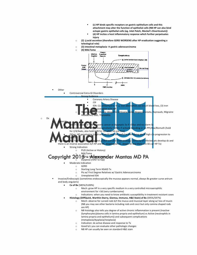
(c) HP binds specific receptors on gastric epithelium cells and this attachment may alter the function of epithelial cells (NB HP can also bind ectopic gastric epithelial cells (eg. Inlet Patch, Meckel’s Diverticulum))
(d) HP incites a host inflammatory response which further perpetuates injury
o (2) ↓acid secre on (therefore GERD WORSENS after HP eradication suggesting a teleological role)
o (3) intestinal metaplasia → gastric adenocarcinoma o (4) MALToma
Other
Controversial Extra‐GI Disorders o Strong Evidence
Coronary Artery Disease ITP IDA: (1) achlorhydria → ↓ iron absorp on, (2) occult blood loss, (3) iron
sequestration w/in HP o Weak Evidence: Chronic Idiopathic Urticaria, Acne, Alopecia Areata, Raynauds, Migraine
Headaches, GBS, Thyroiditis o Dx
General
test if Dz above or FHx of gastric cancer or dyspeptic Sx if <50yo and no alarm Sx
all tests (except serology) lose sensitivity (~25%) if actively bleeding or on PPI/Abx/Bismuth (hold for 2/4/4wks, also hold H2B for 2d while AA are fine)
Other: HP genotyping (cagA and vacA) may be useful in identifying pts at high r/o progression to cancer
Indications for Testing (rationale against global test and treat strategy: minority of infected pts develop dz and there is an inverse association b/t HP and BE, esophageal AC, atopic dz and there are SEs w/ HP Tx)
Strong Indication o PUD (Active or History) o MALToma o Gastric Adenocarcinoma o Dyspesia (refer to top)
Moderate Indication o GERD o Starting Long Term NSAID Tx o Pts w/ First Degree Relatives w/ Gastric Adenocarcinoma o Unexplained IDA
Invasive/Endoscopic (sometimes endoscopically the mucosa appears normal, always Bx greater curve antrum and body angularis)
Cx of Bx (S85%/S100%) o Mech: grow HP in a very specific medium in a very controlled microaerophilic
environment for >3d (very cumbersome) o Indications: when you need to know antibiotic susceptibility in treatment resistant cases
Histology (DiffQuick, Warthin‐Starry, Giemsa, Immuno, H&E Stain) of Bx (S95%/S97%) o Mech: observe for curved rods b/t the mucus and mucosal layer along w/ loss of mucin
(NB you may see other bacteria including rods and cocci but only comma shaped rods are HP)
o NB histology also tells you degree of active chronic inflammation is present (Inactive (lymphocytes/plasma cells in lamina propria and epithelium) vs Active (neutrophils in lamina propria and epithelium)) and subsequent complications (metaplasia/dysplasia/neoplasia)
o Indication: dx active disease and response to Tx o Good b/c you can evaluate other pathologic changes o NB HP can usually be seen on standard H&E stain

Rapid Urease Test (RUT) of Bx (S90%/S95%)
o Trade Names: CLO (Campylobacter Like Organism) Test, HpFast, HUT Test, Pronto Dry, Pyloritek
o Mech: Bx sample is placed on a medium containing urea and a colored pH indicator, if HP is present its urease hydrolyzes urea to bicarb and ammonia increasing the pH over minutes to 24 hours (check at 1hr then send to lab to watch for 24hrs) changing the pH color from yellow to red
o Indications: dx active disease and response to Tx o When would you send a CLO test over histology??? o NB called CLO test b/c HP was originally thought to be “campylobacter‐like”
PCR Non‐Invasive/Non‐Endoscopic
Serum IgG Test (S85%/S80%) o Mech: present after 21d of inoculation, after Tx, titers decrease slowly but never fully go
away remaining positive in about 40% (“serologic scar”) of successfully treated patients after 18 months limiting the usefulness of this test in assessing response to Tx and reinfection (NB poor PPV in low prevalence populations therefore don’t really use in the US)
o Indications: dx active disease during an acute GIB o NB IgA and IgM are not reliable
Fecal Antigen Test (FAT) (S95%/S98%) o Mech: enzyme immunoassay w/ the use of polyclonal anti‐HP Ab against HP antigen in
stool o Indication: diagnosis infection and assess response to Tx
Urea Breath/Blood Test (UBT) (S95%/S95%) o Mech: capsule of urea labeled with 13/14C is ingested and if urease is present the labeled
13/14CO2 that is generated is absorbed into circulation (check bicarb blood levels) and eventually breathed out (check carbon dioxide breath levels) by scintillation counter (NB 13C is preferred in children/pregnant b/c non‐radioactive)
o Indications: dx active disease, assess response to Tx, dx reinfection o Tx
General Principles
If you Tx for a 2nd time check sensitivities w/ Cx of Bx and use different abx
Annual reinfection rate in the US: <1%/yr
SEs can be significant
PPI: Why? increase in gastric pH inhibits HP, BID better than QD, some evidence that H2B are equivalent, all PPIs are the same
Factors decreasing eradication rates: cagA negative HP, certain CYP2C19 polymorphisms increasing clearance of PPIs
Testing for Eradication o Indication: ulcer, persistent dyspeptic Sx, MALToma, gastric AC o How? via functional test (UBT) or structural test (FAT) (unclear if you have to be off of
PPI for FAT test) o NB should not be performed any sooner than 2/4wks after completion of PPI/abx Tx b/c
if sooner then one cannot distinguish b/t HP suppression vs true cure First Line Therapy: PCA/M Triple Therapy (PPI 40mg + Clarithromycin 500mg + Amoxicillin 1g OR
Metronidazole 500mg BID x7d (Prevpac))
75% effective (25% fail b/c of HP resistance, poor compliance, pt demographics (younger age, smoking, prior abx use))
Main Trial: MACH 1 Study Second Line Therapy or if Pen Allergic or Prior Macrolide Exposure: PBMT Quadruple Therapy (PPI BID +
Bismuth Citrate 240mg Subsalicylate 525mg + Metronidazole 250mg + Tetracylcine 500mg QID (except PPI

which is BID) x7d if QID or x14d if BID (Pylera/Helidac, NB does not include the PPI)) A little bit more complex and slightly more SEs, 75% eradication rates after failed first Tx
Third Line Salvage Therapy
Sensitivity testing w/ culture (do after two failed therapies)
If Pen Allergic or Amoxicillin Resistant (1%): Triple Therapy but use Flagyl or Levaquin for Amoxicillin
If Prior Macrolide Exposure (even if distant) or Clarithromycin Resistance (13%): Triple Therapy but use Levaquin 250mg BID (16% resistance) w/ or Rifabutin 150mg BID (not as effective and more SEs) for Clarithromycin
If Metronidazole Resistance (25%) BUT it can be overcome w/ higher doses or using quadruple therapy
“Concomitant Therapy”: PPI + Clarithromycin + Metronidazole + Amoxicillin x5d
“Sequential Therapy”: first 5d course of PPI and Amoxicillin then 5d course of PPI and Clarithromycin and Tinidazole, used in Europe mainly Italy, 95% eradication rates, appears to be effective in clarithromycin resistance strains, reduced cost, less SEs, it appears that flagyl cannot be replaced by tinidazole
Other Infections (other than HP very uncommon) o Bacteria (there are about 128 species of bacteria found in the stomach but the significance of these bacteria is unclear)
Mycobacteria (TB, variable appearance but gastric outlet obstruction is uniquely common) Actinomycosis (ulcerating mass w/ a fistulizing abscess, concurrent TI/Cecal dz is very common) Secondary/Tertiary Syphilis (variable appearance) Polymicrobial Suppurative/Phlegmonous→Necro zing (associated w/ large intake of alcohol, URTI, AIDS,
infected peritoneojugular venous shunts, 70% mortality) o Virus
CMV HSV VZV Measles Enterovirus
o Fungus Candida (often seen w/in ulcers but unclear whether significant or not) Histoplasmosis Zygomycosis Aspergillosis
o Parasite Giardiasis Cryptosporidiosis Strongyloidiasis Anisakiasis Ascariasis Necator (hookworm)
Immune o Autoimmune (refer to HP) o Granulomatous
Infectious (non‐industrialized countries, caseating): TB, Syphilis, Fungal, HP? Non‐Infectious (industrialized countries, non‐caseating): Crohn’s, Sarcoid (stomach is the most common GI
location, must have other organ involvement to confidently make dx), Lymphoma, PUD Complication, Whipple’s, Xanthogranulomatous, Langerhans Cell Histiocytosis, Vasculitis (Churg‐Strauss), foreign body (
o Collagenous (rare w/ only 20 cases reported, adult female, lymphoplasmacytosis in lamina propria and subepithelial collagen w/ some detachment of epithelium, can be associated w/ collagenous microscopic colitis or collagenous sprue)
o Lymphocytic (many times associated w/ Celiac Sprue and HP, appearance can range from normal to nodularity to erosions to volcano like lesions aka varioliform to giant folds, mucosal permeability changes can lead to loss of protein, may be a precursor to lymphoma therefore always exclude)
o Eosinophilic (refer to eosinophilic disorders) o GVHD (usually affects the SI/LI, histology reveals apoptotic bodes in mucosal crypts and intrapetheial vacuoles filled w/
debris) o Allergic (esp cow milk allergy)
Atrophic Gastritis o Multifocal Atrophic Gastritis (MAG) aka Environmental Metaplastic Atrophic Gastritis (EMAG) 2/2 HP o Diffuse Corporal Atrophic Gastritis (DCAG) aka Autoimmune Metaplastic Atrophic Gastritis (AMAG) 2/2 Autoimmune
Destruction or HP (refer to Pernicious Anemia Notes)
Hyperplastic Gastropathies (refer)
Reactive Gastropathies (multifocal damage w/o significant inflammatory infiltrate just some subepithelial hemorrhage and necrosis extending into lamina propria (erosion) or muscularis mucosa (ulcer), b/c there is minimal inflammation they are called reactive gastropathies, intervening mucosa is generally normal unless the insult is severe)

o NSAID and Aspirin (most common, this includes even baby aspirin)
Mechanism
Acute/Minor: local contact effect (hence only PO) occurring shortly after (w/in 15min) ingestion of NSAID, may or may not be the precursor to chronic changes
o NSAIDs uncouple mitochondrial oxidative phosphorylation → decreased ATP → mitochondrial damage w/ increased ROS, disturbed Na/K ratio, leakage of Ca into cytoplasm → loss of control of intracellular jxn w/ increased permeability (loss of blood and protein) → epithelium are exposed to intraluminal contents (bacteria, acids, proteolytic enzymes, etc) → inflamma on
Chronic/Major: systemic effect (hence not just PO but also PR, IV, etc) due to suppression of endogenous mucosal prostaglandin synthesis which are important in maintaining epithelial integrity resulting in erosions to ulcerations (refer to physiology section for more details)
Effects
Analgesics: reduce mild to moderate (not severe) intugemental (not visceral) pain peripherally by blocking synthesis of PGE2 and PGI2 that potentiate pain transduction in nociceptive neuron by activating dormant nociceptors or increasing nocicepter sensitivity to bradykinin, histamine, etc. (released by damaged tissue)
Antipyretics: reduce body temperature by blocking synthesis of PGE2, PGF2_, and PGI2 which act on the hypothalamus to increase body temperature
Anti‐Inflammatory: reduce inflammation by blocking synthesis of all prostaglandins (for some reason a higher doses is needed to achieve this effect vs. the previous two effects)
o Inflammation = (1) Amines (eg. Histamine) (2) Lipids (eg. Eicosinoids) (3) Small Peptides (eg. Bradykinin) (4) Large Peptides (eg. IL‐2) all of these mediators are collectively called autacoids (substances which act on the tissues that formed them)
o Eicosinoid Synthesis: Linoleic Acid (C18:2 essential FA obtained in diet) Membrane
Bound Arachidonic Acid (C20:4) Free Arachidonic Acid via Phospholipase A2 (inhibited steroids)
PGG2 via Cyclooxygenase (COX inhibitors) PGH2 Prostaglandins (PGE2/PGF2/PGI2) & Thromboxanes (TXA2)
Type 1 COX: stimulated by hormones, constitutive expression in stomach/platelets
Type 2 COX: stimulated by cytokines during inflammation, induced expression in many organs including kidney, etc
HPETE via lipoxygenase (?) LTA4 Leukotrienes (LTC4/LTD4/LTB4) & Lipoxins (LXA3/LXB4)
Cancer Prophylaxis: b/c COX‐2 is stimulated by GF and inflammatory cytokines it is believed that it plays a role in the development of cancer and is actually obligatory for colon cancer therefore any COX‐2 inhibitor is a cancer (esp colon) prophylactic
Types
Salicylates o Non‐Prescription: Aspirin aka Acetyl‐Salicylic‐Acid, Alka‐Seltzer, Anacin, Ascriptin,
Aspirin, BC Powder, Goody’s Powder, Bufferin, Excedrin, Pamprin, Pepto‐Bismol, Kaopectate, Vanquish, Sine‐Off, Ecotrin
o Prescription: Aggrenox, Trilisate, Disalcid, Easprin, Endolan, Asacol, Salsate, Salflex, Fiorinal
o Topical: Acne Cleansers, Arthritis Pain Rubs, Dandruff Shampoo, Sun Blocks, Wart Removers, Oil of Wintergreen
o Herbals: Red Oil Chinese, White Flower Oi o IRreversible inhibitors of both cyclooxygenases via acetylation forming salicylate o Non‐acetylated salicylates (mainly salsalate) are less effective and have less effect on
plts and less GI toxicity
Indoleacetic Acid: Indomethacin (Indocin), Sulindac (Clinoril), Tolmetic (Tolmetin), Diclofenac (Voltaren), Etodolac (Lodine), Ketorolac (Toradol), Nabumetone (Relafen)
Gastropathy

o More Specific Dz Indications: Indocin (gout), Clinoril/Tolmetin/Voltaren/Lodine/Relafen (OA), Ketorolac (post‐op pain and pain when pts come into ED as very acute acting)
o More Unique SEs Indomethacin (frontal HA, acute pancreatitis, blood dyscriasis, Etodolac (fluid retention, abnl renal/liver fxn, drug interactions), Ketorolac (increased bleeding, AKI)
Proprionic Acid: Ibuprofen (Motrin, Advil, Nuprin), Naproxen (Naprosyn, Alleve) o generic OTC NSAIDs o Old: Flurbiprofen (Ansaid), Ketoprofen (Orudis), Oxaprozin (Daypro)
Oxicams: Meloxicam (Mobic), Piroxicam (Feldene) o has less GI/Renal SEs and thus is used for more chronic issues
COX‐2 Inhibitors (refer below) S/S & Complications
Asymptomatic until complications
Dyspepsia (50%)
PUD (25%) o Serious Complications (5%)
Risk
Low: no RFs (older age, ulcerogenic NSAIDs, HP infection, etc) = nothing
Mod: 1‐2 RFs = anti‐ulcer agent
High: >3 RFs, h/o complications, concomitant aspirin/steroids/AC = avoid NSAIDs Px
misoprostol (Cytotec) 100,200mcg tab, 100‐200mcg PO QID
o PG‐E agonist o at low doses (100mcg QID) cytoprotective by increasing blood flow and mucus secretion
vs at high doses (200mcg QID) inhibit gastric acid but also causes cramps and diarrhea therefore consider just low dose!!!
o NB causes uterine contraction and thus abortion
AA (not effective)
H2B (only good for DU)
PPIs
Stop NSAIDs or take after meals and use the lowest dose
Avoid steroids, anticoagulants, SSRIs b/c these three specific drugs exacerbate the effects of NSAIDs
Change NSAIDs (the really bad NSAIDs: sulindac, piroxicam, ketorolac, naproxen) o New NSAID (aspirin‐phosphatidylcholine) has less r/o PUD o Enteric Coated NSAIDs eg. ? o Non‐Acetylated NSAIDS eg. salsalate (Salflex, Disalcid, Amigesic) o Non‐Acidic Pro‐Drug NSAIDs eg. nabumetone (Relafen) o COX‐2 Inhibitors eg. celecoxib (Celebrex)
Mech: competitive reversible inhibitor of 2>1 cyclooxygenases therefore they minimally inhibit PG formation in stomach
SEs: sulfa‐like allergic rxn, increased CV dz (Rofecoxib (Vioxx) and Valdecoxib (Bextra) were removed from the market)
NB COX‐2 is up‐regulated when an ulcer is healing therefore avoid these when an ulcer is already present
o Meds (multivitamins?, iron, potassium, fluoride, bisposphonates, macrolides, chemotherapy) o Illicits (ethanol, cocaine) o Portal HTN Gastropathy (refer) o Radiation (refer) o Ischemia (refer) o Gastritis Cystica Profunda (refer to intestine notes) o Cameron’s Erosions (refer) o Aging o Duodenogastric Reflux (DGR)

Etiology
Secondary: s/p partial gastrectomy w/ Billroth reconstruction, truncal vagotomy, pyloroplasty, cholecystectomy, sphyncteroplasty
Primary: no prior surgery, pts likely have some degree of pyloric failure, duodenal dysmotolity and/or delayed gastric emptying
NB physiologic reflux occurs nocturnal and post‐prandial therefore pathogenic reflux must have an increase in rate, volume and/or duration
Mechanism
Reflux of biliary, pancreatic, duodenal secretions into the stomach → disruption of gastric mucuous layer via detergent effects from bile acids → allows for reverse diffusion H+ and pancreatic enzymes (specifically lysolethicin and trypsin) → mucosal damage
Dx
First o Clinical
dyspeptic Sx bilious vomiting post‐cholecystectomy syndrome absence of HP/NSAID/EtOH
o Antral Endoscopy pool of bile w/ underlying bile staining granular, friable, swollen, red mucosa esp around pylorus w/ erosions NB the passage of an endoscope induces a gagging reflex promoting DG
reflux o Antral Histology
non‐specific reactive gastropathy of the antrum (foveolar hyperplasia w/ dilated elongated atypical cystic glands, vascular congestion and dilation, mucosal edema) w/ occasional mild chronic inflammation
Therefore endoscopic and histologic findings are not diagnostic only suggestive
Second o Gastric Aspirate Analysis: directly measure bile acid concentration via an enzymatic
assay (Gold Standard) Problem: cumbersome
o Gastric Scrintigraphy: inject radiolabeled tracer that is secreted into the biliary tract (eg. 99m‐Tc hydroxyiminodiacetic acid ‐ HIDA) and then center gamma camera over stomach
Problem: at best semi‐quantitative, radiation exposure, problems of overlap w/ liver and small bowel
o Gastric pH Monitoring: b/c duodenal contents are alkaline it was theorized that alkaline peaks (pH>7) would indicated DGR
Problem: electrodes commonly used are not accurate above a pH 6, false + w/ alkaline saliva and esophageal bicarb
o Bilitec 2000: endoluminal fiberoptic probe positioned in the gastric body that measures the degree of absorption of 470nm wavelength light (the absorptive peak for bilirubin)
Problem: What is considered the ULN for degree of absorption (Range: 0‐1 w/ >0.14 considered abnormal in most studies)? Does bilirubin always correlate w/ bile acid concentration? Should measurements not be taken post‐prandially and at night? How does diet interfere with results?
Presently it is the first‐line test for confirming a diagnosis of DGR Gastropathy Must be combined w/ pH monitoring as acid causes bilirubin to dimerize
which shifts its wavelength absorption peak to 400nm → in the stomach Bilitec likely underestimates bile reflux (~30% when pH<3.5)
Complication
Gastric Intestinal Metaplasia → Gastric AC o bile acids are not known to be mutagenic but may promote mutagenicity of aromatic
amines or may expose the luminal surface to endoluminal carcinogens o DGR may be the cause for increased r/o AC seen in pts after partial gastrectomy
GERD → Erosive Esophagitis, Stricture/Rings, BE w/ AC o Synergism: reflux of gastric juice combined w/ bile is MORE injurious than gastric juice
alone increasing the r/o complicated GERD (Marshall, et al. Gut. 1997) (Esophageal Injury: acid pH<1.3, acid + pepsin pH <2, acid + pepsin + bile acids <7)
o Suspect in GERD refractory to PPI therapy (~1/4 of pts) or complicated GERD (~1/3 of pts, Atwood, et al. Gut. 1993)
o NB simultaneous esophageal pH/bilirubin monitoring indicates that duodenal juice exposure occurs at ALL pH values such that presence of bile cannot be confidently detected thru pH monitoring alone
Tx (the problem in studying Tx is the definition of pathologic bile acid gastropathy)

1st Medical Tx (anecdotally sucrulfate appears to be the most common Tx used) o Mucosal Protection: sucralfate (aluminum sulfate disaccharide that likely forms a
complex w/ positively charged proteins exposed in damaged mucosa forming a viscous adhesive paste)
o Antisecretory Agents: PPI (as secondary damage is due to acid effects on mucosa, in addition decreasing overall gastric volume may reduce GERD and its complications)
o Prokinetic Agents: metoclopramide o Bile Acid Resins: cholestyramine
2nd Surgical Tx (if medical failure) o Bile Flow Diverting Procedures: Modified Roux‐en‐Y, Duodenal Switch, Henley
Procedure: interpose jejunum b/t stomach and duodenum, Braun Enteroenterostomy: staple the afferent limb followed by side‐to‐side anastomosis of the jejunum
Megoblastic Anemias (Folate and VitB12 Deficiencies)
Mechanism Mech: w/in cells VitB12 is converted to methyl‐cobalamin and adenosyl‐cobalamin which act as coenzymes
o (1) decreased demthylation of methyl‐tetrahydrofolate (THF) impaired pyrimidine synthesis normal RNA but decreased DNA synthesis → ineffec ve erythropoiesis w/ intramedullary hemolysis as cells die in BM (anemia) and potentially other lineages if severe (pancytopenia)
o (2) Decreased Methionine/Succinyl‐CoA and Increased Homocysteine/Methylmalonate → ? (likely responsible for extra‐hematologic effects of vitb12 deficiency)
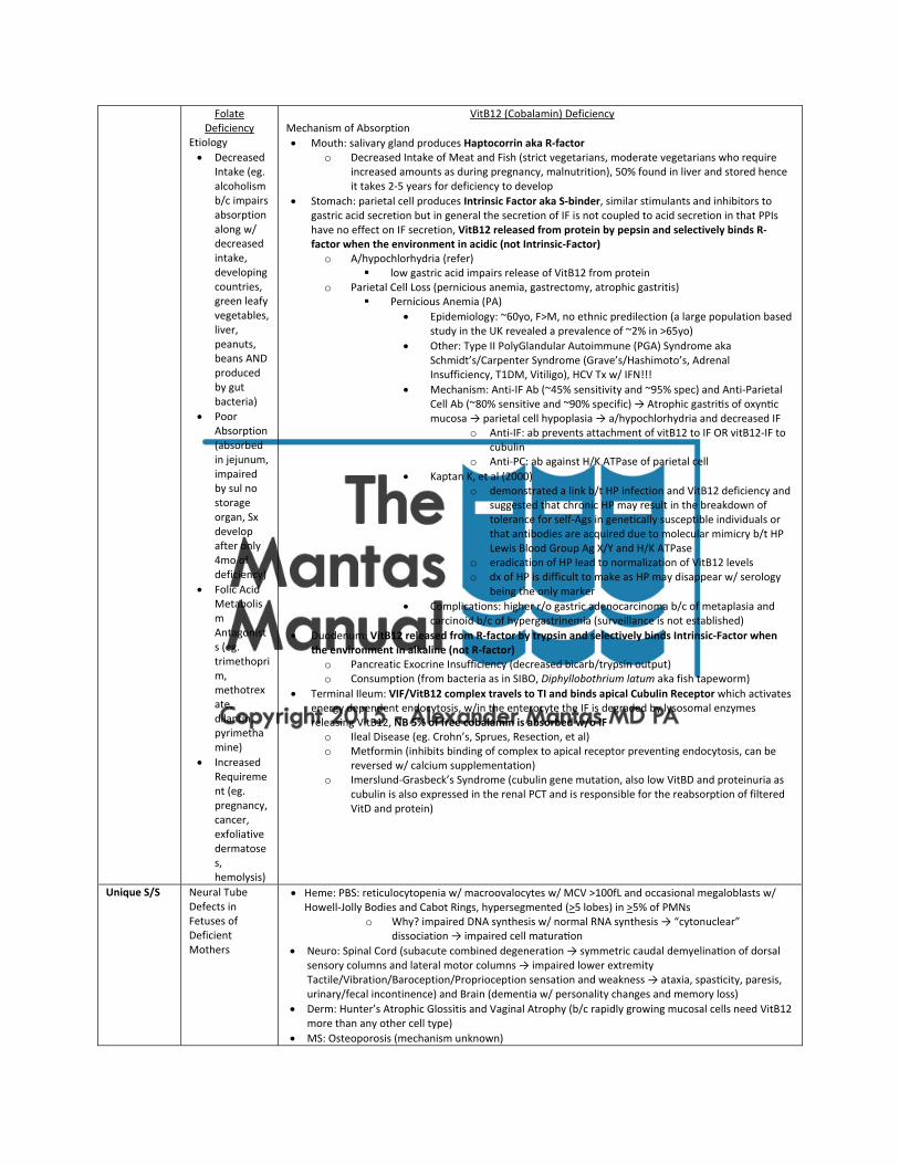
Folate Deficiency
Etiology
Decreased Intake (eg. alcoholism b/c impairs absorption along w/ decreased intake, developing countries, green leafy vegetables, liver, peanuts, beans AND produced by gut bacteria)
Poor Absorption (absorbed in jejunum, impaired by sul no storage organ, Sx develop after only 4mo of deficiency)
Folic Acid Metabolism Antagonists (eg. trimethoprim, methotrexate, dilantin, pyrimethamine)
Increased Requirement (eg. pregnancy, cancer, exfoliative dermatoses, hemolysis)
VitB12 (Cobalamin) DeficiencyMechanism of Absorption
Mouth: salivary gland produces Haptocorrin aka R‐factor o Decreased Intake of Meat and Fish (strict vegetarians, moderate vegetarians who require
increased amounts as during pregnancy, malnutrition), 50% found in liver and stored hence it takes 2‐5 years for deficiency to develop
Stomach: parietal cell produces Intrinsic Factor aka S‐binder, similar stimulants and inhibitors to gastric acid secretion but in general the secretion of IF is not coupled to acid secretion in that PPIs have no effect on IF secretion, VitB12 released from protein by pepsin and selectively binds R‐factor when the environment in acidic (not Intrinsic‐Factor)
o A/hypochlorhydria (refer) low gastric acid impairs release of VitB12 from protein
o Parietal Cell Loss (pernicious anemia, gastrectomy, atrophic gastritis) Pernicious Anemia (PA)
Epidemiology: ~60yo, F>M, no ethnic predilection (a large population based study in the UK revealed a prevalence of ~2% in >65yo)
Other: Type II PolyGlandular Autoimmune (PGA) Syndrome aka Schmidt’s/Carpenter Syndrome (Grave’s/Hashimoto’s, Adrenal Insufficiency, T1DM, Vitiligo), HCV Tx w/ IFN!!!
Mechanism: Anti‐IF Ab (~45% sensitivity and ~95% spec) and Anti‐Parietal Cell Ab (~80% sensitive and ~90% specific) → Atrophic gastri s of oxyn c mucosa → parietal cell hypoplasia → a/hypochlorhydria and decreased IF
o Anti‐IF: ab prevents attachment of vitB12 to IF OR vitB12‐IF to cubulin
o Anti‐PC: ab against H/K ATPase of parietal cell
Kaptan K, et al (2000) o demonstrated a link b/t HP infection and VitB12 deficiency and
suggested that chronic HP may result in the breakdown of tolerance for self‐Ags in genetically susceptible individuals or that antibodies are acquired due to molecular mimicry b/t HP Lewis Blood Group Ag X/Y and H/K ATPase
o eradication of HP lead to normalization of VitB12 levels o dx of HP is difficult to make as HP may disappear w/ serology
being the only marker
Complications: higher r/o gastric adenocarcinoma b/c of metaplasia and carcinoid b/c of hypergastrinemia (surveillance is not established)
Duodenum: VitB12 released from R‐factor by trypsin and selectively binds Intrinsic‐Factor when the environment in alkaline (not R‐factor)
o Pancreatic Exocrine Insufficiency (decreased bicarb/trypsin output) o Consumption (from bacteria as in SIBO, Diphyllobothrium latum aka fish tapeworm)
Terminal Ileum: VIF/VitB12 complex travels to TI and binds apical Cubulin Receptor which activates energy dependent endocytosis, w/in the enterocyte the IF is degraded by lysosomal enzymes releasing VitB12, NB 5% of free cobalamin is absorbed w/o IF
o Ileal Disease (eg. Crohn’s, Sprues, Resection, et al) o Metformin (inhibits binding of complex to apical receptor preventing endocytosis, can be
reversed w/ calcium supplementation) o Imerslund‐Grasbeck’s Syndrome (cubulin gene mutation, also low VitBD and proteinuria as
cubulin is also expressed in the renal PCT and is responsible for the reabsorption of filtered VitD and protein)
Unique S/S Neural Tube Defects in Fetuses of Deficient Mothers
Heme: PBS: reticulocytopenia w/ macroovalocytes w/ MCV >100fL and occasional megaloblasts w/ Howell‐Jolly Bodies and Cabot Rings, hypersegmented (>5 lobes) in >5% of PMNs
o Why? impaired DNA synthesis w/ normal RNA synthesis → “cytonuclear” dissociation → impaired cell matura on
Neuro: Spinal Cord (subacute combined degeneration → symmetric caudal demyelina on of dorsal sensory columns and lateral motor columns → impaired lower extremity Tactile/Vibration/Baroception/Proprioception sensation and weakness → ataxia, spas city, paresis, urinary/fecal incontinence) and Brain (dementia w/ personality changes and memory loss)
Derm: Hunter’s Atrophic Glossitis and Vaginal Atrophy (b/c rapidly growing mucosal cells need VitB12 more than any other cell type)
MS: Osteoporosis (mechanism unknown)

NB nitrous oxide (NO2) (used in anesthesia or abused as a drug) inactivates VitB12 and may precipitate neuropsychiatric deterioration in deficient pts
Schilling Test (obsolete, rarely done anymore as HC/MMA is more sensitive and it is more cost effective to give oral or parental B12 therapy and forego a work‐up, has more historical importance, onerous test requiring creation and storage of radiolabeled reagents)
Phase I: Load pt w/ IM non‐radiolabeled VitB12 so as to saturate tissue stores then administer Oral Radiolabeled VitB12
o Nl 24‐Hour Urine: dietary deficiency o Abln: move to phase II
Phase II: Administer Oral Radiolabeled VitB12 + IF o Nl 24‐Hour Urine: IF deficiency o Abnl: consider other causes and Tx accordingly w/ repeat testing
Labs: Folate (nl 2.7‐17ng/mL) / VitB12 (nl 300‐1200pg/mL, serum levels represent 0.1% of total body content) but if nl but suspicion is high then check below b/c more sensitive
o Homocysteine (nl 5‐14μmol/L) (both)
both B12 and folate are cofactors in the conversion of homocysteine to methionine thus if either is low then homocysteine is high
not specific b/c many disease can cause high homocysteine
more sensitive than vitB12/folate
NB hyperhomocysteinemia has been implicated as a RF for development of atherosclerosis and DVT (controversial)
o Methylmalonic Acid (nl 0.4μmol/L) (VitB12 only)
important to measure to differentiate B12 from folate def o If VitB12 deficiency then check IF Antibody & Parietal Cell Antibody if antibodies are negative then pursue
other causes o Typical order of VitB12 deficiency events: elevated HC/MMA → low VitB12 → hypersegmenta on →
macrocytosis → anemia → neuro Sx
~25% of do not follow this order of events and develops Sx before abnormal labs or have normal hematologic indices w/ neurologic Sx
Treatment Folic Acid 5mg PO Qd x1wk then 1mg PO QD until normal Hct and Folate then 0.4mg PO Qd (which is RDA, 0.6mg for pregnant women)
Normal RDA: 2.6mcg/d
Deficiency: 1000mcg Qd IM x1wk → 1000mcg Qwk IM x1mo → 1000mcg Qmo IM forever as long as the underlying disorder exists (eg. PA)
o PO ≈ IM
a second lower efficiency transport system that does not require IF or a functioning TI exists
very large doses (2000mcg at the same frequency) are needed, nevertheless most give parenteral route until at least neuro Sx resolve
o Route
No IV b/c rapid renal elimination and reported anaphylaxis
SL/IN routes exist but poorly studied and expensive
If PO then separate from meals o NB
Neuro sx begin to resolve at 1wk w/ complete resolution at 3mo (start Tx early as permanent neurologic Sx can occur)
Always follow pts clinically and with labs
hematologic abnormalities can be reversed in VitB12 deficiency when inappropriately Tx w/ folic acid (neuro Sx will not improve)