o nce o ortened o · “terminal ileitis” but because the word “terminal” sounded bad and...
17
IBD (CD/UC/IC) Indeterminate Colitis o colitis that is a combination of both CD and UC, 10% of IBD undergoes a reassignment during the first 2yrs of dx, important to confirm esp if considering surgery History o Morgagni (1761) was first to describe changes to the small intestine consistent with CD o Crohn, Ginzburg, Oppenheimer (1932) was first to recognize Crohn’s as a true pathologic entity, it was first called “terminal ileitis” but because the word “terminal” sounded bad and because there could be colonic involvement “regional enteritis” was used but because Crohn’s often skipped then “granulomatous enterocolitis” was used but because granulomas were not a sina qua non for diagnosis the current “Crohn’s Disease” is used o Current: William Sandborn (Mayo), Gary Lichtenstein (Penn), Stephen Hanauer (Chicago), Sunanda Kane (Mayo), Marla Dubinsky (Cedars) Epidemiology o Incidence 1:1000 (UC) > 1:3000 (CD) NB exact incidence is difficult to estimate (too low b/c diagnosis requires invasive techniques or too high b/c many times infections were incorrectly diagnosed) o Age: bimodal age distribution for diagnosis: 10‐20s and 60‐70s o Gender: F>M but only slight o Course if a pt is in remission the past year there is an 75% chance that the pt will be in remission the next yr VS if a pt has active dz the past year there is a 75% chance that the pt will have active dz the next year overall 15% will have a relapse free course VS 20% will have relapses every year VS 65% will have some years relapse free and some years with relapses VS 5% will have continuous active dz pts are true to their history therefore time will tell (whatever happens during the first few years will likely happen in the future) o Prognosis: lifespan is generally NOT shortened by IBD, if pts die it usually occurs during the first 5yrs after dx RFs o Social: industrialized/urban, higher socioeconomic, higher latitude o Ethnicity: White (North America, Europe) > Non‐White (Hispanic, African, Asian), Eastern European (Ashkenazi) Jews > Eastern European Non‐Jews, Industrialized > Developing Countries o Exposure: smoking (risk for CD vs protective for UC, mechanism is unclear, it could be the nicotine), meds (NSAID, OCP, Isoretinoin, Abx when child, Measles vaccination), diet (Western Diet), other (not breastfed), surgery (appendectomy for appenditicis (not a normal appendix) <20yo is a risk for CD vs protective for UC) o FHx: risk is 15x higher if person has an affected first degree relative, 1/4 pts with IBD have a family member with IBD and usually have similar dz in terms of extent and extra‐GI manifestations, greater genetic component in Crohn’s than UC, Crohn’s (HLA‐DR1‐DQ5) vs UC (HLA‐DR2), greatest RF is a sibling w/ IBD (CD/UC 17‐35x/7‐17x the general population), monozygotic twins have a 58/6% concordance rate for CD/UC, pure Mendelian inheritance is not present Pathogenesis o The gut constantly strives a balance between allowing for commensal enteric flora growth while attacking pathogenic bacteria and this immune tolerance/intolerance occurs via a unique subset of helper T‐cells (Th1 = CD vs Th2 = UC) → in IBD this balance is off (2/2 different genetic loci termed IBD‐#) resulting in a dysregulated immune response to commensal enteric flora or possible an appropriate response to an unrecognized pathogen (1° Mycobacterium paratuberculosis, 2° Measles, Listeria, Chlamydia, Psuedomonas, Reovirus) hence IBD is NOT an autoimmune process IBD‐1 (CD): NOD2/CARD15 gene (Nucleotide binding Oligomerization Domain 2 / CAspase Recruitment Domain 15) on chromosome 16, codes for a protein that senses bacterial cell walls and initiates an inflammatory response, mutations result in an uninitiated or exaggerated inflammatory response, 20% of the normal population have one of these alleles vs 30% of CD pts have one of these alleles therefore low specificity and low sensitivity but clearly this gene is important, 30x/3x increased risk (homo/hetero), associated w/ ileal dz, fibrostenotic course, earlier onset, +FHx IBD‐2 (UC) IBD‐3,4,5 NB the gene that codes for IL‐23 receptor seems the be the next big gene NB germ free rats have decreased incidence of IBD but when introduced to flora IBD manifests NB when you create a diverting ileostomy the area of bowel that does not see stool anymore actually has less disease GI‐Dz o CD Depth: any depth down to transmural Location: focal/asymmetric/transmural/granulomatous tissue, any portion of the GI tract from mouth to anus but usually distal SI to proximal colon (30% SI, 50% SI+C, 20% C) overall terminal ileum is the hallmark location, the rarest location is rectum aka rectal sparing (unlike in UC where rectum is universally affected), interestingly 1/3 of pts have perianal involvement, involvement of esophagus/stomach/duodenum/jejunum is almost always seen only when there is dz of distal SI and colon however inflammation on biopsy may be seen in the absence of gross lesions, upper GI dz is seen in 13% of pts, duodenal Bx is actually the most sensitive tissue to find granulomas, skips/focal both microscopically (eg. within on biopsy one may see a pronounced variable degree of inflammation) and macroscopically (eg. not the entire ileum but different segments, you can have disease with surrounding areas of normal mucosa)
Transcript of o nce o ortened o · “terminal ileitis” but because the word “terminal” sounded bad and...

IBD (CD/UC/IC)
Indeterminate Colitis o colitis that is a combination of both CD and UC, 10% of IBD undergoes a reassignment during the first 2yrs of dx,
important to confirm esp if considering surgery
History o Morgagni (1761) was first to describe changes to the small intestine consistent with CD o Crohn, Ginzburg, Oppenheimer (1932) was first to recognize Crohn’s as a true pathologic entity, it was first called
“terminal ileitis” but because the word “terminal” sounded bad and because there could be colonic involvement “regional enteritis” was used but because Crohn’s often skipped then “granulomatous enterocolitis” was used but because granulomas were not a sina qua non for diagnosis the current “Crohn’s Disease” is used
o Current: William Sandborn (Mayo), Gary Lichtenstein (Penn), Stephen Hanauer (Chicago), Sunanda Kane (Mayo), Marla Dubinsky (Cedars)
Epidemiology o Incidence 1:1000 (UC) > 1:3000 (CD) NB exact incidence is difficult to estimate (too low b/c diagnosis requires invasive
techniques or too high b/c many times infections were incorrectly diagnosed) o Age: bimodal age distribution for diagnosis: 10‐20s and 60‐70s o Gender: F>M but only slight o Course
if a pt is in remission the past year there is an 75% chance that the pt will be in remission the next yr VS if a pt has active dz the past year there is a 75% chance that the pt will have active dz the next year
overall 15% will have a relapse free course VS 20% will have relapses every year VS 65% will have some years relapse free and some years with relapses VS 5% will have continuous active dz
pts are true to their history therefore time will tell (whatever happens during the first few years will likely happen in the future)
o Prognosis: lifespan is generally NOT shortened by IBD, if pts die it usually occurs during the first 5yrs after dx
RFs o Social: industrialized/urban, higher socioeconomic, higher latitude o Ethnicity: White (North America, Europe) > Non‐White (Hispanic, African, Asian), Eastern European (Ashkenazi) Jews >
Eastern European Non‐Jews, Industrialized > Developing Countries o Exposure: smoking (risk for CD vs protective for UC, mechanism is unclear, it could be the nicotine), meds (NSAID, OCP,
Isoretinoin, Abx when child, Measles vaccination), diet (Western Diet), other (not breastfed), surgery (appendectomy for appenditicis (not a normal appendix) <20yo is a risk for CD vs protective for UC)
o FHx: risk is 15x higher if person has an affected first degree relative, 1/4 pts with IBD have a family member with IBD and usually have similar dz in terms of extent and extra‐GI manifestations, greater genetic component in Crohn’s than UC, Crohn’s (HLA‐DR1‐DQ5) vs UC (HLA‐DR2), greatest RF is a sibling w/ IBD (CD/UC 17‐35x/7‐17x the general population), monozygotic twins have a 58/6% concordance rate for CD/UC, pure Mendelian inheritance is not present
Pathogenesis o The gut constantly strives a balance between allowing for commensal enteric flora growth while attacking pathogenic
bacteria and this immune tolerance/intolerance occurs via a unique subset of helper T‐cells (Th1 = CD vs Th2 = UC) → in IBD this balance is off (2/2 different genetic loci termed IBD‐#) resulting in a dysregulated immune response to commensal enteric flora or possible an appropriate response to an unrecognized pathogen (1° Mycobacterium paratuberculosis, 2° Measles, Listeria, Chlamydia, Psuedomonas, Reovirus) hence IBD is NOT an autoimmune process
IBD‐1 (CD): NOD2/CARD15 gene (Nucleotide binding Oligomerization Domain 2 / CAspase Recruitment Domain 15) on chromosome 16, codes for a protein that senses bacterial cell walls and initiates an inflammatory response, mutations result in an uninitiated or exaggerated inflammatory response, 20% of the normal population have one of these alleles vs 30% of CD pts have one of these alleles therefore low specificity and low sensitivity but clearly this gene is important, 30x/3x increased risk (homo/hetero), associated w/ ileal dz, fibrostenotic course, earlier onset, +FHx
IBD‐2 (UC) IBD‐3,4,5 NB the gene that codes for IL‐23 receptor seems the be the next big gene NB germ free rats have decreased incidence of IBD but when introduced to flora IBD manifests NB when you create a diverting ileostomy the area of bowel that does not see stool anymore actually has less
disease
GI‐Dz o CD
Depth: any depth down to transmural Location: focal/asymmetric/transmural/granulomatous tissue, any portion of the GI tract from mouth to anus
but usually distal SI to proximal colon (30% SI, 50% SI+C, 20% C) overall terminal ileum is the hallmark location, the rarest location is rectum aka rectal sparing (unlike in UC where rectum is universally affected), interestingly 1/3 of pts have perianal involvement, involvement of esophagus/stomach/duodenum/jejunum is almost always seen only when there is dz of distal SI and colon however inflammation on biopsy may be seen in the absence of gross lesions, upper GI dz is seen in 13% of pts, duodenal Bx is actually the most sensitive tissue to find granulomas, skips/focal both microscopically (eg. within on biopsy one may see a pronounced variable degree of inflammation) and macroscopically (eg. not the entire ileum but different segments, you can have disease with surrounding areas of normal mucosa)
Gastroduodenal (rare):
Jejunoileitis (rare): multiple stenosis, bacterial overgrowth, protein‐losing enteropathy (usually younger pt)
Ileitis (most common): active inflammation leads to anorexia, diarrhea, weight loss, fever, RLQ pain (often mimics appendicitis)
LI: diarrhea, blood, pain, perianal dz, extra‐GI dz
There is question as to whether isolated granulomatous inflammation of the appendix is Crohn’s or something else
There is evidence that HP‐focal gastritis could suggest CD Early (superficial): neutrophils w/ cryptitis, occurs on mesenteric side, Aphthous Ulcers (tiny ulcers, barely
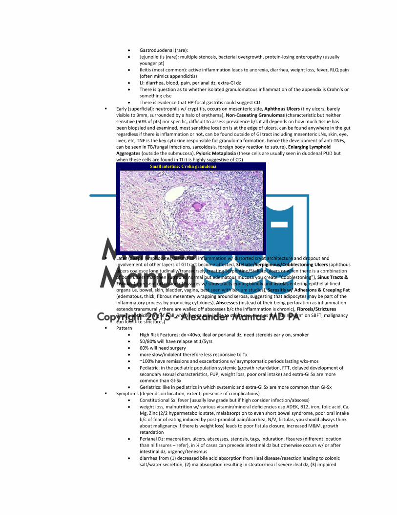
visible to 3mm, surrounded by a halo of erythema), Non‐Caseating Granulomas (characteristic but neither sensitive (50% of pts) nor specific, difficult to assess prevalence b/c it all depends on how much tissue has been biopsied and examined, most sensitive location is at the edge of ulcers, can be found anywhere in the gut regardless if there is inflammation or not, can be found outside of GI tract including mesenteric LNs, skin, eye, liver, etc, TNF is the key cytokine responsible for granuloma formation, hence the development of anti‐TNFs, can be seen in TB/fungal infections, sarcoidosis, foreign body reaction to suture), Enlarging Lymphoid Aggregates (outside the submucosa), Pyloric Metaplasia (these cells are usually seen in duodenal PUD but when these cells are found in TI it is highly suggestive of CD)
Later (deep): lymphocytes/plasma cell inflammation w/ distorted crypt architecture and dropout and
involvement of other layers of GI tract become affected, Stellate/Serpiginous/Cobblestoning Ulcers (aphthous ulcers coalesce longitudinally/transversely creating Serpentine/Stellate ulcers or when there is a combination of both ulcers that then surround normal but edematous mucosa you create “Cobblestoning”), Sinus Tracts & Fistulas (represent extension of fissures w/ sinus tracts ending blindly and fistulas entering epithelial‐lined organs i.e. bowel, skin, bladder, vagina, best seen with barium studies), Serositis w/ Adhesions & Creeping Fat (edematous, thick, fibrous mesentery wrapping around serosa, suggesting that adipocytes may be part of the inflammatory process by producing cytokines), Abscesses (instead of their being perforation as inflammation extends transmurally there are walled off abscesses b/c the inflammation is chronic), Fibrosis/Strictures (irregular thickening of wall which eventually leads to strictures, seen as the “string sign” on SBFT, malignancy can look like strictures)
Pattern
High Risk Features: dx <40yo, ileal or perianal dz, need steroids early on, smoker
50/80% will have relapse at 1/5yrs
60% will need surgery
more slow/indolent therefore less responsive to Tx
~100% have remissions and exacerbations w/ asymptomatic periods lasting wks‐mos
Pediatric: in the pediatric population systemic (growth retardation, FTT, delayed development of secondary sexual characteristics, FUP, weight loss, poor oral intake) and extra‐GI Sx are more common than GI‐Sx
Geriatrics: like in pediatrics in which systemic and extra‐GI Sx are more common than GI‐Sx Symptoms (depends on location, extent, presence of complications)
Constitutional Sx: fever (usually low grade but if high consider infection/abscess)
weight loss, malnutrition w/ various vitamin/mineral deficiencies esp ADEK, B12, iron, folic acid, Ca, Mg, Zinc (2/2 hypermetabolic state, malabsorption to even short bowel syndrome, poor oral intake b/c of fear of eating induced by post‐prandial pain/diarrhea, N/V, fistulas, you should always think about malignancy if there is weight loss) leads to poor fistula closure, increased M&M, growth retardation
Perianal Dz: maceration, ulcers, abscesses, stenosis, tags, induration, fissures (different location than nl fissures – refer), in ¼ of cases can precede intestinal dz but otherwise occurs w/ or after intestinal dz, urgency/tenesmus
diarrhea from (1) decreased bile acid absorption from ileal disease/resection leading to colonic salt/water secretion, (2) malabsorption resulting in steatorrhea if severe ileal dz, (3) impaired
Small intestine: Crohn granuloma

water/electrolyte absorption/secretion from damaged SI/LI, (4) bacterial overgrowth behind strictures, (5) chronic inflammation leading to disordered motility, (6) meds used to Tx CD esp 5‐ASAs, (7) enteroenteric fistulas may allow contents to bypass absorptive surfaces, (8) cancer development
ab pain (much more frequent/severe than in UC, variable in quality depending on cause: (1) inflammation/abscess (2) strictures w/ SBO, (3) serositis) since the TI is the most commonly involved region pts often have RLQ pain mimicking appendicitis
bleeding (mild usually +FOBT if colon is involved, rarely gross as in UC) CD generally behaves in one of three ways but some cases have neither and some cases have all concurrently
or subsequently nevertheless each of these types have characteristic cytokine patterns suggesting different mechanisms (NB better ways at sub grouping CD need to be developed but these groupings are helpful when it comes to talking about outcomes)
(1) Aggressive Inflammatory Fistulizing Penetrating Abscess Disease: inflammation leads to more tissue destruction than fibrosis
(2) Indolent Cicatrizing Fibrostenotic Disease: inflammation leads to more tissue fibrosis than destruction
Surgery
Indications: failure of medical therapy (once a pt is on steroids there is a 40% chance of needing surgery w/in 1yr), intolerable medical SEs, complications (fistulas, strictures, abscess, hemorrhage, cancer)
Pre o Educate: surgery is never curative b/c dz is not isolated but rather skips, surgery will only
be done if it absolutely has to and when done the most conservative approach in terms of length of bowel removed will be done
o in pts w/ “indeterminate colitis” in whom colectomy and IPAA is being considered there is high rate of pouch failure therefore pre‐op work‐up should be done to r/o CD
o Optimize Nutrition o Quit Smoking o Educate pt that surgery is not curative, post‐op meds may be needed, post‐op
surveillance is necessary o Prognosis (“the 3s”): 30% will need surgery w/in 3yrs of dx w/ 65% at some point in their
lifetime, 30% of these will need a second surgery and 30% of these will need a third surgery
Types: ‐ectomy w/ anastomosis or –ostomy to stricturoplasty depending on #, length and severity
Recurrence o Incidence: 10% clinical recurrence and 80% endoscopic recurrence after 1yr at proximal
anastomosis site and neo terminal ileum (mainly seen w/ ileal surgery rarely seen after colectomy)
o RFs: smoking, penetrating dz, h/o prior surgery, refractory to medical therapy prior to surgery, short duration of dz, SI/LI involvement, young age, length of bowel removed
o DDx: true CD recurrence, CMV, C.diff, SIBO, etc o Px
Low Risk: watch Mod Risk: AZA/6‐MP x6mo and Flagyl x3mo (start w/in 2wks of surgery) High Risk: Anti‐TNF (start w/in 4wks of surgery) NB 5‐ASAs are NOT helpful
o Surveillance: ileocolonoscopy at 6mo & 1yr post‐op w/ Bx of anastomosis to detect recurrence and if it occurs then escalate therapy and if not then repeat colonoscopy at Q1yr
Complications
Stenosis o Types: fibrotic strictures, inflammatory stenosis, anastomotic stricture (NB multiple
types can coexist w/in one stenosis) o DDx: adhesions, cancer, hernias, intussusception, impaction, extrinsic compression by
abscess, etc o Location: ileum esp ICV/TI & anus o S/S: obstructive Sx, main cause of hospitalization and surgery, 50% recurrence rate esp
at anastomosis o Dx: imaging, endoscopy, NB all strictures should be Bx b/c some will harbor cancer o Tx: first determine type
Inflammatory stenosis is suggested by elevated inflammatory markers, positive response to the “steroid test”, little pre‐bowel dilation, etc = medical therapy (normal CD meds)
Fibrotic strictures do not have these findings = medical (unclear if steroids/anti‐TNFs have any antifibrogenic effect) therefore endoscopic
(dilation, stricture incision, stents, intralesional steroid injection) → if long stricture, accompanying fistula or failed endoscopic Tx then surgery (resection, stricturoplasty, intestinal bypass, ileostomy)
Cancer o Lymphoma (unclear) o SI (12x increased risk but nonetheless still rare) o Colon (same as UC if colon is involved) o SCC (if chronic cutaneous fistulas)
Serositis → sinus tracts → abscess (if end blindly) vs fistulas (if there are adhesions to other squamous tissue organs) vs perforation
o Toxic Megacolon (same as UC if colon is involved) o Abscess: ¼ of CD pts will get an abscess at some point in their life, Tx: drainage, IV abx o Fistulas: 35%, 2/2 transmural inflammation, in general internal fistulas (don’t forget
about these!!!) are harder to medically close than external fistulas, entero/colo‐enteric/colonic (often asymptomatic except for coloenteric fistulas in which feculent vomiting/eructation can occur, sometimes there is diarrhea w/ long entero‐enteric/colonic), retroperitoneal (can cause psoas abscesses and obstruct ureters), perinal (rarely symptomatic, can fistulize to perianus, labia, scrotum, buttocks, thighs, etc), recto‐vaginal (occurs after TAH allowing direct contact w/o an interfering uterus, foul smelling vaginal discharge or even stool/flatus, dyspareunia), entero‐vesicular (pneumaturia, fecularia, recurrent polymicrobial UTIs), entero‐cutanous (esp after an ab surgery as the fistula follows the planes of surgical dissection)
Dx: MRI (for peri‐anal EUA and EUS)
NB barium fistulography and CT are not effective
NB always r/o concurrent abscess Peri‐Anal Fistulas: Simple (begins low in rectal canal, single opening to skin,
no abscess, no connection to any other structures like vagina, no inflammation) = medical therapy vs Complex (opposite) = medical + surgical therapy
NB Examination Under Anesthesia (EUA) & EUS
NB another classification is the Park’s Classification (used by surgeons, 5 different classes)
Tx
spontaneous cure rate (6‐13%)
abx (Flagyl/Cipro, never long term, complete closure uncommon but Sx recur after Tx)
immunomodulators (AZA/6‐MP)
biologics (infliximab‐ACCENT Trial, adalimumab‐CHARM Trial, certolizumab‐PRECISE Trial, before anti‐TNFs most needed surgery w/ 35% recurrence rate, need to be on it chronically as once stopped fistulas return)
investigations meds (cyclosporine, topical tacrolimus, intralesional infliximab, adipose‐derived stem cell, methotrexate, GM‐CSF, spherical adsorptive carbon, fibrin glues)
surgery (probe fistula during EUA followed by I&D w/ seton placement, if very severe may need fecal diversion)
Goal: no drainage w/ closure, no pain, no sexual impairment o UC
Early: circumferential/continuous inflammation, shallow involving mucosa/submucosa (usually nl mesentery/serosa but sometimes the inflammation can extend thru the wall), limited to rectum extending retrograde through colon (100% (proctitis), 50% to sigmoid (proctosigmoiditis), 30% to left colon (proctocolitis), 20% pan colon (pancolitis)), dz is most severe distally and progressively less severe as you move proximally, interestingly in 10% of pancolitis there can be reflux of colonic material to ileum resulting in distal ileum inflammation “backwash ileitis”, interestingly no anal involvement, inflammation is continuous, NEVER focal or skips (if not endoscopically than microscopically), NB it is important to know that when you Tx the dz can become focal and skip and also 75% of pts w/ L side dz can have appendiceal involvement and patchy cecal inflammation
Late: atrophy and lacking haustral folds looking like a lead pipe, shortening/narrowing 2/2 effects on muscle but stricturing is uncommon, psuedopolyps (regeneration of normal epithelium mainly seen in proximal colon)
Pattern
Some pts p/w more abrupt/acute dz while others have an insidious onset w/ dx 9mo after onset of S/S
50/35/15% p/w mild/mod/severe dz

80% have remissions and exacerbation w/ asymptomatic periods in b/t lasting mos‐yrs, 10% have continual active dz, 10% have just one episode (but you should always question whether these pts have UC or just had an infection)
Extension of dz (distal to pan) occurs in 25% of pts Symptoms (asymptomatic pts can have endoscopic dz)
Constitutional Sx: Fever
Colon: diarrhea, mucus/blood/pus but clots unusual, crampy abdominal pain but not a prominent Sx
Rectum: tenesmus, urgency, incontinence, mucus/blood/pus separate from stool or streaking across stool, sometimes constipation
Complications
Toxic Megacolon o Epidemiology: 5% of flares o Def: acute colonic dilation during an acute attack of colitis, >6cm (transverse) and loss of
haustrations on Xray (usually in the transverse colon), 2/2 extension of inflammation into the muscle layers and neural plexi resulting in loss of tone
NB perforation can occur w/o dilation esp if dz affects the sigmoid colon!!! NB peritonitis S/S can be minimized if pts are on steroids!!!
o RFs: pancolitis, electrolyte imbalance, antimotility agents, procedures such as barium enema and colonoscopy during a severe attack
o Tx: bowel rest, NGT decompression, rectal tube, avoid opiates/antichoincergics, correct electrolytes, instruct pt to redistribute air by moving b/t prone/lateral/supine, abx → 50% resolve w/o surgery however you never want to wait till perforation to do surgery therefore if no improvement in 48‐72hrs do surgery and some also recommend that even if pts improve with medical therapy it is advocated to still do surgery
Strictures o RFs: extensive and longstanding UC (5% of pts) o S/S: alternating D/C o Endoscopy: usually short (2‐3cm), distal to splenic flexure o Bx: muscular hypertrophy not fibrosis (always Bx to r/o cancer)
Colon Cancer o What makes CRC in IBD different than general CRC? higher incidence, increased
frequency of synchronous lesions, does not always proceed from an adenomatous polyp, transition from dysplasia to CRC is faster, broadly infiltrating, more anaplastic, uniformly distributed throughout the colon, flatter, younger
o RFs: duration, extent (NB proctitis carries no risk), degree of inflammation, earlier age of onset, FHx of CRC, backwash ileitis, PSC
NB doesn’t matter if its CD or UC o Surveillance: Q1‐2yrs after 8‐15yrs of dz (depending on extent of dz from pan‐to‐
localized colitis) or immediately and Q1yr if PSC How: random four‐quadrant biopsies Q10cm of the part of the colon that is
affected and also targeted biopsies of any strictures, lesions, masses, etc and if dysplasia is found in these then it is called a Dysplasia Associated Lesion/Mass (DALM) which can be “adenoma like” (polyp like = lower risk) or “non‐adenoma like” (sessile like = higher r/o metachronous/synchronous cancer)
NB polyps can be pseudo, adenomatous or cancer Never do during active disease b/c inflammation looks like dysplasia
o Risk: in general #% risk = #yrs of disease after 10yrs of dz (eg. 15yrs of dz = 15% risk) o Histology: Negative / Indefinite / Positive (low or high) Dysplasia o Tx
Colectomy if any grade of dysplasia on random Bx or non‐adenoma DALM b/c L/HGD has a ~20/40% risk of CRC in some other part of the colon
If you remove an adenoma like DALM in its entirety and it just has LGD and all remaining random Bx are w/o dysplasia then you actually don’t have to do a colectomy rather just follow them as above w/ Q1‐2yrs
3‐6mo f/u if indeterminate dysplasia o Px
In pts w/ PSC start URSO b/c it has been found to reduce the r/o CRC Very controversial whether 5‐ASA decrease risk
Rectovaginal Fistulas otherwise no fistula Surgery (remember that total colectomy CURES the pt)
General
o Indications (20% will need surgery at some point in their life): failure/SEs of medical therapy, non‐traversable stricture, exsanguinating hemorrhage, perforation or toxic megacolon, cancer, growth retardation, control extra‐GI Dz specifically PG
Approach o Partial Colectomy w/ Anastomosis (NEVER do not even in fulminant UC rather do the
operations below) o Subtotal Colectomy w/ Ileostomy or IRA (often done for emergent fulminant UC, if you
do an ileostomy you still need to examine the rectal stump aka Hartman pouch aka mucous fistula)
Ileo Rectal Anastomosis – IRA (easy to perform, continence is best preserved, et al BUT subsequent need for proctectomy in 1/3 of pts w/ continued r/o cancer (5% at 15yrs) and recurrent disease, close endoscopic f/u is imperative)
o Total Proctocolectomy w/ Ileostomy (done for old‐pts) or IPAA (done for young pts) Ileal Pouch Anal Anastomosis – IPAA (two stage process w/ diverting
ileostomy x8‐16wks followed by ileostomy reversal and IPAA formation, some advocate doing a one stage operation if pt is otherwise pretty healthy and no other complications but the r/o infection is higher, not done for CD pts, w/ J/S/W‐pouch (J most common, vary in the number of “back‐and‐forth” connections are made), colorectal mucosa is removed with sphincter preserved, usually 6 BMs/d w/ 1 at night)
Ileostomy Types o Brooke Ileostomy (most common ileostomy where everted mucosa instead of serosa is
sutured to skin, complications: obstruction (cramping, increased output w/ fluid/electrolyte depletion 2/2 proximal ileal dilation w/ increased secretion, demonstrated by inability to pass the little finger?), prestomal ileitis (looks like obstruction but pts also have systemic toxicity, proximal ileum has multiple punched out ulcers, could be a complication of chronic obstruction, may be 2/2 CD), peristomal irritation)
o Koch Pouch Continent Ileostomy (ileal pouch + nipple valve + ileal conduit, allows ileostomies to be continent, appliance aka bag is not needed, one could feed a catheter and drain several times a day, not widely done b/c of complications: valve failure (results in incontinence, seen in 50% of pts during 1st year, a new method w/ a T pouch is being developed), pouchitis, rarely done)
Ileostomy Complications o Increased Output
Normally 1L/d leaves the SI and enters the LI and ileostomy will divert this volume form the body
Over time intestinal adaptation results in a decrease in flow to 750mL/d provided there is enough SI
Pts are able to overcome the 750mL/d loss with increasing oral intake but often they are not able to
High output ileostomy diarrhea occurs when there is >1L/d 2/2 stomal stenosis, partial SBO, bacterial overgrowth, recurrence of the original disease proximal to stoma, medication associated, intraperitoneal infection, however no specific etiology is found in most cases
Pouch Complications (cumulative pouch failure rates range from 4‐10% w/ 75% occurring during 1yr, 12% after 2yrs, 12% after 3yrs)
o Cuff‐itis (when an IPAA is constructed there are two techniques: (1) mucosectomy of the rectal cuff mucosa followed by hand sewn IPAA vs (2) maintain 1‐2cm strip of rectal cuff mucosa followed by stapled IPAA (this is an easier construction and has improved functional outcome by minimizing sphincter injury w/ less seepage and incontinence) unfortunately this cuff is susceptible to UC and unlike pouchitis S/S resemble UC w/ bloody bowel movements)
o Back Wash Ileitis o Polyps, Strictures, et al w/ Outlet Obstruction o Cancer (even w/ complete excision of rectal mucosa during an IPAA there have studies
showing that islands/rests of viable mucosa actually exists in the rectal muscular cuff, furthermore cancer of the ileal pouch has occurred, as such routine endoscopy of the pouch should be done)
o Sexual Dysfunction (refer) o Volume Depletion & Electrolyte Abnormality (the absence of colon leads to decrease
absorption of NaCl and water however this rarely leads to a major overall physiologic problems, tell pts to use salt and drink fluids liberally, normally discharge 300‐800g of effluent per day, 90% is water, kidney stones b/c altered electrolyte/water composition in urine)

o Pelvic Abscess w/ Sepsis o Fistula o Leak o Incontinence o Pouchitis
Epidemiology
occurs exclusively in pts w/ underlying UC (not seen in FAP pts who undergo the same operation)
most frequent long‐term complication
overall prevalence of 50% (40% during the first year) o 50% will have one episode o 40% will have recurrent episodes o 10% will have chronic inflammation
correlates w/ high titers of pANCA!!!! Etiology
Primary/Idiopathic 75% o Dysbiotic Therory (fecal stasis w/ progressive increased
microbial load → colonic metaplasia → return of a “UC‐like” process in which there is an abnormal mucosal immune response to altered luminal microflora)
Natural history may mimic UC (starts out as an acute disease process of bacterial etiology but then turn to a chronic disease of persistent inflammation)
exact flora profile is still uncertain/complicated but what is known is that in No Pouchitis (Anaerobes) vs Pouchitis (Aerobes)
bacterial overgrowth is exacerbated by large pouches w/ anastomotic strictures
o Irritable Pouch Syndrome (pouchitis but pouchoscopy w/ Bx is nl, Tx w/ IBS type meds, DOE)
Secondary (25%) should be suspected and ruled out when pts are dependent or non‐responsive to conventional antibiotic therapy
o Infection CMV (interestingly the majority of pts are
immunocompetent, often pts note additional Sx of fever)
Candida spp Clostridium difficile
o Ischemic Enteritis pouch construction can cause mucosal
ischemia for a variety of reasons including vessel damage during colectomy and vessel tension as the mesentery is lengthened resulting in asymmetric inflammation usually limited to the efferent limb w/ sharp demarcation along the suture line
o NSAID Enteritis o Misdiagnosis (Crohn’s) o Autoimmune
suspect if other autoimmune processes are present
suggested by high titers of pANCA and anti‐CBir1 and extra‐intestinal UC specifically PSC and arthralgias will increase risk
o Celiac Disease Concurrent presence of chronic pouchitis w/
celiac sprue (either de novo or unmasking of latent sprue)
Interestingly improvement in histology of chronic pouchitis can occur after gluten free diet
o Diversion colitis Colitis has been well described in segments
of colon after surgical diversion of the fecal

stream and similar changes can occur in the ileal pouch
It is believed that the deficiency in luminal SCFA and glutamine (key metabolic substrates used by colonic epithelium)
Tx w/ SCFA/glutamine enemas or re‐establishment of bowel continuity w/ pouch excision
S/S
increased volume/Hz of output, hematochezia, ab pain and pelvic discomfort, incontinence w/ nocturnal seepage, tenesmus/urgency
o NB Sx are not specific and can be seen in other pouch disorders including cuffitis
o NB chronic pouchitis has been associated w/ polyp formation which can bleed or become dysplastic
Dx (often empiric Tx w/o pouchoscopy initially)
Pouchoscopy w/ Gastroscope
Histology o Acute + Chronic Inflammation o NB normal ileal adaption to fecal stasis will have a
component of acute and chronic inflammation in addition colonic metaplasia can occur hence the role of Bx is to r/o secondary causes!!!
Imaging o Pouchogram or Pelvic MRI can be helpful in assessing
anatomic abnormalities, abscesses, fistulas, et al to suggest another cause
Tx (acute pouchitis is easily Tx whereas chronic pouchitis is remains difficult to Tx)
Induction w/ Anti‐Microbials: 1° Cipro 1g PO Qd x2wks (more effective and less SEs than Flagyl), 2° Flagyl 15mg/kg/d PO divided TID x2wks, 3° Tinidazole 2000mg PO QD x2wks (newly studied so still consider a third tier Tx)
Refractory (re‐evaluate to r/o secondary cause) o 1st: Different Abx, Longer Term Abx or Combination Abx o 2nd: Budesonide, Mesalamine, 6‐MP, Infliximab o 3rd: reduction in pouch size or pouch excision w/
conversion to permanent ileostomy o 4th: AST‐120 (spherical porous carbon microsphere that
absorbs small weight toxins, inflammatory mediators, bile acids)
Relapse o If infrequent then just Tx w/ induction Tx above b/c
likely abx responsive o If frequent then reTx but add maintenance Tx b/c likely
abx dependent 1° VSL#3 or Rifaximin 2° Chronic Low Dose Antibiotic Cycle
Px
Probiotic: VSL#3
Avoid secondary pouchitis trigger: NSAIDs, broad spectrum antimicrobials, et al
Extra‐GI Dz o General
overall extra‐intestinal symptoms are seen in ¼ of IBD pts w/ most common being MS abnormalities occurs when colonic dz is present but more common in CD degree of extra‐intestinal dz correlates only w/ MS/Derm/Eye = “outer stuff” (not the inner stuff) most present after GI Sx pathogenesis is influx of mononuclear cells activated in gut but homing aberrantly to involved extra‐GI organs
o Skeletal (most common, improves after ileocecal resection) Osteoporosis /Osteomalacia (100%) (not just from steroid use but from Ca/VitD malabsorption, osteoclastic
cytokines, inactivity, etc, check DEXA if you are starting a pt on chronic steroids) Seronegative Spondyloarthropathy & Arthralgias (40%) (most common extra‐GI finding, usually occurs when
IBD relapses, peripheral vs axial)

Clubbing (rare) Pelvic Osteomyelitis from fistulas & psoas abscess (rare) Aseptic Necrosis of the Hip from Steroid Use (rare)
o Mucocutaneous (in general lesions are more common and specific to Crohn’s, most common skin dz is drug reaction esp sulfasalazine)
Oral Disease (10%): Aphthous Ulcers, Lip Fissures, Cobblestone Plaques, Angular Cheilitis, Mucosal Tags, Perioral Erythema, Metallic Dysgeusia, Pyostomatitis Vegetans (pustules, erosions and vegetations of oral mucosa)
Pyoderma Gangrenosum (5%) Erythema Nodosum (5%) (can also be a SE of sulfasalazine) Extension of Crohn’s to Perianal Skin (5%) Metastatic Crohn’s Disease (rare) (granulomatous inflammation of intertriginous areas esp retroauricular,
perineum and inframammary) Other: Sweet’s Syndrome, Leukocytoclastic Vasculitis, Cutaneous Polyarteritis Nodosa, Epidermolysis
Bullosa Acquisita, Pyodermite Vegetante Hallopeau (rare) o Eye
Episcleritis, Scleritis, Uveitis, Iritis (6%) (more common in CD) VitA Deficiency resulting in keratopathy & nigh time blindness (rare) Cataracts from steroids (rare)
o Hepatobiliary Asymptomatic Elevated LFTs (common) (mild increase AST/ALT and AlkPhos during severe attacks that then
normalizes during remission, likely 2/2 steatosis, sepsis, malnutrition, etc) Cholelithiasis (25%) (2/2 bile salt malabsorption, 25% of CD pts, ~2x increased risk compared to general
population) Primary Sclerosing Cholangitis (UC) Hepatic Vein Thrombosis (rare) Autoimmune Hepatitis (rare) Steatosis (rare)
o Renal/GU Calcium Oxalate Nephrolithiasis (?) (CD, 2/2 fat from steatorrhea binding calcium in GI lumen preventing
calcium from binding oxalate, colon can now absorb oxalate, oxalate in the blood is filtered by the kidney, in the tubules oxalate binds calcium preventing calcium reabsorption by the kidney resulting in stone formation, Tx: diminish SI oxalate absorption with low oxalate diet, high fluid intake, K‐Citrate if metabolic acidosis is present, Ca‐Carbonate to bind oxalate)
Uric Acid Nephrolithiasis (?) (CD, 2/2 volume depletion & hypermetabolic state) Nephritic Syndromes (rare)
o Heme Anemia (common) (CD 2/2 iron def, folate def from 5‐ASA, VitB12 def, AOCD, etc vs UC 2/2 GIB, chronic dz,
AOCD, AIHA, etc) Low/High Coag (common) pts are increased r/o for DVT‐PE Low/High WBC/Plt (common) Lymphoma (rare)
o Other Amyloidosis Lung (subclinical dz much more common than is apparent given the commonality of bronchus and gut ALT) Heart (nutrient deficient CM, peri/myo/endo‐carditis, IBD pts are at increased risk for CAD) Pancreas (pancreatitis & insufficiency, drug reaction below)
Dx o Indices
Crohn’s Disease Activity Index – CDAI
Based on S/S and endoscopic findings
Class: symptomatic remission (<150) → mild (150‐220) → mod (220‐450) → severe/fulminant (>450)
NB you can also have “endoscopic remission”, “steroid dependent/refractory” Mayo Clinic Sutherland Ulcerative Colitis Disease Activity Index – UCDAI
Based on S/S and endoscopic findings
Class: symptomatic remission (<2) → ? (2‐10) → severe (>10) o Markers
Prometheus IBD Prognostic (Serology 7 + Genetics) = creates a Probability of Complications (PoC) curve (years after diagnosis‐x vs complications‐y specifically fibrostenotic disease)
Prometheus IBD Serology 7 Panel (levels do not correlate with activity and can remain positive even after total proctocolectomy for UC) = (below)
Prometheus IBD Genetics = SNP 8/12/13 of NOD2/CARD15 (mutations increase risk for more complicating disease)
All are about 50% therefore a “coin flip” and 5% of normal pts have +ASCA, pANCA and/or OmpC!!!

CD UC
ASCA IgA/G (Anti‐Saccharomyces cerevesiae Ab) 60% sensitivity (Ab against microbe, indicating a loss of tolerance to normal commensal enteric flora, Saccharomyces cerevesiae is Baker’s Yeast, + = higher rate of surgery and requiring surgery earlier)
pANCA IFA/DNAse (Perinuclear Anti‐Neutrophil Cytoplasmic Ab) 25% (Ab against self, indicating a loss of tolerance to self)
anti‐OmpC IgA (Outer membrane porin C on E.coli) 45%
Other
anti‐CBir1 (Ab to flagellin on Clostridium)
anti‐I2
pANCA 60%
ASCA 5%
OmpC 2%
o Labs
APRs (ESR/CRP, fecal calprotectin which is a protein found in neutrophils) Blood Counts (refer above) LFTs (refer above) Vitamin (refer above) Hypoalbuminemia
o Imaging MRI (if pts often get multiple images consider getting MRI over CT) SI: Enterography (some do serial exams to follow dz, good to look at the entire wall, good for
fistulas/abscesses/strictures), SBFT, Capsule Endoscopy, Enteroscopy Rectal US (to assess fistulas/abscesses) Tagged WBC Scan (inflammation) XRay (inflamed colon rarely shows feces) EUS (good to assess if disease is transmural)
o Endoscopy Bx both involved and involved mucosa For the colon always determine proximal extent in cm Avoid endoscopy in acute colon inflammation and/or toxic megacolon b/c of r/o perforation (consider
sigmoidoscopy if severe or just trying to rule out C.diff during an exacerbation) Prep esp NaPO4 based preps can cause mucosal changes specifically hyperemia that mimics IBD CD (segmental, rectal sparing, TI, perianal) vs UC Grade: 0 (nl), 1 (erythema, edema, loss of vascularity), 2
(granular), 3 (friable on contact), 4 (spontaneous bleeding, ulceration, purulent exudates, psuedopolyps (small, soft, pale, fleshy))
UC (loss of normal haustral fold pattern, decreased luminal diameter “lead pipe”, shortening of the colon, “featureless”)
Once therapy started for UC follow‐up endoscopy may reveal patchy disease making you think CD Involvement of appendiceal orifice aka cecal patch should not be confused with CD b/c isolated rather it is
actually a rare variant of UC
Tx o General
General
always consider adverse/side effects b/c you can’t cure IBD only try to achieve lasting remission
TID and QID dosing is impossible therefore always do BID dosing
don’t forget meds for symptoms
remember pts can have concurrent IBS Nutrition
Remission: general MV or Rx strength, fish oil, supplement shakes esp if steroid dependent, if pt has short bowel syndrome consider TPN or elemental enteral feeds, if stricturing dz avoid high residue diet, pts w/ SB Dz often develop lactose intolerance, consider nutrition consult
Exacerbation: controversial if bowel rest b/c it deprives the gut of nutrients therefore consider NPO only temporary but after a few days pt will need enteral intake therefore consider elemental and glutamine rich TF formulas and if pt is intolerant to this then consider TPN
CAM
Herbals o Curcumin (Indian spice, turmeric, derived from the root of Curcuma longa, gut anti‐
inflammatory properties) o Omega‐3 FAs (changes cytokine profile to a less inflammatory state but clinically not
effective in IBD) o Boswellia serrate (anti‐inflammatory) o NB aloe, cascara, rhubarb reduces action of steroids
Probiotics o CD: S. boulardii, Trichuris suis

o UC: E.coli Nissle 1917, Bifidobacteria, VSL #3, Fecal Stool Transplant Psych: antidepressants, effects of stress is controversial, join the Crohn’s and Colitis Foundation of America
(www.ccfa.org) Pregnancy
Fertility o Women w/ IBD actually have similar rates of fertility compared to the general
population (a little decreased 2/2 dyspareunia from perineal dz, low libido, ovulatory irregularity, adnexal inflammation) UNTIL they undergo pelvic surgery for their IBD which decreases fertility to 50%!!! b/c of changes in anatomy therefore if a female pt has IBD it is good to advise them to have children sooner, if they need surgery then avoid IPAA rather do ostomy
o Men w/ IBD have similar rates of fertility compared to the general population (a little decreased 2/2 reversible oligospermia/dysmotility from sulfasalazine and 5% have impotence and retrograde ejaculation) and there is no change after surgery (some pts report improvement in Sx)
Disease Activity o Active Dz at conception generally tells you what the dz will be like during pregnancy
If inactive at conception then 1/4 will get a relapse during those 9mo If active at conception then dz follows the rule of thirds
o Active Dz increases r/o preterm birth and spontaneous abortion o Inactive dz still has more adverse outcomes compared to general population but in
general there are no increased r/o congenital abnormalities o Post‐partum relapses are exceedingly rare for some reason o Do VD unless pt is having active perianal dz then do CS
Meds o Absolute Contraindications: MTX, Asacol o Relative Contraindications: Flagyl/Cipro (only after 1st trimester), Steroids (try to use
budesonide, increased r/o PROM, gestational diabetes, infant adrenal insufficiency, cleft palate if used during 1st trimester), Immunomodulators (controversial, if already on it then continue otherwise don’t start), Anti‐TNFs (recent evidence indicates that even though the immune system is developing in fetuses anti‐TNFs can be used in pregnant pts as fetuses can develop correctly w/o TNF, Cimizia is best b/c it has least placental transfer)
o Allowed: 5‐ASAs except Asacol (b/c contains an ingredient (dibutyl phthalate ‐ DBP) which causes birth defects, Sulfasalazine (don’t forget folate supplementation)
Other o PIANO Registry is underway o pregnant women will have increased alk phos and low albumin
Things to Avoid: NSAIDS (COX expression is increased in inflamed colonic mucosa so as to create mucosal healing PGs thus inhibition of COX leads to decreased PGs resulting in increased intestinal permeability)
General Health Care Maintenance
Vaccines: same as any other adults except o Check titers of HAV, HBV, MMR, Varicella before starting therapy (if low then reboost) o no live vaccines (Varicella/Zoster, MMR, intranasal influenza, small pox, typhoid, yellow
fever) when on immunosuppressants (prednisone >20mg >2wks, AZA <3.0mg/kg, 6‐MP >1.5mg/kg, MTX >0.4mg/kg/d, any anti‐TNF, severe protein malnutrition) b/c of the possibility of fatal reactivation (hold for 3mo then give vaccine then wait 3mo before starting)
o there is question whether IBD pts generate enough of a response to a vaccine o ensure all childhood vaccines are UTD o New Adult Vaccines: HPV, IM Influenza, Pneumococcal, HAV, HBV, Meningococcal
PEx: Pap Smear for cervical cancer
Studies: Mammogram, Skin Exam for skin cancer, DEXA
Labs: Q6mo except when you start a new drug then you would check Qmo for 6mo o CBC, CMP, VitB12/Iron/Folate, VitD, FLP, PSA, ESR/CRP, PPD/Gold Qyr o Refer for specific meds
Meds: when using >2 immunosuppressants (eg steroids + anti‐TNFs + immunomodulators) then you need PCP Px w/ Bactrim
Smoking cessation Induction of Remission (very similar for both CD and UC w/ differences noted below)
General o Upside down pyramid or top‐down approach is NOT advocated unlike in rheumatoid
arthritis o If pt needs systemic steroids concurrently start AZA/6MP b/c the goal after remission is
to taper off steroids
o In UC MTX and Abx have no effect but Abx but are often added during severe flares b/c of the complication risk, anti‐TNFs have only mild effect, 5‐ASA have been shown to be better than placebo in mild‐mod dz but no controlled trial has confirmed its effectiveness in severe dz
o Some consider doing a weight based dose for steroids (0.5‐0.75mg/kg)
Mild (Budesonide for SI Dz vs 5‐ASAs‐UC/Flagyl‐CD for Colon Dz) → o NB although widely used large clinical controlled trials show that 5‐ASAs/Abx have very
little effect when compared to placebo o 5‐ASAs are not FDA approved for CD but most still use o Abx are NOT really effective in UC rather they are effective in CD specifically in post‐op
ileal resection, abscess, perianal dz, perineal dz, fistulas, exacerbations Mech: b/c anaerobic flora appear to be important in the pathogenesis of
Crohn’s, anaerobic antibiotics should in theory be helpful, in addition to its antibacterial properties it also has an anti‐inflammatory effect
Mod (Prednisone PO 20mg QD → MTX → an ‐TNFs → an ‐alpha‐4‐integrins) → o NB if no clinical improvement after 2‐4wks and/or clinical remission after 12‐16wks then
escalate o NB for UC only steroid is effective the others (MTX/anti‐TNFs/anti‐alpha‐4‐integrins)
have not been studied
Severe (Hospitalize, always r/o complications, Solu‐Medrol IV 20mg Q8hrs and start only after you r/o infection, if improvement then taper by 5‐10mg Q1‐2wks until 20mg then taper by 2.5‐5mg Q1‐2wk until none w/ call in checks Qmo to see if you should continue taper or to try another agent BUT if no improvement after 3‐5d then escalate → cyclosporine → Infliximab (Active Colitis Trials ‐ ACT‐I/II, rapid effect, will need to continue for maintenance, ACT‐I are pts that failed steroids/thiopurines while ACT‐II pts failed 5‐ASAs) → Surgery (consider tapering down meds to reduce post‐op infection and improve healing)
o cyclosporine/infliximab data is very poor therefore some skip to surgery if no improvement with steroids
o for UC consider resume smoking (~7 cigs/d, not sole therapy, ethics are complicated) or give nicotine patches at a very high dose of 22mg/d but there are SEs of lightheadedness/itching/tremor
o for CD surprisingly there is no good data on the use of ant‐TNFs in severe CD (even though it used in mild CD) therefore don’t use
o 40% of pts will not respond to anti‐TNFs and this is due to (1) Non‐TNF dependent inflammation, (2) Smoking, (3) Polymorphisms of the Ig Fc receptor IIIa resulting in rapid clearance of anti‐TNF
Approach: increase dose and/or change to different anti‐TNF but ultimately try a different class of medicine (eg. natalizumab) or enter into clinical trials
Maintenance of Remission
Goal is to achieve steroid free clinical remission and mucosal healing aka “deep remission”
Some questions o When do we escalate therapy? o If we escalate therapy do we continue early meds?
5‐ASAs
Good: 5‐ASAs inhibit TPMT thus increasing 6‐TGN, 5‐ASAs have possible chemopreventative properties
Bad: increased risk of lymphopenia, more pills to take
try to get pts off steroids/flagyl b/c of the long term SEs hence always transition to AZA/6MP
concurrent use of immunomodulators and biologics is controversial and has changed over time o 2000: always concurrent b/c immunomodulators prevent formation of ab against
biologics and make biologics work better o 2007: there is double risk of SEs (infection, lymphoma, skin cancer) therefore don’t use
together o 2010: combination is better to achieve durable steroid free period and mucosal healing
(SONIC Trial)
consider top down approach if the pt has RFs for a more complicated dz course: younger age, fistulizing/structuring presentation, deep ulcerations, high serologic markers
most pts need to be on maintenance but some have just one very mild episode and might not need to be on maintenance therapy
Exacerbations
1st confirm that active inflammation is present w/ APRs and characterize bowel w/ imaging/endoscopy
2nd consider other diseases if inflammation is not present o IBD Complications: Abscess, Fistula, Stricture, Toxic Megacolon, New Disease Location,
Malignancy
o Bowel Resection Complications: Bile Salt Diarrhea, Steatorrhea, SIBO, Short Bowel Syndrome, Anorectal Sphincter Dysfxn, SBO from Stricture
o Functional: IBS o Medications SEs o Other: Celiac Dz, Lactose Intolerance, Accentuated Gastrocolic Reflex
3rd if IBD then determine the trigger o Infection (check stool studies, can also be non‐GI infections, check for CMV, C.diff,
Ameba) o Meds (abx, NSAIDs but not aspirin) o Toxins (smoking) o Social (stress but controversial)
4th if no trigger then escalate therapy (40% of pts will lose response to anti‐TNFs and this is due to ATIs or low infliximab levels)
o 1st check infliximab trough levels Undetectable = check Antibodies To Infliximab (ATI) aka Human Anti‐
Chimeric Antibodies (HACAs)
+ATIs = change to different anti‐TNF b/c not class effect o NB less likely to form if (1) scheduled vs
single/episodic dose and (2) pts are concurrently on AZA/6‐MP and MTX but controversial esp b/c of the r/o lymphoma
o NB ATIs are associated w/ higher r/o acute/delayed infusion reactions
‐ATIs = intensify infliximab and/or add other immunomodulators Sub‐Therapeutic = intensify infliximab and/or add other
immunomodulators Therapeutic = increase dose and/or change to different anti‐TNF but
ultimately try a different class of medicine (eg. natalizumab) or enter into clinical trials
o similar to induction strategy above but often pts are often already on meds therefore most people additionally add Steroids, Abx (Flagyl/Cipro), and/or Probiotics (VSL#3), continue immunomodulators, if no improvement in 7‐10d than consider cyclosporine/anti‐TNFs/surgery
o 5‐Aminosalicylates (5‐ASAs) aka Mesalamine Mechanism: general topical anti‐inflammatory that is structurally similar to aspirin specifically inhibiting
leukotriene production SEs: HA, fever, rash, paradoxic worsening diarrhea!!!!, various –itis (pancreatitis, hepatitis, pericarditis,
pneumonitis, interstitial nephritis) NB
1‐2% of IBD pts actually worsen when 5‐ASAs are started
some 5‐ASAs are BID‐TID making compliance a big issue
5‐ASA is absorbed in the upper GI tract hence they are modified in various ways to ensure distal delivery b/c they act topically
5‐ASA can be given in a variety of formulations depending on where you want the 5‐ASA to act
takes 2‐4wks to take effect
2.4g mesalamine = 6g sulfasalazine
mesalamine is absorbed and 20% is converted by liver to N‐acetyl‐5‐ASA and then excreted into the urine
oral mesalamine 2.4‐4.8g/d (2.4 maintenance, 3.6 transition, 4.8 induction)
sulfasalazine (Azulfidine) 500mg/tab, 4‐6g/d divided QID (entire colon) o Mech: developed for RA and was found to be helpful in IBD and was the original main
drug, combo of mesalamine + sulfapyridine, azoreductase of colonic flora releases mesalamine from sulfapyridine such that mesalamine acts only in the colon, sulfapyridine has no effect on IBD but has lots of mild SEs b/c it has a sulfa moiety, cheapest of all 5‐ASAs but has SEs, 80% of pts who are intolerant sulfasalazine are able to tolerate mesalamine
o SEs of Sulfapyridine (absorbed by colon, acetylated by liver, excreted into urine) Dose Related (30%): N/V, anorexia, dyspepsia, HA, alopecia, back pain, folate
malabsorption therefore always give folate Non‐Dose Related: F, arthralgia, hypersensitivity rash, arthralgia,
pancytopenia, hemolysis, male infertility w/ reversible decrease in sperm count/motility/morphology, various –itis (pericarditis, pancreatitis, fibrosing alveolitis, hepatitis, colitis, interstitial nephritis), neuropathy, pulmonary eosinophilia

olsalazine (Dipentum) 250mg/tab 500mg BID (dimer of 5‐ASA which is reduced into two molecules by colonic bacteria, entire colon, not used much anymore) NB 1g = 2.4g of mesalamine
balsalazide (Colazal) 750mg/tab 2.25g TID (5‐ASA attached to an inert carrier (4‐aminobenzoyl beta‐alanine) which is separated by colonic bacteria, entire colon, not used much anymore) NB 6.75g = 2.4g of mesalamine
mesalamine (Pentasa) 250,500mg/tab, divided QID (mesalamine coated w/ ethyl‐cellulose capsule that releases with moisture such that it is released slowly from duodenum to colon)
mesalamine (Asacol) 400mg/tab, divided TID (mesalamine coated w/ an acrylate resin that releases when pH >7 which occurs at distal ileum and colon, a long acting form now exists)
mesalamine (Lialda) 1.2g/tab QD or mesalamine (Apriso) 0.375g/tabs QD, the range for Apriso is NOT 2.4‐4.8g but 0.75‐1.5g for Apriso (pH dependent release, entire colon)
balsalazide (Giazo) (entire colon) distal topical mesalamine (most pts are non compliant so if pt has distal colon or rectal disease give these AND
regular oral Lialda/Apriso)
mesalamine (RowASA) 4g/60mL 60mL enema PR Qhs to every third night, let it drip in over 5min, hold for 8hrs (to splenic flexure)
mesalamine (CanASA) 1g/supp 1g suppository PR Qhs to every third night (to 15cm from anus) o Glucocorticoids
budesonide (Entocort) 3mg/tab, start w/ 9mg PO Qam x<8wks then taper dose (85% first pass metabolism hence limited systemic SEs but still 15% risk, ethylcellulose coat breaks down when pH >5.5 hence steroid release in duodenum to R colon)
budesonide (Uceris) hydrocortisone (Cortenema) 100mg/60mL, 60mL enema Qhs x<3wks then taper dose (colon, 10% is absorbed
w/ possible systemic SEs) hydrocortisone (Cortifoam) 10% foam, 1 applicatorful PR QD x<2wks then taper dose (to 15cm from anus)
o Immunomodulators azathioprine‐AZA (Imuran) and 6‐mercaptopruine‐6‐MP (Purinethol) at a 2:1 ratio
6‐TG (active metabolite which when used in purine synthesis inhibits DNA/RNA synthesis,
therapeutic range: 230‐400pmol, >400 = myelosuppression)
6‐MMP (harmful metabolite, obviously no therapeutic range, >5700pmol = hepatitis)
6‐TU (inactive metabolite)
Allopurinol inhibits XO, therefore shunts more 6‐MP to other pathways increasing both 6‐TG but also 6‐MMP
5‐ASAs inhibit TPMT, therefore shunts more 6‐MP to other pathways increasing both 6‐TG Uses: used as a steroid sparing agent, very slow onset in action ~3‐4mo for full effect, duration is unclear but
many pts have been on it for yrs and studies indicate a 50% relapse 3yrs after withdrawal in pts who have been in remission for 5yrs
Adverse Effects: N/V (frequent), opportunistic infection (10%), myelosuppression (5%) and hepatitis (5%) BUT also idiosyncratic pancreatitis (3%, recurs w/ subsequent use, Sx can be very vague therefore check A/L), allergy (2%), lymphoma (very controversial)
Drug Monitoring (approach is variable)
Before you start check both phenotype and genotype (should correspond) o Phenotype (TPMT enzyme activity)
0U/mL = avoid <5U/mL = 0.5/1.0mg/kg 6‐MP/AZA

5‐12U/mL = 1.0/1.5mg/kg 6‐MP/AZA >12U/mL = 1.5/2.0mg/kg 6‐MP/AZA
o Genotype (TPMT alleles) Homozygous Mutation (0.1%) = avoid Heterozygous Mutation (11%) = low dose WT (89%) = normal dose but note that there is great polymorphic variation
even w/in the WT alleles
During therapy check metabolites and toxicity indices o Metabolites: 6‐MMP & 6‐TG but know that toxicity can occur even with normal levels
therefore most focus on checking toxicity indices below o Toxicity Indices: monitor CBC/LFTs Q2wks x2mo then Q3mo if there is no change in labs
but if there is then decrease dose methotrexate (MTX)
Dosing: start 25mg IM/SC Qwk, after 16wks if in remission then decrease to 15mg o co administer 1mg folic acid Qd o data does not support the oral form o there is no blood level that can be measured
Mech o At high doses (used in Tx cancer): folic acid is normally converted to tetrahydrofolate
which is then used to transfer carbons during for the synthesis of purines, pyrimidines, and serine/methionine creating dihydrofolate which is then re‐reduced tetrahydrofolate using dihydrofolate reductase, methotrexate inhibits MAMMALIAN dihydroflate reductase preventing the regeneration of tetrahydrofolate and thus the continued generation of NTs and amino acids for DNA and protein synthesis (NG giving folic acid can reduce SEs from this mechanism and cancer pts can be “rescued” by administering the reduced folate coenzyme, leukovorin)
o At lower doses (used in Tx RA, IBD, etc): MTX is converted to MTX‐polyglutamate which acts to block proliferation of lymphocytes/monocytes (NB nothing you can do which can reduce SEs from this mechanism)
o SEs hepatic damage (follow LFTs Qmo x2mo then Q2mo, the recommendation of
getting a baseline Bx and Bx after a cumulative dose is not recommended anymore, if there is persistent elevated AST in 5/9 tests in a given yr then liver Bx, Bx graded based on Roenigk grading, MTX should be stopped if grade >IIIb, avoid in pts w/ liver dz or RFs for liver dz)
teratogenic & spermicidal (women must be on contraception and men must avoid while conceiving and 3mo after stopping)
hypersensitivity pneumonitis (follow Sx w/ low threshold for checking CXR/PFTs)
myelosuppression (follow CBC Qmo x2mo then Q2mo) stomatitis/N/V/D (take med at bedtime) rash
Immunophilin Inhibitors: cyclosporine/tacrolimus (refer) o Biologics
Anti‐IL‐12/23
Ustekinumab (Stelera) o 2012 NEJM RDBPCT trial demonstrates its effectiveness in CD pts refractory to anti‐TNFs
Anti‐TNFs
Mech: prevents TNF‐alpha (made in liver and acts on macrophages/endothelium) from inducing the synthesis of IL‐1/6 and the adhesion of lymphocyte‐activating molecules and thus reduces inflammatory cells
Types o infliximab (Remicade, chimeric = 75% human) start at 5mg/kg IV at week‐0,2,6 then
Q6‐12wks w/ avg being Q8wks, usually takes <2wks to have effect, if partial response then increase to 10mg/kg Q8wks OR if response but Sx recur in <8wks then change to Q6wks and if still refractory then do both and if still refractory check HACA levels and switch to other anti‐TNF, 2/3 response, ¼ remission, 1/4 have true Tx failure
approved in 1998 CD Trials: ACCENT, REACH UC Trials: ACT NB SONIC study compares AZA vs AZA+IFX vs IFX and found that combo is
better
SONIC (compared AZA vs IFL vs AZA+IFL in CD = primary endpoint was steroid free remission at (AZA 31%, IFN 44%, AZA+IFL 57%) and secondary endpoint was mucosal healing at 56wks (similar results), similar SEs for each arm, decreased infusion reactions

and loss of response to IFL when used in combo w/ AZA, a similar study was done for UC and had the same findings)
o adalimumab (Humira, 100% human) 160mg SC at week‐0 then 80mg SC at week‐2 then 40mg SC at week‐4 then Q2wks NB SC is much slower acting but longer lasting than IV, not based on weight unlike infliximab
approved in 2007 CD Trials: CLASSIC, CHARM, ACCESS, EXTEND UC Trials: ULTRA
o certolizumab pegol (Cimzia, part of an Ig w/ constant region replaced by peg which may affect how it penetrates and retains in tissue) 400mg SC at week‐0,2,4 then 400mg SC Q4wk
CD Trials: PRECiSE, WELCOME UC Trials: none yet
o golimumab (Simponi, human) 200mg SC at week‐0, 100mg SC at week‐1 then 100mg SC Q4wk
CD Trials: none yet UC Trials: PURSUIT
o NB anakinra (Kineret), curcumin (Tumeric), catechins (Green Tea)
GETAID Study looked at stopping IFX if pt had no Sx: 45% relapse at 1yr but 90% re‐Tx achieved remission
SEs (decreases: murine → chimeric → humanized based on the TREAT registry trial) o Arthralgias (common) o Infusion Reactions (higher risk w/ higher ATIs)
Acute (during infusion) Infusion Reaction (if pt has Sx or h/o Sx then slowly infuse over 2hrs and give Steroids/Tylenol/Benadryl to minimize F, flushing, cp, dyspnea, pruritus, HA, dizziness, N, erythema at injection site, related to the degree of Abs made against Remicade) (3%)
IgE Mediated
Non‐IgE Mediated Chronic Reaction
Delayed Hypersensitivity Serum Sickness (2‐12d afterwards, self limited 1‐3d, ? steroids, S/S: rash, sore throat, myalgia, polyarthralgias, dysphagia, urticaria)
Lupus‐Like Reaction: +ANA (35%), + dsDNA (10%), SLE Sx (rare) = stop only if Sx develop not if ab develop
o Infection (4%) mainly when combined w/ steroids (not immunomodulators) and narcotic
analgesics (very interesting!!!) TB reactivation therefore check PPD/Gold and Tx or do not use anti‐TNF normal URIs/PNAs therefore make sure pt is vaccinated and RFs minimized HB/CV reactivation therefore check high risk pts and if + do not use anti‐TNF fungal infections esp disseminated Histo, Coccidio, etc abscess always r/o any active infection esp abscesses before starting NB new epidemiologic studies indicate a specific increased risk of Legionella
and Listeria infections o Worsening or de novo CHF (?) o Hepatotoxicity (?) o Worsening or de novo Demyelinating Dz (?) o Autoimmunity (?) o Cancer (?)
increased w/ concurrent use of immunomodulators but unclear if anti‐TNFs alone increase risk
NHL including Hepatosplenic Natural Killer T‐Cell NHL (mainly young men, uniformly fatal, 3x fold increased risk but it is still 1/1600)
SCC/BCC Skin Cancer o Paradoxic development of psoriasis (very important!!!!)
o Anti‐Homing Molecules (adhesion molecules and chemokine receptors) Anti‐alpha4‐Integrin (natalizumab (Tysabri)) (approved in 2008, category C, adhesion molecule, used in MS
and now being used in CD (ENACT Trial), 300mg IV at week 0,4,8 for induction then ?, SEs: hepatotoxicity, only use as a monotherapy b/c PML seems to be increased with any type of combination therapy, pts must enroll in a mandatory safety f/u program called the TOUCH program, always check anti‐JC Ab before if + then the risk in 1/100 at 2yrs while – is >0.1/100)
NB vedolizumab does not cross BBB therefore no PML (GEMINI Trial) Anti‐CD20, Anti‐IL12, Mucosal Addresin Cell Adhesion Molecule, Beta7, CCR9, etc
o Abx (only good for CD w/ perianal dz, penetrating dz, post‐op)

metronidazole 250,375,500mg/tab, 10‐20mg/kg/d up to 1‐2g/d PO divided TID x3mos, Long Term SEs: peripheral neuropathy, reversible neutropenia (refer for short‐term SEs)
ciprofloxacin clarithromycin
o Probiotics (only good for UC) VSL#3 Trichuris suis
o Other Naltrexone 4.5mg PO QD (low dose) may be effective as an adjuvant therapy in active crohn’s, it is believed
that the endogenous opioid antagonists play a role in tissue repair BM Tx Human Growth Hormone (hGH) Recombinant Granulocyte Macrophage Colony Stimulating Factor (Sargramostim)