Nursing Guidelines on the Care of a child with a Temporary ... · PDF fileNursing Guidelines...
31
Nursing Guidelines on the Care of a child with a Temporary External Pacemaker Version Number 1 Date of Issue 9 th January 2014 Reference Number NGCCTEP-12-2013-ETMLKF-V1 Review Interval 3 yearly Approved By Name: Fionnuala O’ Neill Title: Chairperson Nurse Practice Committee Signature Date Authorised By Name: Rachel Kenna Title: Deputy Director of Nursing Signature Date Author/s Eileen Tiernan, CNF, PICU Marie Lavelle, CNF, St. Theresa’s Kathleen Fitzmaurice, CNF, St Brigid’s Location of Copies On Hospital Intranet and locally in department Document Review History Review Date Reviewed By Signature January 2017 Document Change History Change to Document Reason for Change
-
Upload
vuongxuyen -
Category
Documents
-
view
222 -
download
1
Transcript of Nursing Guidelines on the Care of a child with a Temporary ... · PDF fileNursing Guidelines...

Nursing Guidelines on the
Care of a child with a
Temporary External Pacemaker
Version Number 1
Date of Issue 9th January 2014
Reference Number NGCCTEP-12-2013-ETMLKF-V1
Review Interval 3 yearly
Approved By
Name: Fionnuala O’ Neill
Title: Chairperson Nurse Practice Committee
Signature Date
Authorised By
Name: Rachel Kenna
Title: Deputy Director of Nursing
Signature Date
Author/s
Eileen Tiernan, CNF, PICU
Marie Lavelle, CNF, St. Theresa’s
Kathleen Fitzmaurice, CNF, St Brigid’s
Location of Copies On Hospital Intranet and locally in department
Document Review History
Review Date Reviewed By Signature
January 2017
Document Change History
Change to Document Reason for Change

Nursing Practice Committee December 2013 2
NursingNursingNursingNursing Practice Committee Practice Committee Practice Committee Practice Committee
Nursing Guidelines for the Care of a Child with a
Temporary External Pacemaker
1st
Edition - 2008 Authors
Carmel Gallagher - Course Coordinator HDNS (Children’s Nursing) Janet Coldrick - Clinical Liaison Allocation Officer, NPDU. Eileen Tiernan, Clinical Nurse Facilitator, PICUs
2
nd Edition
Authors Eileen Tiernan, Clinical Nurse Facilitator, PICUs Marie Lavelle, Clinical Nurse Facilitator, St Theresa’s Ward Karen Fitzmaurice, Clinical Nurse Facilitator, St Bridget’s Ward and Tutor CCNE
Issue Date: January 2014 Review Date: January 2017
Contents Page
1. Introduction 2
2. Nursing Care of the Child with a Temporary pacemaker
a) Monitoring
b) Documentation
c) Pacemaker and Wires
d) Electrical Safety
e) Insertion Site
f) Psychological Care
3
3
4
5
7
8
8
3. Trouble Shooting
a) Potential Complications of Temporary External Pacing
8
11
4. Nursing Responsibilities with Assisting with the Removal of Epicardial Pacing Wires
11
5. References 16
6. Appendices 19
Nursing Practice Committee December 2013 3
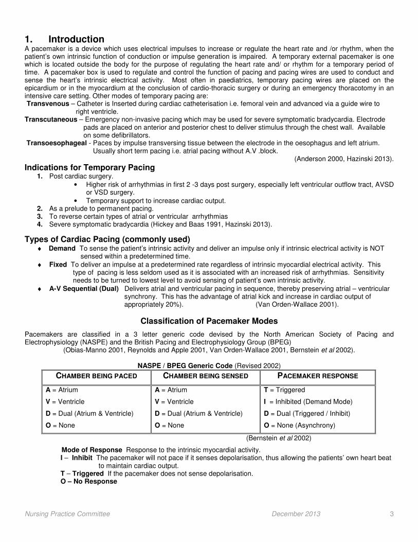
1. Introduction A pacemaker is a device which uses electrical impulses to increase or regulate the heart rate and /or rhythm, when the patient’s own intrinsic function of conduction or impulse generation is impaired. A temporary external pacemaker is one which is located outside the body for the purpose of regulating the heart rate and/ or rhythm for a temporary period of time. A pacemaker box is used to regulate and control the function of pacing and pacing wires are used to conduct and sense the heart’s intrinsic electrical activity. Most often in paediatrics, temporary pacing wires are placed on the epicardium or in the myocardium at the conclusion of cardio-thoracic surgery or during an emergency thoracotomy in an intensive care setting. Other modes of temporary pacing are: Transvenous – Catheter is Inserted during cardiac catheterisation i.e. femoral vein and advanced via a guide wire to right ventricle. Transcutaneous – Emergency non-invasive pacing which may be used for severe symptomatic bradycardia. Electrode pads are placed on anterior and posterior chest to deliver stimulus through the chest wall. Available on some defibrillators. Transoesophageal - Paces by impulse transversing tissue between the electrode in the oesophagus and left atrium. Usually short term pacing i.e. atrial pacing without A.V .block.
(Anderson 2000, Hazinski 2013).
Indications for Temporary Pacing 1. Post cardiac surgery.
• Higher risk of arrhythmias in first 2 -3 days post surgery, especially left ventricular outflow tract, AVSD or VSD surgery.
• Temporary support to increase cardiac output. 2. As a prelude to permanent pacing. 3. To reverse certain types of atrial or ventricular arrhythmias 4. Severe symptomatic bradycardia (Hickey and Baas 1991, Hazinski 2013).
Types of Cardiac Pacing (commonly used) ♦ Demand To sense the patient’s intrinsic activity and deliver an impulse only if intrinsic electrical activity is NOT sensed within a predetermined time.
♦ Fixed To deliver an impulse at a predetermined rate regardless of intrinsic myocardial electrical activity. This type of pacing is less seldom used as it is associated with an increased risk of arrhythmias. Sensitivity needs to be turned to lowest level to avoid sensing of patient’s own intrinsic activity.
♦ A-V Sequential (Dual) Delivers atrial and ventricular pacing in sequence, thereby preserving atrial – ventricular synchrony. This has the advantage of atrial kick and increase in cardiac output of appropriately 20%). (Van Orden-Wallace 2001).
Classification of Pacemaker Modes
Pacemakers are classified in a 3 letter generic code devised by the North American Society of Pacing and Electrophysiology (NASPE) and the British Pacing and Electrophysiology Group (BPEG)
(Obias-Manno 2001, Reynolds and Apple 2001, Van Orden-Wallace 2001, Bernstein et al 2002).
NASPE / BPEG Generic Code (Revised 2002)
CHAMBER BEING PACED CHAMBER BEING SENSED PACEMAKER RESPONSE
A = Atrium
V = Ventricle
D = Dual (Atrium & Ventricle)
O = None
A = Atrium
V = Ventricle
D = Dual (Atrium & Ventricle)
O = None
T = Triggered
I = Inhibited (Demand Mode)
D = Dual (Triggered / Inhibit)
O = None (Asynchrony)
(Bernstein et al 2002)
Mode of Response Response to the intrinsic myocardial activity. I – Inhibit The pacemaker will not pace if it senses depolarisation, thus allowing the patients’ own heart beat to maintain cardiac output. T – Triggered If the pacemaker does not sense depolarisation. O – No Response
Nursing Practice Committee December 2013 4
The mode of pacing selected depends on the patient’s inherent heart rate and rhythm and the function of the atria and ventricles. The mode used will be the one which will best optimise cardiac output. The most common temporary pacing is:
♦ AAI - Atrial Pacing
♦ VVI - Ventricular Pacing
♦ DDD - A.V Sequential (Dual) Pacing
The Pacing Circuit Pulse Generator (Pacing Box) This contains the energy source and electrical circuitry to provide an electrical stimulus to maintain the specified rate. It also recognises and evaluates the heart’s intrinsic rhythm. The pacing circuit has terminals for pacemaker wire connection of bi-polar leads. Bipolar leads measure electrical potential between 2 lead wires in contact with the heart. Lead / Wire / Electrode This transmits the patients’ rhythm to the pulse generator and also carries an electrical stimulus, between the pulse generator and the chamber being paced. The electrode needs a negative (output) pole (the tip) and a positive (ground) pole (the insulator) which enables a current to flow between the pulse generator and the heart. Epicardial wires may be placed after cardiac surgery on the epicardium or placed transvenously through guided insertion of specialised catheters at cardiac catheter (Reynolds and Apple 2001, Hazinski 2013).
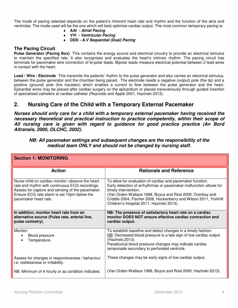
2. Nursing Care of the Child with a Temporary External Pacemaker
Nurses should only care for a child with a temporary external pacemaker having received the necessary theoretical and practical instruction to practice competently, within their scope of All nursing care is given with regard to guidance for good practice practice (An Bord Altranais, 2000, OLCHC, 2002).
NB: All pacemaker settings and subsequent changes are the responsibility of the medical team ONLY and should not be changed by nursing staff.
Section 1: MONITORING
Action
Rationale and Reference
Nurse child on cardiac monitor: observe the heart rate and rhythm with continuous ECG recordings. Assess for capture and sensing of the pacemaker. Ensure ECG rate alarm is set 10pm below the pacemaker heart rate.
To allow for evaluation of cardiac and pacemaker function. Early detection of arrhythmias or pacemaker malfunction allows for timely intervention, (Van Orden-Wallace 1998, Boyce and Rost 2000, Overbay and Criddle 2004, Fischer 2008, Hockenberry and Wilson 2011, Yorkhill Children’s Hospital 2011, Hazinski 2013).
In addition, monitor heart rate from an alternative source (Pulse rate, arterial line, pulse oximetry).
NB: The presence of satisfactory heart rate on a cardiac monitor DOES NOT ensure effective cardiac contraction and cardiac output.
Monitor:
• Blood pressure
• Temperature. Assess for changes in responsiveness / behaviour i.e. restlessness or irritability. NB: Minimum of 4 hourly or as condition indicates
To establish baseline and detect changes in a timely fashion. NB: Decreased blood pressure is a late sign of low cardiac output (Hazinski,2013). Paradoxical blood pressure changes may indicate cardiac tamponade secondary to perforated ventricle. These changes may be early signs of low cardiac output. (Van Orden-Wallace 1998, Boyce and Rost 2000, Hazinski 2013).

Nursing Practice Committee December 2013 5
Assess tissue perfusion:
• Peripheral pulses (strong or weak)
• Capillary refill (brisk or sluggish)
• Warmth of extremities NB: Minimum of 4 hourly or as condition indicates
Tissue perfusion depends on adequate cardiac output. These are early signs of low cardiac output. (Boyce and Rost 2000, Hazinski 2013).
Assess rate and regularity of respirations. Monitor colour and oxygen saturations to establish parameters for same. Administer oxygen if ordered and clinically indicated
To establish baseline and detect changes in a timely fashion. Increased respiratory rate, dyspnoea or cough may be indications of increasing heart failure (Van Orden-Wallace 1998, Hockenberry and Wilson 2011).
Maintain strict fluid balance chart.
To provide information about fluid balance. Large positive balance and diminished urine output may indicate worsening heart failure (Van Orden-Wallace 1998, Hazinski 2013).
Action
Rationale and Reference
Monitor serum electrolytes (as per medical team). Monitor acid-base balance (as per medical team). Inform medical team of changes in patient’s condition or laboratory findings. Document same.
Electrolyte imbalance may interfere with electrical activity of the heart (Reiswig-Timothy and Rodeman 2004, Hockenberry and Wilson 2011). Pacing thresholds can be affected by acid-base balance (Hazinski, 2013). To allow for timely interventions by medical team (Van Orden-Wallace 1998, An Bord Altranais 2002).
Assess bowel function daily. Prevent constipation.
To allow timely interventions in preventing constipation. Straining on defaecation may reduce cardiac output (Van Orden-Wallace 1998).
SECTION 2: DOCUMENTATION
Action
Rationale and Reference
Check pacemaker settings against doctors’ order and document same in nursing notes Verify pacemaker settings and record the following information on vital signs flow sheet of Clinical Information management System (CIMS) (Appendix I). NB: Minimum once per shift and following all changes to settings.
To ensure correct settings of mode, rate, sensitivity and output. To have baseline settings in case of alterations. Documentation provides continuity of care when information is shared (Scheider-Hickey and Bass 1991, An Bord Altranais 2002, Fischer 2008, Hazinski 2013). To promote and facilitate continuity of care and good communication through effective documentation. (Schneider-Hickey and Baas 1999, An Bord Altranais 2002, Hazinski 2013).

Nursing Practice Committee December 2013 6
� Patient’s name and hospital number �Date and time �Pacemaker mode �Rate �Atrial output �Atrial sense �Ventricular output �Ventricular sense �A.V. delay �Battery change (date) �Battery voltage �Pacing wires
� Secured to patient � Entry site dry � Secured to pacemaker
�Pacing spike(s) on monitor �Heart rate from arterial line or alternative source �Ensure lock on Document make and model of external pacemaker and any changes.
(Appendix I, II and III). NB: pacing spikes may not be visible on telemetry (PICU only). Assists in tracking pacemaker malfunction.
SECTION 3: PACEMAKER AND WIRES
Action
Rationale and Reference
Assess integrity and security of pacing wires, ensuring no loose connections or wire fractures (minimum once per shift). NB: Take extra care when moving patient. Ensure wires are secure and pacemaker box and leads are supported.
To ensure good pacemaker connection and prevent disconnection. To prevent accidental changes to settings. (Appendix IV). (Martin and Aragen 1992, Hazinski 1999, Dwyer 2001, Reynolds and Apple 2001, Dwyer and Bauer 2010).
Ensure the pacemaker box is secure Ensure the cables are secure. NB: Pacemaker should be visible at all times. If pacemaker is dropped or becomes damaged it should be replaced immediately by the medical team and sent to clinical engineers for evaluation.
To prevent strain and accidental disconnection or dislodgement of pacing wires and damage to the pacemaker box. (Keenan 1995, Cottle 1997, Dwyer, 2001, Overbay and Criddle 2004, Reiswig-Timothy and Rodeman 2004). To ensure the pacemaker is functioning correctly.
If alternative pacemaker is required contact PICU 2
nd Floor first and then theatre dept. or clinical
engineer for replacement. (Clinical Engineers: Bleep 465 / 008, Ext 6465. Out of Hours via switchboard)
Inform cardiothoracic, medical / surgical team via bleep or out of hour’s telephone number via switch board. Also contact consultant in charge.
To ensure timely replacement of pacemaker.
Battery Use 9 volt alkaline batteries only. NB: DO NOT USE rechargeable batteries Record battery voltage at beginning of shift and following insertion of new battery.
Risk of low capacity and unstable charge which may cause a pacemaker malfunction (St Jude Medical 2011). To ascertain battery status.

Nursing Practice Committee December 2013 7
When battery is in use, the battery should be changed when battery depletion symbol displays only one blinking segment and warning message ‘Change battery!’ appears. This is repeated every 10 minutes. Ask cardiothoracic team to change the battery (Appendix III).
Figure 1: Battery Symbol Cardiothoracic (medical / surgical) team change all temporary external pacemaker batteries After inserting a new battery, the device Model 3085 needs 30 minutes to recharge its internal power capacitor in order to perform the bridging function again.
Cardiothoracic (medical / surgical) team to change pacemaker battery:
• With each new patient and then minimum of every 3 days
Label rear of pacemaker, with date the battery was last changed, nurses initials and document same in the nursing notes. Ensure safe disposal of battery. Have a replacement 9 volt battery available at the bedside at all times.
Critical Battery Depletion The nurse should avoid this occurring by organising battery change earlier. When critical battery depletion occurs the battery symbol will be empty and blinking. The warning message; ‘Hurry up! Change battery!’ will display. This is repeated every 2 minutes. Battery will need to be replaced immediately. Ensure manufacturer’s user manual is always available for reference, in an area that all staff are aware of and have access to.
To allow cardiothoracic (medical / surgical) team to replace battery in a timely fashion. Battery change level is reached. There is approximately 24 hours reserve of battery life on Model 3085 if pacemaker mode set on standard setting (St Jude Medical 2011). To minimise risk and create a safe environment should interruption of pacing / complications occur during the procedure. NB: during battery changeover the pacemaker provides a minimum of 30 seconds additional power for extra safety. Battery change should take place WITHOUT DELAY but avoid undue haste (Jude Medical 2011).
To ensure, fully charged battery in situ. NB: AV sequentional pacing exhausts a battery more quickly than ventricular demand pacing.
(Hazinski 2013). To have replacement in case of battery failure (Dwyer 2001, Mater Misericordiae University Hospital 2011, Yorkhill Children’s Hospital 2011, Hazinski 2013).
To minimise risk and ensure the infant/ child receives continuous and uninterrupted pacing (St Jude Medical 2011). Critical battery change level has been reached and immediate battery change is required (St Jude Medical 2011). Readily available for reference. Increased familiarity with pacemaker (Dwyer 2001, St Jude Medical 2011).
Nursing Practice Committee December 2013 8
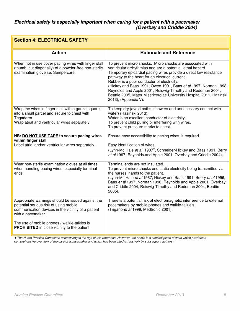
Electrical safety is especially important when caring for a patient with a pacemaker (Overbay and Criddle 2004)
Section 4: ELECTRICAL SAFETY
Action
Rationale and Reference
When not in use cover pacing wires with finger stall (thumb, cut diagonally) of a powder-free non-sterile examination glove i.e. Sempercare.
To prevent micro shocks. Micro shocks are associated with ventricular arrhythmias and are a potential lethal hazard. Temporary epicardial pacing wires provide a direct low resistance pathway to the heart for an electrical current. Rubber is a poor conductor of electricity. (Hickey and Baas 1991, Owen 1991, Baas et al 1997, Norman 1998, Reynolds and Apple 2001, Reiswig-Timothy and Rodeman 2004, Beattie 2005, Mater Misericordiae University Hospital 2011, Hazinski 2013), (Appendix V).
Wrap the wires in finger stall with a gauze square, into a small parcel and secure to chest with Tegaderm. Wrap atrial and ventricular wires separately. NB: DO NOT USE TAPE to secure pacing wires within finger stall Label atrial and/or ventricular wires separately.
To keep dry (avoid baths, showers and unnecessary contact with water) (Hazinski 2013). Water is an excellent conductor of electricity. To prevent child pulling or interfering with wires. To prevent pressure marks to chest. Ensure easy accessibility to pacing wires, if required. Easy identification of wires.
(Lynn-Mc Hale et al 1987*, Schneider-Hickey and Baas 1991, Berry et al 1997, Reynolds and Apple 2001, Overbay and Criddle 2004).
Wear non-sterile examination gloves at all times when handling pacing wires, especially terminal ends.
Terminal ends are not insulated. To prevent micro shocks and static electricity being transmitted via the nurses’ hands to the patient. (Lynn-Mc Hale et al 1987, Hickey and Baas 1991, Beery et al 1996, Baas et al 1997, Norman 1998, Reynolds and Apple 2001, Overbay and Criddle 2004, Reiswig-Timothy and Rodeman 2004, Beattie 2005).
Appropriate warnings should be issued against the potential serious risk of using mobile communication devices in the vicinity of a patient with a pacemaker. The use of mobile phones / walkie-talkies is PROHIBITED in close vicinity to the patient.
There is a potential risk of electromagnetic interference to external pacemakers by mobile phones and walkie-talkie’s (Trigano et al 1999, Medtronic 2001).
�The Nurse Practice Committee acknowledges the age of this reference. However, the article is a seminal piece of work which provides a comprehensive overview of the care of a pacemaker and which has been cited extensively by subsequent authors.
Nursing Practice Committee December 2013 9
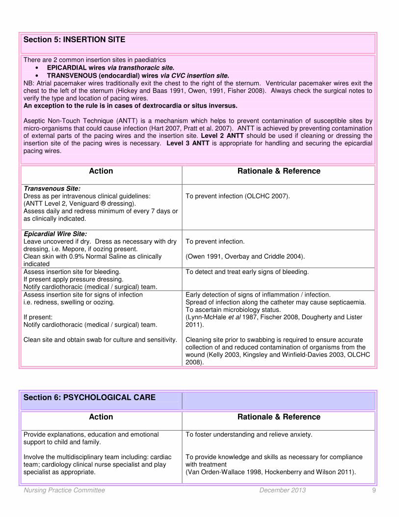
Section 5: INSERTION SITE
There are 2 common insertion sites in paediatrics
• EPICARDIAL wires via transthoracic site.
• TRANSVENOUS (endocardial) wires via CVC insertion site. NB: Atrial pacemaker wires traditionally exit the chest to the right of the sternum. Ventricular pacemaker wires exit the chest to the left of the sternum (Hickey and Baas 1991, Owen, 1991, Fisher 2008). Always check the surgical notes to verify the type and location of pacing wires. An exception to the rule is in cases of dextrocardia or situs inversus. Aseptic Non-Touch Technique (ANTT) is a mechanism which helps to prevent contamination of susceptible sites by micro-organisms that could cause infection (Hart 2007, Pratt et al. 2007). ANTT is achieved by preventing contamination of external parts of the pacing wires and the insertion site. Level 2 ANTT should be used if cleaning or dressing the insertion site of the pacing wires is necessary. Level 3 ANTT is appropriate for handling and securing the epicardial pacing wires.
Action
Rationale & Reference
Transvenous Site: Dress as per intravenous clinical guidelines: (ANTT Level 2, Veniguard ® dressing). Assess daily and redress minimum of every 7 days or as clinically indicated.
To prevent infection (OLCHC 2007).
Epicardial Wire Site: Leave uncovered if dry. Dress as necessary with dry dressing, i.e. Mepore, if oozing present. Clean skin with 0.9% Normal Saline as clinically indicated
To prevent infection. (Owen 1991, Overbay and Criddle 2004).
Assess insertion site for bleeding. If present apply pressure dressing. Notify cardiothoracic (medical / surgical) team.
To detect and treat early signs of bleeding.
Assess insertion site for signs of infection i.e. redness, swelling or oozing. If present: Notify cardiothoracic (medical / surgical) team. Clean site and obtain swab for culture and sensitivity.
Early detection of signs of inflammation / infection. Spread of infection along the catheter may cause septicaemia. To ascertain microbiology status. (Lynn-McHale et al 1987, Fischer 2008, Dougherty and Lister 2011). Cleaning site prior to swabbing is required to ensure accurate collection of and reduced contamination of organisms from the wound (Kelly 2003, Kingsley and Winfield-Davies 2003, OLCHC 2008).
Section 6: PSYCHOLOGICAL CARE
Action Rationale & Reference
Provide explanations, education and emotional support to child and family. Involve the multidisciplinary team including: cardiac team; cardiology clinical nurse specialist and play specialist as appropriate.
To foster understanding and relieve anxiety. To provide knowledge and skills as necessary for compliance with treatment (Van Orden-Wallace 1998, Hockenberry and Wilson 2011).

Nursing Practice Committee December 2013 10
3. Trouble Shooting
Most troubleshooting associated with pacemaker systems is related to changes in the patient’s medical condition or misinterpretation of normal pacemaker function. In all instances it is vital to assess the patient and identify the cause.
♦ It is essential for nurses to contact the cardiothoracic (medical/ surgical) team IMMEDIATELY, for early and timely intervention.
There are four potential problems which can exist during pacing: 1. Failure to Fire 2. Failure to Capture / Pace 3. Under Sensing 4. Over Sensing
1. Failure to Fire
Failure to fire is characterised by the loss of output from the pulse generator, which is identifiable by an abnormally slow heart rate or asystole. Intervention should be specific to the problem found in the pacemaker system. If failure to fire
cannot be corrected emergency measures may need to be initiated. Failure to fire related to pacemaker malfunction is
rare. It is more likely to be related to settings, connections or changing thresholds. Contact Cardiothoracic (medical / surgical) Team IMMEDIATELY
Problem Intervention
Loose connection or disconnection between lead wire, cables and pacemaker
Ensure connections are secure
Fracture / dislodgement of lead wire Assess integrity of lead wires and replace as necessary NB: Remember the skin can be used as a new or extra positive lead.
Low pacemaker battery Insert new battery
Failure of pacemaker pulse generator Replace pacemaker generator. Contact Cardiothoracic Team (medical / surgical).
Over sensing (not common in paediatrics i.e. P wave high, mainly occurs in adults, unless the sense thresholds have been set too low)
Contact Cardiothoracic Team (medical/surgical) to assess sensitivity and decrease if necessary
(Lynn-McHale et al 1987)
2. Failure to Capture / Pace
Capture occurs when the myocardium responds to the pacing stimulus by depolarising i.e. P wave or QRS wave. Failure to capture occurs when the myocardium fails to respond to a pacing stimulus. It will be seen as the pacing spike(s), not been followed by a P wave or QRS complex.
Contact Cardiothoracic Team (medical / surgical) IMMEDIATELY Possible causes for increased pacing threshold:
• Inflammation or fibrosis at electrode site
• Increased serum Potassium or Calcium
• Acid base imbalances
• Medications i.e. Verapamil or Propanolol
• Fibrillation or flutter Problem Intervention
Loose connection between lead wire, cables, and pacemaker.
Ensure connections are secure.
Fracture / insulation break of lead wire. Displacement of lead wire.
Assess integrity. Contact Cardiothoracic Team (medical / surgical) who will replace it if required. NB: The skin can be used as a new positive electrode.
Low pacemaker battery.
Battery replaced by Cardiothoracic Team (medical / surgical).
Failure of pulse generator Pulse generator replaced by cardiothoracic team.
Increased pacing threshold/ inadequate output (energy) for depolarisation.
Contact Cardiothoracic Team (medical/surgical). Who will reassess pacing threshold and identify and treat the underlying physiological disturbances.
Nursing Practice Committee December 2013 11
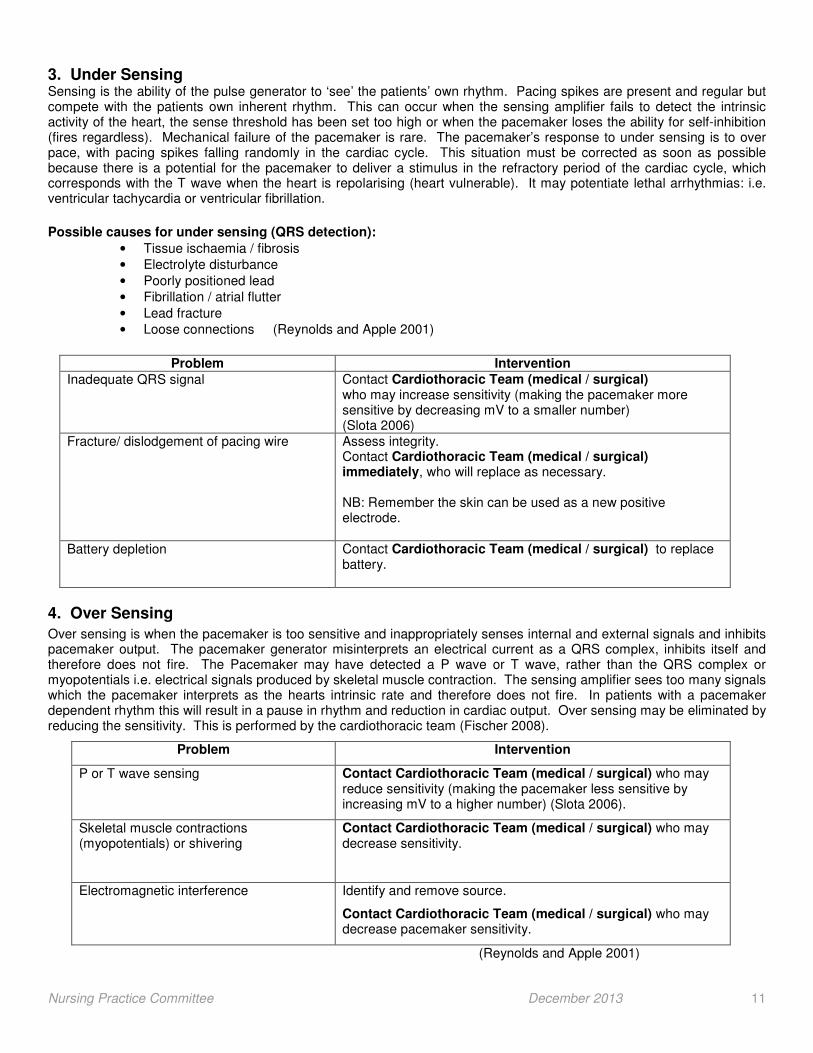
3. Under Sensing Sensing is the ability of the pulse generator to ‘see’ the patients’ own rhythm. Pacing spikes are present and regular but compete with the patients own inherent rhythm. This can occur when the sensing amplifier fails to detect the intrinsic activity of the heart, the sense threshold has been set too high or when the pacemaker loses the ability for self-inhibition (fires regardless). Mechanical failure of the pacemaker is rare. The pacemaker’s response to under sensing is to over pace, with pacing spikes falling randomly in the cardiac cycle. This situation must be corrected as soon as possible because there is a potential for the pacemaker to deliver a stimulus in the refractory period of the cardiac cycle, which corresponds with the T wave when the heart is repolarising (heart vulnerable). It may potentiate lethal arrhythmias: i.e. ventricular tachycardia or ventricular fibrillation.
Possible causes for under sensing (QRS detection):
• Tissue ischaemia / fibrosis
• Electrolyte disturbance
• Poorly positioned lead
• Fibrillation / atrial flutter
• Lead fracture
• Loose connections (Reynolds and Apple 2001)
Problem Intervention
Inadequate QRS signal
Contact Cardiothoracic Team (medical / surgical) who may increase sensitivity (making the pacemaker more sensitive by decreasing mV to a smaller number) (Slota 2006)
Fracture/ dislodgement of pacing wire
Assess integrity. Contact Cardiothoracic Team (medical / surgical) immediately, who will replace as necessary. NB: Remember the skin can be used as a new positive electrode.
Battery depletion
Contact Cardiothoracic Team (medical / surgical) to replace battery.
4. Over Sensing
Over sensing is when the pacemaker is too sensitive and inappropriately senses internal and external signals and inhibits pacemaker output. The pacemaker generator misinterprets an electrical current as a QRS complex, inhibits itself and therefore does not fire. The Pacemaker may have detected a P wave or T wave, rather than the QRS complex or myopotentials i.e. electrical signals produced by skeletal muscle contraction. The sensing amplifier sees too many signals which the pacemaker interprets as the hearts intrinsic rate and therefore does not fire. In patients with a pacemaker dependent rhythm this will result in a pause in rhythm and reduction in cardiac output. Over sensing may be eliminated by reducing the sensitivity. This is performed by the cardiothoracic team (Fischer 2008).
Problem Intervention
P or T wave sensing Contact Cardiothoracic Team (medical / surgical) who may reduce sensitivity (making the pacemaker less sensitive by increasing mV to a higher number) (Slota 2006).
Skeletal muscle contractions (myopotentials) or shivering
Contact Cardiothoracic Team (medical / surgical) who may decrease sensitivity.
Electromagnetic interference Identify and remove source.
Contact Cardiothoracic Team (medical / surgical) who may decrease pacemaker sensitivity.
(Reynolds and Apple 2001)
Nursing Practice Committee December 2013 12
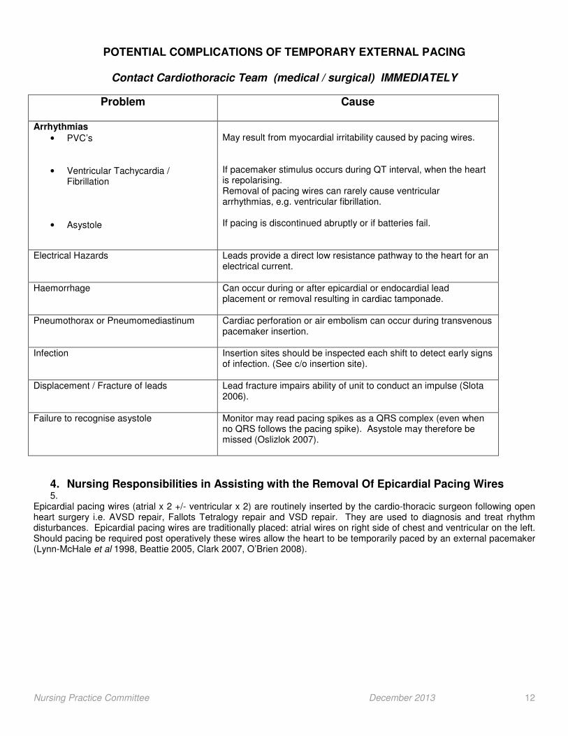
POTENTIAL COMPLICATIONS OF TEMPORARY EXTERNAL PACING
Contact Cardiothoracic Team (medical / surgical) IMMEDIATELY
Problem
Cause
Arrhythmias
• PVC’s
• Ventricular Tachycardia / Fibrillation
• Asystole
May result from myocardial irritability caused by pacing wires. If pacemaker stimulus occurs during QT interval, when the heart is repolarising. Removal of pacing wires can rarely cause ventricular arrhythmias, e.g. ventricular fibrillation. If pacing is discontinued abruptly or if batteries fail.
Electrical Hazards
Leads provide a direct low resistance pathway to the heart for an electrical current.
Haemorrhage
Can occur during or after epicardial or endocardial lead placement or removal resulting in cardiac tamponade.
Pneumothorax or Pneumomediastinum
Cardiac perforation or air embolism can occur during transvenous pacemaker insertion.
Infection
Insertion sites should be inspected each shift to detect early signs of infection. (See c/o insertion site).
Displacement / Fracture of leads Lead fracture impairs ability of unit to conduct an impulse (Slota 2006).
Failure to recognise asystole Monitor may read pacing spikes as a QRS complex (even when no QRS follows the pacing spike). Asystole may therefore be missed (Oslizlok 2007).
4. Nursing Responsibilities in Assisting with the Removal Of Epicardial Pacing Wires 5.
Epicardial pacing wires (atrial x 2 +/- ventricular x 2) are routinely inserted by the cardio-thoracic surgeon following open heart surgery i.e. AVSD repair, Fallots Tetralogy repair and VSD repair. They are used to diagnosis and treat rhythm disturbances. Epicardial pacing wires are traditionally placed: atrial wires on right side of chest and ventricular on the left. Should pacing be required post operatively these wires allow the heart to be temporarily paced by an external pacemaker (Lynn-McHale et al 1998, Beattie 2005, Clark 2007, O’Brien 2008).
Nursing Practice Committee December 2013 13
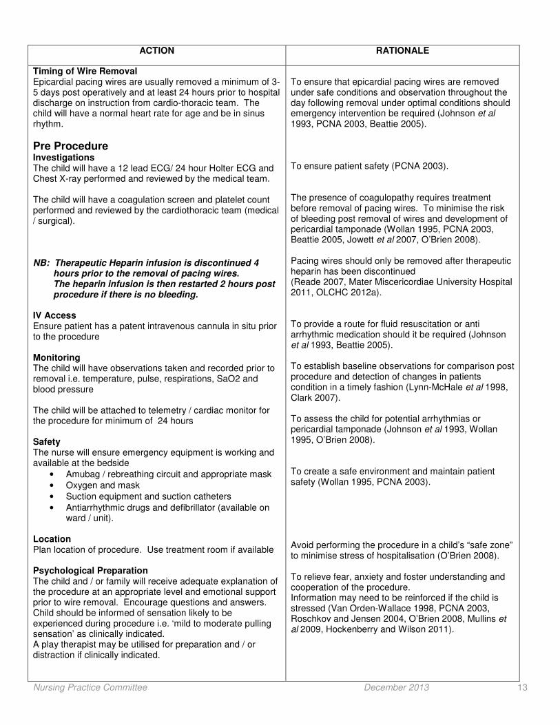
ACTION RATIONALE
Timing of Wire Removal Epicardial pacing wires are usually removed a minimum of 3-5 days post operatively and at least 24 hours prior to hospital discharge on instruction from cardio-thoracic team. The child will have a normal heart rate for age and be in sinus rhythm.
Pre Procedure Investigations The child will have a 12 lead ECG/ 24 hour Holter ECG and Chest X-ray performed and reviewed by the medical team. The child will have a coagulation screen and platelet count performed and reviewed by the cardiothoracic team (medical / surgical). NB: Therapeutic Heparin infusion is discontinued 4 hours prior to the removal of pacing wires. The heparin infusion is then restarted 2 hours post procedure if there is no bleeding. IV Access Ensure patient has a patent intravenous cannula in situ prior to the procedure Monitoring The child will have observations taken and recorded prior to removal i.e. temperature, pulse, respirations, SaO2 and blood pressure The child will be attached to telemetry / cardiac monitor for the procedure for minimum of 24 hours Safety The nurse will ensure emergency equipment is working and available at the bedside
• Amubag / rebreathing circuit and appropriate mask
• Oxygen and mask
• Suction equipment and suction catheters
• Antiarrhythmic drugs and defibrillator (available on ward / unit).
Location Plan location of procedure. Use treatment room if available Psychological Preparation The child and / or family will receive adequate explanation of the procedure at an appropriate level and emotional support prior to wire removal. Encourage questions and answers. Child should be informed of sensation likely to be experienced during procedure i.e. ‘mild to moderate pulling sensation’ as clinically indicated. A play therapist may be utilised for preparation and / or distraction if clinically indicated.
To ensure that epicardial pacing wires are removed under safe conditions and observation throughout the day following removal under optimal conditions should emergency intervention be required (Johnson et al 1993, PCNA 2003, Beattie 2005). To ensure patient safety (PCNA 2003). The presence of coagulopathy requires treatment before removal of pacing wires. To minimise the risk of bleeding post removal of wires and development of pericardial tamponade (Wollan 1995, PCNA 2003, Beattie 2005, Jowett et al 2007, O’Brien 2008). Pacing wires should only be removed after therapeutic heparin has been discontinued (Reade 2007, Mater Miscericordiae University Hospital 2011, OLCHC 2012a). To provide a route for fluid resuscitation or anti arrhythmic medication should it be required (Johnson et al 1993, Beattie 2005). To establish baseline observations for comparison post procedure and detection of changes in patients condition in a timely fashion (Lynn-McHale et al 1998, Clark 2007). To assess the child for potential arrhythmias or pericardial tamponade (Johnson et al 1993, Wollan 1995, O’Brien 2008). To create a safe environment and maintain patient safety (Wollan 1995, PCNA 2003). Avoid performing the procedure in a child’s “safe zone” to minimise stress of hospitalisation (O’Brien 2008). To relieve fear, anxiety and foster understanding and cooperation of the procedure. Information may need to be reinforced if the child is stressed (Van Orden-Wallace 1998, PCNA 2003, Roschkov and Jensen 2004, O’Brien 2008, Mullins et al 2009, Hockenberry and Wilson 2011).

Nursing Practice Committee December 2013 14
Pain Relief Administer analgesia and sedation if required as prescribed by the medical team as per ‘Procedural Analgesia and Sedation’ Algorithm (Appendix VI). Sedation will always be given in conjunction with analgesia. Assess pain score. Positioning The child will be positioned supine or alternatively at 30-45o angle if not possible in bed for the procedure. Ensure privacy in older child / adolescent.
Procedure Responsility for removal of pacing wires The cardio-thoracic team are responsible for removal of the epicardial pacing wires. Equipment
• Dressing trolley
• Dressing pack including sterile gloves and gauze
• 0.05% Chlorhexidine solution
• Opsite occlusive dressing
• Stitch cutter Cardio-thoracic Surgeon will wash hands using a Aseptic Non-touch Technique (ANTT) (level 2) and put on sterile gloves
The nurse will decontaminate hands and assist doctor in laying dressing trolley. Nurse will remove dressing around pacing wires to expose pacing wires and then repeat handwashing. The doctor will clean around pacing wire sites with Chlorhexidine 0.05%. The atrial pacing wires are usually removed first if present and ventricular wires last. The holding suture of the pacing wire is released using a stitch cutter.
Holding the pacing wire near to the chest it will be pulled with a smooth, continuous, downward, pulling motion, exerting gentle traction until release from the epicardium is felt.
The tip of the epicardial pacing wire is inspected for intactness and pieces of myocardial tissue. The procedure is repeated by the doctor for each additional pacing wire(s).
To provide comfort and minimise pain. Patients have reported ‘mild to moderate pulling sensation’ on epicardial pacing wire removal (Mullins et al 2009, Mater Misericordiae University Hospital 2011, OLCHC 2012b). To ensure correct positioning for removal of epicardial pacing wires. Semi upright position is often preferred in children as it is often associated with less anxiety (Wollan 1995, Clark 2007, O’Brien 2008; Beattie 2008). Procedure only performed by Cardiothoracic Team because of the potential complications that may occur following the procedure (Roschkov and Jensen 2004, O’Brien 2008). To prevent cross infection, universal precautions (PCNA 2003). Epicardial pacing wires provide a direct low resistant pathway to the heart and patient may receive micro shocks due to static electricity (Wollan 1995, Beattie 2005).
To minimise transmission of organisms (O’Brien 2008). To allow complete visualisation of pacing wire site and holding suture (O’Brien 2008). Reduces risk of infection (O’Brien 2008). This allows pacing of the ventricle to restore cardiac output in the event of a symptomatic arrhythmia, following removal of atrial pacing wires. To reduce the risk of trauma. Jerking or pulling against resistance may cause bleeding. (Wollan 1995, Sheehan et al 2001, PCNA 2003, Clark 2007). (Clark 2007). To ensure that the entire wire has been removed and determine the risk of infection, migration or haemorrhage (Johnson et al 1993, Wollan 1995, Clark 2007, Beattie 2008).
Nursing Practice Committee December 2013 15
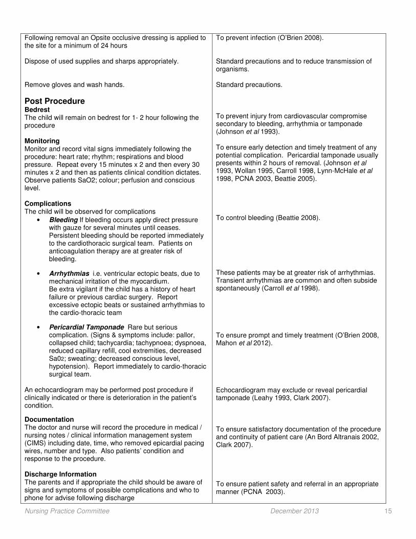
Following removal an Opsite occlusive dressing is applied to the site for a minimum of 24 hours Dispose of used supplies and sharps appropriately. Remove gloves and wash hands.
Post Procedure Bedrest The child will remain on bedrest for 1- 2 hour following the procedure Monitoring Monitor and record vital signs immediately following the procedure: heart rate; rhythm; respirations and blood pressure. Repeat every 15 minutes x 2 and then every 30 minutes x 2 and then as patients clinical condition dictates. Observe patients SaO2; colour; perfusion and conscious level. Complications The child will be observed for complications
• Bleeding If bleeding occurs apply direct pressure with gauze for several minutes until ceases. Persistent bleeding should be reported immediately to the cardiothoracic surgical team. Patients on anticoagulation therapy are at greater risk of bleeding.
• Arrhythmias i.e. ventricular ectopic beats, due to mechanical irritation of the myocardium. Be extra vigilant if the child has a history of heart failure or previous cardiac surgery. Report excessive ectopic beats or sustained arrhythmias to the cardio-thoracic team
• Pericardial Tamponade Rare but serious complication. (Signs & symptoms include: pallor, collapsed child; tachycardia; tachypnoea; dyspnoea, reduced capillary refill, cool extremities, decreased Sa02; sweating; decreased conscious level, hypotension). Report immediately to cardio-thoracic surgical team.
An echocardiogram may be performed post procedure if clinically indicated or there is deterioration in the patient’s condition.
Documentation The doctor and nurse will record the procedure in medical / nursing notes / clinical information management system (CIMS) including date, time, who removed epicardial pacing wires, number and type. Also patients’ condition and response to the procedure. Discharge Information The parents and if appropriate the child should be aware of signs and symptoms of possible complications and who to phone for advise following discharge
To prevent infection (O’Brien 2008). Standard precautions and to reduce transmission of organisms. Standard precautions. To prevent injury from cardiovascular compromise secondary to bleeding, arrhythmia or tamponade (Johnson et al 1993). To ensure early detection and timely treatment of any potential complication. Pericardial tamponade usually presents within 2 hours of removal. (Johnson et al 1993, Wollan 1995, Carroll 1998, Lynn-McHale et al 1998, PCNA 2003, Beattie 2005). To control bleeding (Beattie 2008). These patients may be at greater risk of arrhythmias. Transient arrhythmias are common and often subside spontaneously (Carroll et al 1998). To ensure prompt and timely treatment (O’Brien 2008, Mahon et al 2012).
Echocardiogram may exclude or reveal pericardial tamponade (Leahy 1993, Clark 2007). To ensure satisfactory documentation of the procedure and continuity of patient care (An Bord Altranais 2002, Clark 2007). To ensure patient safety and referral in an appropriate manner (PCNA 2003).

Nursing Practice Committee December 2013 16
Retained Wire Lead or Fragments Ensure retained wire lead or fragments are communicated to ward nursing staff on transfer documentation as clinically indicated. It should be clearly documented in the patient’s medical and nursing notes also. Instruct parent to check childs’ temperature daily until next out patient appointment and report temperature > 38oc Advise parent regarding the long term need to inform doctor regarding any possible signs of infection i.e. malaise, chills, fever and signs of infection at epicardial pacing wire exit sites Instruct parent to inform all attending doctors and dentists of retained pacing wire
There is increased risk of infection as they create an open wound through the skin which communicates with the pericardial space. Complications from retained epicardial wires have been described in the literature i.e. localised abscess / fistula to infective endocarditis. Complications have been reported to occur up to many years later. Ensure satisfactory communication and continuity of care (An Bord Altranais 2002, Yorkhill Children’s Hospital 2011, Shaikhrezai et al 2012). Early detection of infected epicardial pacing wire (Johnson et al 1993). To ensure prompt and timely treatment of any infection at pacing wire sites or due to retained epicardial pacing wire There is a potential risk of endocarditis and doctor or dentist may decide to administer prophylactic antibiotics prior to any invasive procedure (Johnson et al 1993).

Nursing Practice Committee December 2013 17
References
An Bord Altranais (2000) Scope of Nursing and Midwifery Practice Framework. An Bord Altranais: Dublin. An Bord Altranais (2002) Recording Clinical Practice, Guidance to Nurses and Midwives. An Bord Altranais: Dublin. Anderson, D.M. (2000) Dorlands Illustrated Medical Dictionary, 29
th Edition. W.B. Saunders: Philadelphia.
Baas, L.S. Beery, T.A. and Hickey, C (1997) Care of pacemaker electrodes in intensive care and telemetry units. American Journal of Critical Care 6(4): 302-311. Beattie, S. (2005) Epicardial wires. Modern Medicine Available online www.modernmedicine.com/modernmedicine?Hands-On+help/Epicardial-wires/ . Accessed December 3rd, 2012 Beery, T.A. Baas, L.S. and Hickey, C.S. (1996) Infectious precautions with temporary leads: a descriptive study. Heart and Lung 25(3): 182-189. Bernstein, A.D. Daubert, J.C. Fletcher, R.D. Hayes, D.L. Luderitz, B. Reynolds, D.W. Schoenfeld, M.H. and Sutton, R. (2002) The revised NAPSE / BPEG Generic Code for antibradycardia, adaptive-rate and multisite pacing. Pacing Clinical Physiology, 25: 260-264. Boyle, J. and Rost, A.K. (2000) Present status of cardiac pacing: a nursing perspective. Critical Care Nursing Quarterly 23(1): 1-19. Carroll, K.C. Reeves, L.M. Anderson, G. Ray, F.M. Clopton, P.L. Shively, M. and Tarazi, R.Y. (1998) Risks associated with removal of ventricular epicardial pacing wires after cardiac surgery. American Journal of Critical Care 7 (6):444-9 Clark, L. (2007) Bedside nurses removing epicardial pacer wires: from concept to practice. Canadian Journal of Cardiovascular Nursing 17(1): 27-30. Cottle, S. (1997) Temporary transvenous cardiac pacing. Nursing Times 93(48): 48-51. De Vooght, (1999) Pacemaker leads: performance and progress. American Journal of Cardiology 11(8): 187D-191D. Dougherty, L. and Lister, S (eds) (2011) The Royal Marsden Hospital Manual of Clinical Nursing Procedures, 8
th Edition.
Wiley-Blackwell: London. Dwyer, D. (2001) Medical device adverse events and the temporary invasive cardiac pacemaker. International Journal of Trauma Nursing 7(2): 70-73. Dwyer, D. and Bauer, K. (2010) Take the lead on safety with temporary cardiac pacing. Nursing, 40(3): 63-64. Fischer, M. (2008) Transvenous and epicardial pacing: monitoring. In Verger, J.T. and Lebet, R.M. (eds) ACCN Procedure Manuel for Pediatric Acute and Critical Care. Saunders Elsevier: St Louis, 375-382. Hart, S. (2007) Using an aseptic technique to reduce the risk of infection. Nursing Standard 21(47): 43-48. Hazinski, M. F. (ed) (2013) Nursing Care of the critically Ill Child, 3
rd Edition. Elsevier Mosby: St Louis.
Hickey, C.S. and Baas, L.S. ( 1991) Temporary cardiac pacing. AACN. Clinical Issues 2(1): 107-117. Hockenberry, M.J. and Wilson, D. (eds) (2011) Wong’s Nursing care of Infant’s and Children, 9
th Edition. Elsevier Mosby:
St Louis. Johnson, L.G. Brown, O.P. and Alligood, M.R. (1993) Complications of epicardial pacing wire removal. Journal of Cardiovascular Nursing 7 (2): 32-40. Jowett, V. Hayes, N. Sridharan, S. Rees, P. and Macrae, D. (2007) Timing of removal of pacing wires following paediatric cardiac surgery. Cardiology in the Young, 17(5): 512-516.

Nursing Practice Committee December 2013 18
Keenan, J. (1995) Temporary cardiac pacing. Nursing Standard 9(20): 50-51. Kelly, F. (2003) Infection Control: validity and reliability in wound swabbing. British Journal of Nursing 12(16): 959-60, 962-964. Kingsley, A. and Winfield-Davies, S. (2003) Audit of wound swab sampling: why protocols could improve practice. Professional Nurse 18(6): 338-343. Lynn-McHale, D. Riggs, K. and Thurman, L. (1987) Epicardial pacing after cardiac surgery. Critical Care Nurse 11(8): 62-77. Lynn-McHale, D.J. Riggs, K.L. and Thurman, L. (1998) Epicardial pacing after cardiac surgery. Critical Care Nurse 11 (8): 62-74. Mahon, L. Bena, J.F. Morrison, S.M. and Albert, N.M. (2012) Cadiac tampobade after removal of temporary pacer wires. American Journal of Critical Care 21(6): 432-440. Martin, M. and Aragon, D. (1992) Temporary DDD pacing: evaluating haemodynamic performance. Dimensions of Critical Care Nursing 11(4): 191-200. Mater Misericordiae University Hospital (2011) Guidelines for the Care and Removal of Temporary Epicardial Pacing Wires Post Cardiac Surgery. MMUH: Dublin. Medtronic. (2001) Medtronic Dual Chamber Temporary Pacemaker: Technical Manual. Medtronic: Minneapolis. Mullins, M.H. Roschkov, M.N. Jensen, L. Moore, G. and Smith, A. (2009) Sensations during removal of epicardial pacing wires after coronary artery bypass graft surgery. Heart and Lung, 38(5): 377-381. Norman, E.M. (1998) Avoiding electrical hazards. American Journal of Nursing 98(6): 16GG-16HH. Obias-Mango, D. (2001) Unconventional applications in pacemaker therapy. Advanced Practice in Acute and Critical Care 12(1), 127-139. O’Brien.P. (2008) Epicardial pacing wire removal:Perform. In Trivits-Verger, J.T. and Lebet, R.M. (eds) AACN Procedure Manuel for Pediatric, Acute and Critical Care. Saunders Elsevier: St Louis. 383-389. Oslizlok, P. (2007) Personal Communication to Pacemaker Guidelines Project Group, 13
th December 2007. Our Lady’s
Children’s Hospital, Crumlin, Dublin. OLCHC (2007) Intravenous Guidelines for Nursing Staff. Our Lady’s Children’s Hospital, Crumlin, Dublin. OLCHC (2008) Guidelines on Performing a Wound Swab. Our Lady’s Children’s Hospital, Crumlin, Dublin. OLCHC (2012a) Anti Thrombotic Central Line Guidelines. Our Lady’s Children’s Hospital, Crumlin, Dublin. OLCHC (2012b) Procedural Analgesia and Sedation in PICU / HDU. Our Lady’s Children’s Hospital, Crumlin, Dublin. Overbay, D. and Criddle, L. (2004) Mastering temporary invasive cardiac pacing. Critical Care Nurse 24(3): 23-32. Owen, A. (1991) Keeping pace with temporary pacemakers. Nursing 21(4): 58-64. Paediatric Cardiac Nurses Association (PCNA) (2003) PCNA National Standard for Temporary Epicardial Pacing Wire Removal in Children. PCNA: London. Pratt, R.J. Pellowe, C.M. Wilson, J.A. Loveday, H.P. Harper, P.J. Jones, S.R.L.J. McDougall, C. and Wilcox, M.H. (2007) National evidence-based guidelines for preventing healthcare-associated infections in NHS hospitals in England. Journal of Hospital Infection 65 (supplement1), S1-S49. Reade, M.C. (2007) Temporary epicardial pacing after cardiac surgery: a practical review. Part 1: General considerations in the management of epicardial pacing. Anaesthesia 62: 264-271.
Nursing Practice Committee December 2013 19
Reiswig-Timothy, P. and Rodeman, R.N. (2004) Temporary pacemakers in critically ill patients. ACCN Clinical Issues 15(3): 305-325. Reynolds, J. and Apple, S. (2001) A systematic approach to pacemaker assessment. AACN Clinical Issues 12(1), 114-126. Roschkov, S. and Jensen, L. (2004) Coronary artery bypass graft patients’ pain perception during epicardial pacing wire removal. Canadian Journal of Cardiovascular Nursing, 14(3):32-38. Schneider Hickey, C. and Baas, L.S. (1991) Temporary cardiac pacing. AACN Clinical Issues 2(1): 107-117. Shaikhrezai, K. Khorsandi, M. Patronis, M. and Prasaad, S. (2012) Is it safe to cut pacing wires flush with he skin instead of removing them? Interactive Cardiovascular and Thoracic Surgery, 15:1047-1051. Sheehan, K. Tometzki, A. and Tsai-Goodman, B. (2001) National Audit of Temporary Epicardial Wire Removal. Unpublished, Bristol Royal Children’s Hospital. Slota, M.C. (ed) (2006) Core Curriculum for Paediatric Critical Care Nursing, 2
nd Edition. Saunders Elsevier: St Louis.
St Jude Medical (2011) Model 3085 Dual Chamber (DDD) External Pulse Generator, User Manual. Osypka Medical: La Jolla, California. Trigano, A. J. Azoulay, A. Rochdi, M. and Campillo, A. (1999) Electromagnetic interference of external pacemakers by walkie-talkies and digital cellular phones: experimental study. PACE 22: 588-593. Van Orden-Wallace, C.J. (1998) Dual chamber pacemakers in the management of severe heart failure. Critical Care Nurse 18(2): 57-66. Van Orden-Wallace, C.J. (2001) Diagnosing and treating pacemaker syndrome. Critical Care Nurse 21(1): 24-31, 35. Wollan, D.L. (1995) Removal of epicardial pacing wires: an expanded role for nurses. Progress in Cardiovascular Nursing 10 (4): 21-26. Xia, B. Thakur, M.D. Shah, C.P. and Hoon, V.K. (1998) Permanent cardiac pacing. Emergency Clinics of North America 16(2): 419-462. Yorkhill Children’s Hospital (2011) Practice Advisory for Temporary Epicardial Pacing after Cardiac Surgery. Yorkhill Children’s Hospital: Glasgow.

Nursing Practice Committee December 2013 20
APPENDIX I: PACEMAKER CONTROLS
Nursing Practice Committee December 2013 21
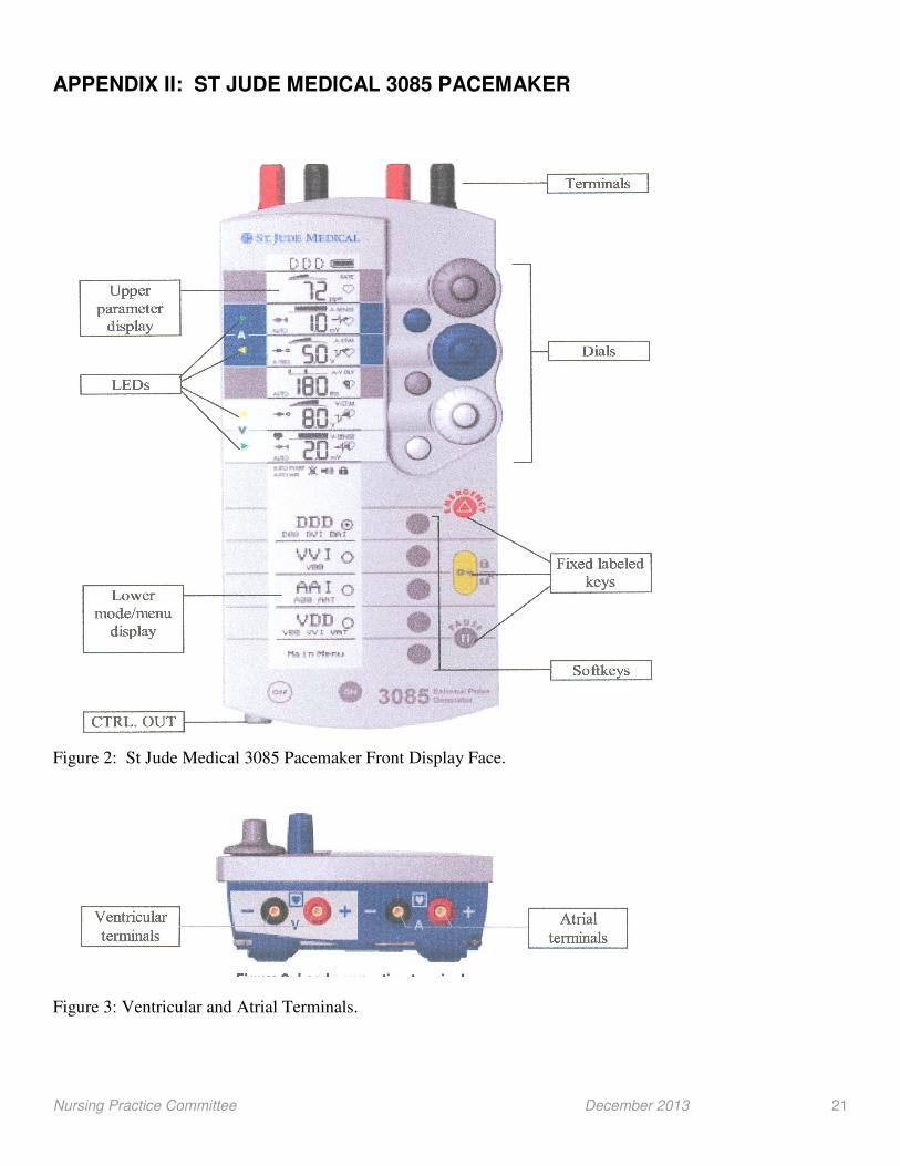
APPENDIX II: ST JUDE MEDICAL 3085 PACEMAKER
Figure 2: St Jude Medical 3085 Pacemaker Front Display Face.
Figure 3: Ventricular and Atrial Terminals.
Nursing Practice Committee December 2013 22
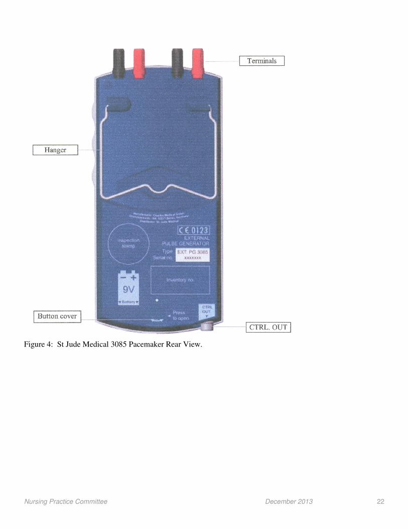
Figure 4: St Jude Medical 3085 Pacemaker Rear View.
Nursing Practice Committee December 2013 23
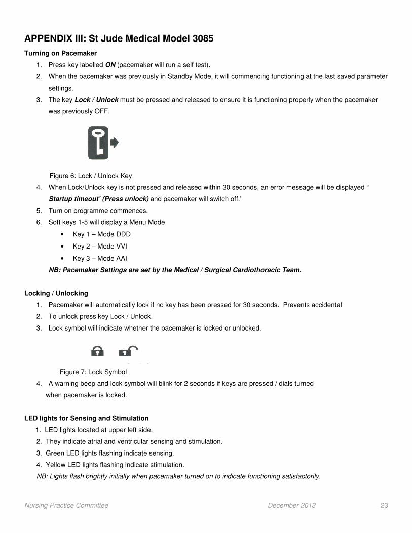
APPENDIX III: St Jude Medical Model 3085
Turning on Pacemaker
1. Press key labelled ON (pacemaker will run a self test).
2. When the pacemaker was previously in Standby Mode, it will commencing functioning at the last saved parameter
settings.
3. The key Lock / Unlock must be pressed and released to ensure it is functioning properly when the pacemaker
was previously OFF.
Figure 6: Lock / Unlock Key
4. When Lock/Unlock key is not pressed and released within 30 seconds, an error message will be displayed ‘
Startup timeout’ (Press unlock) and pacemaker will switch off.’
5. Turn on programme commences.
6. Soft keys 1-5 will display a Menu Mode
• Key 1 – Mode DDD
• Key 2 – Mode VVI
• Key 3 – Mode AAI
NB: Pacemaker Settings are set by the Medical / Surgical Cardiothoracic Team.
Locking / Unlocking
1. Pacemaker will automatically lock if no key has been pressed for 30 seconds. Prevents accidental
2. To unlock press key Lock / Unlock.
3. Lock symbol will indicate whether the pacemaker is locked or unlocked.
Figure 7: Lock Symbol
4. A warning beep and lock symbol will blink for 2 seconds if keys are pressed / dials turned
when pacemaker is locked.
LED lights for Sensing and Stimulation
1. LED lights located at upper left side.
2. They indicate atrial and ventricular sensing and stimulation.
3. Green LED lights flashing indicate sensing.
4. Yellow LED lights flashing indicate stimulation.
NB: Lights flash brightly initially when pacemaker turned on to indicate functioning satisfactorily.

Nursing Practice Committee December 2013 24
Emergency Key
Pressing the emergency button key will commence pacemaker stimulation at emergency settings.
Pause Pacemaker
Pressing the pause button key will disable pacemaker stimulation as long as it is pressed.
Turning Off the Pacemaker
1. Press lock/ unlock key.
2. Press OFF key.
3. A soft key power-off menu will display.
•••• Press key 1 – OFF (with no storage). Actual settings are not saved
•••• Press Key 2 – Stand-by with data stored.
NB: No battery power is consumed in the stand-by mode.
(St Jude Medical 2011).
Nursing Practice Committee December 2013 25
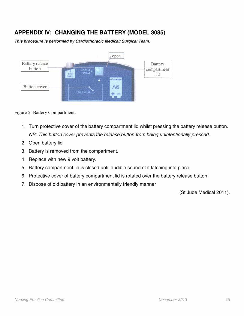
APPENDIX IV: CHANGING THE BATTERY (MODEL 3085)
This procedure is performed by Cardiothoracic Medical/ Surgical Team.
Figure 5: Battery Compartment.
1. Turn protective cover of the battery compartment lid whilst pressing the battery release button.
NB: This button cover prevents the release button from being unintentionally pressed.
2. Open battery lid
3. Battery is removed from the compartment.
4. Replace with new 9 volt battery.
5. Battery compartment lid is closed until audible sound of it latching into place.
6. Protective cover of battery compartment lid is rotated over the battery release button.
7. Dispose of old battery in an environmentally friendly manner
(St Jude Medical 2011).

Nursing Practice Committee December 2013 26
APPENDIX V: Securing Epicardial Pacing Wires when not in use.
Equipment
• Non-disposable gloves
• Gauze
• Tegaderm dressing
• Labels
1. Cut the thumb off a non-disposable glove. 3. Wearing gloves, wrap the two NB: Thumb has a wider opening. pacing wires around your 2
nd
and 3rd
fingers.
4. and 5. Pacing wires now form a small roll. 6. Insert pacing wire roll into at Bottom of the thumb of the previously cut non-
disposable glove.
7. Wires in thumb of glove,now .8. Open one sheet of gauze 9. Wrap gauze around wires in form a small parcel. under the wires in the glove. the glove.
NB: Gauze protects skin and ensures comfort.

Nursing Practice Committee December 2013 27
10. The gauze forms a small parcel 11 Apply tegaderm dressing 12. Ensure tegaderm dressing around wires in the glove. over the gauze. Secures gauze to skin at all edges. Apply second dressing PRN.
13. Label wires ‘trouser leg’ is 14. Repeat procedure with second NB: Atrial wires are on the right set of wires if required. and ventricular wires on the left.

Nursing Practice Committee December 2013 28
APPENDIX VI: Procedural Analgesia and Sedation in PICU / HDU (OLCHC 2012).

Nursing Practice Committee December 2013 29
APPENDIX VI: PACEMAKER GLOSSARY
A Arrhythmia. An abnormal rhythm of the heart (too slow, too fast, or uneven), which can cause the heart to pump less effectively. In pacing, any rhythm disturbance. Examples include bradycardia, tachycardia, any markedly irregular rhythm, block or the presence of premature contractions. A. V Delay. Atrio-ventricular delay in a dual chamber pacing mode. The AV delay is the period between an atrial event (paced or sensed) and a paced ventricular event. In DDD pacing the AV delay is generally programmed to 120 -150 milliseconds (msec) depending on the patient’s age, allowing a heart rate of up to 140-150 / minute. If the heart rate is higher the AV interval needs to be reduced (Oslizlok, 2007; Wood, 2007).
C Capture. The successful depolarisation and contraction of a cardiac chamber caused by the pacemaker's output pulse. One-to-one capture occurs when each pacemaker output pulse results in a contraction. Cardiac Cycle. One complete heartbeat. Seen on the ECG as a P wave, a QRS complex and a T wave. Cardiac Output. The volume of blood, measured in litres, ejected by the heart per minute. Cardiac output is determined by multiplying the heart rate and the stroke volume.
F Fibrillation. A type of cardiac arrhythmia characterised by rapid, unsynchronised quivering of atria or ventricles. Atrial fibrillation may be asymptomatic, but ventricular fibrillation is typically fatal if not corrected within minutes.
I Intrinsic. An intrinsic beat is a naturally occurring heartbeat. Intrinsic rate is the patient's own heart rate. Sometimes called native. Inhibition The effect of pulse suppression when pacemaker in a demand mode and senses a cardac depolarization.
L Lead The insulated wire plus electrode(s) and terminal pin used to connect the pulse generator to the cardiac tissue. The lead carries the stimulus from the pulse generator to the heart and in demand modes, relays intrinsic cardiac signals back to the sense amplifier of the pulse generator. A single-chamber pulse generator requires one lead, while a dual-chamber pulse generator usually requires two (one for the atrium, the other for the ventricle). Lead Dislodgement. The detachment of the pacing lead from the intracardiac location to which it had been positioned.
M Microshock Low-voltage electrical current or static electricity which can pass from the nurse and into the patient. As little as 0.1mA has the potential to cause ventricular fibrillation.
O Output. The electrical stimulus delivered by the pulse generator and usually defined in terms of pulse amplitude (V) and pulse width (ms). (In pacing, output used alone usually refers to electrical output of the device, while the term cardiac output is used for blood throughput of the heart.) Maximum 10 volts (Wood, 2007). Output usually set 3 times output (pacing) threshold. Output (Pacing) Threshold The minimum electrical stimulus needed to consistently elicit a cardiac depolarisation (capture) and expressed in millivolts (mV). Usually 2 mV or less.
Over Sensing Detection by the pulse generator's sense amplifier of inappropriate electrical stimulus. The over sensed signal may or may not be visible on a surface EGG. Over sensing can often be corrected by making the pacemaker less sensitive (increasing the mV value), programming to a triggered mode or by the judicious programming of the refractory period.

Nursing Practice Committee December 2013 30
P Premature Ventricular Contractures (PVCs) A ventricular contraction initiated by an ectopic focus which occurs earlier than the next expected normal ventricular contraction. Also known as ‘ventricular ectopic beats’ or ventricular premature beats (VPBs). R Refractory. (1) Inability of tissue to respond to a stimulus. (2) Inability of a pacemaker to respond to an incoming signal. Refractory Period.
(1) The length of time the myocardium is incapable of responding to a stimulus. (2) In pacing, an interval or timing cycle following a sensed or paced event during which the sense amplifier will not respond to incoming signals. Dual-chamber pacemakers have separate refractory periods for each chamber (atrial and ventricular). In most modern pacemakers, the refractory periods are programmable values.
S Sensing. The ability of the pacemaker to recognise and respond to electrical activity in the heart. How the pacemaker responds to sensed signals depends on its programmed mode and parameters. Sensitivity. A pacemaker parameter which determines the amplitude of signals to which the device's sense amplifiers will respond. Sensitivity is stated in millivolts (mV). Note that the higher the mV value, the lower the sensitivity. Thus the lower the mV value, the more sensitive the device. Average setting is 2, lowest 1mV (Wood, 2007).
Sensitivity Threshold The minimum atrial or ventricular intracardiac signal amplitude required to inhibit or trigger a demand pacemaker, expressed in millivolts. Sensitivity is usually 2-3 times more sensitive than sensitivity threshold (i.e. divide threshold by 3). Spike. A small but sharply vertical deflection that appears on the surface ECG indicating that a pacemaker output was delivered. It is caused by the brief discharge of electricity produced by the pacemaker to stimulate the heart. In some
situations, a pacemaker spike may not appear clearly on an ECG.
T Telemetry. The transmission of signals or data from one electronic unit to another by radiowaves or other means (Medtronic, 2003). Temporary Lead. A pacing lead intended for short-term use, usually with an external pacemaker. Temporary leads may be epicardial or transvenous. A temporary lead does not have a fixation mechanism, allowing it to be easily removed when it is no longer required.
U Under Sensing. Occurs if the pacemaker fails to sense the P or R wave and thus inappropriately timed impulses may result.

Nursing Practice Committee December 2013 31
References Beattie, S. (2005) Epicardial wires. Modern Medicine Available online www.modernmedicine.com/modernmedicine?Hands-On+help/Epicardial-wires/ . Accessed December 3rd, 2012 Medtronic (2003) Pacing Glossary. Medtronic: Minneapolis.. Oslizlok, P. (2007) Personal Communication to Pacemaker Guidelines Project Group, 13
th December 2007. Our Lady’s Children’s
Hospital, Crumlin, Dublin. Pacesetter (2004) Glossary. Available from: http://www.studio-delos.com/glossary (Accessed 5
th November, 2007) Internet.
St Jude Medical (2011) Model 3085 Dual Chamber Pulse Generator, User Manual. Osypka Medical: La Jolla, California. Wood, F. (2007) Personal Communication to Pacemaker Guidelines Project Group, 15
th January 2007. Our Lady’s Children’s
Hospital, Crumlin, Dublin.
Acknowledgements We wish to acknowledge and thank all those who have been involved in developing and reviewing this guideline.
Approval by Cardiac Team
I have read and approve the Nursing Practice Committee’s ‘Nursing Guidelines on Care of the Child with an External Temporary Pacemaker’.
Mr L. Nolke, Consultant Cardio-Thoracic Surgeon
© 2013, Our Lady’s Children’s Hospital, Crumlin, Dublin 12. All rights reserved. No part of this publication may be reproduced, stored in a retrieval system or transmitted in any form or by any means without the prior written permission of the copyright holder. Our Lady’s Children’s Hospital Crumlin makes no representation, express or implied, with regard to the accuracy of the information contained in this publication and cannot accept any legal responsibility for any errors or omissions that may be made.