Nucleotides and Nucleic Acids - Web Publishing and Nucleic Acids ... the two solutions are mixed and...
66
CHAPTER 8 Nucleotides and Nucleic Acids
Transcript of Nucleotides and Nucleic Acids - Web Publishing and Nucleic Acids ... the two solutions are mixed and...
CHAPTER 8 Nucleotides and Nucleic Acids
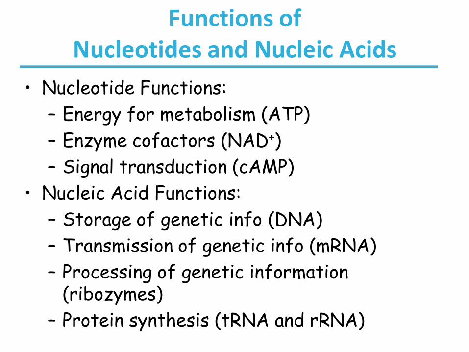
Functions of Nucleotides and Nucleic Acids
• Nucleotide Functions:
– Energy for metabolism (ATP)
– Enzyme cofactors (NAD+)
– Signal transduction (cAMP)
• Nucleic Acid Functions:
– Storage of genetic info (DNA)
– Transmission of genetic info (mRNA)
– Processing of genetic information (ribozymes)
– Protein synthesis (tRNA and rRNA)
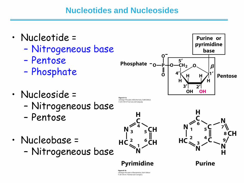
Nucleotides and Nucleosides
• Nucleotide = – Nitrogeneous base – Pentose – Phosphate
• Nucleoside = – Nitrogeneous base – Pentose
• Nucleobase = – Nitrogeneous base

Phosphate Group
• Negatively charged at neutral pH
• Typically attached to 5’ position
– Nucleic acids are built using 5’-
triphosphates
• ATP, GTP, TTP, CTP
– Nucleic acids contain one phosphate
moiety per nucleotide
• May be attached to other positions
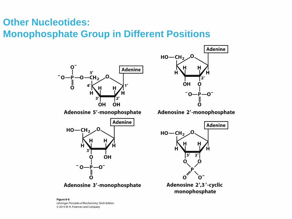
Other Nucleotides:
Monophosphate Group in Different Positions
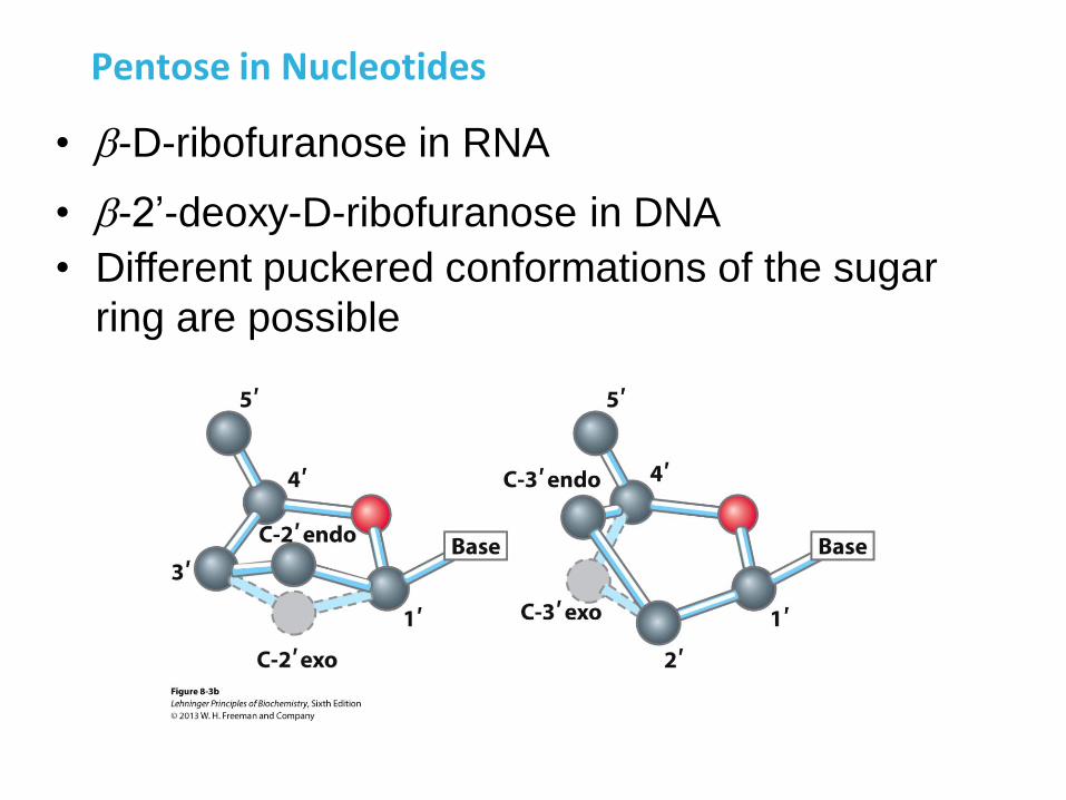
Pentose in Nucleotides
• -D-ribofuranose in RNA
• -2’-deoxy-D-ribofuranose in DNA
• Different puckered conformations of the sugar
ring are possible
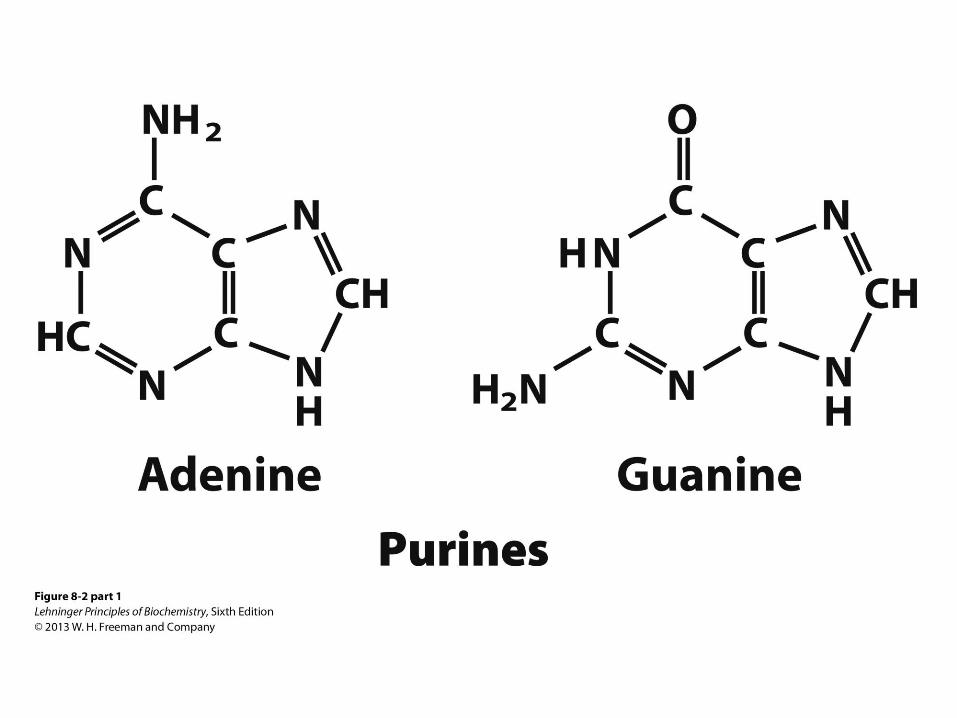
Purine Bases
• Adenine and guanine are found in both
RNA and DNA
• Also good H-bond donors and acceptors
• Adenine pKa at N1 is 3.8
• Guanine pKa at N7 is 2.4
• Neutral molecules at pH 7
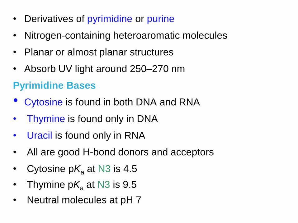
• Derivatives of pyrimidine or purine
• Nitrogen-containing heteroaromatic molecules
• Planar or almost planar structures
• Absorb UV light around 250–270 nm
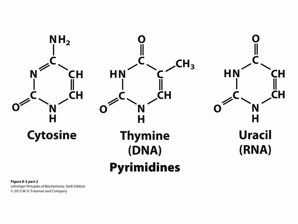
Pyrimidine Bases
• Cytosine is found in both DNA and RNA
• Thymine is found only in DNA
• Uracil is found only in RNA
• All are good H-bond donors and acceptors
• Cytosine pKa at N3 is 4.5
• Thymine pKa at N3 is 9.5
• Neutral molecules at pH 7
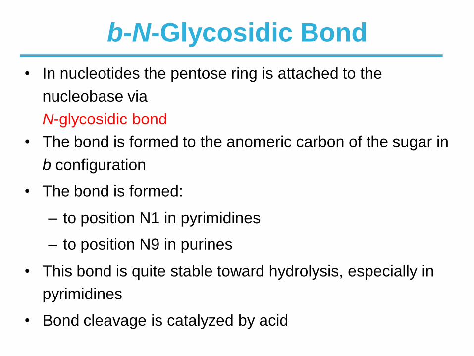
b-N-Glycosidic Bond
• In nucleotides the pentose ring is attached to the
nucleobase via
N-glycosidic bond
• The bond is formed to the anomeric carbon of the sugar in
b configuration
• The bond is formed:
– to position N1 in pyrimidines
– to position N9 in purines
• This bond is quite stable toward hydrolysis, especially in
pyrimidines
• Bond cleavage is catalyzed by acid
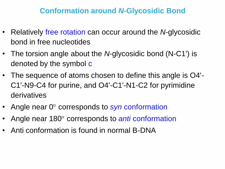
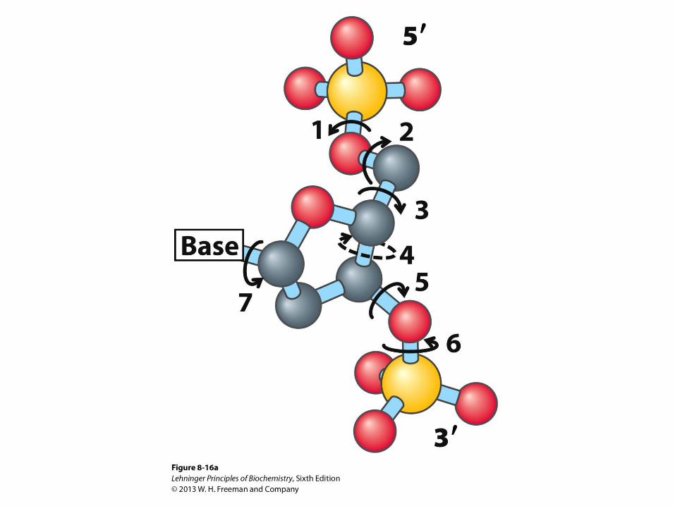
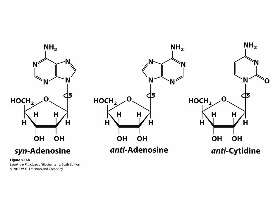
Conformation around N-Glycosidic Bond
• Relatively free rotation can occur around the N-glycosidic
bond in free nucleotides
• The torsion angle about the N-glycosidic bond (N-C1') is
denoted by the symbol c
• The sequence of atoms chosen to define this angle is O4'-
C1'-N9-C4 for purine, and O4'-C1'-N1-C2 for pyrimidine
derivatives
• Angle near 0 corresponds to syn conformation
• Angle near 180 corresponds to anti conformation
• Anti conformation is found in normal B-DNA
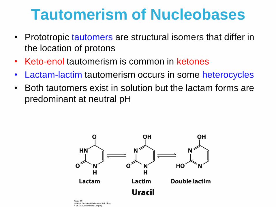
Tautomerism of Nucleobases
• Prototropic tautomers are structural isomers that differ in
the location of protons
• Keto-enol tautomerism is common in ketones
• Lactam-lactim tautomerism occurs in some heterocycles
• Both tautomers exist in solution but the lactam forms are
predominant at neutral pH
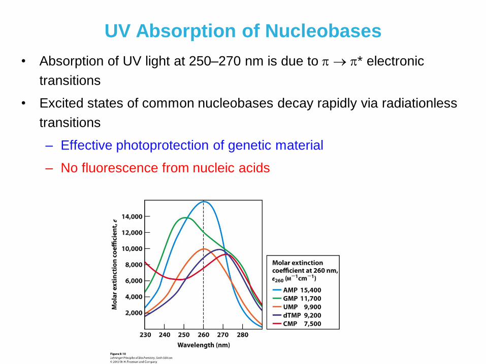
UV Absorption of Nucleobases
• Absorption of UV light at 250–270 nm is due to * electronic
transitions
• Excited states of common nucleobases decay rapidly via radiationless
transitions
– Effective photoprotection of genetic material
– No fluorescence from nucleic acids
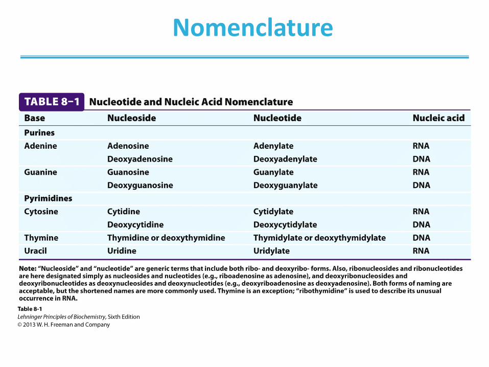
Nomenclature
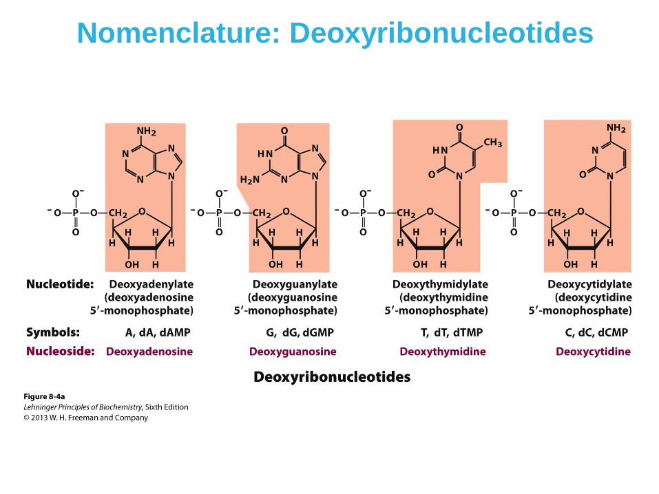
Nomenclature: Deoxyribonucleotides
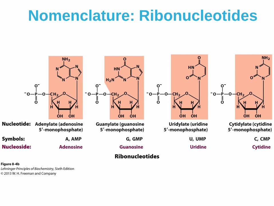
Nomenclature: Ribonucleotides
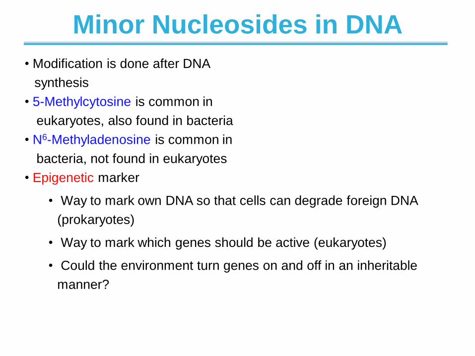
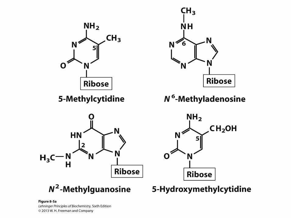
Minor Nucleosides in DNA
• Modification is done after DNA
synthesis
• 5-Methylcytosine is common in
eukaryotes, also found in bacteria
• N6-Methyladenosine is common in
bacteria, not found in eukaryotes
• Epigenetic marker
• Way to mark own DNA so that cells can degrade foreign DNA
(prokaryotes)
• Way to mark which genes should be active (eukaryotes)
• Could the environment turn genes on and off in an inheritable
manner?
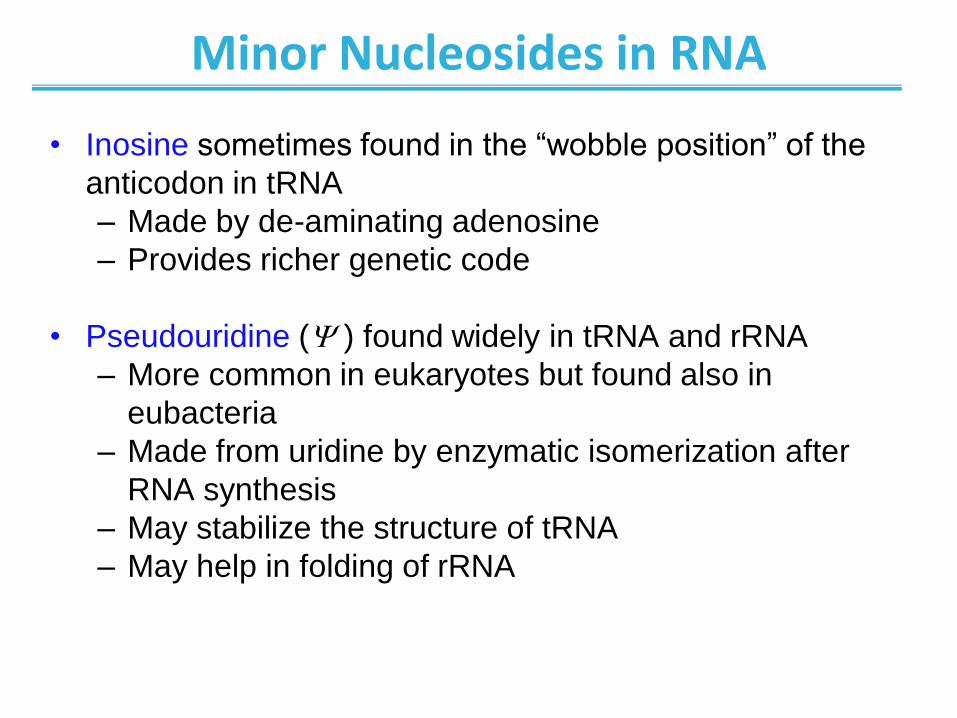
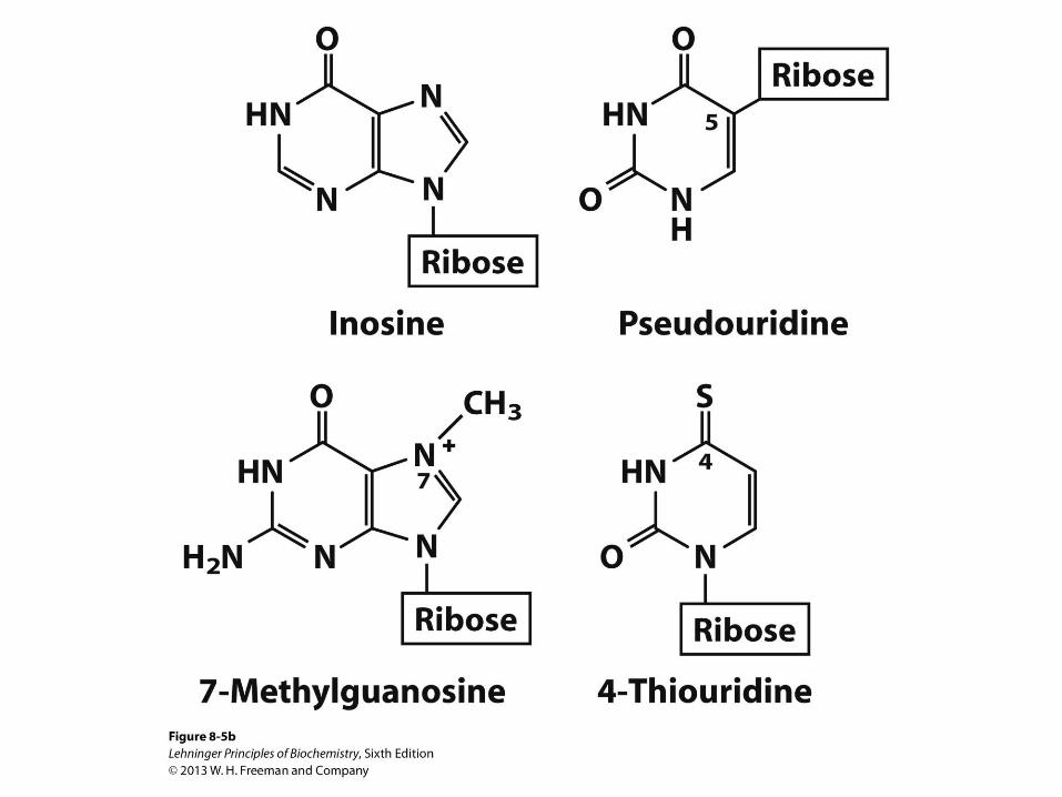
Minor Nucleosides in RNA
• Inosine sometimes found in the “wobble position” of the
anticodon in tRNA
– Made by de-aminating adenosine
– Provides richer genetic code
• Pseudouridine ( ) found widely in tRNA and rRNA
– More common in eukaryotes but found also in
eubacteria
– Made from uridine by enzymatic isomerization after
RNA synthesis
– May stabilize the structure of tRNA
– May help in folding of rRNA
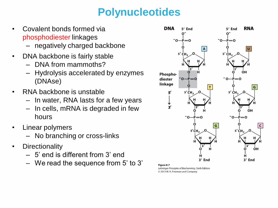
Polynucleotides
• Covalent bonds formed via
phosphodiester linkages
– negatively charged backbone
• DNA backbone is fairly stable
– DNA from mammoths?
– Hydrolysis accelerated by enzymes
(DNAse)
• RNA backbone is unstable
– In water, RNA lasts for a few years
– In cells, mRNA is degraded in few
hours
• Linear polymers
– No branching or cross-links
• Directionality
– 5’ end is different from 3’ end
– We read the sequence from 5’ to 3’
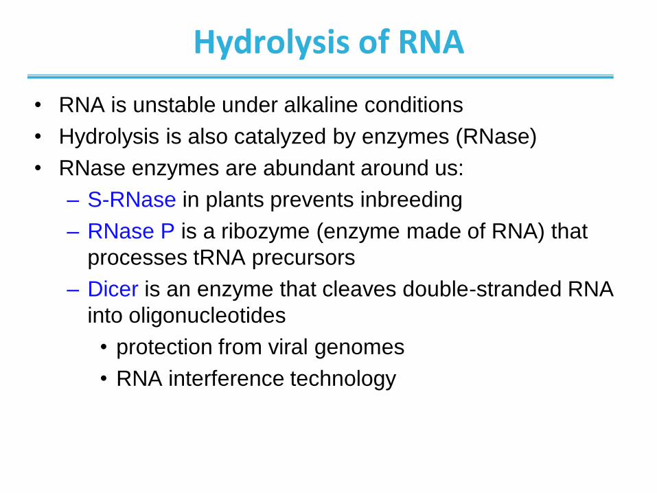
Hydrolysis of RNA
• RNA is unstable under alkaline conditions
• Hydrolysis is also catalyzed by enzymes (RNase)
• RNase enzymes are abundant around us:
– S-RNase in plants prevents inbreeding
– RNase P is a ribozyme (enzyme made of RNA) that
processes tRNA precursors
– Dicer is an enzyme that cleaves double-stranded RNA
into oligonucleotides
• protection from viral genomes
• RNA interference technology
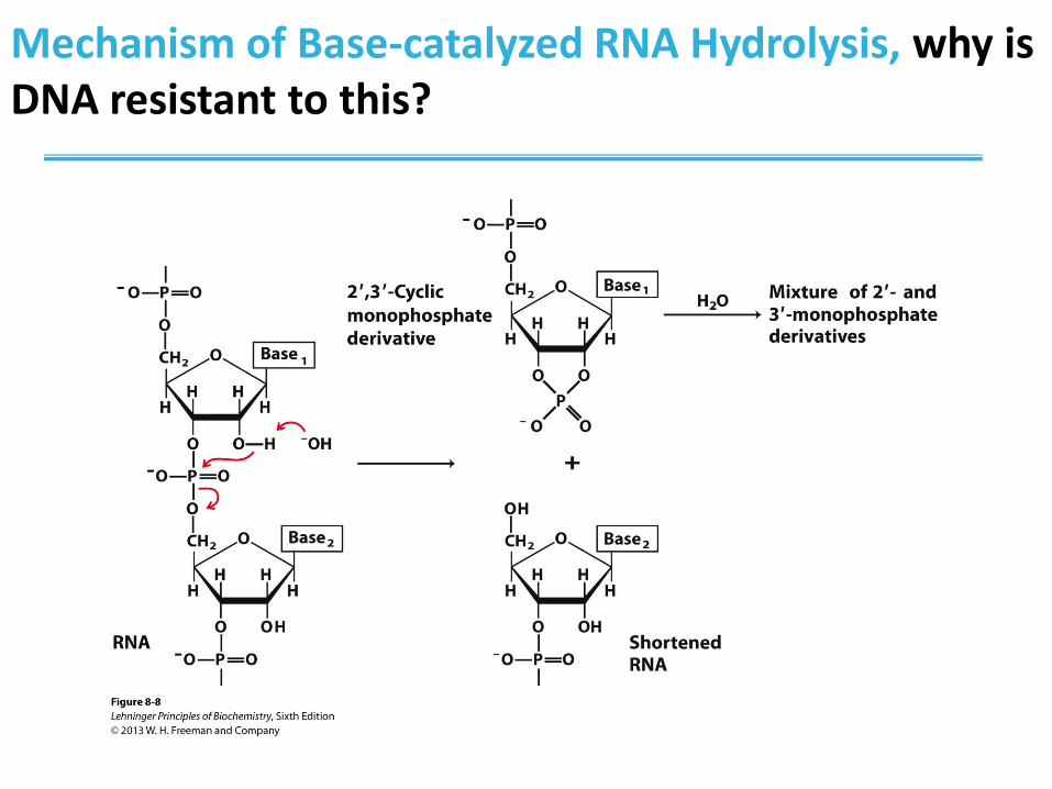
Mechanism of Base-catalyzed RNA Hydrolysis, why is DNA resistant to this?
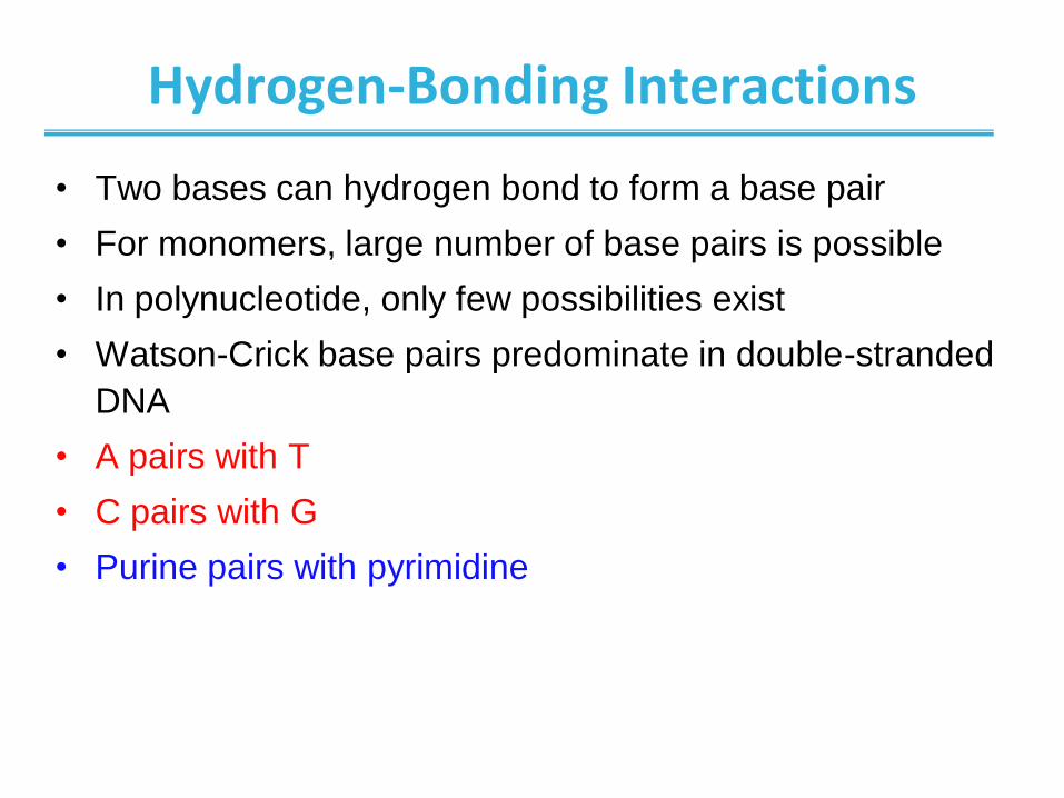
Hydrogen-Bonding Interactions
• Two bases can hydrogen bond to form a base pair
• For monomers, large number of base pairs is possible
• In polynucleotide, only few possibilities exist
• Watson-Crick base pairs predominate in double-stranded
DNA
• A pairs with T
• C pairs with G
• Purine pairs with pyrimidine
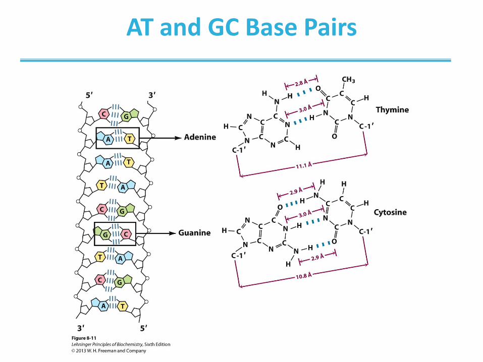
AT and GC Base Pairs
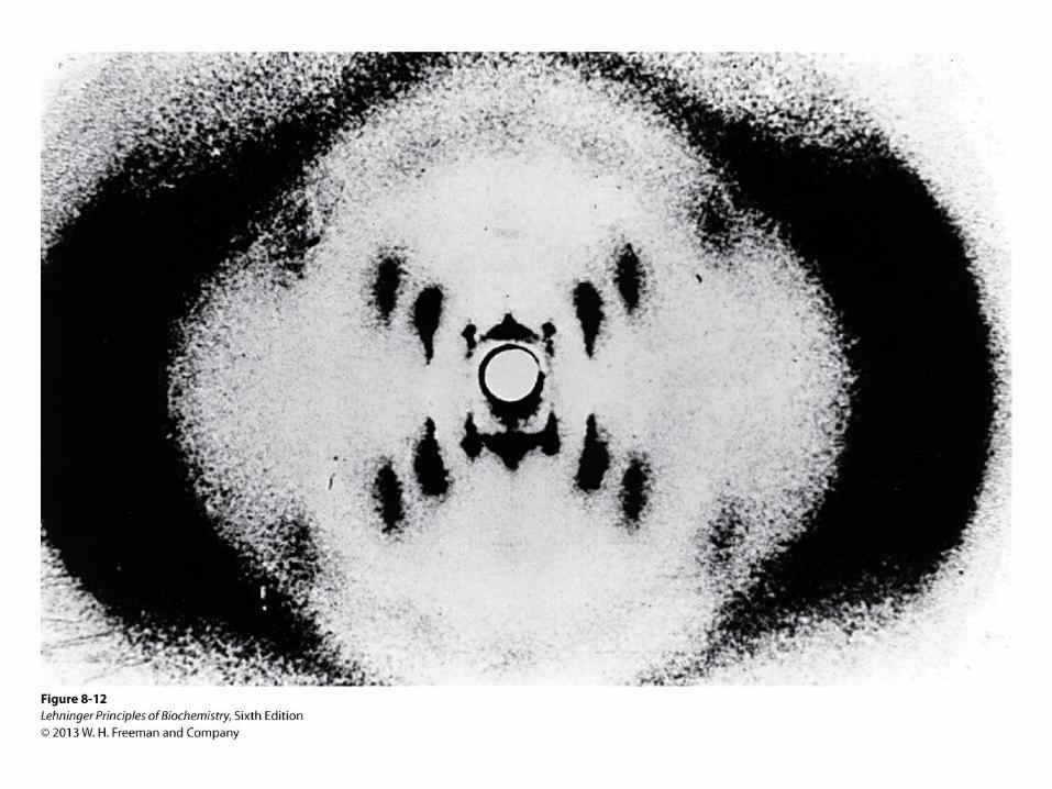
Discovery of DNA Structure
• One of the most important discoveries in biology
• Why is this important?
“This structure has novel features which are of considerable
biological interest” ―Watson and Crick, Nature, 1953
• Good illustration of science in action –Missteps in the path to a discovery
–Value of knowledge
–Value of collaboration
–Cost of sharing your data too early
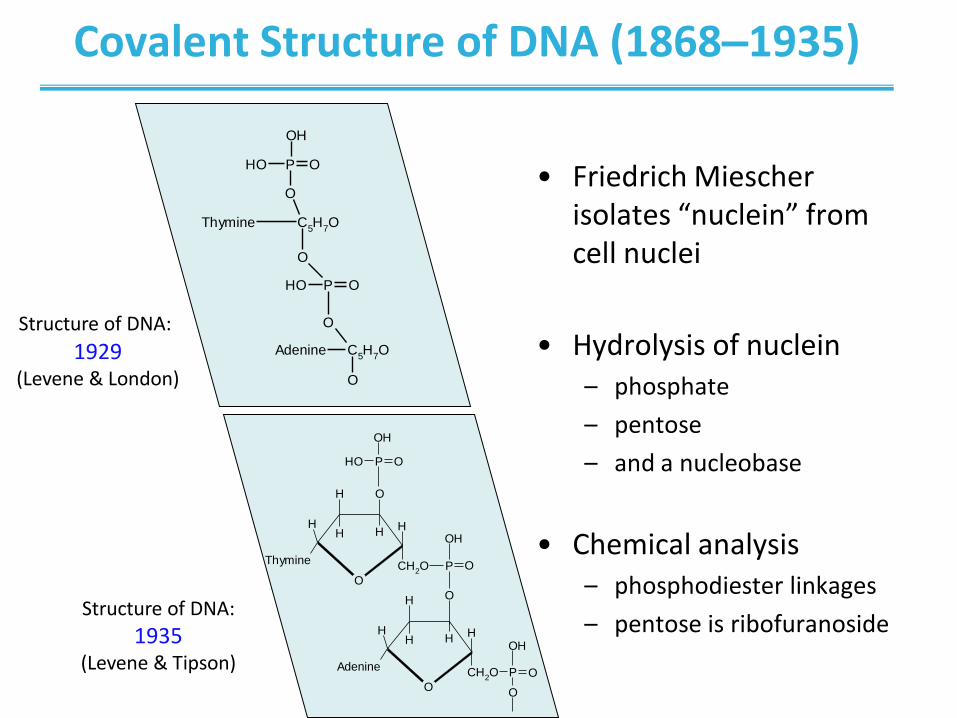
Covalent Structure of DNA (1868–1935)
• Friedrich Miescher isolates “nuclein” from cell nuclei
• Hydrolysis of nuclein – phosphate
– pentose
– and a nucleobase
• Chemical analysis – phosphodiester linkages
– pentose is ribofuranoside
O
OH
H HH
Thymine
H
CH2O P
OH
O
O
P OOH
OH
O
H
H HH
Adenine
H
CH2O P
OH
O
O
Structure of DNA: 1929
(Levene & London)
Structure of DNA:
1935 (Levene & Tipson)
C5H
7OThymine
O
P
O
POH
OH
O
O
O
OH
C5H
7OAdenine
O

Road to the Double Helix
• Franklin and Wilkins
–“Cross” means helix
–“Diamonds” mean
that the phosphate-
sugar backbone
is outside
– Calculated helical
parameters
• Watson and Crick
– Missing layer means
alternating pattern
(major & minor groove)
– Hydrogen bonding:
A pairs with T
G pairs with C
Double helix fits the data!
Watson, Crick, and Wilkins shared
1962 Nobel Prize Franklin died in 1958
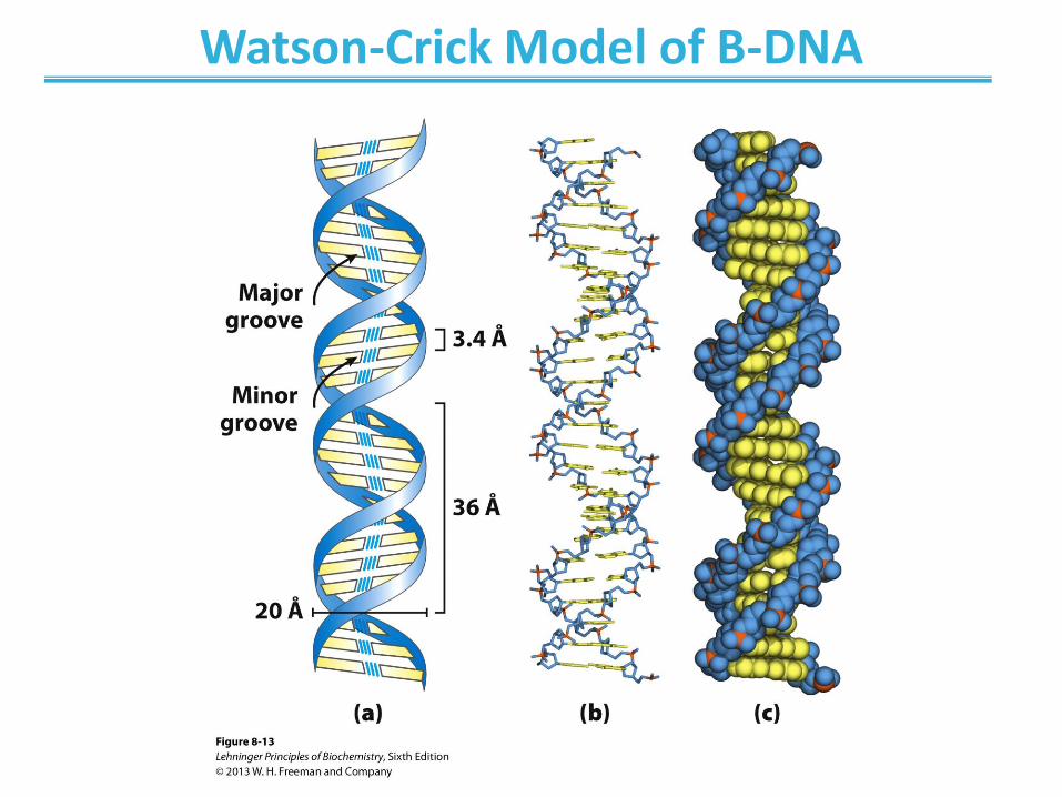
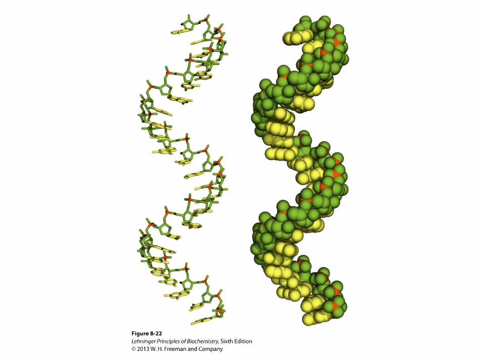
Watson-Crick Model of B-DNA
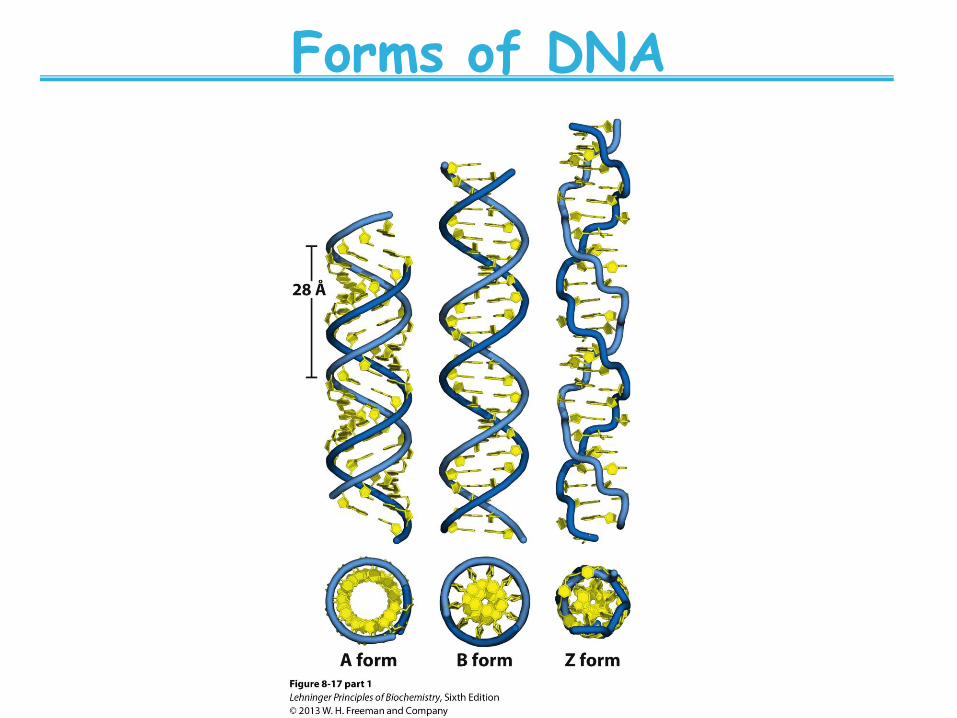
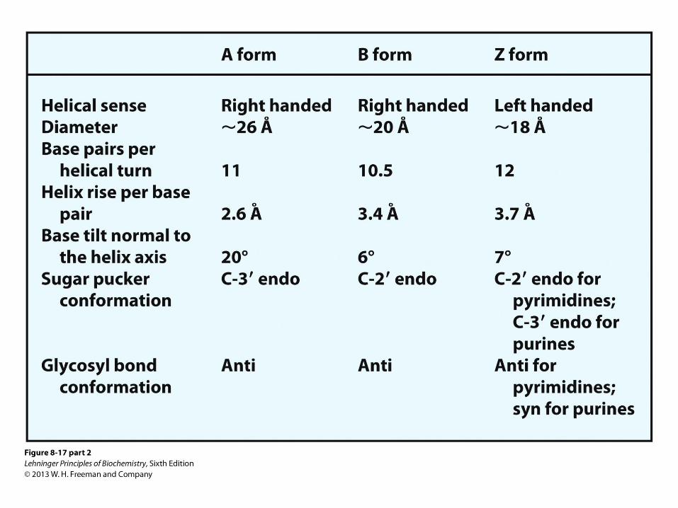
Forms of DNA
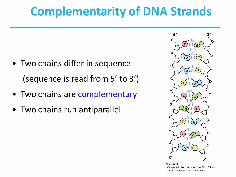
Complementarity of DNA Strands
• Two chains differ in sequence
(sequence is read from 5’ to 3’)
• Two chains are complementary
• Two chains run antiparallel
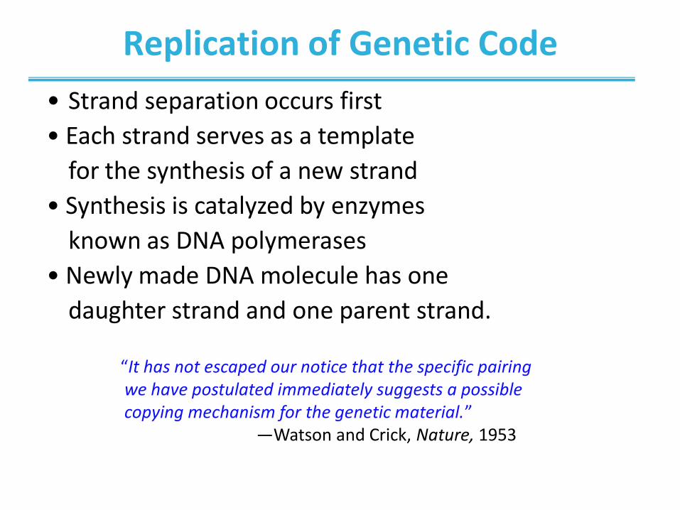
Replication of Genetic Code
“It has not escaped our notice that the specific pairing we have postulated immediately suggests a possible copying mechanism for the genetic material.” ―Watson and Crick, Nature, 1953
• Strand separation occurs first
• Each strand serves as a template
for the synthesis of a new strand
• Synthesis is catalyzed by enzymes
known as DNA polymerases
• Newly made DNA molecule has one
daughter strand and one parent strand.
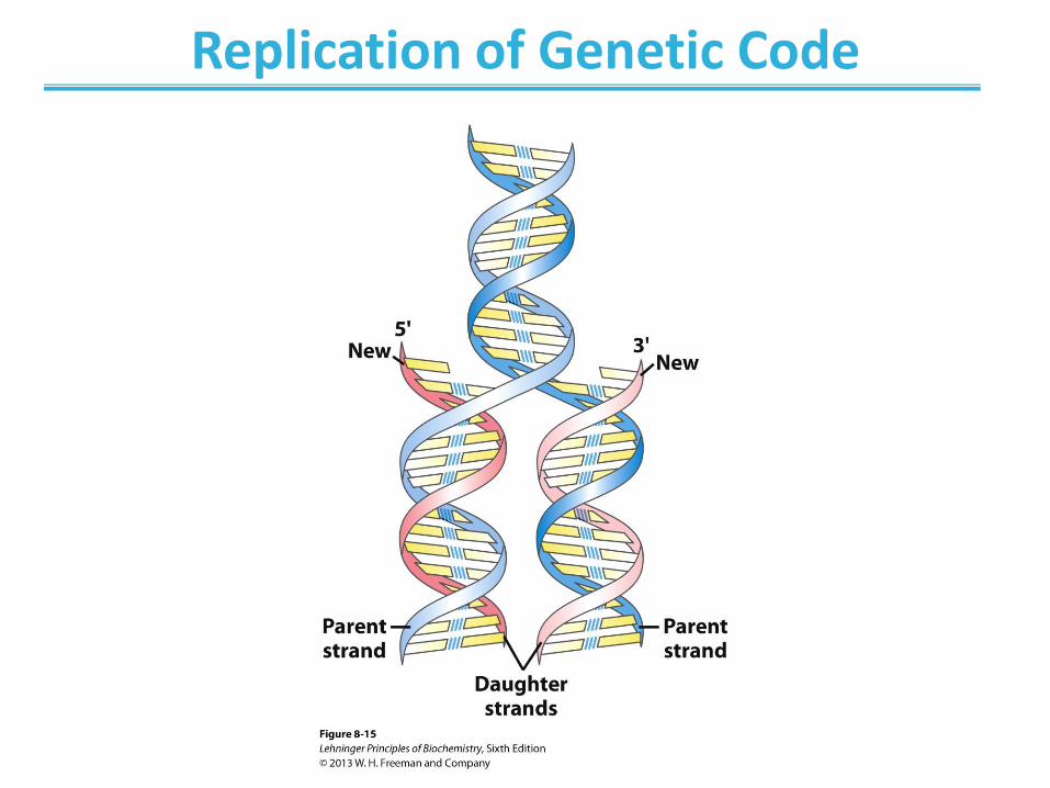
Replication of Genetic Code
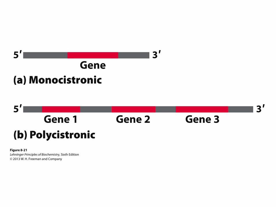
Messenger RNA: Code Carrier for the Sequence of Proteins
• Is synthesized using DNA template
• Contains ribose instead of deoxyribose
• Contains uracil instead of thymine
• One mRNA may code for more than one protein
• Together with transfer RNA (tRNA) transfers
genetic information from DNA to proteins
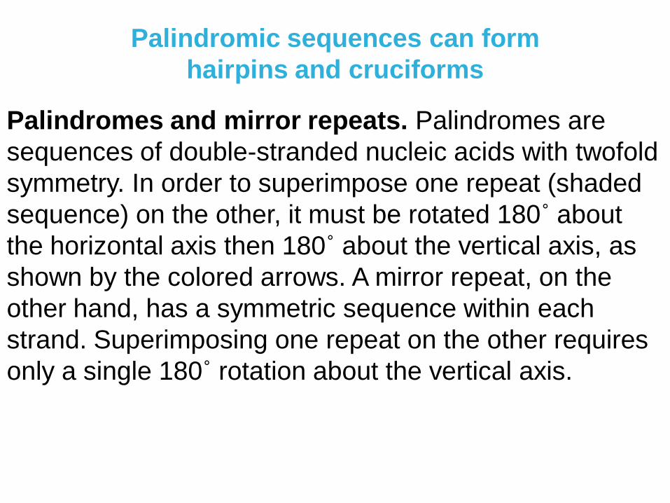
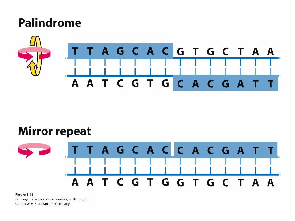
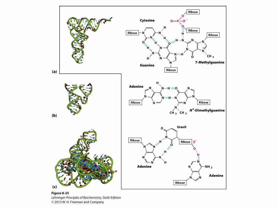
Palindromic sequences can form
hairpins and cruciforms
Palindromes and mirror repeats. Palindromes are
sequences of double-stranded nucleic acids with twofold
symmetry. In order to superimpose one repeat (shaded
sequence) on the other, it must be rotated 180˚ about
the horizontal axis then 180˚ about the vertical axis, as
shown by the colored arrows. A mirror repeat, on the
other hand, has a symmetric sequence within each
strand. Superimposing one repeat on the other requires
only a single 180˚ rotation about the vertical axis.
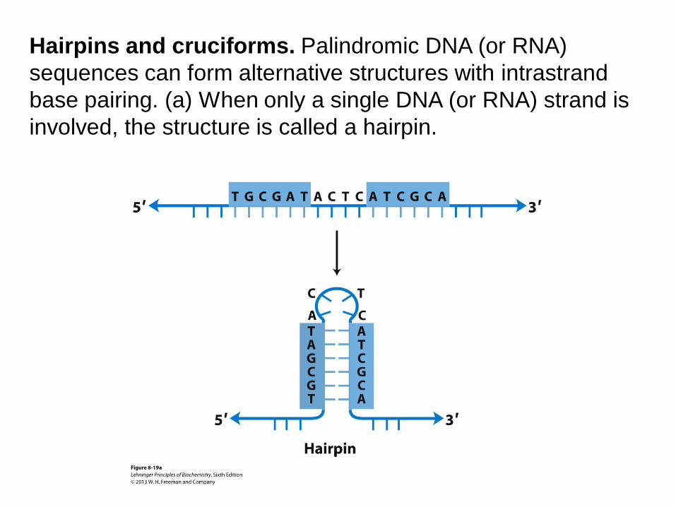
Hairpins and cruciforms. Palindromic DNA (or RNA)
sequences can form alternative structures with intrastrand
base pairing. (a) When only a single DNA (or RNA) strand is
involved, the structure is called a hairpin.
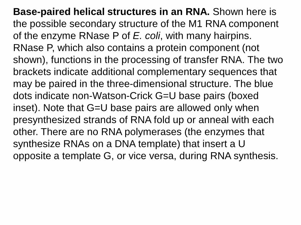
Base-paired helical structures in an RNA. Shown here is
the possible secondary structure of the M1 RNA component
of the enzyme RNase P of E. coli, with many hairpins.
RNase P, which also contains a protein component (not
shown), functions in the processing of transfer RNA. The two
brackets indicate additional complementary sequences that
may be paired in the three-dimensional structure. The blue
dots indicate non-Watson-Crick G=U base pairs (boxed
inset). Note that G=U base pairs are allowed only when
presynthesized strands of RNA fold up or anneal with each
other. There are no RNA polymerases (the enzymes that
synthesize RNAs on a DNA template) that insert a U
opposite a template G, or vice versa, during RNA synthesis.
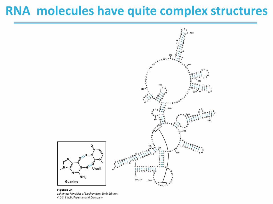
RNA molecules have quite complex structures
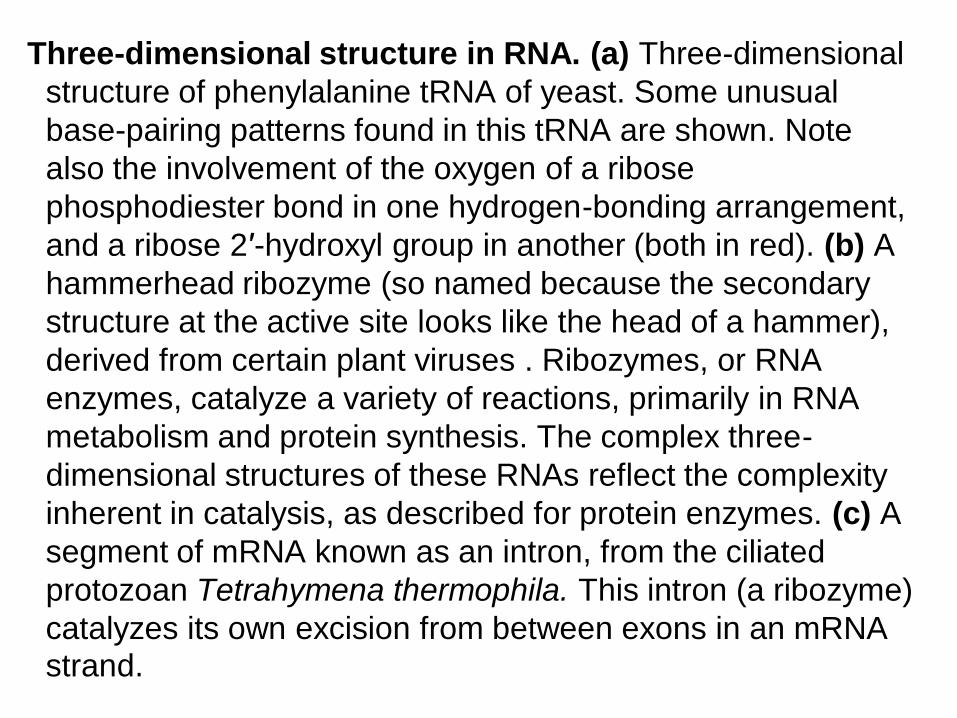
Three-dimensional structure in RNA. (a) Three-dimensional
structure of phenylalanine tRNA of yeast. Some unusual
base-pairing patterns found in this tRNA are shown. Note
also the involvement of the oxygen of a ribose
phosphodiester bond in one hydrogen-bonding arrangement,
and a ribose 2′-hydroxyl group in another (both in red). (b) A
hammerhead ribozyme (so named because the secondary
structure at the active site looks like the head of a hammer),
derived from certain plant viruses . Ribozymes, or RNA
enzymes, catalyze a variety of reactions, primarily in RNA
metabolism and protein synthesis. The complex three-
dimensional structures of these RNAs reflect the complexity
inherent in catalysis, as described for protein enzymes. (c) A
segment of mRNA known as an intron, from the ciliated
protozoan Tetrahymena thermophila. This intron (a ribozyme)
catalyzes its own excision from between exons in an mRNA strand.
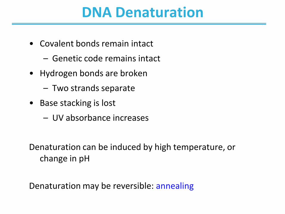
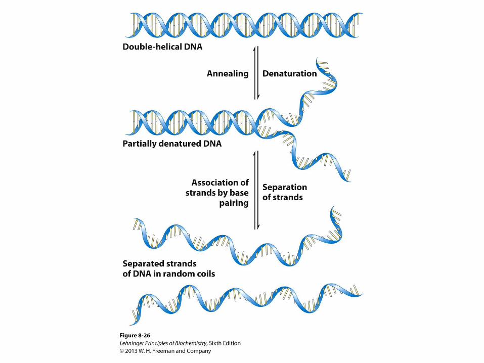
DNA Denaturation
• Covalent bonds remain intact
– Genetic code remains intact
• Hydrogen bonds are broken
– Two strands separate
• Base stacking is lost
– UV absorbance increases
Denaturation can be induced by high temperature, or change in pH
Denaturation may be reversible: annealing
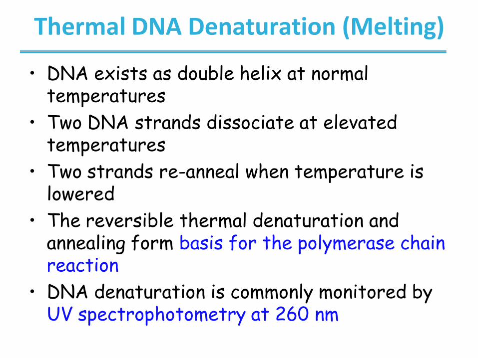
Thermal DNA Denaturation (Melting)
• DNA exists as double helix at normal temperatures
• Two DNA strands dissociate at elevated temperatures
• Two strands re-anneal when temperature is lowered
• The reversible thermal denaturation and annealing form basis for the polymerase chain reaction
• DNA denaturation is commonly monitored by UV spectrophotometry at 260 nm
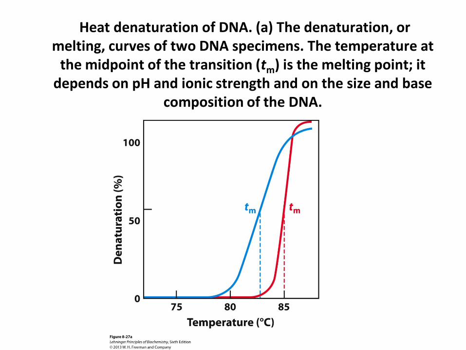
Heat denaturation of DNA. (a) The denaturation, or melting, curves of two DNA specimens. The temperature at
the midpoint of the transition (tm) is the melting point; it depends on pH and ionic strength and on the size and base
composition of the DNA.
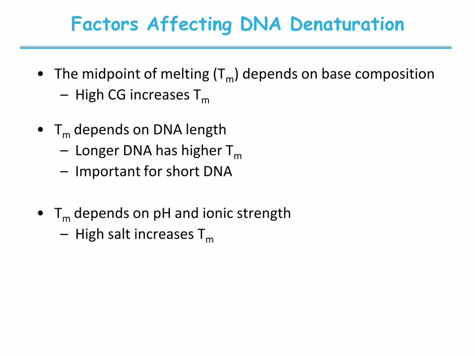
Factors Affecting DNA Denaturation
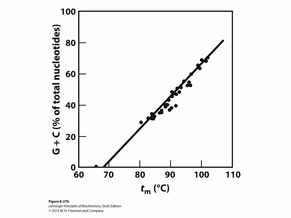
• The midpoint of melting (Tm) depends on base composition
– High CG increases Tm
• Tm depends on DNA length
– Longer DNA has higher Tm
– Important for short DNA
• Tm depends on pH and ionic strength
– High salt increases Tm
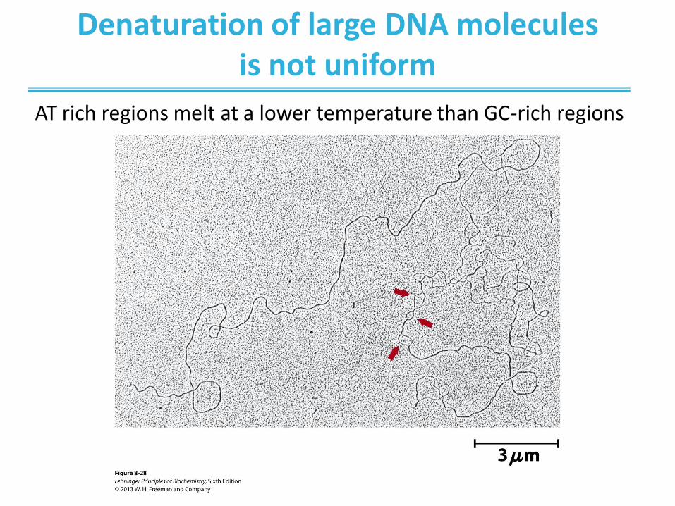
Denaturation of large DNA molecules is not uniform
AT rich regions melt at a lower temperature than GC-rich regions
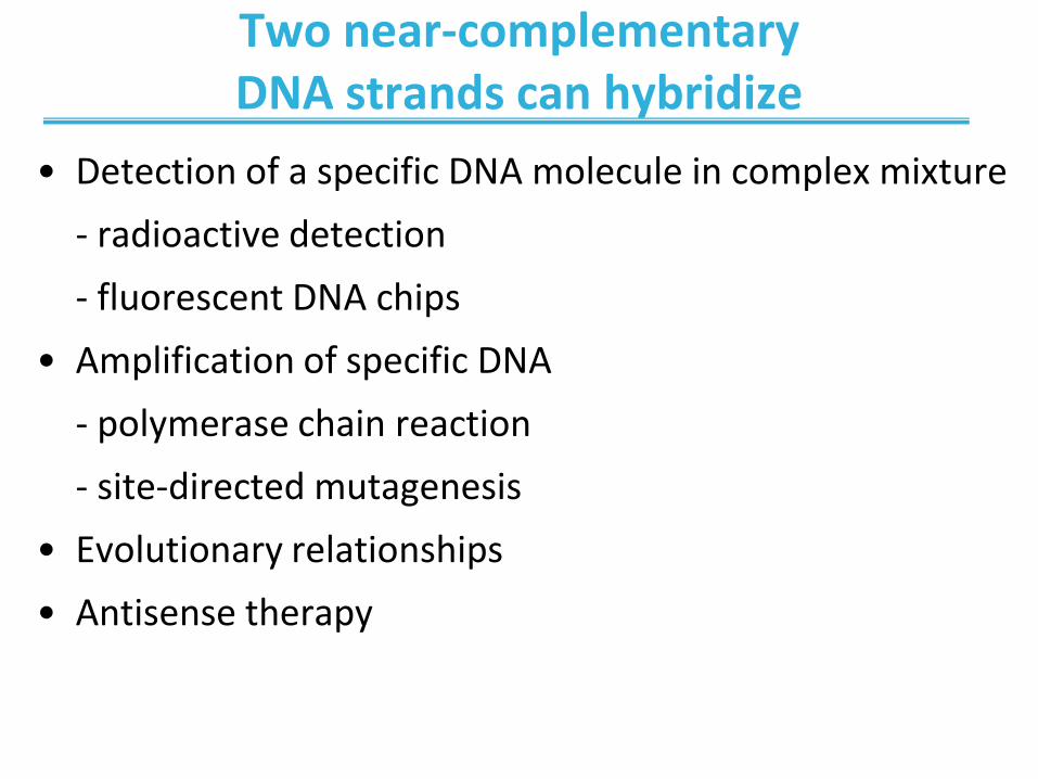
Two near-complementary DNA strands can hybridize
• Detection of a specific DNA molecule in complex mixture
- radioactive detection
- fluorescent DNA chips
• Amplification of specific DNA
- polymerase chain reaction
- site-directed mutagenesis
• Evolutionary relationships
• Antisense therapy
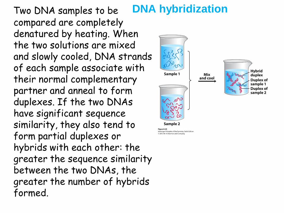
DNA hybridization Two DNA samples to be compared are completely denatured by heating. When the two solutions are mixed and slowly cooled, DNA strands of each sample associate with their normal complementary partner and anneal to form duplexes. If the two DNAs have significant sequence similarity, they also tend to form partial duplexes or hybrids with each other: the greater the sequence similarity between the two DNAs, the greater the number of hybrids formed.
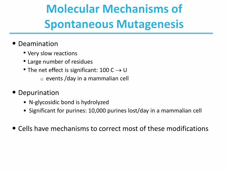
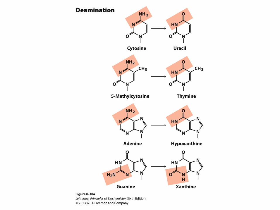
Molecular Mechanisms of Spontaneous Mutagenesis
• Deamination • Very slow reactions
• Large number of residues
• The net effect is significant: 100 C U
o events /day in a mammalian cell
• Depurination • N-glycosidic bond is hydrolyzed
• Significant for purines: 10,000 purines lost/day in a mammalian cell
• Cells have mechanisms to correct most of these modifications

Some well-characterized nonenzymatic reactions of
nucleotides.
Depurination, in which a purine is lost by hydrolysis of the N-β-glycosyl bond. Loss of pyrimidines via a similar reaction occurs, but much more slowly. The resulting lesion, in which the deoxyribose is
present but the base is not, is called an abasic site or an AP site (apurinic site or, rarely, apyrimidinic site). The deoxyribose remaining after depurination is readily converted from the β-furanose to the aldehyde form (see Fig. 8–3), further destabilizing the DNA at this position.
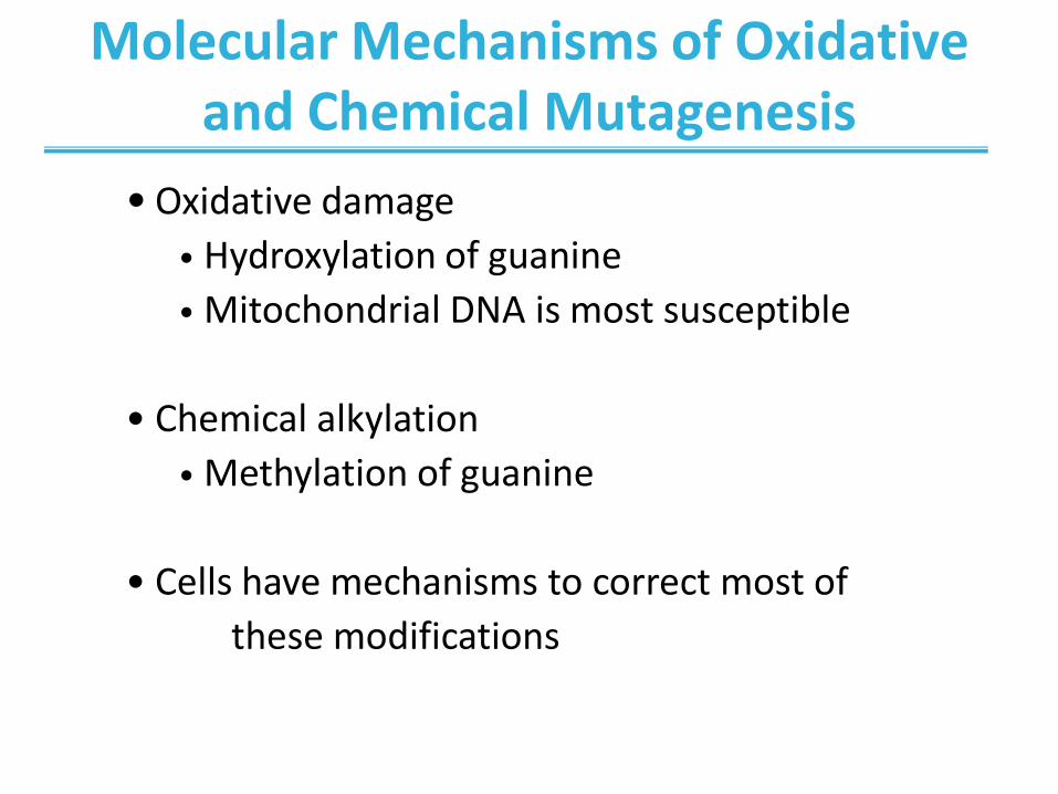
Molecular Mechanisms of Oxidative and Chemical Mutagenesis
• Oxidative damage
• Hydroxylation of guanine
• Mitochondrial DNA is most susceptible
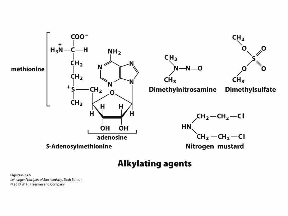
• Chemical alkylation
• Methylation of guanine
• Cells have mechanisms to correct most of
these modifications
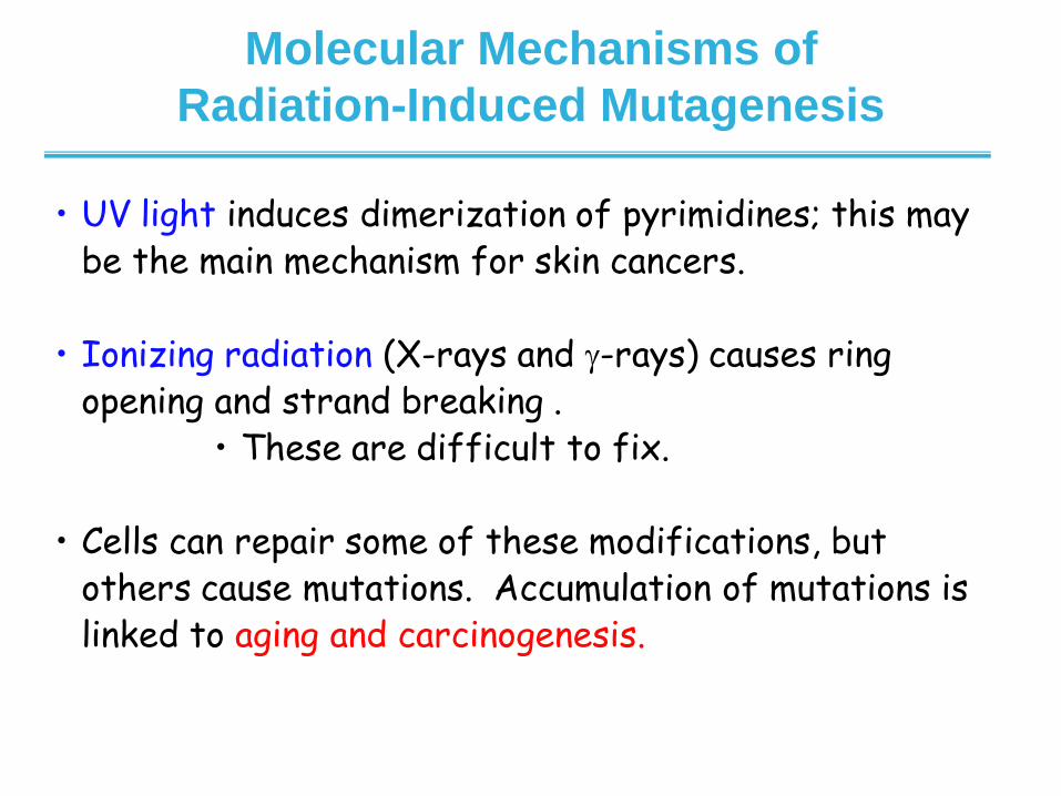
• UV light induces dimerization of pyrimidines; this may be the main mechanism for skin cancers.
• Ionizing radiation (X-rays and -rays) causes ring
opening and strand breaking . • These are difficult to fix.
• Cells can repair some of these modifications, but
others cause mutations. Accumulation of mutations is linked to aging and carcinogenesis.
Molecular Mechanisms of
Radiation-Induced Mutagenesis
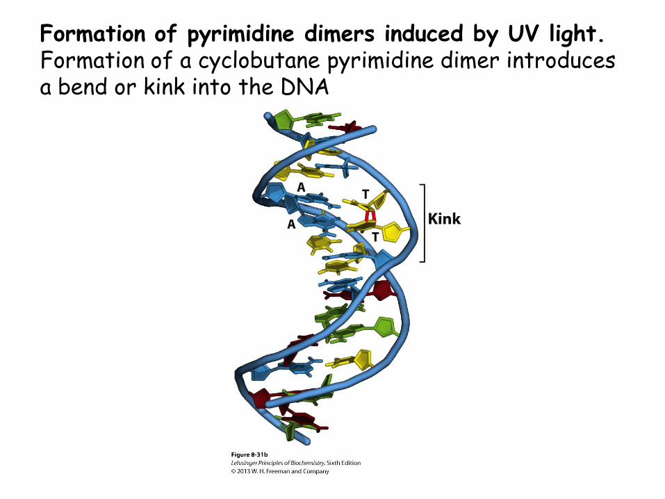
Formation of pyrimidine dimers induced by UV light. Formation of a cyclobutane pyrimidine dimer introduces a bend or kink into the DNA
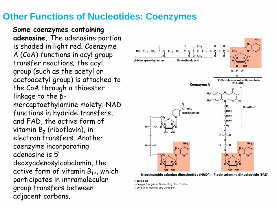
Other Functions of Nucleotides: Coenzymes
Some coenzymes containing adenosine. The adenosine portion is shaded in light red. Coenzyme A (CoA) functions in acyl group transfer reactions; the acyl group (such as the acetyl or acetoacetyl group) is attached to the CoA through a thioester linkage to the β-mercaptoethylamine moiety. NAD functions in hydride transfers, and FAD, the active form of vitamin B2 (riboflavin), in electron transfers. Another coenzyme incorporating adenosine is 5’-deoxyadenosylcobalamin, the active form of vitamin B12, which participates in intramolecular group transfers between adjacent carbons.