
Nuclease cleavage rRNA Operon 7 copies/genome in E. coli.
57
-
date post
22-Dec-2015 -
Category
Documents
-
view
272 -
download
2
Transcript of Nuclease cleavage rRNA Operon 7 copies/genome in E. coli.


Nuclease cleavage
rRNA Operon7 copies/genome in E. coli

CCA Nucleotidyl transferase(template independent)
Splicing
Nuclease cleavage

A selection of the modified nucleosides that occur in tRNAs together with their standard abbreviations.


Three-dimensional structure of yeast tRNAPhe deduced from x-ray diffraction analysis. The shape resembles a twisted L.
CCA

Figure 32-12 Tertiary base pairing interactions in yeast tRNAPhe.
Page
129
6

A selection of the modified nucleosides that occur in tRNAs together with their standard abbreviations.
Pag
e 12
94

Molecular Recognition of Codons in mRNA by tRNA
• The codon sequence is complementary with the anticodon sequence
• The codon in mRNA base pairs with the anticodon in mRNA via hydrogen bonding
• The alignment of two RNA segments is antiparallel

Inosinate in the Anticodon in Some tRNAs is “Wobble”
• Inosinate can hydrogen bond with three different nucleotides (A, U, C)
• This interaction is weaker than typical Watson Crick base pairing


Allowed Wobble Pairing Combinations in the Third Codon–Anticodon Position.

Wobble pairing. U · G and I · A wobble pairs. Both have been observed in X-ray structures.
WOBBLE ONLY OCCURS IN POSITION
3! !!!!!!!!!!!!!!!!!!!!!!

Pairing relationship of codon and anticodon

mRNA/tRNA binding is antiparallel
• 5’
5’ 3’mRNA
tRNA
CCA-amino acid (3’end)5’end
Codon: 1 2 3
Anticodon 3 2 1
Anticodon 3’---GUI----5’ Codon 5’----CAU----3’
Wobble base

Protein Synthesis Involves Five Stages
• Activation of amino acids– Enzymatic synthesis of aminoacyl tRNA molecules
• Initiation of translation– Binding of mRNA and N-formylmethionine to ribosome
• Elongation– Binding of aminoacyl tRNAs to ribosome
– Formation of peptide bonds
• Termination and ribosome recycling– Termination codon in mRNA reaches ribosome
• Folding and post-translational processing– Catalyzed by a variety of enzymes

Synthesis of Aminoacylated tRNAs: Aminoacyl AMP
Aminoacyl-tRNA Synthetases
• Each enzyme binds a specific amino acid and the matching tRNA
• Most cells contain twenty different aminoacyl-tRNA synthetases, one for each amino acid
• Some cells contain less than 20 synthetases; in this case one amino acid is converted to another after charging the tRNA


Aminoacylation of tRNA by aminoacyl-tRNA synthetases.
Step 1 is formation of an aminoacyl adenylate, which remains bound to the active site.
What does this remind you of??

In step 2, the aminoacyl group is transferred to the
tRNA. The mechanism of this step is somewhat
different for the two classes of
aminoacyl-tRNA synthetases.
Class IClass II

The Second Genetic Code
• Matching each amino acid with correct tRNA can be viewed as the “second genetic code”
• The “code” is in molecular recognition of a specific tRNA molecule by a specific synthetase
• Only a few nucleotides in tRNA confer the binding specificity– Anticodon region
– Other regions (G•U in Ala-tRNA)

Structural elements of tRNAAla that are required for recognition by Ala-tRNA synthetase. (a) The tRNAAla structural elements recognized by the Ala-tRNA synthetase are unusually simple. A single G=U base pair (pink) is the only element needed for specific binding and aminoacylation. (b) A short synthetic RNA mini-helix, with the critical G=U base pair but lacking most of the remaining tRNA structure. This is aminoacylated specifically with alanine almost as efficiently as the complete tRNAAla.

Gln-tRNA synthetase from E. coli, a typical monomeric class I synthetase

X-Ray structure of E. coli GlnRS · tRNAGln · ATP. tRNA and ATP wireframe; tRNA sugar–phosphates, bases, ATP.
Pag
e 13
00

PDB molecule of the month

Rossmann fold (alternating αβ structures) from a decarboxylase

Figure 32-19a Comparison of the modes by which GlnRS and AspRS bind their cognate
tRNAs.
Page
130
2
(b) AspRS, a Class II synthetase.(a) GlnRS, a Class I synthetase.

Different Approaches to the Same ProblemIn this picture, five complexes of an aminoacyl-tRNA synthetase with tRNA are shown, aligned so that the tRNA molecules are in the same orientation. Notice that the enzymes approach the tRNA from different angles. The isoleucine, valine and glutamine enzymes cradle the tRNA, gripping the anticodon loop (at the bottom in each tRNA), and placing the amino-acid acceptor end of the tRNA in the active site (at the top right in each tRNA). These all share a similar protein framework, known as "Type I," approaching the tRNA similarly and adding the amino acid to the last 2' hydroxyl group in the tRNA.
The phenlyalanine and threonine enzymes are part of a second class of enzymes, known as "Type II." They approach the tRNA from the other side, and add the amino acid to the 3’ hydroxyl on the last tRNA base. Type I enzymes contain a Rossmann
fold in the catalytic domain

Cartoon comparison of the putative aminoacylation and editing modes of IleRS ·
tRNAIle.
Page
130
4
http://pubs.acs.org/doi/full/10.1021/ja9095208

Composition of E. coli Ribosome
• Ribosome is a Non-covalent Assembly of Many Proteins and a Few RNA Molecules


30S and 50S Subunits of Bacterial Ribosome
• Subunits are identified by their sedimentation coefficients (Svedberg units)
• A, P, and E sites are locations for binding of tRNA molecules


Ribosomal RNA Molecules have Complex Secondary Structures


22 Genetically Coded Amino Acids
• 20 genetically encoded amino acids are common in all organisms
• Selenocysteine is formed after charging an UGA(stop)-recognizing tRNA with serine in both bacteria and eukaryotes
• Pyrrolysine is directly attached to its tRNA that recognizes UAG(stop) codon by some archae

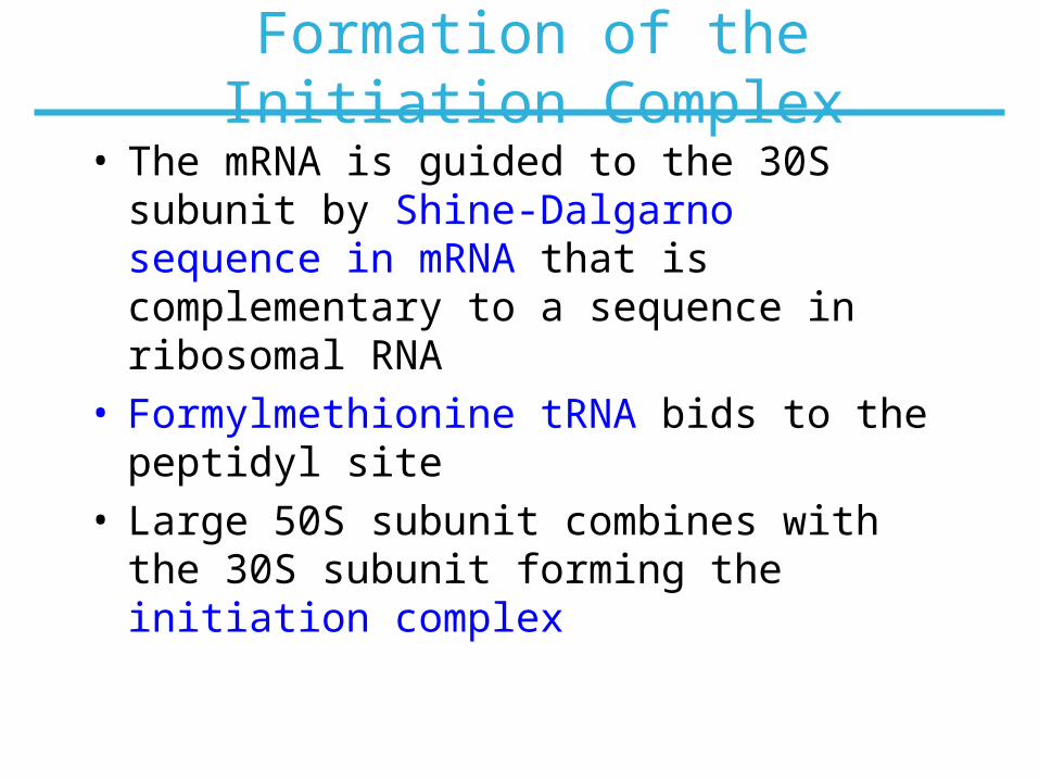
Formation of the Initiation Complex
• The mRNA is guided to the 30S subunit by Shine-Dalgarno sequence in mRNA that is complementary to a sequence in ribosomal RNA
• Formylmethionine tRNA bids to the peptidyl site
• Large 50S subunit combines with the 30S subunit forming the initiation complex


Several Protein Factors are Involved in Initiation


First Elongation Step
• Aminoacyl tRNA binds to the elongation factor Tu that also carries GTP
• The EF-Tu-GTP complex with second amino acid on its tRNA binds to the aminoacyl site
• After GTP hydrolysis EF-Tu-GDP leaves the ribosome


Formation of the Peptide Bond
• The 23S RNA ribozyme is the catalyst


Termination of Protein Synthesis
• When the stop codon (UAG) reaches ribosome a release factor binds to the A site
• The linkage between nascent polypeptide and tRNA in the P site is cleaved
• Protein, mRNA, and deacylatd tRNA dissociate from ribosome


Coupling of Transcription and Translation in Bacteria


Chapter 27: Summary
• The primary genetic code for protein synthesis is in triplets of nucleotides in mRNA that recognize the anticodon of tRNA
• The secondary genetic code for protein synthesis is in recognition features between tRNA and the enzyme that attaches the correct amino acids to this tRNA
• The mRNA binds to the ribosome and its codons are exposed to the aminoacyl-tRNA binding site
• The protein synthesis from charged aminoacyl tRNA substrates is catalyzed by a ribozyme in the ribosome
In this chapter, we learned that:


![The conserved Fanconi anemia nuclease Fan1 and the SUMO E3 … · 2017. 2. 23. · FAN1 (Fanconi anemia-associated nuclease 1, or FANCD2/FANCI-associated nuclease 1) [13–18]. Human](https://static.fdocuments.us/doc/165x107/60c9d965c710eb0d72008d0e/the-conserved-fanconi-anemia-nuclease-fan1-and-the-sumo-e3-2017-2-23-fan1-fanconi.jpg)
