

NTB-A, a new activating receptor in T cells that regulates ... · expansion and Th1-type cytokine...
40
NTB-A, a new activating receptor in T cells that regulates autoimmune disease Patricia A. Valdez 1 , Hua Wang 1 , Dhaya Seshasayee 1 , Menno Van Lookeren Campagne 1 , Austin Gurney 2 , Wyne P. Lee 1 , and Iqbal S. Grewal 1 1 Department of Immunology and 2 Molecular Biology, Genentech Inc. South San Francisco, CA 94080 Correspondence to: Iqbal S. Grewal Ph.D. Department of Immunology Genentech Inc. 1 DNA Way 1 JBC Papers in Press. Published on February 26, 2004 as Manuscript M312313200 Copyright 2004 by The American Society for Biochemistry and Molecular Biology, Inc. by guest on April 3, 2019 http://www.jbc.org/ Downloaded from
Transcript of NTB-A, a new activating receptor in T cells that regulates ... · expansion and Th1-type cytokine...

NTB-A, a new activating receptor in T cells that regulates
autoimmune disease
Patricia A. Valdez1, Hua Wang1, Dhaya Seshasayee1, Menno Van Lookeren Campagne1,
Austin Gurney2, Wyne P. Lee1, and Iqbal S. Grewal1
1Department of Immunology and 2Molecular Biology, Genentech Inc. South San Francisco, CA
94080
Correspondence to:
Iqbal S. Grewal Ph.D.
Department of Immunology
Genentech Inc.
1 DNA Way
1
JBC Papers in Press. Published on February 26, 2004 as Manuscript M312313200
Copyright 2004 by The American Society for Biochemistry and Molecular Biology, Inc.
by guest on April 3, 2019
http://ww
w.jbc.org/
Dow
nloaded from
South San Francisco, CA 94080
Tel: (650) 225 3239
Fax: (650) 225 8221
Email: [email protected]
Summary
The CD28 co-stimulatory pathway is well established for T cell activation; however, results
from CD28 -/- mice suggest the existence of additional co-stimulatory pathways. Here we
report the further characterization of a new member of the CD2 superfamily, NTB-A, important
in T cell co-stimulation. NTB-A is expressed on T cells and its expression is upregulated on
activated cells. Triggering of NTB-A with monoclonal antibodies in the absence of CD28
signals leads to T cell proliferation and IFN-γ secretion but not IL-4. Crosslinking of NTB-A
also induces phosphorylation of NTB-A and the association of SAP, the protein absent in X-
linked lymphoproliferative disease. T helper cells differentiated by crosslinking NTB-A and
CD3 developed predominately into Th1 cells, not Th2 cells. In vivo blocking of NTB-A
interactions with its ligands by using soluble NTB-A-Fc fusion protein inhibits B cell isotype
switching to IgG2a and IgG3, commonly induced by Th1-type cytokines. Importantly,
treatment of mice with NTB-A-Fc delays the onset of antigen-induced experimental allergic
encephalomyelitis (EAE) in MBP-TCR transgenic mice, suggesting a role in T cell mediated
autoimmune disease. Regulation of IFN-γ secretion, and not IL-4 in vitro, as well as inhibition
Th1 cell induced isotype switching and attenuation of EAE, indicates that NTB-A is important
for Th1 responses. The observation that crosslinking of NTB-A induces T cell activation,
2
by guest on April 3, 2019
http://ww
w.jbc.org/
Dow
nloaded from
expansion and Th1-type cytokine production suggests NTB-A is a novel co-stimulatory
receptor. The identification of NTB-A as a regulator of T cell response paves the way to provide
novel therapeutic approaches for modulation of the immune response.
3
by guest on April 3, 2019
http://ww
w.jbc.org/
Dow
nloaded from
Introduction
T cell activation requires two distinct signals: the first signal through the T cell receptor (TCR)
which maintains antigen specificity, and a second co-stimulatory signal, from another receptor
such as CD28 (1). Interaction of CD28 with its ligands B7.1 (CD80) and B7.2 (CD86) expressed
on activated antigen presenting cells (APCs) is thought to provide T cells with necessary signals
for proper activation and development into effector T cells. Although CD28 is considered a
primary receptor for the second signal, CD28 deficient mice can still mount an antigen-specific
T cell response, suggesting the existence of other co-stimulatory molecules (2,3). For instance,
CD8+ cytotoxic T cells are still functional in CD28 deficient mice and these mice also exhibit
delayed-type hypersensitivity in response to viral infection. Several other receptors in the CD28
family have been subsequently cloned in recent years. ICOS is a positive regulatory receptor in
the family, which can provide co-stimulatory signals to T cells, although ICOS does not activate
naïve T cells and is thought to be involved primarily in T helper cell development (4-6). The
current data in the field indicates that there may be other positive regulatory receptors yet to be
identified.
In addition to CD28 family members, other receptors such as those belonging to the CD2
superfamily can also provide the second signal for co-stimulation. Included in the CD2
superfamily are CD2 (LFA-2), CD48, CD58 (LFA-3), CD84, CD150 (SLAM), and CD244
(2B4) (7-14). The CD2 family of receptors and ligands are expressed throughout the immune
system and play an important role in immune responses. Although CD2 can act as a co-
stimulatory molecule on T cells, CD2 deficient mice exhibit normal T cell responses, suggesting
4
by guest on April 3, 2019
http://ww
w.jbc.org/
Dow
nloaded from
a redundant role for CD2 in T cell co-stimulation (15). The importance of CD2 becomes much
clearer when analyzed in the context of CD28 deficiency. CD2-/-/CD28-/- double knockout
mice exhibit a much more severe T cell activation defect when compared to CD28 deficient
mice, underscoring the important contribution of CD2 in T cell co-stimulation (16). Still, in the
absence of both of these receptors, T cells can be activated under certain circumstances. Clearly,
other molecules in addition to CD28 and CD2 are involved in early T cell activation.
Another CD2 superfamily member, CD150, can also provide a co-stimulatory signal to T
cells (10-12). CD150 is included in a subfamily of receptors that are characterized by
extracellular immunoglobulin-like repeats and intracellular binding sites for SAP (SH2D1A),
the protein associated with X-linked lymphoproliferative disease (XLP) (9,17-23). The basis
for the lymphoproliferation defect seen in XLP patients has been attributed to defects in T cell
and NK cell function, although there is no clear understanding of how SAP deficiency
contributes to this disease at this time. A better understanding of SAP will come from studying
the function of receptors associated with SAP. The most well-studied SAP-associated receptor
is CD150. CD150 can provide a co-stimulatory signal to T cells, inducing proliferation and
IFN-γ expression. There is also evidence for CD150 involvement in Th1 cell responses,
suggesting the importance of this molecule in inflammation and autoimmune disease (10-12,24).
Recently, CD84, another CD2/CD150 family member was found to possess co-stimulatory
activity on T cells (25). Clearly this family of receptors plays an important role in co-
stimulation and lymphocyte regulation, based on its role in lymphocyte activation and
association with XLP.
5
by guest on April 3, 2019
http://ww
w.jbc.org/
Dow
nloaded from

Given the important role of CD2 superfamily members in the immune system, we sought
to identify novel members of this family. In our search, we have identified a protein referred to
as NTB-A (for T cell COstimulatory Molecule), which shares high amino acid similarity with
CD2/CD150 family members. NTB-A is identical to NTB-A and SF2000, cloned by Bottino et
al. and Fraser et al., respectively, therefore we refer to our molecule as NTB-A(26) (27). We
show that NTB-A enhances the Th1 cell phenotype and crosslinking of NTB-A induces
phosphorylation of NTB-A and SAP association. An NTB-A-Fc fusion protein inhibits Th1
cytokine-induced isotype switching and attenuates EAE, indicating that NTB-A is important for
in vivo T cell responses including Th1 cell mediated autoimmune disease.
6
by guest on April 3, 2019
http://ww
w.jbc.org/
Dow
nloaded from

Experimental Procedures
Generation of anti-NTB-A monoclonal antibody and NTB-A fusion proteins
NTB-A-His consists of the first 226 amino acids of NTB-A, which comprise the extracelluar
region of the protein, fused to six His residues. NTB-A-Fc consists the first 226 amino acids of
NTB-A fused to the Fc portion of murine IgG1. For monoclonal antibody generation, mice
were immunized with NTB-A-His protein. Several hybridomas were screened for recognition
of NTB-A. Specificity of the antibody was confirmed by ELISA binding to NTB-A-Fc as well
as by binding to 293 cells transfected with full-length NTB-A.
Homotypic binding of NTB-A
Human NTB-A-His or 19A-His were coated onto Nunc Maxisorb plates at 2 µg/ml and washed
off. After blocking in 10% FCS, NTB-A-Fc or 19A-Fc was titrated on the plate starting at 20
ug/ml. Binding of the Fc fusion proteins were detected using Fc specific goat anti-human IgG
(KPL). For detection of cell surface binding, full length human NTB-A or human 19A was
transfected into 293T cells. Forty-eight hours after transfection, the cells were washed in PBS,
and incubated with 5 µg/ml NTB-A-Fc plus 5% normal goat serum for one hour. Cells were
then washed, fixed in 4% paraformaldehyde for 15 minutes. Biotinylated goat anti-human Fc
antibody (Jackson Immunoresearch) was added to the cells at 1:400 dilution, and incubated for
30 minutes, followed by washing in PBS. Cy3-conjugated streptavidin (Jackson
Immunoresearch) was used at 1:400 dilution for detection. Cell staining was analyzed using a
7
by guest on April 3, 2019
http://ww
w.jbc.org/
Dow
nloaded from
40X objective and SPOT camera.
NTB-A co-stimulation assay
Costar 96-well U-bottom plates were coated overnight with 0.1 µg/ml anti-CD3 antibody
(Pharmingen), 1 µg/ml anti-CD28 (Pharmingen), 1 µg/ml IgG1, or 1 µg/ml anti-NTB-A
antibody generated against the extracellular domain of human NTB-A. Human CD4+ T cells
were purified from whole blood by negative selection using the CD4 T cell MACs separation kit
(Miltyni Biotec). The resulting CD4 T cell population was 95% pure. Naïve CD4 T cells were
separated by positive selection of CD45RA+ cells. The remaining CD4 T cells were saved as the
memory CD4 T cell fraction. Naïve and memory T cells were plated at 0.5x10⁶ T cells per well
in RPMI. On the third day, supernatant was removed for cytokine analysis (by ELISA or
Luminex, Upstate Biotechnology) and the cells were pulsed for 16 hours with [3H]thymidine.
For Jurkat cell stimulation, Costar plates were coated as described above and 0.5x10⁶ Jurkat
cells were stimulated for 24 hours. At 24 hours, the cells were analysed by FACS for expression
of the activation marker, CD69 (Pharmingen).
Co-stimulation of mouse T cells was carried out by coating a plate with 0.1 µg/ml anti-CD3
antibody (Pharmingen), 1 µg/ml anti-CD28 (Pharmingen), 1 µg/ml IgG1, or 1 µg/ml anti-NTB-
A that cross-reacted to mouse form. CD4 T cells were isolated from the spleen of a C57/B6
mouse by negative selection using the CD4 T cell isolation kit from Miltyni Biotec. 0.5x10⁶
8
by guest on April 3, 2019
http://ww
w.jbc.org/
Dow
nloaded from

CD4 T cells were activated for 3 days, and on the third day, supernatant was removed for cytokine
analysis and the cells were pulsed with [3H]thymidine.
T helper cell differentiation
Naïve human CD4 T cells were isolated from peripheral blood by negative selection and
Automacs sorting. Cells were either stimulated with anti-CD3 coated at 2 µg/ml plus anti-
CD28 coated at 1 µg/ml (Pharmingen) or with anti-CD3 coated at 2 µg/ml plus anti-NTB-A
coated at 1 µg/ml. For Th0 conditions, cells were cultured with IL-2, anti-INF-γ, anti-IL-12,
and anti-IL-4 antibody. For Th1 conditions, cells were cultured with IL-2, IL-12, and anti-
IL-4 in RPMI. For Th2 conditions, cells were cultured with IL-2, IL-4, anti-IL-12, and anti-
INF-γ in RPMI. Concentrations of antibodies and cytokines were as follows: IL-2: 2 ng/ml;
IL-4: ng/ml; IL-12: 5 ng/ml; anti-IFN-γ: 1 µg/ml; anti-IL-12: 1 µg/ml; anti-IL-4: 1 µg/ml
(R&D Systems). Three days after the initial activation, the T cells were washed and let to rest
with only IL-2 in the media. After 7 days, cells were restimulated for 24 hours with anti-CD3
only coated at 2 µg/ml in 96 plate wells. The cell number per well was 0.5 x 10⁶. Supernatants
were then removed for cytokine analysis by Luminex (Upstate Biotechnology).
NTB-A immunoprecipitation and Western blots
107 Jurkat T cells were treated with pervanadate for 15 minutes at 37C and lysed in RIPA buffer
with 1% NP-40 and protease and phosphatase inhibitors. Lysates were pre-cleared with 10 µg
9
by guest on April 3, 2019
http://ww
w.jbc.org/
Dow
nloaded from

mIgG1 and protein G; then NTB-A was immunoprecipitated using 10 µg of a mixture of IgG1
monoclonal antibodies raised against the extracellular domain of NTB-A. LAIR was also
immunoprecipitated using the monoclonal antibody DX26 (Pharmingen). Beads were washed
with RIPA buffer with 0.1% NP-40. Association of SAP was detected by Western blot (Santa
Cruz Biotechnology), along with SHP-1 and SHP-2 (Upstate Biotechnology). Human T cells
were isolated by AutoMACS sorting of CD4 positive cells from blood. 4 x 10⁷ T cells were
used per condition. Cells were first incubated with 8 µg anti-CD3 (Pharmingen) and 8 µg anti-
NTB-A antibodies. Cells were then washed, let to rest in PBS, then stimulated with 4 µg goat
anti-mouse Fc antibody (Jackson Immunoresearch) for 2 minutes to crosslink the receptors.
Immunoprecipitation and Western blot were carried out as described above.
In vivo T dependent B cell responses
C57/B6 mice were immunized I.P. with 100 ug NP(23)-KLH emulsified 1:1 in Complete
Freund’s Adjuvant. Mice were injected I.P. daily with 100 ug mNTB-A-Fc fusion protein or
mIgG1 as a control. After 10 days, serum was obtained and NP-specific immunoglobulins were
measured by ELISA. Nunc maxisorb plates were coated overnight with NP(23)-BSA at
10ug/ml. After washing and blocking with 10% FCS, dilutions of serum were then added to the
plate for two hours. After washing the serum from the plate, biotinylated anti-isotype specific
antibodies were added to the plate. The antibodies used included biotinylated anti-IgM, anti-
IgG1, anti-IgG2a, and anti-IgG3 (Pharmingen). Detection was carried out with SA-HRP and
TMB substrate.
10
by guest on April 3, 2019
http://ww
w.jbc.org/
Dow
nloaded from

Induction of EAE
Two groups of MBP TCR transgenic mice were immunized with 10 µg Ac1-11/CFA
subcutaneously on day 0. One group of 8 mice was injected with 100 µg mNTB-A-Fc I.P. daily
and the other group of 9 mice was injected with 100 ug mIgG1 daily. On days 1 and 2, mice
were given 200 ng of pertussis toxin I.P. Mice were evaluated daily for signs of disease.
Disease scores were as follows: 0: no disease; 1: tail droop or hind limb weakness; 2: paralysis
involving one limb; 3: paralysis involving two limbs; 4: death.
11
by guest on April 3, 2019
http://ww
w.jbc.org/
Dow
nloaded from

Results
Characterization of NTB-A expression
In a search for novel CD2 family members, we identified a protein referred to as NTB-A (for T
cell COstimulatory Molecule). Shortly thereafter, this novel protein was described by Bottino et
al. and Fraser et al. as NTB-A and SF2000, respectively (26) (27). NTB-A has been show to
play a role in NK cell activity, however a physiological role in T cells has yet to be examined.
Like other family members NTB-A has two predicted immunoglobulin-like repeats in the
extracellular domain as well as three tyrosine motifs in the intracellular domain. Two of these
domains bear the TxYxxV/I motif, which is commonly found in the cytoplasmic domains of
CD2 family members and act as a SAP-binding domain in CD150. The murine form of NTB-A
was also cloned based on sequence homology with the human form and is identical to Ly108
(28). The amino acid similarity between the mouse and human NTB-A is 46% and the two
TxYxxV/I motifs are highly conserved between species. Using both the human and murine form
of NTB-A, our studies define a novel role for NTB-A in T cell activation.
Expression of human NTB-A on cell lines was evaluated using a monoclonal antibody
raised against the extracellular domain of NTB-A. Our monoclonal antibody detected NTB-A
expression on several T cells lines including Jurkat, MOLT3, and MOLT4 (Fig. 1A). Some, but
not all, B cell lines were also positive for NTB-A expression, including Raji, Ramos, and Daudi
cell lines. No cell surface expression of NTB-A was detected on other cell lines including a
breast adinocarcinoma (MDA231), a monocyte line (THP-1), and a kidney epithelial cell line
(293). In addition to cells lines, cell surface expression of NTB-A on human peripheral T cells
12
by guest on April 3, 2019
http://ww
w.jbc.org/
Dow
nloaded from

was also detected (Fig. 1B). Stimulation of T cells with anti-CD3 and anti-CD28 antibodies led
to upregulation of NTB-A surface expression at 24 hours (data not shown), with even higher
expression seen at 48 hours post-activation (Fig. 1B). Human peripheral B cells also express
NTB-A, and NTB-A expression increased upon stimulation with anti-CD40 and IL-4 (Fig.
1C). High expression of NTB-A on CD4+CD45RO+ memory T cells was also observed (Fig.
1D). Our data indicate that NTB-A is expressed on both naïve and activated lymphocytes.
Homotypic binding of NTB-A
Homotypic interaction has been demonstrated for CD150 (29,30), and it is suggested that other
closely related family members (CD84 and Ly9), may also exhibit homotypic binding (25). To
determine whether NTB-A can also act as a self-ligand, we looked for NTB-A:NTB-A
association in a number of assays. First, the ability of NTB-A to bind to cell-surface-expressed
NTB-A was analysed by transfection of 293T cells with the full length form of human NTB-A
or human 19A, a member of the CD150 family of receptors (Fig. 2A). The transfected cells were
incubated with an NTB-A-Fc fusion protein. Binding of NTB-A-Fc to the cell surface was
observed only in 293T cells transfected with NTB-A, and not in cells transfected with 19A,
suggesting that NTB-A can bind to cell-surface expressed NTB-A and that this interaction is
specific. No significant binding of NTB-A-Fc was found to cells transfected with other CD2
family members, including CD150, BLAME, or 2B4 (data not shown). In an ELISA assay, we
find that NTB-A-Fc can specifically associate with NTB-A-His protein coated on a plate (Fig.
2B). 19A-Fc does not associate with NTBA-His bound to a plate, and NTBA-Fc does not bind
13
by guest on April 3, 2019
http://ww
w.jbc.org/
Dow
nloaded from

to 19A-His bound to a plate. BIAcore assays were also carried out to determine the affinity of
His-tagged NTB-A protein for itself. Homotypic binding of His-NTB-A was found to be a
very weak interaction (Kd>100 µM) (data not shown). Interestingly, the CD150-CD150
interaction has been reported to be very weak as well, at 200 µM (29). Our data indicate that
NTB-A can bind to itself very weakly, although because the interaction is to weak, we cannot
rule out the existence of other ligands for NTB-A.
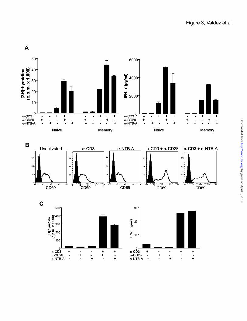
Co-stimulation of T cells by NTB-A
To determine whether NTB-A is capable of providing a co-stimulatory signal to T cells, we
isolated naïve and memory human peripheral CD4 T cells and stimulated them with anti-NTB-
A antibody and a sub-optimal dose of anti-CD3 antibody. Anti-CD3 treatment alone only
induced minimal proliferation in the naïve T cells, while higher proliferation was seen in the
memory T cells since these cells do not require co-stimulation for activation (Fig. 3A).
Treatment with anti-NTB-A or anti-CD28 antibody alone did not induce proliferation or IFN-γ
secretion in naïve or memory T cells. The combination of anti-NTB-A with a sub-optimal dose
of anti-CD3 in the absence of anti-CD28 antibody led to a substantial level of proliferation that
was higher than anti-CD3 alone and slightly lower than the proliferation induced with anti-CD3
plus anti-CD28 treatment. Increase in IFN-γ expression corresponded to the increase in
proliferation induced by anti-CD3 plus anti-NTB-A treatment. The effect of anti-NTB was
more dramatic in the naïve T cells, presumably due to the fact that memory cells respond very
strongly to anti-CD3 treatment alone. IFN-γ secretion induced by anti-CD3 treatment was not
14
by guest on April 3, 2019
http://ww
w.jbc.org/
Dow
nloaded from
augmented by the addition of anti-NTB-A. The ability of NTB-A to activate Jurkat T cells was
assessed by culturing Jurkat cells on plates coated with anti-CD3, anti-CD28, anti-NTB-A, or a
combination of these antibodies. Twenty-four hours after activation, Jurkat cells that were
stimulated with anti-CD3 and anti-NTB-A upregulated expression of the activation marker,
CD69 (Fig. 3B). Co-stimulation of murine CD4 T cells was also carried out using antibody that
cross-reacts to the murine form of NTB-A (Fig. 3C). We find similar results as with human T
cells, including induced proliferation and IFN-γ expression with NTB-A co-stimulation,
indicating the murine and human forms of NTB-A function similarly in T cells. Thus, our
results suggest that NTB-A is capable of co-stimulating T cells to a level similar to CD28.
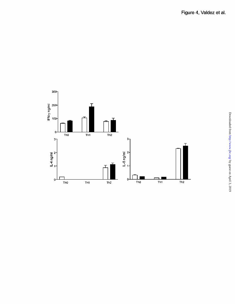
NTB-A promotes Th1 cell phenotype
Given that NTB-A cross-linking can induce proliferation and INF-γ secretion, we next sought
to determine whether NTB-A influences Th1 or Th2 cell differentiation. Purified human CD4 T
cells were stimulated with anti-CD3 and anti-NTB-A under Th1-inducing conditions or under
Th2-inducing conditions. This stimulation was independent of a CD28 signal because no anti-
CD28 agonistic reagents were used where anti-NTB-A antibody was used. For comparison,
cells were also stimulated with anti-CD3 and anti-CD28 under appropriate Th1 or Th2
conditions. After three days of stimulation, the cells were washed and rested for four days with
IL-2. After that time period, all cells were restimulated with anti-CD3 only. Th1 cells
stimulated with anti-NTB-A secreted more IFN-γ compared to Th1 cells stimulated with anti-
CD28, and Th0 cells also had slightly higher levels of INF-γ (Fig. 4). Levels of IL-4 and IL-5,
15
by guest on April 3, 2019
http://ww
w.jbc.org/
Dow
nloaded from

Th2-type cytokines, were comparable in all samples, indicating that NTB-A had no effect
distinguishable from CD28 on the differentiation of Th2 cells. These results suggest that NTB-
A helps to enhance the Th1 cell phenotype by inducing more Th1-type cytokine expression.
Interestingly, effects of NTB-A in enhancing differentiation of T cells to Th1 were completely
independent of CD28 signals.
Phosphorylation of NTB-A and SAP association
We have demonstrated that cross-linking of CD3 and NTB-A on T cells leads to proliferation
and IFN↑γ expression. We next sought to analyze the molecular events associated with CD3 and
NTB-A cross-linking. A hallmark of CD2 family members is the presence of two to four SAP-
binding motifs (TxYxxV/I) in the intracellular domain; SAP can bind to the both the
phosphorylated and non-phosphorylated forms of CD150, though SAP has a higher affinity for
the phosphorylated form (31,32). In addition to binding SAP, CD150 has also been reported to
bind SHP-2 (22,24,33-35). Both human and mouse NTB-A have two SAP-binding motifs in
the intracellular domain. To determine whether NTB-A also associates with these CD150-
associated proteins, Jurkat T cells were treated with pervanadate to induce phosphorylation of
NTB-A. NTB-A was immunoprecipitated from cell lysates and the association of SAP, SHP-1,
and SHP-2 was determined by western blot. SAP association with NTB-A was not detected in
untreated cells. However, SAP did associate with NTB-A in pervanadate treated cells,
suggesting a phosphorylation-dependent association of SAP with NTB-A. SHP-1 and SHP-2,
although expressed in Jurkat cells, were not found associated with NTB-A in either untreated or
16
by guest on April 3, 2019
http://ww
w.jbc.org/
Dow
nloaded from

in pervanadate-treated cells (Fig. 5A). This lack of association with SHP-1 or SHP-2 suggests
a signaling mechanism distinct from CD150 in T cells. The inhibitory receptor LAIR does
associate with SHP-1 an SHP-2 in pervanadate-treated Jurkat cells, ruling out the possibility
that SHP-1 and SHP-2 are not capable of being immunoprecipitated in our system (Fig, 5A).
To study the signaling events following co-stimulation with NTB-A in T cells, we next
analyzed the phosphorylation of NTB-A induced by crosslinking CD3 and NTB-A in freshly
isolated human T cells (Fig. 5B). NTB-A is phosphorylated upon cross-linking of CD3 and
NTB-A in human T cells as shown by immunoprecipitation of NTB-A and western blotting
with an anti-phosphotyrosine antibody. Interestingly, SAP does not associate with NTB-A in
untreated human T cells, but SAP does associate with co-stimulation-induced phosphorylated
NTB-A. Similar to Jurkat cells, SHP1 and SHP2 do not associate with NTB-A in human T
cells although both proteins are expressed. These finding suggest a mechanism by which cross-
linking of NTB-A induces NTB-A phosphorylation and SAP association in T cells. It is
possible that these signaling events then lead to proliferation and IFN-γ secretion.
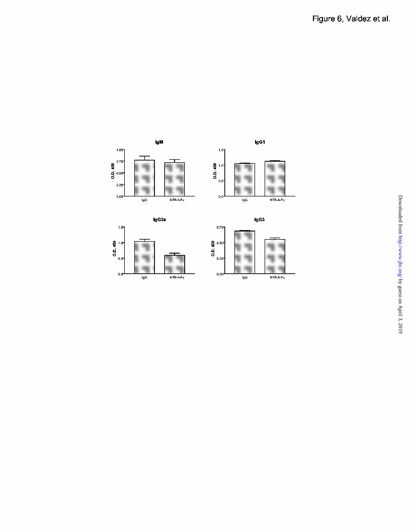
NTB-A-Fc inhibits Th1 cell-induced isotype switching
Our data thus far suggests that NTB-A may act to induce Th1 T cell responses. We next
analysed whether NTB-A plays a role in T-dependent B cell responses. To block a possible
ligand interaction with NTB-A in vivo, we used a fusion protein consisting of the extracelluar
portion of murine NTB-A fused to an Fc domain (mNTB-A-Fc). C57/B6 mice were
immunized with NP(23)-KLH and and treated daily with mNTB-A-Fc or mIgG1 as a control.
17
by guest on April 3, 2019
http://ww
w.jbc.org/
Dow
nloaded from

Ten days after immunization, serum was analysed for the expression of NP-specific
immunoglobulin isotypes (Figure 6). Generally, Th2 type cytokines such as IL-4 induce the
increase of switching to the IgG1 isotype and suppress the switching to IgG2a and IgG3. Th1
type cytokines such as IFN-γ suppress switching to IgG1 and increase switching to IgG2a and
IgG3. Mice treated with mNTB-A-Fc demonstrated decreased levels of IgG2a and IgG3, and
increased levels of IgG1, which is indicative of the presence of Th2-type cytokines such as IL-
4. These results suggest that mNTB-A-Fc suppressed B cell isotype switching to IFN-γ
induced isotypes, possibly by inhibiting the activation of Th1 cells during immunization.
Although we have demonstrated a homotypic interaction for NTB-A, the weak binding affinity
suggests that there may be an additional ligand for NTB-A. The addition of the fusion protein in
vivo may act to block this ligand in vivo and prevent the activation and development of Th1
cells.
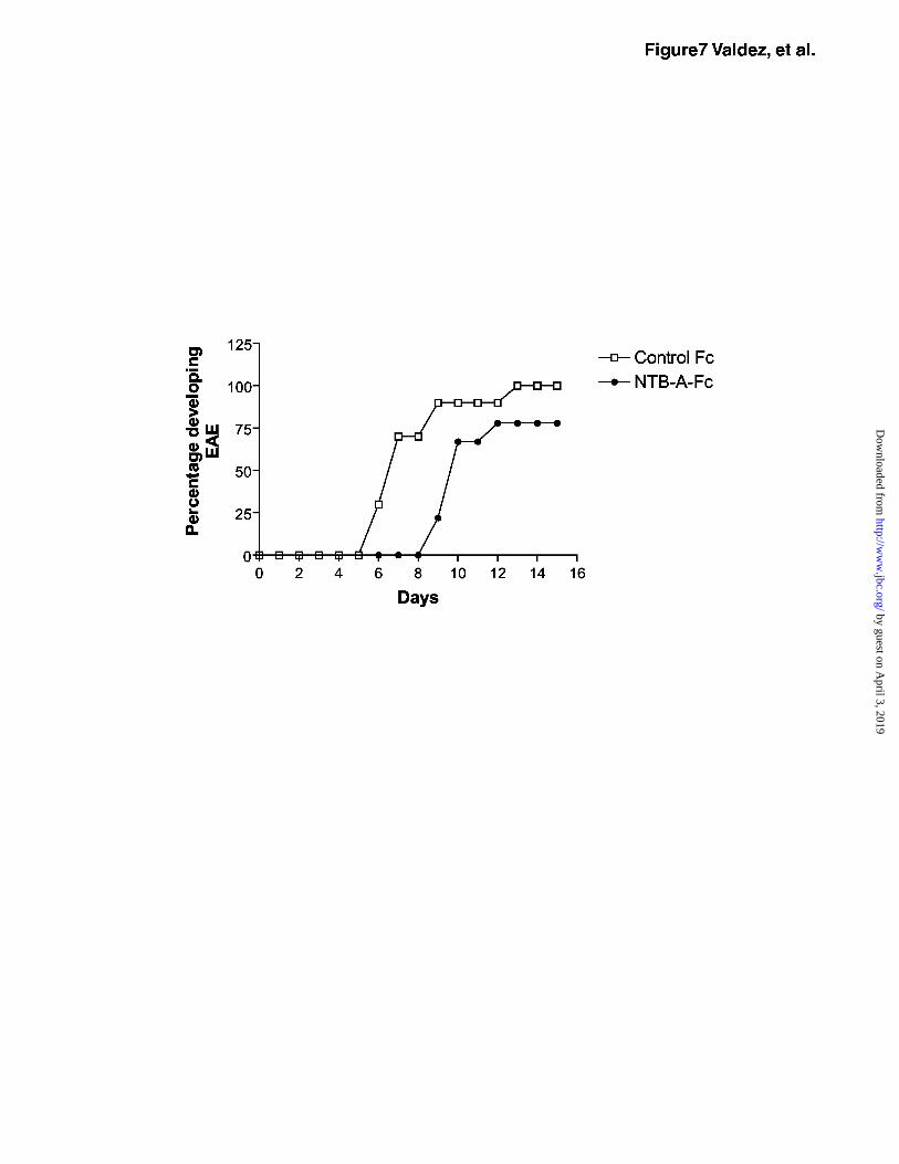
NTB-A-Fc delays onset of EAE
The increase in IFN↑γ expression induced by NTB-A suggests that NTB-A may act to
skew T cell responses to a Th1 response. To determine the effect of NTB-A in Th1-mediated
autoimmune disease, we analyzed the effect of an NTB-A-Fc fusion protein in a murine model
for multiple sclerosis, experimental autoimmune encephalomyelitis (EAE). Myelin Basic
Protein (MBP) TCR transgenic mice were immunized with Ac1-11 (NH2-terminal peptide from
MBP) and treated daily with either NTB-A-Fc or mIgG1 control protein. Disease progression
was monitored each day and the onset of disease began at day 5 in the control mice (Fig. 7).
18
by guest on April 3, 2019
http://ww
w.jbc.org/
Dow
nloaded from

Mice treated with mNTB-A-Fc showed a delayed onset of disease at day 8 and 20% of the mice
never developed disease. The ability of mNTB-A-Fc to delay the onset of EAE in MBP TCR
transgenic mice suggests that the fusion protein may dampen effector functions of T cells since
EAE is a predominately a Th1 cell mediated disease. Based on the ability of NTB-A-Fc to
decrease Th1 cytokine-induced isotype switching, we hypothesize that NTB-A-Fc delays EAE
development by inhibition of the generation of Th1 responses.
19
by guest on April 3, 2019
http://ww
w.jbc.org/
Dow
nloaded from

Discussion
Recognition of antigen by TCR is not sufficient for proper T cell activation. Additional co-
stimulatory molecules are required, and this provides the basis for the ‘two signal model’ of
lymphocyte activation. Members of the CD28/B7 family of co-stimulatory molecules are
generally considered to provide the major pathway for co-stimulation. However, data including
analysis of CD28 deficient T cells suggest that other co-stimulatory molecules exist that can
provide the second signal for T cells. The CD2 superfamily has been implicated in T cell co-
stimulation. Included in this superfamily is the CD150 sub-family of receptors, which associate
with SAP. Human patients who lack expression of SAP have major defects in cell mediated
immunity and succumb to massive lymphocyte infiltration to multiple organs, a syndrome called
X-linked proliferative disease (XLP). Thus, the CD2/CD150 family members may play a
significant part in cellular immunity.
We have identified a new T cell function for the CD2 family member, NTB-A. We find
that NTB-A can co-stimulate T cells to proliferate and secrete IFN↑γ. NTB-A cross-linking
leads to phosphorylation of the receptor and association of SAP, but not SHP-1 or SHP-2 in
human T cells. Our data suggest that NTB-A plays a role in Th1 cell responses since Th1 cells
stimulated with NTB-A secrete more Th1 and not Th2 type cytokines. An NTB-A-Fc fusion
protein inhibits IFN-γ induced isotype switching. NTB-A-Fc also delays development and
severity of EAE, a mouse model for multiple sclerosis.
NTB-A contains two ITSM (TxYxxV/I) motifs in its intracellular domain. These motifs
have been shown to bind SAP in other receptors including CD150 and 2B4. Co-stimulation
20
by guest on April 3, 2019
http://ww
w.jbc.org/
Dow
nloaded from

induced by cross-linking of CD3 and NTB-A leads to early phosphorylation of NTB-A and
association of SAP, but not SHP-1 or SHP-2. SAP is composed largely of an SH2 domain, and
it was previously thought to block interaction of SH2-domain containing proteins such as SHP-
2 (22). Unlike CD150, NTB-A does not associate with SHP-2. Lack of association of NTB-A
with SHP-2 in T cells suggests that the regulation of NTB-A signaling and IFN-γ production is
distinct from CD150. Recently, it has been suggested that SAP may act to recruit other
molecules such as FynT (19), and this could help drive the specificity of the signal through
different SAP-associated receptors. It will be interesting to determine whether FynT also
associates with NTB-A. Given that XLP patients are deficient in SAP, understanding the
mechanism by which SAP helps regulate signals through NTB-A may give us insight into the
pathology of XLP disease. Our preliminary data suggests that T cells from XLP patients cannot
be activated through NTB-A, suggesting an important role for SAP and NTB-A in T cell
activation.
We have demonstrated a critical role for NTB-A in T cell activation and disease using a
NTB-A-Fc fusion protein. NTB-A-Fc was able to suppress T-dependent B cell class
switching to Th1 cytokine-induced isotypes, most likely by blocking interaction with a NTB-A
ligand and interfering with activation of T cells to Th1-type cells. The contribution of NTB-A
to early activation events versus later effector functions is not clear. We do find that naïve cells
can respond to NTB-A cross-linking and that initial cross-linking with NTB-A skews
development to the Th1 cell type. Stimulation of Th1 or Th2 cell clones with NTB-A has little
effect (data not shown), suggesting that NTB-A may play a greater role in the early phase of
21
by guest on April 3, 2019
http://ww
w.jbc.org/
Dow
nloaded from
activation. When tested in a Th1-mediated autoimmune disease model, EAE, NTB-A-Fc
delayed the onset of disease. These results further suggest that NTB-A may play an important
role in T cell activation and effector functions. The block in disease by NTB-A-Fc is not
complete, and it may be the case that NTB-A-Fc does not block NTB-A activity completely.
As we have shown, NTB-A exhibits weak homotypic interaction, similar to CD150 and other
family members. It is possible that NTB-A-Fc may provide a weak activation signal while
blocking a stronger signal at the same time. Thus, disease is not completely blocked during EAE
progression. Another possibility for the lack of a complete block in disease is that other
molecules important for T cell functions are present. Other co-stimulatory molecules such as
ICOS have been implicated in autoimmunity (36-39). How NTB-A regulates T cell function
along with other co-stimulatory molecules will be an interesting focus. Our results suggest an
important role for NTB-A in T cell mediated functions, including T cell activation and T cell-
mediated autoimmune disease.
The identification of NTB-A as a new SAP-associated receptor may help to further
understand the XLP disease process in human patients. Our findings implicate a role for NTB-A
in T cell activation, suggesting a possible mechanism by which SAP deficient T cells fail in their
response to pathogens, including virally infected cells and tumor cells. The identification of
NTB-A and new SAP-associated receptors, as well as understanding the signaling mechanisms
involved, will help shed new light on XLP disease. Given its role as a co-stimulatory molecule,
inducing proliferation and IFN↑γ secretion by T cells, as well as the ability of the fusion protein
to suppress Th1-induced isotype switching and EAE, NTB-A is a potential target for therapeutic
22
by guest on April 3, 2019
http://ww
w.jbc.org/
Dow
nloaded from

intervention in Th1-type autoimmune disease as well as inflammation.
23
by guest on April 3, 2019
http://ww
w.jbc.org/
Dow
nloaded from

Acknowledgements
We wish to thank Betty Li for her technical support; the Genentech Antibody Technology
group, DNA sequencing lab, FACS lab, and protein purification group for their support; Andy
Chan and lab members for discussion; Laura de Forge for cytokine analysis; Henry Lowman for
Biacore measurements; Peter Gribling, Kathy Nguyen, and Yifan Zhang for in vivo work; Amy
Shen and Zhonghua Lin for technical assistance; and Charles A. Janeway Jr. for MBP-TCR
transgenic mice.
24
by guest on April 3, 2019
http://ww
w.jbc.org/
Dow
nloaded from
References
1. Carreno, B. M., and Collins, M. (2002) Annu Rev Immunol 20, 29-53.
2. Lucas, P. J., Negishi, I., Nakayama, K., Fields, L. E., and Loh, D. Y. (1995) J Immunol
154, 5757-5768.
3. Shahinian, A., Pfeffer, K., Lee, K. P., Kundig, T. M., Kishihara, K., Wakeham, A.,
Kawai, K., Ohashi, P. S., Thompson, C. B., and Mak, T. W. (1993) Science 261, 609-
612.
4. Tafuri, A., Shahinian, A., Bladt, F., Yoshinaga, S. K., Jordana, M., Wakeham, A.,
Boucher, L. M., Bouchard, D., Chan, V. S., Duncan, G., Odermatt, B., Ho, A., Itie, A.,
Horan, T., Whoriskey, J. S., Pawson, T., Penninger, J. M., Ohashi, P. S., and Mak, T. W.
(2001) Nature 409, 105-109.
5. Sharpe, A. H., and Freeman, G. J. (2002) Nat Rev Immunol 2, 116-126.
6. Dong, C., Juedes, A. E., Temann, U. A., Shresta, S., Allison, J. P., Ruddle, N. H., and
Flavell, R. A. (2001) Nature 409, 97-101.
7. Seed, B., and Aruffo, A. (1987) Proc Natl Acad Sci U S A 84, 3365-3369.
8. Staunton, D. E., and Thorley-Lawson, D. A. (1987) Embo J 6, 3695-3701.
9. de la Fuente, M. A., Pizcueta, P., Nadal, M., Bosch, J., and Engel, P. (1997) Blood 90,
2398-2405.
10. Cocks, B. G., Chang, C. C., Carballido, J. M., Yssel, H., de Vries, J. E., and Aversa, G.
(1995) Nature 376, 260-263.
11. Aversa, G., Chang, C. C., Carballido, J. M., Cocks, B. G., and de Vries, J. E. (1997) J
25
by guest on April 3, 2019
http://ww
w.jbc.org/
Dow
nloaded from

Immunol 158, 4036-4044.
12. Aversa, G., Carballido, J., Punnonen, J., Chang, C. C., Hauser, T., Cocks, B. G., and De
Vries, J. E. (1997) Immunol Cell Biol 75, 202-205.
13. Davis, S. J., and van der Merwe, P. A. (1996) Immunol Today 17, 177-187.
14. Valiante, N. M., and Trinchieri, G. (1993) J Exp Med 178, 1397-1406.
15. Killeen, N., Stuart, S. G., and Littman, D. R. (1992) Embo J 11, 4329-4336.
16. Green, J. M., Karpitskiy, V., Kimzey, S. L., and Shaw, A. S. (2000) J Immunol 164,
3591-3595.
17. Mathew, P. A., Garni-Wagner, B. A., Land, K., Takashima, A., Stoneman, E., Bennett,
M., and Kumar, V. (1993) J Immunol 151, 5328-5337.
18. Sandrin, M. S., Gumley, T. P., Henning, M. M., Vaughan, H. A., Gonez, L. J., Trapani, J.
A., and McKenzie, I. F. (1992) J Immunol 149, 1636-1641.
19. Latour, S., Gish, G., Helgason, C. D., Humphries, R. K., Pawson, T., and Veillette, A.
(2001) Nat Immunol 2, 681-690.
20. Morra, M., Howie, D., Grande, M. S., Sayos, J., Wang, N., Wu, C., Engel, P., and
Terhorst, C. (2001) Annu Rev Immunol 19, 657-682.
21. Nichols, K. E., Koretzky, G. A., and June, C. H. (2001) Nat Immunol 2, 665-666.
22. Sayos, J., Wu, C., Morra, M., Wang, N., Zhang, X., Allen, D., van Schaik, S.,
Notarangelo, L., Geha, R., Roncarolo, M. G., Oettgen, H., De Vries, J. E., Aversa, G.,
and Terhorst, C. (1998) Nature 395, 462-469.
23. Veillette, A. (2002) Sci STKE 2002, E8.
26
by guest on April 3, 2019
http://ww
w.jbc.org/
Dow
nloaded from

24. Castro, A. G., Hauser, T. M., Cocks, B. G., Abrams, J., Zurawski, S., Churakova, T.,
Zonin, F., Robinson, D., Tangye, S. G., Aversa, G., Nichols, K. E., de Vries, J. E., Lanier,
L. L., and O’Garra, A. (1999) J Immunol 163, 5860-5870.
25. Martin, M., Romero, X., de la Fuente, M. A., Tovar, V., Zapater, N., Esplugues, E.,
Pizcueta, P., Bosch, J., and Engel, P. (2001) J Immunol 167, 3668-3676.
26. Bottino, C., Falco, M., Parolini, S., Marcenaro, E., Augugliaro, R., Sivori, S., Landi, E.,
Biassoni, R., Notarangelo, L. D., Moretta, L., and Moretta, A. (2001) J Exp Med 194,
235-246.
27. Fraser, C. C., Howie, D., Morra, M., Qiu, Y., Murphy, C., Shen, Q., Gutierrez-Ramos, J.
C., Coyle, A., Kingsbury, G. A., and Terhorst, C. (2002) Immunogenetics 53, 843-850.
28. Peck, S. R., and Ruley, H. E. (2000) Immunogenetics 52, 63-72.
29. Mavaddat, N., Mason, D. W., Atkinson, P. D., Evans, E. J., Gilbert, R. J., Stuart, D. I.,
Fennelly, J. A., Barclay, A. N., Davis, S. J., and Brown, M. H. (2000) J Biol Chem 275,
28100-28109.
30. Punnonen, J., Cocks, B. G., Carballido, J. M., Bennett, B., Peterson, D., Aversa, G., and
de Vries, J. E. (1997) J Exp Med 185, 993-1004.
31. Li, S. C., Gish, G., Yang, D., Coffey, A. J., Forman-Kay, J. D., Ernberg, I., Kay, L. E.,
and Pawson, T. (1999) Curr Biol 9, 1355-1362.
32. Poy, F., Yaffe, M. B., Sayos, J., Saxena, K., Morra, M., Sumegi, J., Cantley, L. C.,
Terhorst, C., and Eck, M. J. (1999) Mol Cell 4, 555-561.
33. Howie, D., Simarro, M., Sayos, J., Guirado, M., Sancho, J., and Terhorst, C. (2002)
27
by guest on April 3, 2019
http://ww
w.jbc.org/
Dow
nloaded from

Blood 99, 957-965.
34. Lewis, J., Eiben, L. J., Nelson, D. L., Cohen, J. I., Nichols, K. E., Ochs, H. D.,
Notarangelo, L. D., and Duckett, C. S. (2001) Clin Immunol 100, 15-23.
35. Shlapatska, L. M., Mikhalap, S. V., Berdova, A. G., Zelensky, O. M., Yun, T. J., Nichols,
K. E., Clark, E. A., and Sidorenko, S. P. (2001) J Immunol 166, 5480-5487.
36. Sperling, A. I., and Bluestone, J. A. (2001) Nat Immunol 2, 573-574.
37. Ozkaynak, E., Gao, W., Shemmeri, N., Wang, C., Gutierrez-Ramos, J. C., Amaral, J.,
Qin, S., Rottman, J. B., Coyle, A. J., and Hancock, W. W. (2001) Nat Immunol 2, 591-
596.
38. Rottman, J. B., Smith, T., Tonra, J. R., Ganley, K., Bloom, T., Silva, R., Pierce, B.,
Gutierrez-Ramos, J. C., Ozkaynak, E., and Coyle, A. J. (2001) Nat Immunol 2, 605-611.
39. Gonzalo, J. A., Tian, J., Delaney, T., Corcoran, J., Rottman, J. B., Lora, J., Al-garawi,
A., Kroczek, R., Gutierrez-Ramos, J. C., and Coyle, A. J. (2001) Nat Immunol 2, 597-
604.
28
by guest on April 3, 2019
http://ww
w.jbc.org/
Dow
nloaded from

Figure 1. NTB-A is a CD150 family member expressed in T cells.
(A) Cell surface expression of NTB-A on cell lines. Expression was detected by FACS using a
labeled monoclonal antibody raised against the extracellular domain of NTB-A.
(B) NTB-A expression on resting and activated T cells. Expression was detected on human T
cells using the same antibody described above. Cells were stimulated for 24 (data not shown)
and 48 hours with plate-bound anti-CD3 (0.1 ug/ml) and anti-CD28 (1 ug/ml). Cells from the
same culture were also stained for CD69 expression. The dark histogram is the isotype control.
(C) NTB-A expression on resting and activated B cells. Cells were activated for 48 hours in the
presence of anti-CD40 (10 µg/ml) and IL-4 (50 ng/ml). Cells from the same culture were also
stained for CD80 expression.
(D) High levels of NTB-A expression is detected in human CD4+CD45RO+ memory T cells as
shown by FACS analysis.
Figure 2. NTB-A binds to itself in a homotypic fashion.
(A) Surface staining of NTB-A on transfected cells. Full length NTB-A or 19A was transfected
into 293T cells. NTB-A-NTB-A association was detected with NTB-A-Fc, and a Cy3 labeled
anti-human Fc antibody (left panels). Expression of NTB-A or 19A in transfected cells was
detected using FITC-labeled anti-NTB-A or anti-19A monoclonal antibodies (right panels).
(B) ELISA assay for NTB-A-NTB-A interaction. Human NTB-A-His or 19A-His were
coated onto plates at 2 ug/ml and association was detected using NTB-A-Fc or 19A-Fc with a
goat anti-human IgG (Fc specific) secondary antibody. The closed square represents NTB-A-
29
by guest on April 3, 2019
http://ww
w.jbc.org/
Dow
nloaded from
Fc binding to coated NTB-A-His. The open square represents 19A-Fc binding to coated NTB-
A-His. The triangle represents NTB-A-Fc binding to coated 19A-His.
Figure 3. NTB-A co-stimulates T cells and induces IFN-γ expression.
(A) Co-stimulation induced by anti-NTB-A antibody. Naïve and memory human CD4 T cells
isolated from blood were activated with combinations of anti-CD3 (0.1 µg/ml), anti-CD28 (1
µg/ml), and anti-NTB-A antibodies (1 µg/ml) coated on a plate. 0.5 x 10⁶ T cells were used per
well in a 96-well plate. After 3 days of activation, proliferation of human T cells was measured
by [3H] thymidine incorporation. The secretion of IFN-γ by these same samples was measured
by ELISA, along with IL-4 (data not shown).
(B) Activation of Jurkat T cells with anti-CD3 and anti-NTB-A antibody. Jurkat cells were
activated using coated antibodies as described above. 0.5 x 10⁶ Jurkat T cells were used per well
in a 96-well plate. After 24 hours of activation, the Jurakat cells were analysed by FACS for the
expression of CD69. The black histogram is the isotype control.
(C) Co-stimulation of murine T cells using a cross-reacting anti-NTB-A antibody. The
concentrations of antibodies used were: anti-CD3 (0.1 µg/ml), anti-CD28 (1 µg/ml), and anti-
NTB-A (1 µg/ml). The NTB-A antibody was raised against the human protein, but cross-reacts
to the mouse protein. CD4 T cells isolated from spleen were activated at 0.5 x 10⁶ cells per well
in a 96 well plate. On the third day of activation, the cells were pulsed with [3H] thymidine and
30
by guest on April 3, 2019
http://ww
w.jbc.org/
Dow
nloaded from
supernatant was removed for ELISA analysis of IFN-γ secretion.
Figure 4. NTB-A enhances the Th1 cell phenotype.
Human Th1/ Th2 differentiation. CD4 T cells were initially stimulated with anti-CD3 and anti-
CD28 antibodies with Th1 or Th2 inducing cytokines (white bars) or they were stimulated with
anti-CD3 and anti-NTB-A antibodies with Th1 or Th2 inducing cytokines (black bars). Three
days later, cells were rested with only IL-2. Seven days after the initial stimulation, cells were
restimulated with anti-CD3 antibody only for 24 hours and secretion of INF-γ, IL-4, and IL-5
was analyzed.
Figure 5. Anti-CD3 and anti-NTB-A crosslinking leads to phosphorylation of NTB-A, and
association of SAP, but not SHP-1 or SHP-2 in T cells.
(A) Jurkat T cells were treated with pervanadate, lysed, and NTB-A was immunoprecipitated
using a mixture of monoclonal antibodies to NTB-A. Control immunoprecipitations included
LAIR and murine IgG1 (isotype control). SAP, SHP-1, SHP-2, and NTB-A were detected by
Western blot. Lanes 1 and 2 are whole lysate; lanes 3 and 4 are IgG1 IP; lanes 5 and 6 are NTB-
A IP; and lanes 7 and 8 are LAIR IP.
(B) Co-stimulation of human peripheral T cells. Cells were stimulated by cross-linking CD3
and NTB-A for 30 seconds. NTB-A was immunoprecipitated and phosphorylation of NTB-A
was determined by western blotting with a phospotyrosine antibody. Association of SAP, SHP-
1, and SHP-2 was detected by western blot. The asterisk indicates Ig heavy chain. Lanes 1 and
31
by guest on April 3, 2019
http://ww
w.jbc.org/
Dow
nloaded from

2 are whole lysate; lanes 3 and 4 are NTB-A IP; and lanes 5 and 6 are IgG1 control IP.
Figure 6. NTB-A-Fc treatment suppresses B cell isotype switching to Ig2a and IgG3.
C57/B6 mice immunized with NP(23)-KLH were treated daily with 100 ug of either mNTB-A-
Fc or with mIgG1 as a control. T-dependent B cell responses were measured by analysing
serum immunoglobulin levels by ELISA. Immunoglobulin isotypes shown here are IgM, IgG1,
IgG2a, and IgG3.
Figure 7. mNTB-A-Fc delays the onset of EAE. MPB TCR transgenic mice were treated daily
with 100 µg mNTB-A-Fc or mIgG1. On day 0, mice were injected with the Ac1-11 peptide,
followed by pertussis toxin on day 1 and 2. Disease was monitored daily and the percentage of
mice developing disease each day was plotted.
32
by guest on April 3, 2019
http://ww
w.jbc.org/
Dow
nloaded from

Gurney, Wyne P. Lee and Iqbal S. GrewalPatricia A. Valdez, Hua Wang, Dhaya Seshasayee, Menno van Lookeren Campagne, Austin
NTB-A, a new activating receptor in T cells that regulates autoimmune disease
published online February 26, 2004J. Biol. Chem.
10.1074/jbc.M312313200Access the most updated version of this article at doi:
Alerts:
When a correction for this article is posted•
When this article is cited•
to choose from all of JBC's e-mail alertsClick here
by guest on April 3, 2019
http://ww
w.jbc.org/
Dow
nloaded from