Nowa1p and Nowa2p: Novel Putative RNA Binding Proteins Involved in trans-Nuclear Crosstalk in...
13
Current Biology, Vol. 15, 1616–1628, September 20, 2005, ©2005 Elsevier Ltd All rights reserved. DOI 10.1016/j.cub.2005.07.033 Nowa1p and Nowa2p: Novel Putative RNA Binding Proteins Involved in trans-Nuclear Crosstalk in Paramecium tetraurelia Mariusz Nowacki, 1,2 Wlodzimierz Zagorski-Ostoja, 2 and Eric Meyer 1, * 1 Laboratoire de Génétique Moléculaire Centre National de la Recherche Scientifique Unité Mixte de Recherche 8541 Ecole Normale Supérieure 46 rue d’Ulm 75005 Paris France 2 Institute of Biochemistry and Biophysics, PAS Pawinskiego 5a 02-106 Warszawa Poland Summary Background: The germline genome of ciliates is extens- ively rearranged during development of a new somatic macronucleus from the germline micronucleus, a pro- cess that follows sexual events. In Paramecium tetraur- elia, single-copy internal eliminated sequences (IESs) and multicopy transposons are eliminated, whereas cellular genes are amplified to w800 n. For a subset of IESs, introduction of the IES sequence into the mater- nal (prezygotic) macronucleus specifically inhibits exci- sion of the homologous IES in the developing zygotic macronucleus. This and other homology-dependent maternal effects have suggested that rearrangement patterns are epigenetically determined by an RNA- mediated, trans-nuclear comparison, involving the RNA interference pathway, of germline and somatic genomes. Results: We report the identification of novel develop- mentally regulated RNA binding proteins, Nowa1p and Nowa2p, which are required for the survival of sexual progeny. Green fluorescent protein (GFP) fusions show that Nowa1p accumulates into the maternal macronu- cleus shortly before meiosis of germline micronuclei and is later transported to developing macronuclei. No- wa1p/2p depletion impairs the elimination of transpo- sons and of those IESs that are controlled by maternal effects, confirming the existence of distinct IES classes. Conclusions: The results indicate that Nowa proteins are essential components of the trans-nuclear-cross- talk mechanism that is responsible for epigenetic pro- gramming of genome rearrangements. We discuss im- plications for the current models of genome scanning in ciliates, a process related to the formation of hetero- chromatin by RNA interference in other eukaryotes. Introduction The discovery of RNA interference has provided a new paradigm for gene-specific regulation in eukaryotes, whereby short RNAs are used as specificity modules to target protein complexes to homologous transcripts. It was then realized that such homology-dependent pro- *Correspondence: [email protected] cesses are not limited to mRNA degradation or transla- tion, but can also regulate chromatin structure through the targeting of epigenetic modifications to specific re- gions of the genome [1]. Some of the recent evidence has come from the study of ciliates: There is increasing evidence that maternal RNAs are involved in specifying the conspicuous genome rearrangements that occur during development in these organisms, a process that probably underlies long-known cases of non-Mende- lian inheritance [2]. These unicellular eukaryotes harbor two different kinds of nuclei, germline micronuclei and somatic macro- nuclei. The diploid micronuclei are transcriptionally si- lent during vegetative growth but undergo meiosis dur- ing sexual events to transmit the germline genome to the next generation. The highly polyploid macronuclei (w800 n in P. tetraurelia) have somatic functions: They are responsible for gene expression throughout the life cycle but are lost or degraded after fertilization, as new micro- and macronuclei develop from mitotic copies of the zygotic nucleus. During the development of a new macronucleus, the diploid zygotic genome is amplified and extensively rearranged: Chromosomes are frag- mented into shorter molecules capped by de novo telo- mere addition, and numerous IESs are excised from these acentric macronuclear “chromosomes” [3–5]. P. tetraurelia IESs are short (26–882 bp), single-copy, noncoding elements that are precisely excised from coding and noncoding sequences. They are invariably flanked by two 5#-TA-3# dinucleotides, one of which is left in the macronuclear sequence after excision. IESs contain no other strictly conserved motif, although their ends often resemble the consensus TAYAGYNR [6, 7]. The local sequence context appears to be important for the staggered double-strand cuts that initiate exci- sion [8], but the degenerate consensus does not con- tain sufficient information to direct the reproducible ex- cision of the >50,000 different IESs in the haploid genome, suggesting that epigenetic mechanisms are involved [9]. Some IESs are indeed sensitive to homology-depen- dent maternal effects: Introduction of an IES sequence into the vegetative macronucleus, prior to meiosis, can inhibit excision of the homologous IES very specifically in the sexual progeny of transformed clones when a new macronucleus develops from the wild-type micro- nucleus [10]. The effect was observed for one-third of tested IESs, hereafter called maternally controlled IESs (mcIESs), and it varies quantitatively among these [11]. In the most efficient cases, a moderate IES copy num- ber in the maternal macronucleus will inhibit excision in a greater number of chromosome copies in the new macronucleus. This quickly results in retention of the IES in 100% of macronuclear copies, a stable epige- netic state that is maternally (cytoplasmically) inherited in the following sexual generations. The fragmentation of chromosomes during macro- nuclear development appears to be the consequence of a second, imprecise mode of DNA elimination that removes larger regions of the Paramecium germline ge-
-
Upload
mariusz-nowacki -
Category
Documents
-
view
212 -
download
0
Transcript of Nowa1p and Nowa2p: Novel Putative RNA Binding Proteins Involved in trans-Nuclear Crosstalk in...
Current Biology, Vol. 15, 1616–1628, September 20, 2005, ©2005 Elsevier Ltd All rights reserved. DOI 10.1016/j.cub.2005.07.033
Nowa1p and Nowa2p: Novel PutativeRNA Binding Proteins Involved in trans-NuclearCrosstalk in Paramecium tetraurelia
Mariusz Nowacki,1,2 Wlodzimierz Zagorski-Ostoja,2
and Eric Meyer1,*1Laboratoire de Génétique MoléculaireCentre National de la Recherche ScientifiqueUnité Mixte de Recherche 8541Ecole Normale Supérieure46 rue d’Ulm75005 ParisFrance2 Institute of Biochemistry and Biophysics, PASPawinskiego 5a02-106 WarszawaPoland
Summary
Background: The germline genome of ciliates is extens-ively rearranged during development of a new somaticmacronucleus from the germline micronucleus, a pro-cess that follows sexual events. In Paramecium tetraur-elia, single-copy internal eliminated sequences (IESs)and multicopy transposons are eliminated, whereascellular genes are amplified to w800 n. For a subset ofIESs, introduction of the IES sequence into the mater-nal (prezygotic) macronucleus specifically inhibits exci-sion of the homologous IES in the developing zygoticmacronucleus. This and other homology-dependentmaternal effects have suggested that rearrangementpatterns are epigenetically determined by an RNA-mediated, trans-nuclear comparison, involving the RNAinterference pathway, of germline and somatic genomes.Results: We report the identification of novel develop-mentally regulated RNA binding proteins, Nowa1p andNowa2p, which are required for the survival of sexualprogeny. Green fluorescent protein (GFP) fusions showthat Nowa1p accumulates into the maternal macronu-cleus shortly before meiosis of germline micronucleiand is later transported to developing macronuclei. No-wa1p/2p depletion impairs the elimination of transpo-sons and of those IESs that are controlled by maternaleffects, confirming the existence of distinct IES classes.Conclusions: The results indicate that Nowa proteinsare essential components of the trans-nuclear-cross-talk mechanism that is responsible for epigenetic pro-gramming of genome rearrangements. We discuss im-plications for the current models of genome scanningin ciliates, a process related to the formation of hetero-chromatin by RNA interference in other eukaryotes.
Introduction
The discovery of RNA interference has provided a newparadigm for gene-specific regulation in eukaryotes,whereby short RNAs are used as specificity modules totarget protein complexes to homologous transcripts. Itwas then realized that such homology-dependent pro-
*Correspondence: [email protected]
cesses are not limited to mRNA degradation or transla-tion, but can also regulate chromatin structure throughthe targeting of epigenetic modifications to specific re-gions of the genome [1]. Some of the recent evidencehas come from the study of ciliates: There is increasingevidence that maternal RNAs are involved in specifyingthe conspicuous genome rearrangements that occurduring development in these organisms, a process thatprobably underlies long-known cases of non-Mende-lian inheritance [2].
These unicellular eukaryotes harbor two differentkinds of nuclei, germline micronuclei and somatic macro-nuclei. The diploid micronuclei are transcriptionally si-lent during vegetative growth but undergo meiosis dur-ing sexual events to transmit the germline genome tothe next generation. The highly polyploid macronuclei(w800 n in P. tetraurelia) have somatic functions: Theyare responsible for gene expression throughout the lifecycle but are lost or degraded after fertilization, as newmicro- and macronuclei develop from mitotic copies ofthe zygotic nucleus. During the development of a newmacronucleus, the diploid zygotic genome is amplifiedand extensively rearranged: Chromosomes are frag-mented into shorter molecules capped by de novo telo-mere addition, and numerous IESs are excised fromthese acentric macronuclear “chromosomes” [3–5].P. tetraurelia IESs are short (26–882 bp), single-copy,noncoding elements that are precisely excised fromcoding and noncoding sequences. They are invariablyflanked by two 5#-TA-3# dinucleotides, one of which isleft in the macronuclear sequence after excision. IESscontain no other strictly conserved motif, although theirends often resemble the consensus TAYAGYNR [6, 7].The local sequence context appears to be importantfor the staggered double-strand cuts that initiate exci-sion [8], but the degenerate consensus does not con-tain sufficient information to direct the reproducible ex-cision of the >50,000 different IESs in the haploidgenome, suggesting that epigenetic mechanisms areinvolved [9].
Some IESs are indeed sensitive to homology-depen-dent maternal effects: Introduction of an IES sequenceinto the vegetative macronucleus, prior to meiosis, caninhibit excision of the homologous IES very specificallyin the sexual progeny of transformed clones when anew macronucleus develops from the wild-type micro-nucleus [10]. The effect was observed for one-third oftested IESs, hereafter called maternally controlled IESs(mcIESs), and it varies quantitatively among these [11].In the most efficient cases, a moderate IES copy num-ber in the maternal macronucleus will inhibit excisionin a greater number of chromosome copies in the newmacronucleus. This quickly results in retention of theIES in 100% of macronuclear copies, a stable epige-netic state that is maternally (cytoplasmically) inheritedin the following sexual generations.
The fragmentation of chromosomes during macro-nuclear development appears to be the consequenceof a second, imprecise mode of DNA elimination thatremoves larger regions of the Paramecium germline ge-

RNA Binding Proteins in trans-Nuclear Crosstalk1617
nome, including repeated sequences such as transpo-sons and minisatellites [12]. These deletions are oftenhealed by the addition of telomeric repeats at theheterogeneous ends of flanking sequences, although atsome sites, the same imprecise events can alterna-tively lead to the rejoining of flanking sequences. Suchinternal deletions occur by recombination between twoshort direct repeats that vary in position but alwayscontain at least one TA dinucleotide. Imprecise dele-tions may also be determined by epigenetic mecha-nisms because they can be induced experimentally inmost, if not all, regions of the genome by a processinvolving RNA interference [2]. Homology-dependentsilencing of any endogenous gene can be achievedduring vegetative growth either by high-copy-number,untranslatable transgenes [13] or by feeding cells withbacteria producing homologous double-stranded RNA(dsRNA) [14]. In both cases, vegetative silencing corre-lates with the accumulation of short interfering RNAs(siRNAs) w23 nt in length [15]. These siRNAs persistduring sexual events and quantitatively correlate withimprecise deletions of the homologous zygotic gene inthe developing new macronucleus, suggesting thatthey can target these deletions in a homology-dependent manner.
Once induced by siRNAs, the deletion of a givengene in all macronuclear copies can be reproduced insubsequent sexual generations without the need for si-lencing transgenes or new dsRNA, providing furtherevidence for homology-dependent regulation of impre-cise deletions [2]. The targeted gene becomes part ofan alternative DNA-elimination program that is mater-nally inherited during sexual reproduction. The sponta-neous deletion of the gene in the developing macronu-cleus is determined by its absence in the maternalmacronucleus because transformation of the latter witha homologous transgene will rescue normal amplifica-tion in sexual progeny [16, 17], provided the transgenedoes not produce siRNAs [15]. This rescue effect isanalogous to the maternal inhibition of mcIES excision.Taken together, these observations suggest that epige-netic programming of DNA elimination involves a trans-nuclear, RNA-mediated comparison of the zygotic ge-nome to be rearranged with the previously rearrangedversion contained in the maternal macronucleus, whichis still present in the cytoplasm at that stage.
Very similar homology-dependent effects wereshown to control DNA elimination in the related ciliateTetrahymena thermophila [18–20], and recent resultshave indicated that the comparison of germline and so-matic genomes may also involve the RNA-interferencepathway. Developmental rearrangements were shownto depend on a Dicer-like protein that produces endog-enous short RNAs from the meiotic micronucleus andon a Piwi-like protein that was found to associate withthese short RNAs in parental as well as in zygoticmacronuclei [21–23]. Much remains to be learned inboth ciliates about the RNA and protein partners in-volved in this fascinating process. Here, we report theidentification in Paramecium of novel RNA binding pro-teins that accumulate in the maternal macronucleusbefore meiosis and are later transferred to developingmacronuclei. The proteins are required for eliminationof transposons and mcIESs but apparently not for that
of other IESs, and their properties suggest that theyplay an essential role in the mechanism of trans-nucleargenome comparison.
Results
NOWA1 and NOWA2 Are Expressed during EarlyStages of Sexual EventsThe NOWA1 gene (accession number AJ876761) wascloned by serendipity during a search for P. tetraureliaproteins with short repeats similar to those of the PrP27prion. Translation of the intronless coding sequenceyields a 1024 amino acid (aa) protein containing twodistinct domains separated by a stretch of asparagineresidues (Figure 1A and Figure S1 in the SupplementalData available with this article online). The large N-ter-minal domain (NTD), predicted to be disordered, con-tains alternating blocks of two types of short repeats.In the PrP27-like “GGWG” repeats, regularly spacedtryptophan residues are surrounded by small and/or polaramino acids. The glycine- and arginine-rich “FRG” re-peats also contain an aromatic residue (phenylalanine);these repeats are reminiscent of the RGG boxes pres-ent in many RNA binding proteins. The smaller C-ter-minal domain (CTD) is separated from the NTD by astretch of asparagine residues; it is predicted to beglobular and does not show significant similarity to anyprotein in the databases. A highly similar paralogousgene, NOWA2 (accession number CR933367), was iden-tified by Southern blotting (Figure 1A and Figure S1).
The patterns of expression of these genes during thelife cycle were examined by northern-blot analysis oftotal-RNA samples. NOWA1 and NOWA2 mRNAs can-not be detected in vegetatively growing cells but arestrongly expressed at an early stage of autogamy (aself-fertilization sexual process). Figure 1B shows thatmRNA levels peak well before meiosis of the micronu-clei and fragmentation of the old macronucleus and de-crease steadily thereafter. Expression was also exam-ined during conjugation, the reciprocal fertilization ofcomplementary mating types (Figure 1C). NOWA1/2mRNA cannot be detected in starved, sexually reactivecells of either mating type but are already very abun-dant 2 hr after the mixing of mating types, as meiosisbegins. The more rapid decline of mRNA levels, whichare reduced 5-fold by 4.5 hr postmixing, can be attrib-uted to the fact that conjugation is much more synchro-nous than autogamy in mass cultures.
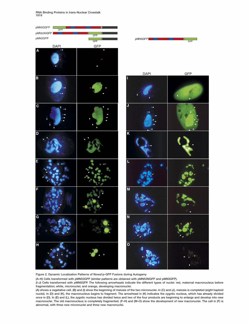
Dynamic Nuclear-Localization Pattern Drivenby the C-Terminal DomainTo study the subcellular localization of Nowa1p, dif-ferent GFP-fusion constructs were microinjected intothe macronucleus of vegetative cells (Figure 2). All con-structs contained the natural NOWA1 regulatory se-quences (163 bp upstream of the initiator ATG and 50bp downstream of the stop codon). P. tetraurelia in-tergenic sequences are extremely short [24], and thesesegments were found to be sufficient for proper regula-tion of the constructs. Figures 2A–2H show the resultsobtained with pMN53GFP, in which the GFP is fused atthe N terminus of Nowa1p. GFP fluorescence could notbe detected during vegetative growth of the trans-

Current Biology1618
Figure 1. Structure and Expression of the NOWA1 and NOWA2Genes
(A) The drawings show the alternating blocks of GGWG and FRGrepeats in the NTD (examples of repeat sequences are given below;the full sequences can be seen in Figure S1). N represents a stretchof 23 Asn residues interspersed with Tyr and His residues (reducedto three Asn residues in Nowa2p).(B) Northern-blot analysis of vegetative cells (“V”) and different timepoints during a mass autogamy (same as ND7 control culture ofFigure 5). EA (early autogamy) indicates cells committed for autog-amy, macronucleus not yet fragmented. The other time points ana-lyzed are 5, 7.5, 30, and 45 hr (t = 0 is the beginning of macro-nuclear fragmentation). For each time point, the fraction of cells indifferent developmental stages can be seen in Figure S2. The blotwas successively hybridized with NOWA1-, NOWA2-, and 17SrRNA-specific probes. The histogram shows normalization of theNOWA1 signal with the rRNA signal.(C) Northern-blot analysis of starved, sexually reactive cells (SR,mating-type O; mating-type E is not shown but gave the same re-sult) and different time points after the mixing of complementarymating types (t = 2–7.5 hr).
frbflGtmviTf
i2nrtagstartpfwtwtri(
rFacttidnstsoriop
AIBmttnsrt
ormed clones, but it specifically appeared in the mac-onucleus when autogamy was induced by starvation,efore meiosis of the micronuclei. Consistent with the
act that commitment to autogamy occurs before theast vegetative division [25], some dividing cells withFP-labeled macronuclei were observed upon starva-
ion. When such cells were isolated into rich medium,eiosis always followed completion of the ongoing di-
ision (not shown), confirming that the fusion constructs expressed only when cells are committed to meiosis.he GFP signal remained stable during macronucleusragmentation, which follows meiosis (Figure 2C–2E).
GFP was never detected in germline micronuclei dur-ng vegetative growth (Figure 2A) or meiosis (FiguresB and 2C) and up to the first division of the zygoticucleus (Figure 2D). Soon after the second division, itapidly accumulated in two of the four products, ashese began to enlarge and develop into macronuclearnlagen (Figure 2E). GFP fluorescence then pro-ressively increased in the developing anlagen andimultaneously decreased in the fragments of the ma-ernal macronucleus, where it became more punctatend finally disappeared (Figures 2F–2H). Note that inare cases where cells developed three anlagen andhree micronuclei (which may be an effect of theMN53GFP fusion; see Expression of pMN53GFP Af-
ects Macronuclear Development), the three anlagenere labeled (Figure 2F). The same dynamic localiza-
ion pattern was observed when the GFP sequenceas inserted within the asparagine stretch separating
he two domains (pMN53NGFP) or when the NTD wasemoved from the latter construct (pMN3GFP), indicat-ng that the CTD is sufficient to drive GFP localizationdata not shown).
Removal of the CTD from the pMN53NGFP constructesulted in a different localization pattern (pMN5GFP,igure 2). GFP first appeared in the cytoplasm as wells in the macronucleus but not visibly in the micronu-lei (Figure 2I); during meiosis, it concentrated slowly inhe macronucleus (Figure 2J); remained confined therehroughout the process of fragmentation, never enter-ng the new macronuclei (Figures 2K–2N); and finallyisappeared from the fragments of the maternal macro-ucleus, albeit more slowly than the previous GFP fu-ions (Figure 2O). Finally, a construct containing onlyhe GFP coding sequence under the control of theame NOWA1 regulatory sequences was expressednly during autogamy and produced a GFP signal thatemained cytoplasmic throughout the process, indicat-ng that the specific expression of NOWA1 at the onsetf sexual processes is entirely determined by very shortromoter and/or terminator sequences.
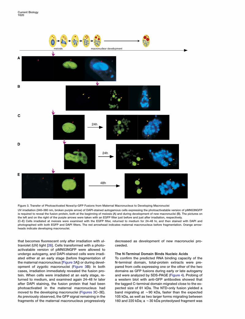
Photoactivatable Nowa1p-GFP Fusions Transported between Nucleiecause the bulk of NOWA1 expression occurs beforeeiosis, it is unlikely that the relocalization of CTD-con-
aining GFP fusions during development could be dueo degradation of the protein accumulated in the mater-al macronucleus and direct targeting of newly synthe-ized protein to the developing macronuclei. To test di-ectly whether the protein can be transported betweenhese nuclei, we used a photoactivatable GFP variant
RNA Binding Proteins in trans-Nuclear Crosstalk1619
Figure 2. Dynamic Localization Patterns of Nowa1p-GFP Fusions during Autogamy
(A–H) Cells transformed with pMN53GFP (similar patterns are obtained with pMN53NGFP and pMN3GFP).(I–J) Cells transformed with pMN5GFP. The following arrowheads indicate the different types of nuclei: red, maternal macronucleus beforefragmentation; white, micronuclei; and orange, developing macronuclei.(A) shows a vegetative cell. (B) and (I) show the beginning of meiosis of the two micronuclei. In (C) and (J), meiosis is completed (eight haploidnuclei). In (D) and (K), the macronucleus begins to fragment. The arrowhead in (K) indicates the zygotic nucleus, which has already dividedonce in (D). In (E) and (L), the zygotic nucleus has divided twice and two of the four products are beginning to enlarge and develop into newmacronuclei. The old macronucleus is completely fragmented. (F–H) and (M–O) show the development of new macronuclei. The cell in (F) isabnormal, with three new micronuclei and three new macronuclei.
Current Biology1620
that becomes fluorescent only after irradiation with ul-traviolet (UV) light [26]. Cells transformed with a photo-activatable version of pMN53NGFP were allowed toundergo autogamy, and DAPI-stained cells were irradi-ated either at an early stage (before fragmentation ofthe maternal macronucleus [Figure 3A]) or during devel-opment of zygotic macronuclei (Figure 3B): In bothcases, irradiation immediately revealed the fusion pro-tein. When cells were irradiated at an early stage, re-turned to medium, and examined again 24–48 hr laterafter DAPI staining, the fusion protein that had beenphotoactivated in the maternal macronucleus hadmoved to the developing macronuclei (Figures 3C–3E).As previously observed, the GFP signal remaining in thefragments of the maternal macronucleus progressively
dc
TTNpdaatpb11
Figure 3. Transfer of Photoactivated Nowa1p-GFP Fusions from Maternal Macronucleus to Developing Macronuclei
UV irradiation (340–380 nm, broken purple arrow) of DAPI-stained autogamous cells expressing the photoactivatable version of pMN53NGFPis required to reveal the fusion protein, both at the beginning of meiosis (A) and during development of new macronuclei (B). The pictures onthe left and on the right of the purple arrows were taken with an EGFP filter just before and just after irradiation, respectively.(C–E) Cells irradiated at meiosis were examined with the EGFP filter, returned to medium for 24–48 hr, and then stained with DAPI andphotographed with both EGFP and DAPI filters. The red arrowhead indicates maternal macronucleus before fragmentation. Orange arrow-heads indicate developing macronuclei.
ecreased as development of new macronuclei pro-eeded.
he N-Terminal Domain Binds Nucleic Acidso confirm the predicted RNA binding capacity of the-terminal domain, total-protein extracts were pre-ared from cells expressing one or the other of the twoomains as GFP fusions during early or late autogamynd were analyzed by SDS-PAGE (Figure 4). Probing ofwestern blot with anti-GFP antibodies showed that
he tagged C-terminal domain migrated close to the ex-ected size of 61 kDa. The NTD-only fusion yielded aand migrating at w90 kDa, faster than the expected05 kDa, as well as two larger forms migrating between60 and 220 kDa; a w30 kDa proteolysed fragment was

RNA Binding Proteins in trans-Nuclear Crosstalk1621
Figure 4. Western-Blot Analysis of Total Cell Proteins from ClonesTransformed with pMN3GFP or pMN5GFP or from an UninjectedControl Clone during Early or Late Autogamy
E represents early autogamy, L represents late autogamy, and Mrepresents molecular weight markers.(A) Anti-GFP polyclonal antibodies revealed with the ECL kit. Ar-rowheads indicate the different GFP-positive forms produced frompMN5GFP: 1–2 indicate the two large forms migrating between 160and 200 kDa; 3 indicates the 90 kDa form, slightly faster than theexpected 105 kDa; 4 indicates the w30 kDa product that may rep-resent cleaved GFP.(B–E) The membrane (or a duplicate) was then successively incu-bated (in the presence of excess cold double-stranded DNA) withthe following various 32P-labeled probes: (B), RNA/DNA oligonucle-otide duplex; (C), RNA/RNA oligonucleotide duplex; (D), single-stranded DNA oligonucleotide; and (E), single-stranded RNA oligo-nucleotide.
consistently observed in early-autogamy samples,even at high concentrations of protease inhibitors.
The same membrane or a duplicate was then incu-bated with various 32P-labeled oligonucleotide probesin the presence of excess duplex DNA (see Experimen-tal Procedures). In the early-autogamy samples, thetwo larger forms of the N-terminal domain fusion, butnot the w90 kDa form or the C-terminal domain fusion,were able to bind single-stranded (ss) RNA, double-stranded (ds) RNA, and ssDNA, as well as an RNA/DNAduplex. It is not possible to quantitate relative affinitiesbecause the half-lives of these different probes duringincubation were different. However, when compared tothe background of endogenous proteins, the two largeforms appeared to be more strongly labeled by theRNA/DNA duplex (Figure 4B) than by other probes.
Although the pattern was not changed after reduc-tion of disulfide bridges, the apparent size of the topband (w210 kDa) suggests that it could be a covalentdimer of the NTD fusion. The smaller of the large forms,which binds three of the probes equally well but givesa weaker GFP signal, might then represent a dimer ofthe NTD fusion with a free NTD produced by proteolysisof the NTD fusion and/or endogenous Nowa1p/2p. At a
later stage of autogamy, the two large forms appearedto have lost much of their nucleic acid binding capacity(Figure 4). However, the significance of this is unclearbecause the NTD-only fusion remained confined to thefragments of the old macronucleus, whereas at thatstage the protein should have been transferred to de-veloping macronuclei.
Expression of pMN53GFP AffectsMacronuclear DevelopmentClones expressing large amounts of the pMN53GFP fu-sion during autogamy (as judged from the intensity ofGFP fluorescence, see Figure 2) yielded progeny withsevere developmental defects that often led to death.Surviving postautogamous clones showed reducedgrowth rate, altered cell morphology, and O-to-E mat-ing-type change (data not shown). Death and develop-mental defects were specific effects of the fusion pro-tein; postautogamous progeny of cells transformedwith high copy numbers of the untagged NOWA1 genewere perfectly healthy and only occasionally showedmating-type change. Interestingly, mating-type deter-mination in the developing macronucleus is known tobe epigenetically controlled by the maternal macronu-cleus [2], suggesting that Nowa1p is involved in thismaternal effect. The other GFP-fusion constructs rarelycaused any developmental defect in postautogamousprogeny, even at high copy numbers, suggesting thatonly pMN53GFP had dominant-negative effects (see Si-lencing of NOWA1/2 Results in Postautogamous Le-thality).
Autogamy of pMN53GFP-transformed cells also oc-casionally resulted in macronuclear regeneration. Thisprocess occurs at a low frequency when developmentof new macronuclei aborts, allowing some cells to re-generate a fully functional macronucleus from a frag-ment of the old macronucleus. Vegetative clones pro-duced by macronuclear regeneration had wild-typegrowth rates and morphology and did not change mat-ing type, indicating that Nowa1p is required only formacronuclear development (see Discussion).
Silencing of NOWA1/2 Resultsin Postautogamous LethalityTo study the effects of Nowa1p depletion, we used thefeeding technique to induce RNA interference [14].Cells were fed an E. coli strain producing dsRNA ho-mologous to an 846 bp NOWA1-gene sequence encod-ing the C-terminal domain. This dsRNA can also be ex-pected to silence NOWA2 because the two genes are87% identical in that region, with a total of 237 bp ofperfect identity in segments R 23 bp [15]. A controlculture was fed an E. coli strain producing dsRNA ho-mologous to ND7, a gene required for trichocyst dis-charge [27]. Consistent with the known expression pat-terns, NOWA1 dsRNA feeding did not have any visibleeffect during vegetative growth, whereas ND7 dsRNAfeeding resulted in a completely mutant phenotype(nondischarge) in less than three to four divisions.
We then induced autogamy in both cultures by lettingthe cells starve after 3–4 vegetative divisions in thefeeding media, and total-RNA and -DNA samples wereextracted at different times. The first samples (early au-

Current Biology1622
togamy) were taken as cells were just beginning tostarve, and the t = 0 reference time points were arbitrar-ily defined when DAPI staining revealed the first signsof macronuclear fragmentation in 20%–30% of cells,about 25 hr later (Figure S2). Commitment to autogamyoccurs at a fixed point of the w6 hr cell cycle [25], put-ting a severe limit on synchrony in mass cultures. Fur-thermore, the speed with which the food supply be-comes exhausted is quite variable in such experiments.Thus, although a larger fraction of cells showed com-plete macronuclear fragmentation between 2.5 and 7.5hr in the ND7 culture, the difference is not significant.The development of new macronuclei followed a similartime course between 20 and 45 hr in both cultures (Fig-ure S2).
To check the efficiency of silencing, we analyzedRNA samples on northern blots (Figure 5). In the controlsilencing of ND7, NOWA1 and NOWA2 mRNAs werevery abundant in the early-autogamy time point andgradually declined afterward, whereas ND7 mRNA levelswere strongly reduced throughout autogamy. In con-trast, NOWA1 and NOWA2 mRNAs could not be de-
vnpNts(n
SoTmeapdeasn
Figure 5. Northern-Blot Analysis of NOWA1/2 and ND7 Silencing
Total-RNA samples were extracted from vegetative cultures grownon Klebsiella (V, no silencing), from early autogamy of the samecultures after three to four divisions in ND7 or NOWA1 dsRNA-pro-ducing E. coli media (EA, cells were starved and committed forautogamy but maternal macronuclei were not yet fragmented) orfrom different time points after the onset of macronuclear fragmen-tation (t = 5, 7.5, 30, and 45 hr; the percentage of cells in the dif-ferent developmental stages for each point can be seen in Fig-ure S2).(A–D) RNA samples were run on an agarose gel, blotted, and hy-bridized with a probe of the CTD of NOWA1 (A), with a NOWA2-specific oligonucleotide probe (B), with a probe specific for theND7 gene (C), or with an oligonucleotide probe of the 17S rRNA(D). The histograms above the blots show the relative levels ofNOWA1/2 and ND7 signals after normalization with the rRNAsignals.(E and F) RNA samples were run on a 15% polyacrylamide-urea gel,blotted, and hybridized with the same NOWA1 and ND7 probes,revealing the doublet formed by the sense (top) and antisense (bot-tom) strands of the w23 nt siRNAs.
tftaabd
woNtsNbscasohnNps(pwa
qctnfmNsrd
ected during early autogamy after NOWA1 dsRNAeeding. NOWA1 silencing remained quite efficienthroughout autogamy, whereas NOWA2 silencing waslmost completely relieved about 30 hr after the early-utogamy time point (t = 5 hr, see Figure 5B): This maye due to the limited homology between the NOWA1sRNA inducer and NOWA2 mRNA.The presence of silencing-associated w23 nt siRNAsas also tested on northern blots after electrophoresisf the same RNA samples on acrylamide-urea gels.D7 siRNAs were specifically detected as a doublet in
he ND7 feeding culture and appeared to remain con-tant during autogamy (Figure 5F). Hybridization withD7 single-stranded probes showed that the top andottom bands of the doublet contain sense and anti-ense siRNAs, respectively. A comparison with 23 ntontrol RNA oligonucleotides representing sense andntisense ND7 sequences indicated that both siRNAtrands are probably 23 nt long, the slower migrationf the sense strand being entirely accounted for by itsigher purine content (data not shown). Similarly, w23t NOWA1 siRNAs were specifically detected in theOWA1 feeding culture, although their amounts ap-eared to decrease as autogamy proceeded. The anti-ense strand also migrated faster than the sense strandas confirmed with strand-specific probes) but ap-eared to be present in much lower amounts, as if itere depleted in the process of degrading largemounts of NOWA1/2 mRNAs.To determine whether expression of NOWA1/2 is re-
uired for the development of functional new macronu-lei, we took 54 postautogamous cells from each cul-ure after the latest time point and isolated them intoormal medium bacterized with Klebsiella. All 54 clonesrom the ND7 feeding culture were viable and had nor-al growth rates. In contrast, all 54 clones from theOWA1 feeding culture died after a few vegetative divi-ions, as the nonreplicating fragments of the old mac-onucleus were diluted out by random distribution toaughter cells. Thus, although new macronuclei de-eloping in conditions of Nowa1p/2p depletion have aormal appearance, they appear to be unable to sup-ort vegetative growth. In other experiments whereOWA1 silencing was only partial, a fraction of postau-
ogamous clones survived and showed phenotypesimilar to those induced by overexpression of pMN53GFPnot shown), suggesting that the latter has a dominant-egative effect.
ilencing of NOWA1/2 Impairs Eliminationf Micronuclear-Specific Sequenceshe progression of genome rearrangements duringacronuclear development was examined by South-
rn-blot analysis of the total DNA samples from bothutogamous cultures. In wild-type cells, transient am-lification of micronucleus-specific sequences can beetected at intermediate time points because DNAlimination in developing macronuclei starts only afterfew endoreplication cycles [28]. This was indeed ob-
erved for two multicopy class II transposons (O. Gar-ier, A. Le Mouël, M. Prajer, and E.M., unpublished data)
RNA Binding Proteins in trans-Nuclear Crosstalk1623
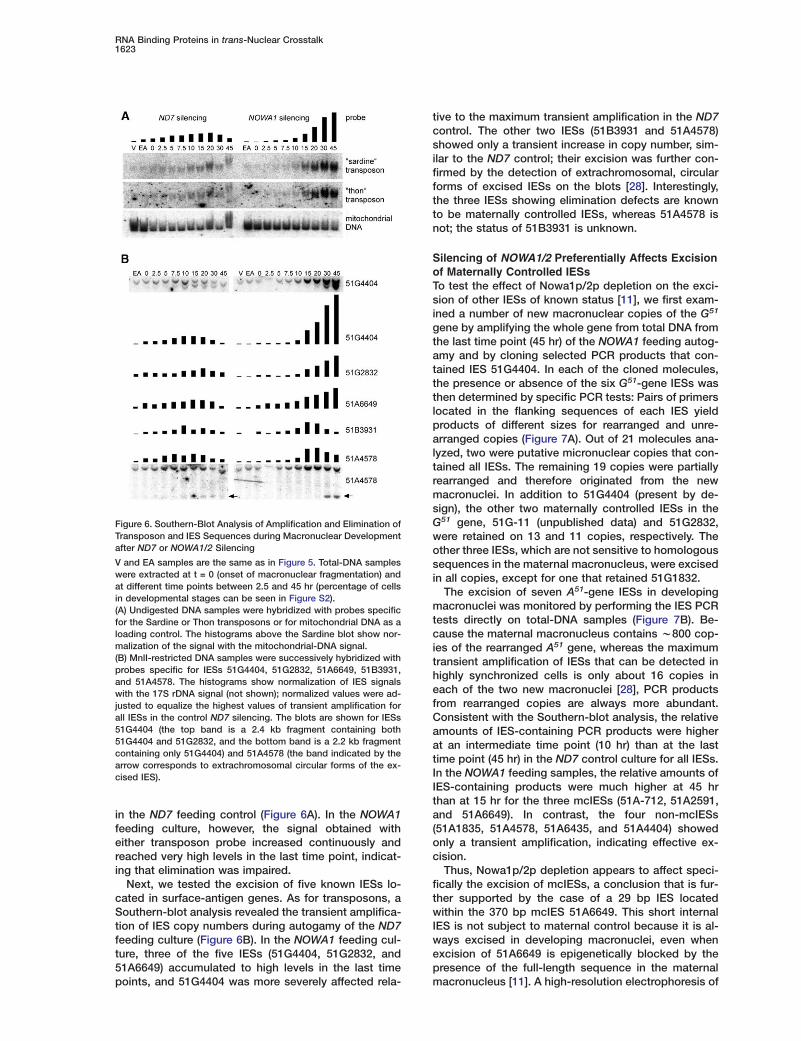
Figure 6. Southern-Blot Analysis of Amplification and Elimination ofTransposon and IES Sequences during Macronuclear Developmentafter ND7 or NOWA1/2 Silencing
V and EA samples are the same as in Figure 5. Total-DNA sampleswere extracted at t = 0 (onset of macronuclear fragmentation) andat different time points between 2.5 and 45 hr (percentage of cellsin developmental stages can be seen in Figure S2).(A) Undigested DNA samples were hybridized with probes specificfor the Sardine or Thon transposons or for mitochondrial DNA as aloading control. The histograms above the Sardine blot show nor-malization of the signal with the mitochondrial-DNA signal.(B) MnlI-restricted DNA samples were successively hybridized withprobes specific for IESs 51G4404, 51G2832, 51A6649, 51B3931,and 51A4578. The histograms show normalization of IES signalswith the 17S rDNA signal (not shown); normalized values were ad-justed to equalize the highest values of transient amplification forall IESs in the control ND7 silencing. The blots are shown for IESs51G4404 (the top band is a 2.4 kb fragment containing both51G4404 and 51G2832, and the bottom band is a 2.2 kb fragmentcontaining only 51G4404) and 51A4578 (the band indicated by thearrow corresponds to extrachromosomal circular forms of the ex-cised IES).
in the ND7 feeding control (Figure 6A). In the NOWA1feeding culture, however, the signal obtained witheither transposon probe increased continuously andreached very high levels in the last time point, indicat-ing that elimination was impaired.
Next, we tested the excision of five known IESs lo-cated in surface-antigen genes. As for transposons, aSouthern-blot analysis revealed the transient amplifica-tion of IES copy numbers during autogamy of the ND7feeding culture (Figure 6B). In the NOWA1 feeding cul-ture, three of the five IESs (51G4404, 51G2832, and51A6649) accumulated to high levels in the last timepoints, and 51G4404 was more severely affected rela-
tive to the maximum transient amplification in the ND7control. The other two IESs (51B3931 and 51A4578)showed only a transient increase in copy number, sim-ilar to the ND7 control; their excision was further con-firmed by the detection of extrachromosomal, circularforms of excised IESs on the blots [28]. Interestingly,the three IESs showing elimination defects are knownto be maternally controlled IESs, whereas 51A4578 isnot; the status of 51B3931 is unknown.
Silencing of NOWA1/2 Preferentially Affects Excisionof Maternally Controlled IESsTo test the effect of Nowa1p/2p depletion on the exci-sion of other IESs of known status [11], we first exam-ined a number of new macronuclear copies of the G51
gene by amplifying the whole gene from total DNA fromthe last time point (45 hr) of the NOWA1 feeding autog-amy and by cloning selected PCR products that con-tained IES 51G4404. In each of the cloned molecules,the presence or absence of the six G51-gene IESs wasthen determined by specific PCR tests: Pairs of primerslocated in the flanking sequences of each IES yieldproducts of different sizes for rearranged and unre-arranged copies (Figure 7A). Out of 21 molecules ana-lyzed, two were putative micronuclear copies that con-tained all IESs. The remaining 19 copies were partiallyrearranged and therefore originated from the newmacronuclei. In addition to 51G4404 (present by de-sign), the other two maternally controlled IESs in theG51 gene, 51G-11 (unpublished data) and 51G2832,were retained on 13 and 11 copies, respectively. Theother three IESs, which are not sensitive to homologoussequences in the maternal macronucleus, were excisedin all copies, except for one that retained 51G1832.
The excision of seven A51-gene IESs in developingmacronuclei was monitored by performing the IES PCRtests directly on total-DNA samples (Figure 7B). Be-cause the maternal macronucleus contains w800 cop-ies of the rearranged A51 gene, whereas the maximumtransient amplification of IESs that can be detected inhighly synchronized cells is only about 16 copies ineach of the two new macronuclei [28], PCR productsfrom rearranged copies are always more abundant.Consistent with the Southern-blot analysis, the relativeamounts of IES-containing PCR products were higherat an intermediate time point (10 hr) than at the lasttime point (45 hr) in the ND7 control culture for all IESs.In the NOWA1 feeding samples, the relative amounts ofIES-containing products were much higher at 45 hrthan at 15 hr for the three mcIESs (51A-712, 51A2591,and 51A6649). In contrast, the four non-mcIESs(51A1835, 51A4578, 51A6435, and 51A4404) showedonly a transient amplification, indicating effective ex-cision.
Thus, Nowa1p/2p depletion appears to affect speci-fically the excision of mcIESs, a conclusion that is fur-ther supported by the case of a 29 bp IES locatedwithin the 370 bp mcIES 51A6649. This short internalIES is not subject to maternal control because it is al-ways excised in developing macronuclei, even whenexcision of 51A6649 is epigenetically blocked by thepresence of the full-length sequence in the maternalmacronucleus [11]. A high-resolution electrophoresis of

Current Biology1624
Figure 7. Analysis of Excision of mcIESs and Other IESs in the G51
and A51 Genes
(A) Analysis of cloned individual copies of the G51 gene from de-veloping macronuclei after NOWA1/2 silencing. Black boxes in thetop line represent the six IESs (not to scale). Numbered lines showthe results of six specific IES PCR tests performed on each of 19cloned copies. A black box indicates that the IES was retained; abroken line indicates that the IES was excised.(B) Specific PCR tests performed directly on total-DNA samplesfrom the ND7 (t = 10 and 45 hr) and NOWA1/2 (t = 15 and 45 hr)silencing cultures. Representative gels are shown for IESs 51A2591and 51A4404. Amounts of IES-containing (mic) and IES-excised(mac) PCR products were quantified after ethidium-bromide stain-ing; the histograms show the relative amounts of IES-containingproducts (mic/mac) in the four DNA samples for all tested IESs.
Naatmlmomfn
tacItIvpmrttshemirnctwm
NoWangidtipctdi5m
the 51A6649 PCR products showed that the 29 bp IESwas completely excised from all copies of 51A6649 thatwere maintained in the new macronucleus as a resultof NOWA1/2 silencing (data not shown).
Discussion
Nowa Proteins Are Required for Developmentof Functional MacronucleiThe very similar paralogous genes NOWA1 and NOWA2
spcaaaptl5
are tightly regulated during the P. tetraurelia life cycle.
orthern blots and GFP fusions have shown that theyre briefly but massively expressed only in cells thatre committed to meiosis. Commitment occurs afterhe mixing of sexually reactive cells of complementaryating types in the case of conjugation but before the
ast vegetative division in the case of autogamy. Thisakes NOWA1/2 expression the earliest known markerf commitment to meiosis. However, silencing experi-ents have shown that these genes are not required
or meiosis per se or for any of the cytologically visibleuclear events of fertilization and early development.The lethal effect of Nowa1p/2p depletion on postau-
ogamous clones may be entirely attributable to the re-rrangement defects observed in developing macronu-lei: Retention of a significant fraction of the w50,000ESs in the genome would certainly produce a nonfunc-ional macronucleus unable to support vegetative life.ndeed, postautogamous clones died only after a fewegetative divisions, when gene expression is sup-osed to switch from the fragments of the maternalacronucleus, which are progressively diluted out by
andom segregation to daughter cells, to the fully ma-ure zygotic macronucleus. Furthermore, the pheno-ypic effects of the pMN53GFP fusion were only ob-erved in true postautogamous progeny, whereas theeterokaryons that resulted from macronuclear regen-ration and contained zygotic micronuclei and maternalacronuclei did not show the mutant phenotypes. This
ndicates that the essential functions of Nowa1p/2p areequired only for normal development of zygotic macro-uclei. The frequent occurrence of O-to-E mating-typehange in true progeny further suggests that these pro-eins are involved in the epigenetic mechanism byhich the maternal macronucleus controls develop-ental alternatives in zygotic macronuclei [2].
owa Proteins Are Involved in the Recognitionf a Subset of Eliminated Sequencese have shown that the silencing of NOWA1/2 during
utogamy impairs the elimination of a subset of micro-ucleus-specific sequences. It is not known whether allermline copies of the two class II transposons exam-
ned are affected, but the analysis of a number of indivi-ual IESs showed that Nowa1p/2p depletion preferen-ially affects the excision of mcIESs. The depletionnduced by NOWA1 dsRNA feeding was not complete,articularly for Nowa2p, and a fraction of mcIES copiesould still be excised; quantitative analyses indicatedhat this partial depletion affected different mcIESs toifferent extents. Non-mcIESs were correctly excised,
ncluding the 29 bp IES that is located within mcIES1A6649. Although the possibility remains that non-cIESs would also be affected in the complete ab-
ence of Nowa1p/2p, the effects of partial depletionrovide strong support for the existence of distinct IESlasses in P. tetraurelia and further indicate that the re-rrangement defects observed in our time-coursenalysis are not due to a general delay of genome re-rrangements. It is also likely that Nowa1p/2p depletionermanently impairs the excision of mcIESs, ratherhan just delaying it until after the last time point ana-yzed, because at least some mcIESs (51G2832 and1G4404) were found to be occasionally maintained in
the mature macronuclei of vegetatively growing post-

RNA Binding Proteins in trans-Nuclear Crosstalk1625
autogamous clones surviving the expression of thedominant-negative pMN53GFP fusion (data not shown).
There is currently no evidence that mcIESs and otherIESs are excised by distinct endonucleolytic machiner-ies. No significant difference was observed betweenthe two classes in the geometry of the double-strandbreaks that can be transiently detected at their bound-aries (4-base 5# overhangs centered on the TA dinucle-otides, as well as 3-base 5# overhangs that are believedto be processed forms of the former) ([8]; A. Gratias, S.Malinsky, and M. Bétermier, personal communication).The only known difference is that the excision ofmcIESs, but not that of other IESs, depends on a trans-nuclear comparison of the germline genome with thematernal macronuclear genome; transient double-strand breaks cannot be detected at the boundaries ofan mcIES when the homologous sequence is presentin the maternal macronucleus, as observed for 51G4404(G. Lepère, S. Duharcourt, and M. Bétermier, personalcommunication). Nowa proteins are therefore likely tobe involved in the mechanism of trans-nuclear compar-ison. This conclusion is further supported by the factthat the sensitivities of different mcIESs to Nowa1p/2pdepletion quantitatively parallels their sensitivities tohomologous maternal sequences, 51G4404 being themost sensitive in both cases [11].
The recognition of germline transposons may alsodepend on such a comparison. Indeed, there is indirectevidence that their imprecise elimination is determinedby homology-dependent effects, both in Paramecium[12] and in T. thermophila [29]. In the latter, similar ef-fects are also thought to result in the elimination of neobacterial genes introduced into the micronuclear ge-nome when these are absent from the parental macro-nucleus [20, 30].
Properties of Nowa ProteinsThe NTD and CTD domains of Nowa proteins have dis-tinct properties. We have shown that the Nowa1p CTDis necessary and sufficient both for the rapid accumula-tion of newly synthesized protein in the maternal mac-ronucleus, prior to meiosis of the micronuclei, and laterfor the transfer of the same protein molecules to de-veloping macronuclei, as demonstrated by the use of aphotoactivatable GFP fusion. The Nowa2p CTD is 90%identical and 99% similar to that of Nowa1p, and threepotential nuclear-localization signals, as well as a high-scoring coiled-coil region, are conserved. The Nowa1pand Nowa2p NTDs differ mostly by the lengths of somerepeat blocks (see Figure S1). Overall, the high sim-ilarity and identical expression patterns of the two par-alogous proteins suggest that they have redundantfunctions.
Of the two types of short repeats that make up theNowa1p/2p NTD, the FRG type resembles the arginine/glycine-rich boxes that mediate RNA binding in a num-ber of proteins. Such motifs are also present in severalproteins, such as dFXR and VIG in Drosophila [31] andTbAGO1 in trypanosomes [32, 33], that have been im-plicated in RNAi. The GGWG type may also bind RNAbecause it is similar to the repeats present in an RNAbinding domain of the PrP27 prion [34]. Northwesternblots of a GFP fusion containing only the Nowa1p NTD
confirmed its predicted ssRNA binding capacity andshowed that it can also bind ssDNA, dsRNA, and anRNA/DNA duplex, although the data do not allow us tocompare the binding affinities for these different sub-strates. We did not examine the binding of the Nowa1pNTD to different sequences, but because our assayswere performed with an arbitrarily chosen oligonucleo-tide sequence, it is unlikely that the NTD shows muchsequence specificity.
We could not directly identify the nucleic acids boundby Nowa1p in vivo because all attempts to immuno-precipitate the full-length fusion pMN53NGFP with anti-GFP antibodies failed, yielding only proteolysed frag-ments. Intriguingly, the GFP-positive bands that boundnucleic acids in north- and southwestern assays mi-grated at about twice the size expected for thepMN5GFP fusion, even after SDS denaturation and re-duction of disulfide bridges, whereas a band migratingslightly faster than the expected size did not bind theprobes. This suggests that covalent modifications (pos-sibly including dimerization) may regulate nucleic acidbinding by the NTD; future studies will clearly be re-quired to better understand its biochemical and bind-ing properties.
Possible Roles of Nowa Proteins in RNA-Mediated,trans-Nuclear CrosstalkWe cannot formally exclude the possibility that Nowaproteins are transcription factors required for the ex-pression of other genes specifically involved in theelimination of mcIESs and transposons. However, thehigh expression levels and homogeneous distributionof Nowa1p/2p throughout chromatin (indicated by thecolocalization of fusion GFP and DAPI signals) do notsupport a specific action on a few genes and suggestinstead that they interact with many (possibly all) geno-mic sequences. Furthermore, it is difficult to imaginewhy such transcription factors should be massivelytransferred from maternal to zygotic macronuclei, evenif the putative Nowa1p/2p-regulated genes were ex-pressed from both nuclei, because maternal genes canbe expressed in the fragments of the old macronucleusfor at least two cell cycles after meiosis [35]. Thus, be-cause there is a perfect overlap between the subsetof IESs that are known to be sensitive to homology-dependent maternal effects and the subset that is af-fected by Nowa1p/2p depletion, the massive transferof these proteins between nuclei is likely to reflect amore direct role in the RNA-mediated, trans-nuclearcrosstalk implied by these effects. The properties ofNowa proteins are consistent with a role in non-se-quence-specific RNA transport; in what follows, weconsider possible mechanisms of action in relation todifferent models for genome comparison.
In the simplest of these models, maternal macro-nuclear sequences would produce specialized noncod-ing transcripts that would be transported to the de-veloping macronucleus, where they would target, onmacronuclear-destined sequences, specific epigeneticmarks that ensure their maintenance. Initially proposedto account for the maternal inhibition of mcIES excision[11], this model could also explain the rescue of mater-nally inherited gene deletions, as well as their inductionby the silencing-associated w23 nt siRNAs: These could
target deletions indirectly, by destroying the protective
Current Biology1626
maternal transcripts [15]. If Nowa proteins were involvedin the transport or action of these protective RNAs, in-terfering with their function would be expected to result inconstitutive excision of mcIESs, but NOWA1/2 silencinginstead resulted in constitutive retention.
Thus, if Nowa proteins indeed transport RNAs to thedeveloping macronucleus, these are more likely to in-clude molecules required for the direct recognition ofmcIESs. This would fit a different model that was sug-gested by the likely role of endogenous short RNAsspecifically produced by the meiotic micronucleus in T.thermophila [22]. These scan (scn) RNAs appear to beexported to the parental macronucleus, where they areassumed to scan the genome by pairing interactions.The scnRNAs that cannot pair with homologous se-quences would then be selectively transported to de-veloping macronuclei to target histone H3 K9 methyla-tion on homologous sequences, resulting in thespecific elimination of micronuclear sequences that areabsent from the parental macronucleus. A similarmechanism may operate in P. tetraurelia because en-dogenous short RNAs, distinct from the w23 nt siRNAs,are also produced from the micronuclear genome dur-ing meiosis (G. Lepère, M.N., S. Duharcourt, and E.M.,unpublished data). This scnRNA/maternal-DNA scan-ning model could explain the maternal inhibition ofmcIES excision and the rescue of maternally inheritedgene deletions, but it is more difficult to reconcile withthe fact that silencing maternal genes by RNA interfer-ence induces the deletion of homologous zygoticgenes; the presence of the DNA sequence in the mater-nal macronucleus should still prevent homologousscnRNAs (or siRNAs, if functionally equivalent) from de-leting these genes in developing macronuclei.
All known homology-dependent maternal effects canbe explained by combining features from both models.We have proposed that the scnRNAs from the micronu-cleus be compared with an RNA copy of the maternalgenome rather than with DNA itself [15]. In cases ofsilencing, the degradation of maternal RNAs by thew23 nt siRNAs would mimic the absence of the gene,allowing scnRNAs to target deletions in the developingmacronucleus. A similar scnRNA/maternal-RNA scan-ning model was also considered in T. thermophila [18].In such a mechanism, the Nowa proteins could be re-quired at any step upstream of the final recognition ofmcIESs and transposons by homologous scnRNAs; therecognition of non-mcIESs may not depend on shortRNAs or may involve short RNAs that are not selectedby the scanning of maternal sequences. Nowa1p-GFPfusions were not detected in micronuclei, and prelimi-nary results indicate that Nowa proteins are not re-quired for the production of scnRNAs during meiosis(M.N. and E.M., unpublished data). As noted above, onepossible function may be the transport of selectedscnRNAs to developing macronuclei. Another intriguingpossibility is suggested by the resemblance of the NTDshort repeats to those present in proteins known tohave strand-annealing activity [36–38]: The Nowa pro-teins could also be required for the intense pairing ac-tivity implied by the scanning process in the maternalmacronucleus and in developing macronuclei.
CTsmtaiapohgaskiabhc
E
PAsIbaGp
CNfApoasnfsambNamuTta
SFpiitaddβ
MPmcjmm
onclusionshis study establishes a mechanistic link between exci-ion of maternally controlled IESs and elimination ofulticopy transposons, suggesting that their recogni-
ion depends on a common trans-nuclear, RNA-medi-ted pathway that appears to involve conserved RNA-
nterference factors. The identification of Nowa proteinss novel components expands our knowledge of thisathway and opens the way for more detailed studiesf RNA-transport and -pairing interactions. Althoughomology-dependent maternal effects on the zygoticenome have so far been firmly established only in cili-tes, a recent study of the hothead mutant in Arabidop-is has led to the provocative hypothesis that plantseep an extragenomic memory of ancestral sequences
n the form of RNA molecules, which may later be useds templates to convert genomic sequences [39]. It wille interesting to see whether structural or functionalomologs of the Nowa proteins are involved in similar,ryptic mechanisms in other eukaryotes.
xperimental Procedures
aramecium Strains, Cultivation, Conjugation, and Autogamyll experiments were carried out with the entirely homozygoustrain 51. Cells were grown in a wheat grass powder (WGP; Pinesnternational, Lawrence, KS) infusion medium bacterized the dayefore use with Klebsiella pneumoniae, unless otherwise stated,nd supplemented with 0.8 mg/l of β-sitosterol (Merck, Darmstadt,ermany). Cultivation and autogamy were carried out at 27°C asreviously described [11].
onstructs and ProbesOWA1 constructs were obtained by cloning a 3287 bp MfeI-HincII
ragment (including nucleotides 10–3284 of accession numberJ876761) into the pCRScriptAmpSK+ vector (Stratagene). In theMN53GFP fusion construct, a homemade EGFP coding sequenceptimized for Paramecium codon usage was inserted immediatelyfter the NOWA1 initiating ATG. For pMN53NGFP, the same EGFPequence was inserted within the asparagine stretch, after engi-eering restriction sites. pMN5GFP and pMN3GFP were obtainedrom pMN53NGFP by deleting the NOWA1 CTD and NTD codingequences, respectively. The GFP and fusion sequences are avail-ble upon request. The constructs used in the silencing experi-ents were obtained by cloning PCR products (a 397 bp fragmentetween positions 1330 and 1726 of accession number Y07803 forD7, and an 846 bp fragment between positions 2401 and 3246 ofccession number AJ876761 for NOWA1) into the “feeding” plas-id and E. coli strain described in [14]. The same fragments were
sed for NOWA1 and ND7 probes on Southern and northern blots.he NOWA2-specific probe (a 100 nt oligonucleotide), as well asransposon, IES, rDNA, and mitochondrial-DNA probes are avail-ble upon request.
ilencing by dsRNA Feedingeeding media were prepared by inoculating precultures of the ap-ropriate feeding strains into WGP Paramecium medium contain-
ng ampicillin at 0.1 mg/ml and growing them overnight with shak-ng at 37°C. On the next day, the culture was diluted 10-fold intohe same medium. After 1 hr of incubation at 37°C, IPTG was addedt a final concentration of 0.5 mM to induce the synthesis ofsRNA, and the culture was incubated overnight at 37°C. The me-ium was cooled to 27°C and supplemented with 0.8 mg/l of-sitosterol just before use.
icroinjectionsaramecium cells from a single caryonidal clone in each experi-ent were microinjected in Volvic mineral water (Volvic, France)
ontaining 0.2% bovine serum albumin (BSA), under an oil film (Nu-ol), while they were visualized with a phase-contrast inverted
icroscope (Axiovert 35M, Zeiss). Column-purified (Qiagen) plas-ids containing NOWA-GFP fusion constructs were linearized

RNA Binding Proteins in trans-Nuclear Crosstalk1627
within the vector sequence, filtered on a 0.22 �m Ultrafree-MC filter(Millipore), precipitated with ethanol, and dissolved in filtered water.Approximately 5 pl of a 5 mg/ml solution were delivered into themacronucleus.
Protein Extraction, Electrophoresis, and BlottingCell pellets were resuspended in one volume of 5% SDS with“Complete” protease inhibitor with EDTA (Roche), boiled for 10 min,and frozen immediately. For western blots, total-protein samples(w5.103 cells/lane) were separated by SDS-PAGE electrophoresisand transferred to Optitran membranes (Schleicher & Schuell).Blots were revealed with the milk-powder standard procedure [40]with polyclonal anti-GFP antibodies and were visualized with anti-rabbit IgG-HRP (Amersham) and the ECL reagent (Amersham). Forsouth- and northwestern blots, western-blot membranes were in-cubated at room temperature (RT) in northwestern buffer (25 mMHepes [pH 7.9], 5 mM MgCl2, 50 mM NaCl, 0.5 mM EDTA, 0.025%Tween 20, 1 mM DTT) for 30 min and then in northwestern bufferwith 1% BSA for 1 hr before and 1 hr after the addition of salmonsperm DNA at 50 �g/ml. 32P-labeled oligonucleotide probes werepurified on MicroSpin G-25 columns (Amersham) and added to thebuffer. After incubation for 1.5 hr at RT, blots were washed twicefor 10 min in northwestern buffer at RT prior to autoradiography.The ssRNA probe was oligonucleotide rND7-1 (5#-GUGCUAGGAGGGAAAUGCAAGAG-3#). The dsRNA was obtained by annealingrND7-1 with rND7-2 (5#-CUUGCAUUUCCCUCCUAGCACGU-3#).The ssDNA probe was dND7-2 (5#-CTAGACGTGCTAGGAGGGAAATGCAAGAGT-3#), and the RNA/DNA duplex was obtained by an-nealing rND7-1 with dND7-2.
DNA and RNA ExtractionTwo-hundred- to four-hundred-milliliter cultures of exponentiallygrowing cells at w1000 cells/ml or of autogamous cells at w3000cells/ml were centrifuged. For DNA extraction, cell pellets werewashed in 10 mM tris-HCl (pH 7.0), resuspended in one volume ofthe same buffer, and quickly added to four volumes of lysis solution(0.44 M EDTA [pH 9.0], 1% SDS, 0.5% N-laurylsarcosine [Sigma],and 1 mg/ml proteinase K [Merck]). The lysates were incubated at55°C for at least 12 hr, gently extracted with phenol, and dialysedtwice against TE (10 mM tris-HCl, 1 mM EDTA, pH 8.0) containing20% ethanol and once against TE. RNA was extracted from un-washed cell pellets with the TRIzol (Invitrogen) procedure, modifiedby the addition of glass beads.
Southern and Northern BlotsDNA and RNA electrophoreses were carried out according to stan-dard procedures [40]. DNA (1–2 �g per sample) was transferredfrom agarose gels to Hybond N+ membranes (Amersham) in 0.4 NNaOH after depurination in 0.25 N HCl. For agarose northerns, RNA(w10 �g) was separated in 2.2 M formaldehyde gels, transferred toHybond N+ membrane in 20× SSC buffer, and UV crosslinked. Forshort-RNA northerns, RNA (w20 �g) was denatured and run on15% polyacrylamide-urea gels (Long Ranger; BioWhittaker Molec-ular Applications, Rockland, ME), transferred to Hybond N+ undervacuum in 20× SSC, and UV crosslinked. Hybridization was carriedout in 7% SDS, 0.5 M sodium phosphate, 1% BSA, and 1 mM EDTA(pH 7.2) at 60°C (decreasing to 30°C for oligonucleotide probes).Double-stranded probes were labeled by random priming with [α-32P]dATP (3000 Ci/mmol, Amersham). Oligonucleotide probes were la-beled with [γ-32P] ATP (5000 Ci/mmol, Amersham) and T4 poly-nucleotide kinase. Membranes were then washed for at least 30min in 0.2× SSC, 0.1% SDS at 60°C (or RT for short-RNA northernblots) prior to image-plate exposure.
Supplemental DataSupplemental Data include two figures and are available with thisarticle online at: http://www.current-biology.com/cgi/content/full/15/18/1616/DC1/.
Acknowledgments
We thank all members of the lab for fruitful discussions, L. Sperlingfor critical reading of the manuscript, and R. Kissmehl for the gift
of anti-GFP antibodies. M. Nowacki was the recipient of fellow-ships from the French-Polish Centre of Plant Biotechnology, theCentre of Excellence in Molecular Biotechnology/IBB Polish Acad-emy of Sciences, the Ministère de l’Education Nationale, and theAssociation pour la Recherche sur le Cancer. Work in our lab wassupported by the Ministère de l’Education Nationale, de la Re-cherche et de la Technologie (ACI Microbiologie FNS 2003#MIC0322 and ACI BCMS 2004 #BCMS287), the Association pourla Recherche sur le Cancer (grant #3608), and the Comité de Parisde la Ligue Nationale contre le Cancer (grant #R03/75-99).
Received: May 9, 2005Revised: July 3, 2005Accepted: July 14, 2005Published: September 20, 2005
References
1. Matzke, M.A., and Birchler, J.A. (2005). RNAi-mediated path-ways in the nucleus. Nat. Rev. Genet. 6, 24–35.
2. Meyer, E., and Garnier, O. (2002). Non-Mendelian inheritanceand homology-dependent effects in ciliates. Adv. Genet. 46,305–337.
3. Jahn, C.L., and Klobutcher, L.A. (2002). Genome remodeling inciliated protozoa. Annu. Rev. Microbiol. 56, 489–520.
4. Klobutcher, L.A., and Herrick, G. (1997). Developmental ge-nome reorganization in ciliated protozoa: The transposon link.Prog. Nucleic Acid Res. Mol. Biol. 56, 1–62.
5. Yao, M.C., Duharcourt, S., and Chalker, D.L. (2002). Genome-wide rearrangements of DNA in ciliates. In Mobile DNA II, N.L.Craig, R. Craigie, M. Gellert, and A.M. Lambowitz, eds. (Wash-ington, D.C.: American Society for Microbiology), pp. 730–758.
6. Gratias, A., and Betermier, M. (2001). Developmentally pro-grammed excision of internal DNA sequences in Parameciumaurelia. Biochimie 83, 1009–1022.
7. Klobutcher, L.A., and Herrick, G. (1995). Consensus invertedterminal repeat sequence of Paramecium IESs: Resemblanceto termini of Tc1-related and Euplotes Tec transposons. NucleicAcids Res. 23, 2006–2013.
8. Gratias, A., and Betermier, M. (2003). Processing of double-strand breaks is involved in the precise excision of Parame-cium internal eliminated sequences. Mol. Cell. Biol. 23, 7152–7162.
9. Meyer, E., and Duharcourt, S. (1996). Epigenetic programmingof developmental genome rearrangements in ciliates. Cell 87,9–12.
10. Duharcourt, S., Butler, A., and Meyer, E. (1995). Epigenetic self-regulation of developmental excision of an internal eliminatedsequence on Paramecium tetraurelia. Genes Dev. 9, 2065–2077.
11. Duharcourt, S., Keller, A.M., and Meyer, E. (1998). Homology-dependent maternal inhibition of developmental excision of in-ternal eliminated sequences in Paramecium tetraurelia. Mol.Cell. Biol. 18, 7075–7085.
12. Le Mouël, A., Butler, A., Caron, F., and Meyer, E. (2003). Devel-opmentally regulated chromosome fragmentation linked to im-precise elimination of repeated sequences in Paramecia. Eu-karyot. Cell 2, 1076–1090.
13. Ruiz, F., Vayssie, L., Klotz, C., Sperling, L., and Madeddu, L.(1998). Homology-dependent gene silencing in Paramecium.Mol. Biol. Cell 9, 931–943.
14. Galvani, A., and Sperling, L. (2002). RNA interference by feed-ing in Paramecium. Trends Genet. 18, 11–12.
15. Garnier, O., Serrano, V., Duharcourt, S., and Meyer, E. (2004).RNA-mediated programming of developmental genome re-arrangements in Paramecium tetraurelia. Mol. Cell. Biol. 24,7370–7379.
16. Kim, C.S., Preer, J.R., Jr., and Polisky, B. (1994). Identificationof DNA segments capable of rescuing a non-Mendelian mutantin Paramecium. Genetics 136, 1325–1328.
17. You, Y., Scott, J., and Forney, J. (1994). The role of macro-nuclear DNA sequences in the permanent rescue of a non-

Current Biology1628
Mendelian mutation in Paramecium tetraurelia. Genetics 136,1319–1324.
18. Chalker, D.L., Fuller, P., and Yao, M.C. (2005). Communicationbetween parental and developing genomes during Tetrahy-mena nuclear differentiation is likely mediated by homologousRNAs. Genetics 169, 149–160.
19. Chalker, D.L., and Yao, M.C. (1996). Non-Mendelian, heritableblocks to DNA rearrangement are induced by loading the so-matic nucleus of Tetrahymena thermophila with germ line-lim-ited DNA. Mol. Cell. Biol. 16, 3658–3667.
20. Yao, M.C., Fuller, P., and Xi, X. (2003). Programmed DNA dele-tion as an RNA-guided system of genome defense. Science300, 1581–1584.
21. Mochizuki, K., Fine, N.A., Fujisawa, T., and Gorovsky, M.A.(2002). Analysis of a piwi-related gene implicates small RNAsin genome rearrangement in Tetrahymena. Cell 110, 689–699.
22. Mochizuki, K., and Gorovsky, M.A. (2004). Conjugation-specificsmall RNAs in Tetrahymena have predicted properties of scan(scn) RNAs involved in genome rearrangement. Genes Dev. 18,2068–2073.
23. Mochizuki, K., and Gorovsky, M.A. (2005). A Dicer-like proteinin Tetrahymena has distinct functions in genome rearrange-ment, chromosome segregation, and meiotic prophase. GenesDev. 19, 77–89.
24. Zagulski, M., Nowak, J.K., Le Mouel, A., Nowacki, M., Migdal-ski, A., Gromadka, R., Noel, B., Blanc, I., Dessen, P., Wincker,P., et al. (2004). High coding density on the largest Parameciumtetraurelia somatic chromosome. Curr. Biol. 14, 1397–1404.
25. Berger, J.D. (1986). Autogamy in Paramecium. Cell cycle stage-specific commitment to meiosis. Exp. Cell Res. 166, 475–485.
26. Patterson, G.H., and Lippincott-Schwartz, J. (2002). A photoac-tivatable GFP for selective photolabeling of proteins and cells.Science 297, 1873–1877.
27. Skouri, F., and Cohen, J. (1997). Genetic approach to regulatedexocytosis using functional complementation in Paramecium:Identification of the ND7 gene required for membrane fusion.Mol. Biol. Cell 8, 1063–1071.
28. Betermier, M., Duharcourt, S., Seitz, H., and Meyer, E. (2000).Timing of developmentally programmed excision and circular-ization of Paramecium internal eliminated sequences. Mol.Cell. Biol. 20, 1553–1561.
29. Wuitschick, J.D., and Karrer, K.M. (2003). Diverse sequenceswithin Tlr elements target programmed DNA elimination in Tet-rahymena thermophila. Eukaryot. Cell 2, 678–689.
30. Liu, Y., Song, X., Gorovsky, M.A., and Karrer, K.M. (2005). Elimi-nation of foreign DNA during somatic differentiation in Tetrahy-mena thermophila shows position effect and is dosage depen-dent. Eukaryot. Cell 4, 421–431.
31. Caudy, A.A., Myers, M., Hannon, G.J., and Hammond, S.M.(2002). Fragile X-related protein and VIG associate with theRNA interference machinery. Genes Dev. 16, 2491–2496.
32. Durand-Dubief, M., and Bastin, P. (2003). TbAGO1, an argo-naute protein required for RNA interference, is involved in mito-sis and chromosome segregation in Trypanosoma brucei. BMCBiol. 1, 2.
33. Shi, H., Ullu, E., and Tschudi, C. (2004). Function of the Trypa-nosome Argonaute 1 protein in RNA interference requires theN-terminal RGG domain and arginine 735 in the Piwi domain.J. Biol. Chem. 279, 49889–49893.
34. Gabus, C., Derrington, E., Leblanc, P., Chnaiderman, J., Dor-mont, D., Swietnicki, W., Morillas, M., Surewicz, W.K., Marc, D.,Nandi, P., et al. (2001). The prion protein has RNA binding andchaperoning properties characteristic of nucleocapsid proteinNCP7 of HIV-1. J. Biol. Chem. 276, 19301–19309.
35. Berger, J.D. (1973). Nuclear differentiation and nucleic acidsynthesis in well-fed exconjugants of Paramecium aurelia.Chromosoma 42, 247–268.
36. Baechtold, H., Kuroda, M., Sok, J., Ron, D., Lopez, B.S., andAkhmedov, A.T. (1999). Human 75-kDa DNA-pairing protein isidentical to the pro-oncoprotein TLS/FUS and is able to pro-mote D-loop formation. J. Biol. Chem. 274, 34337–34342.
37. Gabus, C., Auxilien, S., Pechoux, C., Dormont, D., Swietnicki,W., Morillas, M., Surewicz, W., Nandi, P., and Darlix, J.L. (2001).The prion protein has DNA strand transfer properties similar toretroviral nucleocapsid protein. J. Mol. Biol. 307, 1011–1021.
3
3
4
A
Tqs
8. Kumar, A., and Wilson, S.H. (1990). Studies of the strand-annealing activity of mammalian hnRNP complex protein A1.Biochemistry 29, 10717–10722.
9. Lolle, S.J., Victor, J.L., Young, J.M., and Pruitt, R.E. (2005). Ge-nome-wide non-mendelian inheritance of extra-genomic infor-mation in Arabidopsis. Nature 434, 505–509.
0. Sambrook, J., and Russell, D.W. (2001). Molecular Cloning: ALaboratory Manual, 3rd ed. (Cold Spring Harbor, N.Y.: ColdSpring Harbor Laboratory Press).
ccession Numbers
he EMBL accession numbers for the NOWA1 and NOWA2 se-uences reported in this paper are AJ876761 and CR933367, re-pectively.