novel episode IV - medkorat.in.th episode IV.pdf · Levator Palpebra receives bilateral innervation...
62
1 LOCALIZATION NEUROLOGY EPISODE IV EYE MOVEMENT AND FOOT DROP
Transcript of novel episode IV - medkorat.in.th episode IV.pdf · Levator Palpebra receives bilateral innervation...

1
LOCALIZATION
NEUROLOGY
EPISODE IV EYE MOVEMENT AND FOOT DROP
2
EPISODE IV2012
EYE MOVEMENTLOCALIZATION NEUROLOGY
PAWUT MEKAWICHAI MD
DEPARTMENT of MEDICINE
MAHARAT NAKORNRAJSIMA HOSPITAL

ABNORMAL
EYE MOVEMENT
SUPRANUCLEAR
INFRANUCLEAR
INTRINSIC
EXTRINSIC
SUPRANUCLEAR LESION

Centers: Cerebrum, Cerebellum, Brainstem
Three Control Networks
� Saccade System (finding)
Fast eye movement toward contralateral visual space
� Pursuit System (follow)
Slow eye movement toward ipsilateral visual space
� Vestibule-Ocular System (Doll’s eye)
Maintains image stability during head movement
– Enable the two eyes to conjugate
SUPRANUCLEAR CONTROL

Saccade System (finding)
purpose = to bring objects of interest onto the fovea
Pathways
Initiation :
contralateral frontal lobe
(Brodmann area 8)
frontal eye field gaze center
Decussates: lower midbrain
Ends: contralateral PPRF
SUPRANUCLEAR CONTROL-saccade

Pursuit System (follow)
purpose = to hold image of moving target on the fovea
Pathways:
Initiation :
ill-defined origin in parieto-temporal-
occipital junction
-probable double
End: ipsilateral PPRF
SUPRANUCLEAR CONTROL-pursuit

cold water: simulates a destructive vestibular lesion
jerk nystagmus with slow phase to
ipsilateral side, jerk to opposite side
warm water: simulates an irritative vestibular lesion
jerk nystagmus to the ipsilateral side
caloric testing = COWS (cold opposite / warm same)
Vestibulo-Ocular System (Doll’s eye/Caloric test)
purpose = to hold images of world steady on the retina with rapid, brief head
rotations
SUPRANUCLEAR CONTROL-VOS

Brainstem Gaze Centers
�Vertical Gaze Center:
Midbrain
� Horizontal Gaze Center:
Pons
Paramedian Pontine
Reticular Formation
(PPRF)
SUPRANUCLEAR CONTROL
MLF MLF

Cortical Input:
Paramedian Pontine Reticular Formation: PPRF
� The zone surrounding CN VI nucleus
� Combines the various eye movement commands
- Sends integrated signal to ocular motor nuclei
� Receives input from:
- Contralateral frontal cortex:
regulates saccades
- Ipsilateral parietooccipital cortex:
regulates pursuits
� Lesions
- Destructive
- Irritative
SUPRANUCLEAR CONTROL
ABNORMAL EYE MOVEMENT
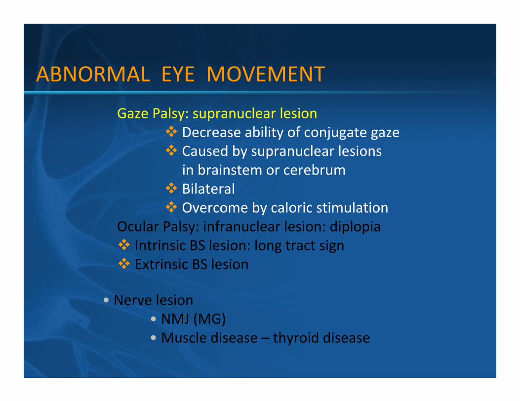
Gaze Palsy: supranuclear lesion
� Decrease ability of conjugate gaze
� Caused by supranuclear lesions
in brainstem or cerebrum
� Bilateral
� Overcome by caloric stimulation
Ocular Palsy: infranuclear lesion: diplopia
� Intrinsic BS lesion: long tract sign
� Extrinsic BS lesion
• Nerve lesion
• NMJ (MG)
• Muscle disease – thyroid disease

SUPRANUCLEAR LESION
Hemispheric lesion
� Destructive: produce bilateral deviation toward side
of the lesion & away from hemiparesis side
� Irritative: motor seizures = gaze out side of lesion
Midbrain Lesions:
� Affect the center responsible for
voluntary upward gaze
� Produces upward gaze paralysis
� Parinaud’s Syndrome
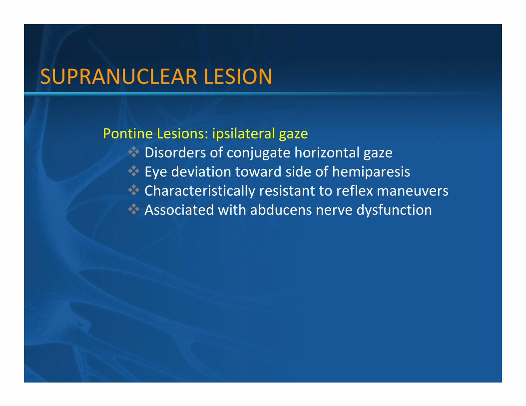
Pontine Lesions: ipsilateral gaze
� Disorders of conjugate horizontal gaze
� Eye deviation toward side of hemiparesis
� Characteristically resistant to reflex maneuvers
� Associated with abducens nerve dysfunction
SUPRANUCLEAR LESION

Internuclear Ophthalmoplegia:
� Lesions of the medial longitudinal fasiculus (MLF)
� Conjugate gaze of CN III & CN IV is uncoupled
� Excursion of the adbucting eye is full & adduction of the
contralateral eye is impaired
� Cannot be overcome by caloric stimulation
� Distinguished from CN III palsy by the preservation of
adduction w/ convergence
� Cause: small vessel disease, demyelination
SUPRANUCLEAR LESION

One and a Half Syndrome:
� Lesions of the medial longitudinal fasciculus
( MLF) and paraabducen nucleus
� Conjugate gaze of CN III & CN IV is uncoupled
� Affected eye cannot move horizontally
� Unaffected eye cannot abduct
� Cannot be overcome by caloric stimulation
� Distinguished from CN III palsy by the preservation of
adduction w/ convergence
� Cause: small vessel disease, demyelination
SUPRANUCLEAR LESION

nystagmus
INO
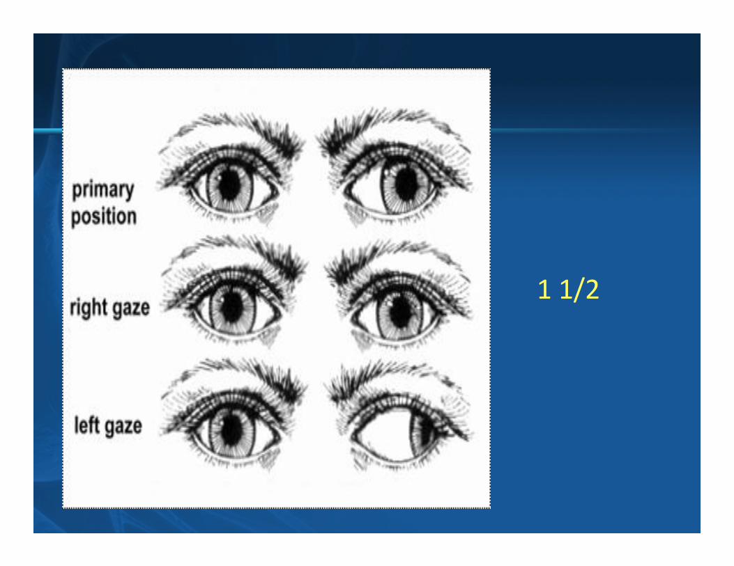
1 1/2
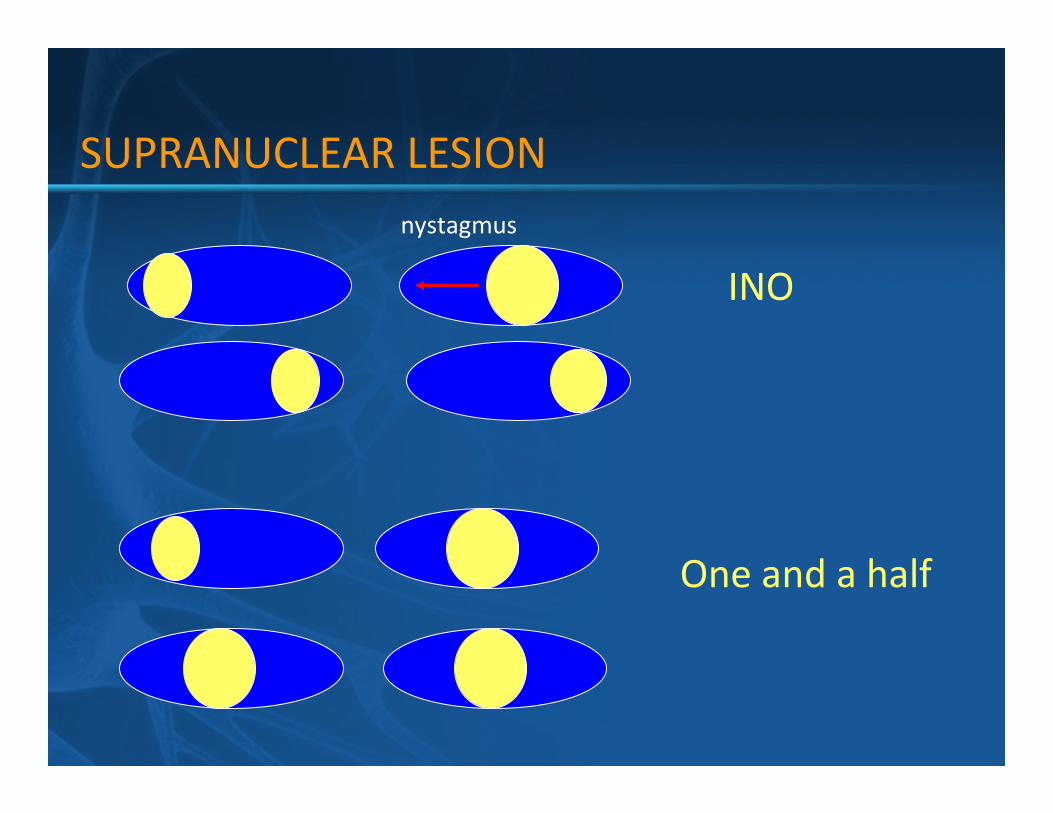
INO
One and a half
nystagmus
SUPRANUCLEAR LESION

MLF
PPRF
VIVI
III III
PPRF
LEFT RIGHT
RIGHT CORTEX LEFT CORTEX
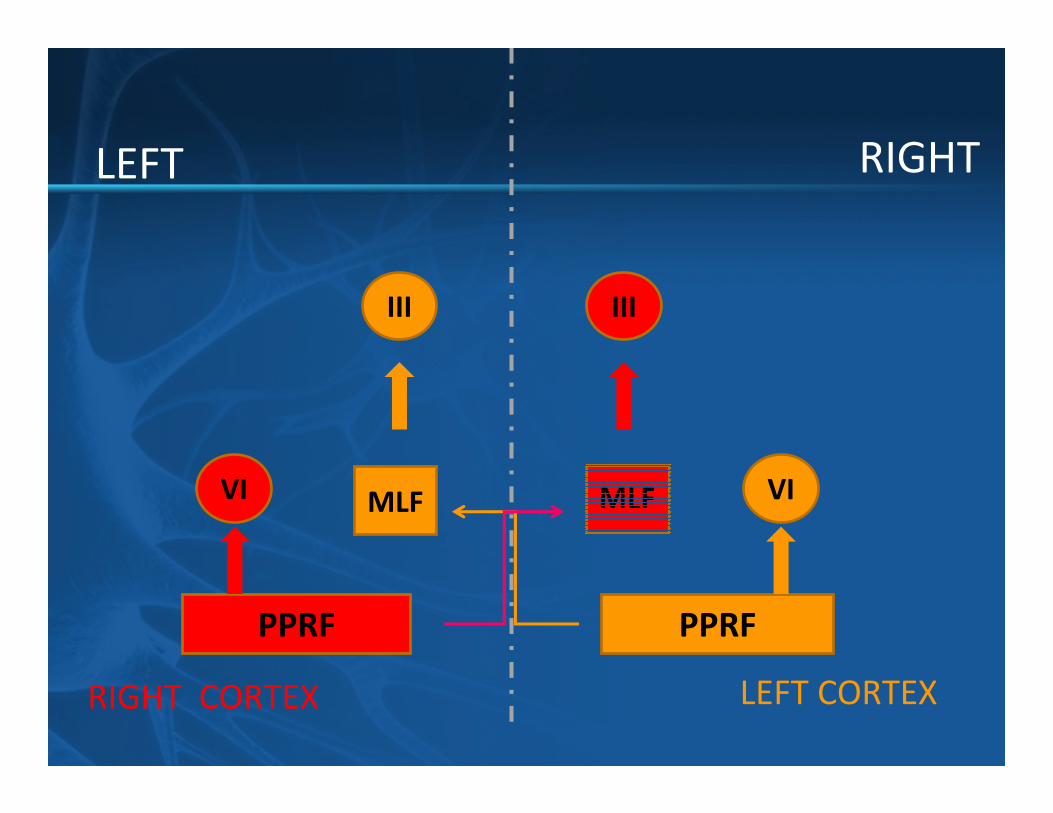
MLF
PPRF
VIVI
III III
PPRF
LEFT RIGHT
RIGHT CORTEX LEFT CORTEX

INFRANUCLEAR LESION

Gaze Palsy: supranuclear lesion
� Decrease ability of conjugate gaze
� Caused by supranuclear lesions in brainstem or cerebrum
� Bilateral
� Overcome by caloric stimulation
Ocular Palsy: infranuclear lesion: diplopia
� Intrinsic BS lesion: long tract sign
� Extrinsic BS lesion
• Nerve lesion
• NMJ (MG)
• Muscle disease – thyroid disease
ABNORMAL EYE MOVEMENT

FORAMEN SYNDROME
SUBARACHNOIDAL SPACE
NERVE LESION

Cranial Nerves:
� CN III, IV, & VI
� SO4 LR6
EXTRAOCCULAR MUSCLE
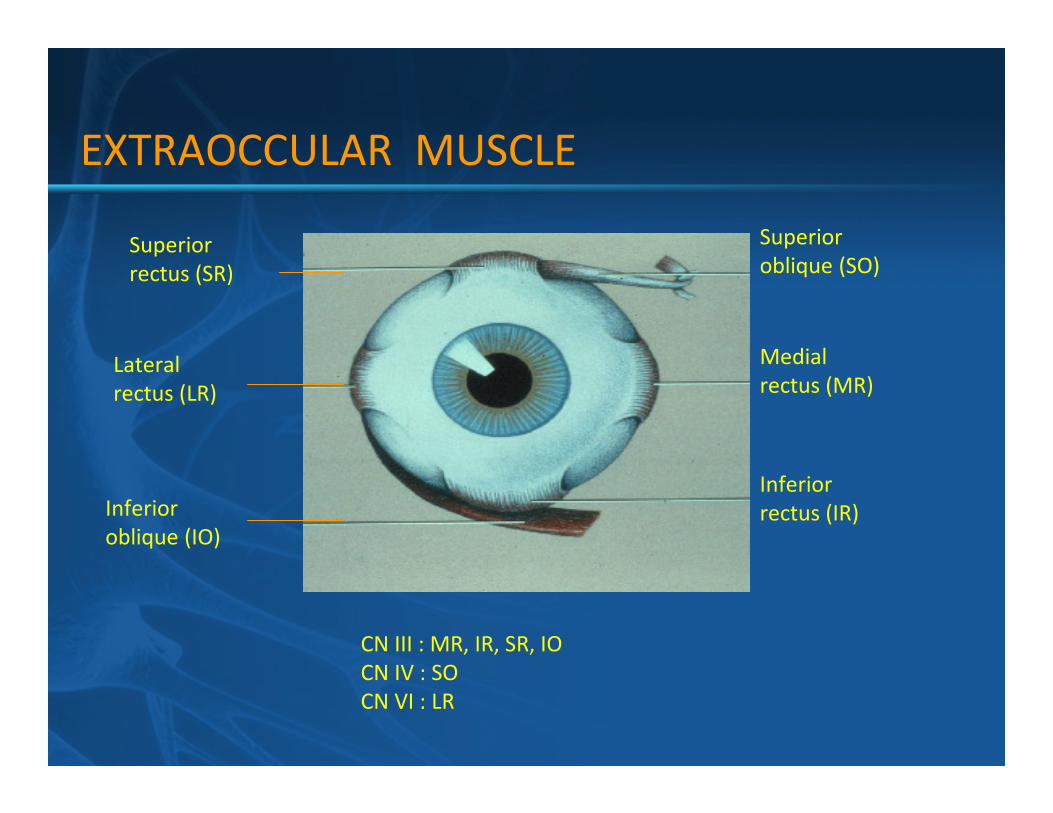
CN III : MR, IR, SR, IO
CN IV : SO
CN VI : LR
Medial
rectus (MR)
Inferior
rectus (IR)Inferior
oblique (IO)
Lateral
rectus (LR)
Superior
oblique (SO)Superior
rectus (SR)
EXTRAOCCULAR MUSCLE
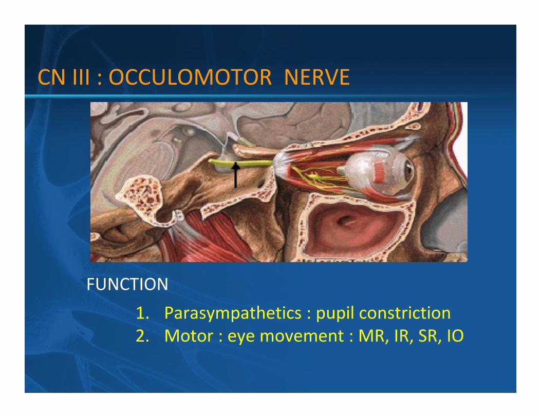
CN III : OCCULOMOTOR NERVE
FUNCTION
1. Parasympathetics : pupil constriction
2. Motor : eye movement : MR, IR, SR, IO

CLINICLAL
Pupil: fixed and dilated
Resting: laterally
Movement: lateral direction only
CN III : OCCULOMOTOR NERVE

CN III Nucleus:
� Superior Rectus receives fibers from
contralateral oculomotor nucleus
� Levator Palpebra receives bilateral innervation
CN III : OCCULOMOTOR NERVE

CN III Nerve Lesion vs Nuclear Lesion
� Nerve Lesion
– Unilateral Ophthalmoplegia
– Ipsilateral Ptosis
– Ipsilateral Pupillary Paralysis
� Nuclear Lesion
– Bilateral Ophthalmoplegia
– Bilateral Ptosis
– Ipsilateral Pupillary Paralysis
� General: diplopia, deviation down & out
CN III : OCCULOMOTOR NERVE

-CN III + superior cerebellar peduncle =
Nothnagel’s syndrome
- CN III + red nucleus = Benedikt’s syndrome
- CN III + cerebral peduncle = Weber’s syndrome
-CN III + superior cerebellar peduncle + red
nucleus =
Claude syndrome
Fascicular syndromes of the CN III nerve
CN III : OCCULOMOTOR NERVE

80% of diabetic CN III palsies are pupil sparing
95% of compressive CN III palsies have pupil involvement
Nuclear CN III palsies
- very rare
Uncal herneation syndrome of CN III nerve
- CN III passes along free edge of tentorium cerebelli
Posterior communicating artery aneurysm
- most common cause of painful, non-traumatic
ISOLATED CN III PALSY
CN III: OCCULOMOTOR NERVE

CN III:
� Nerve Lesions:
– Pituitary adenoma
– 1o or Metastatic Tumors, lymphoma
– Inflammation/infection
- Posterior Communicating artery aneurysm
- Ischemia (DM)
� Nuclear Lesions:
– Ischemia
– Central Demyelinating Disorders (MS)
CN III: OCCULOMOTOR NERVE

nerve carrying motor fiber to superior
oblique muscle
CN IV : TROCHEAR NERVE

nerve carrying motor fiber to superior oblique muscle
CN IV : TROCHEAR NERVE

unable to distinguish between nerve or nuclear lesions
� Ophthalmologic
� Excyclodeviation of the eye
� Vertical Diplopia
- Widest separation occurs w/ gaze away from
lesion
CN IV : TROCHEAR NERVE
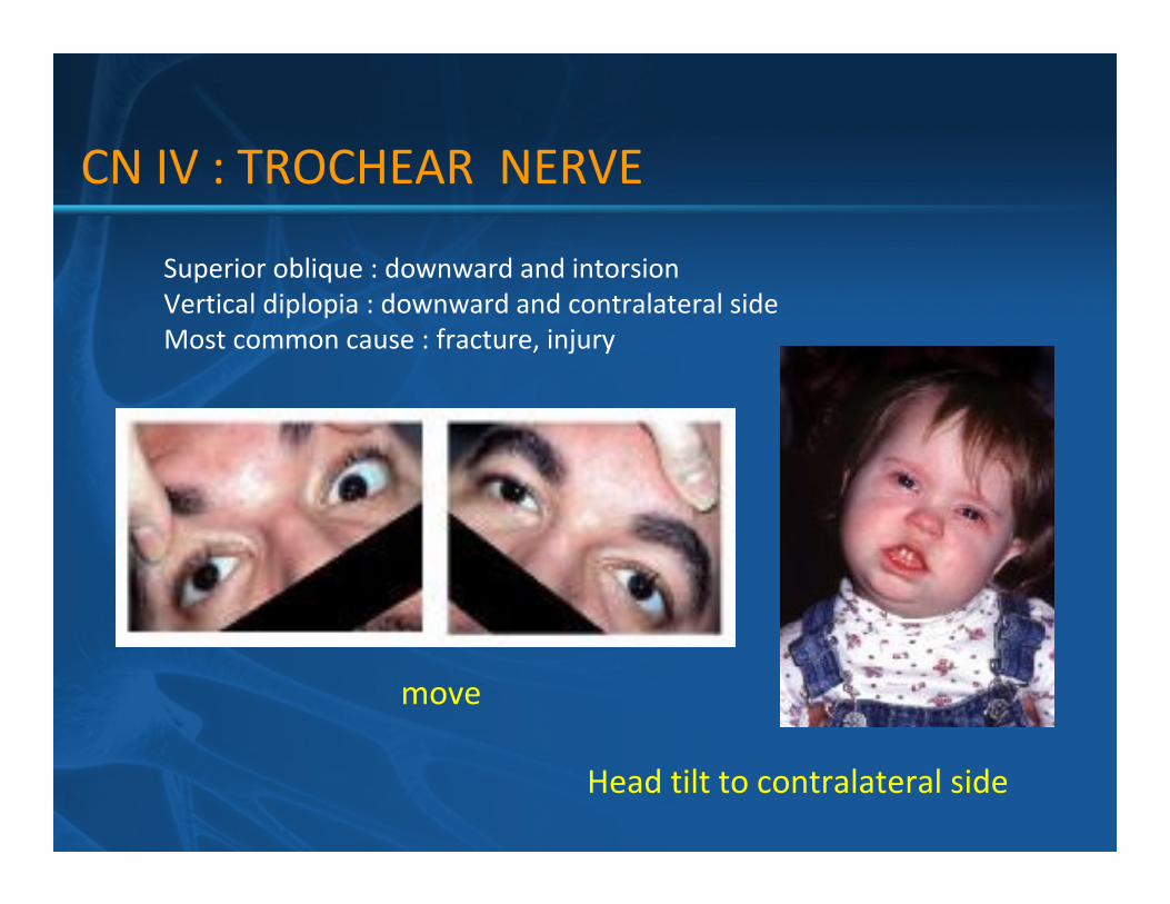
CN IV : TROCHEAR NERVE
Superior oblique : downward and intorsion
Vertical diplopia : downward and contralateral side
Most common cause : fracture, injury
move
Head tilt to contralateral side

CN IV:
� Nerve Lesions:
– Head Trauma
– Ischemia
– Inflammation
– Pituitary Adenoma
� Nuclear Lesions:
– Ischemia
– Central Demyelinating Disorders
– Inflammation
CN IV : TROCHEAR NERVE
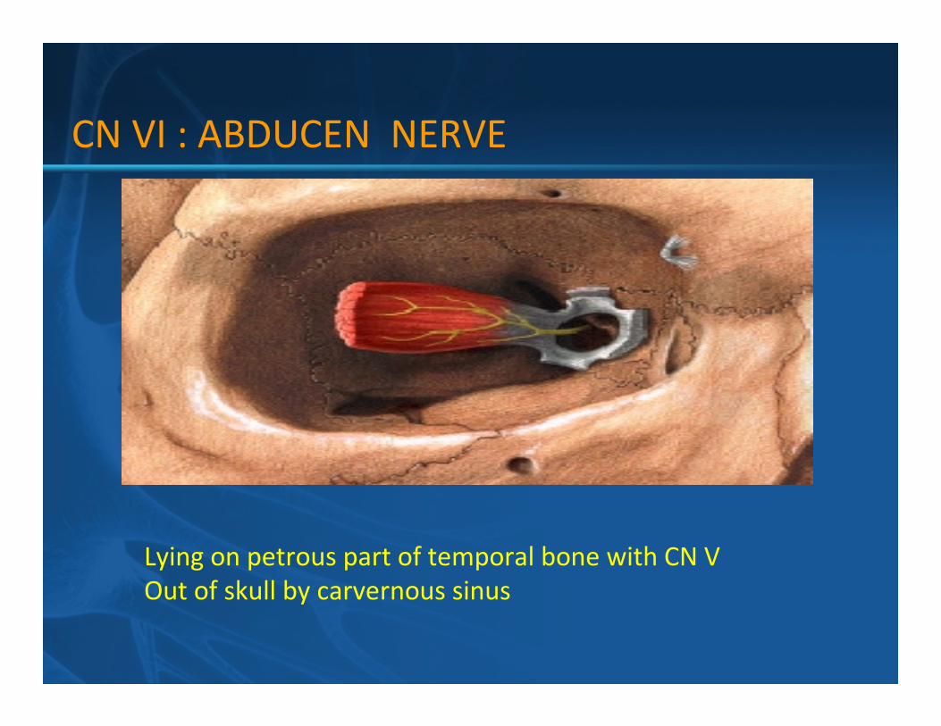
CN VI : ABDUCEN NERVE
Lying on petrous part of temporal bone with CN V
Out of skull by carvernous sinus

CN VI : ABDUCEN NERVE
Nucleus: lower part of pons
Closed relation with fiber of CN VII
Pass medial lemniscus and pyramidral tract

VI nerve + VII nerve + cerebral peduncle
medial pontine syndrome (Millard-Gubler syndrome)
VI nerve + cerebral peduncle
Raymonds syndrome
VI n. + V n. + VII n. + VIII n. + sympathetics
lateral pontine syndrome (Fovilles syndrome)
FASICULAR LESION
CN VI : ABDUCEN NERVE

SUBARACHNOID SPACE
� Elevated ICP CN VI palsy: false localizing sign
� Petrous apex syndrome of the VI nerve
passes under the petrosphenoidal ligament
petrous apex pathology may result in
VI+ VIII + VII + facial pain (V) = Gradenigo’s syndrome
true Gradenigo’s syndrome = otidis media complicated by petritis /
abscess
pseudo-Gradenigo’s syndrome = NPCA, CPA mass
CN VI : ABDUCEN NERVE

Nerve Lesions:
– Meningeal tumors
– Pituitary Adenoma
– Inflammation
- Increase intracranial pressure
Nuclear Lesions:
– Ischemia (pontine infarction)
– Central Demyelinating Disorders
– Inflammation
CN VI : ABDUCEN NERVE

� Cavernous sinus = III, IV, V1,V2,VI
� Superior orbital fissure = III, IV, V1, VI
� Orbital apex = II, III, IV, VI, V1
� Cerebellopontine angle = V, VII, VIII, (IX)
(acoustic neuroma, meningioma)
� Jugular foramen = IX, X, XI
(tumor, aneurysm)
FORAMEN SYNDROME

V1
V2
V3
III
IV
VI
CARVERNOUS SINUS
CN III
CN IV
V1
V2
V3

Superior orbital fissure
III, IV, VI, V1Carvernous sinus
III, IV, VI, V1, V2
Apex of
petrous bone
V, VI

Jugular foramen (IX, X, XI)Foramen
rotundum
(V2)
Foramen ovale
(V3)
Foramen spinosum
(middle meningeal a.)
Hypoglossal canal (XII)
Front
back

� Infection
Carvernous sinus thrombosis
Chronic granulomatous infection: TB, Fungal
� Vascular
CC fistular
Dural AVM
Aneurysm of intracarvernous part of carotid a. eg. posterior communicating a. aneurysm – CN III
FORAMEN SYNDROME

� Mass
Direct extension from skull base: CA nasopharynx
Metastasis: breast, lung
Granulomatous: Wegener’s granulomatosis
Hematologic: lymphoma, leukemia
Extension from sella tumor
� Idiopathic inflammatory (Tolosa-Hunt)
� Pseudotumor Occuli
FORAMEN SYNDROME

SUBARACHNOIDAL SPACE
� Meningeal inflammationMeningitis: TB, Bacterial, Fungus
Metastasis: Carcinomatous meningitis
Hematologic: lymphoma, leukemia
GBS (Miller-Fisher variant)
� Idiopathic pachy meningitis
� Menigioma en plaque
� Cranial neuritis – post viral, ischemic
� Diplopia is dysconjugated eye movement
� Supranuclear or infranuclear lesion
� Supranuclear lesion = lesion at gaze center
(midbrain or pons): INO, 1 ½
- sudden onset
� Infranuclear lesion = lesion at brain stem, cranial nerve
NMJ, muscle
DIPLOPIA

DIPOLPIA
Supranuclear Infranuclear
INO
1 1/2
Long tract sign
IntraaxialExtraaxial
Exclude NMJ, muscle
groupUngroup
Foramen syndrome Subarachnoidal space

52
EPISODE IV 2012
FOOT DROPLOCALIZATION NEUROLOGY
PAWUT MEKAWICHAI MD
DEPARTMENT of MEDICINE
MAHARAT NAKORNRAJSIMA HOSPITAL
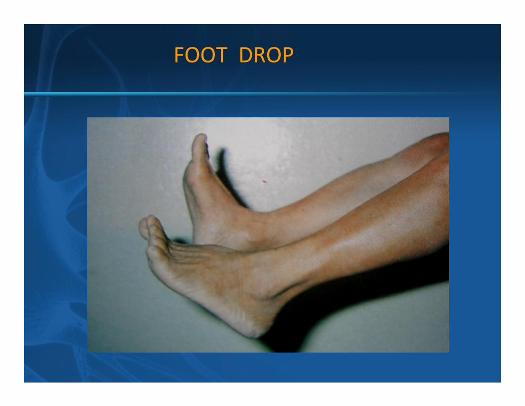
FOOT DROP

FOOT DROP

UMN lesion
� Spinal cord
� Motor cortex
LMN lesion
� Nerve root: L5
� Lumbosacral plexus
� Sciatic n.
� Peroneal n. (common, deep)
� Peripheral neuropathy: CMT
FOOT DROP

Action Muscle Root Nerve
Hip flexor Iliopsoas L 1,2 Femoral
Knee extensor Quadriceps L 2,3 Femoral
Ankle inversion Tibialis posterior L 4,5 Tibial
Ankle dorsiflex Tibialis anterior L 4,5 Peroneal
Toe extensor EHL L5, S1 Peroneal
Ankle eversion Peroneus L5, S1 Peroneal
Ankle plantarflex Gastrosoleus S 1,2 Tibial
Knee flexor Hamstrings S 1,2 Sciatic
FOOT DROP/root lesion

Action Muscle Root Nerve
Hip flexor Iliopsoas L 1,2 Femoral
Hip adductor Adductors L 2,3 Obturator
Hip abductor G. Medius L 4,5 Supr. Gluteal
Hip extensor G.Maximus L5, S1 Inf r. Gluteal
FOOT DROP/root lesion
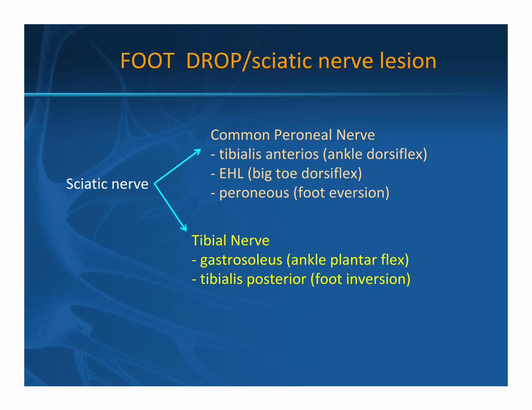
FOOT DROP/sciatic nerve lesion
Sciatic nerve
Common Peroneal Nerve
- tibialis anterios (ankle dorsiflex)
- EHL (big toe dorsiflex)
- peroneous (foot eversion)
Tibial Nerve
- gastrosoleus (ankle plantar flex)
- tibialis posterior (foot inversion)

COMMOM PERONEAL
� Peroneus longus
� Peroneus brevis
DEEP PERONEAL
� Tibialis anterior
� EDL/B
� EHL
FOOT DROP/peroneal nerve lesion

Commom peroneal
Deep peroneal
FOOT DROP/peroneal nerve lesion
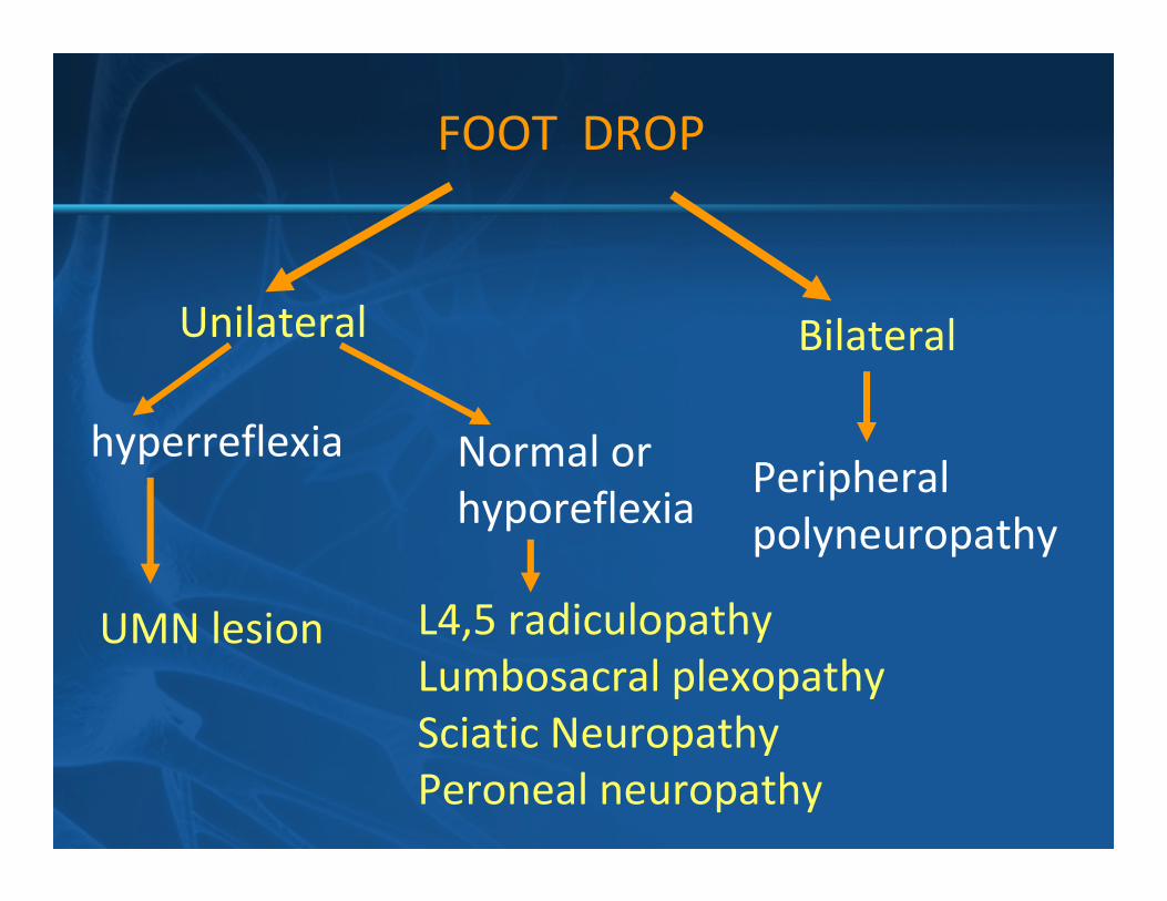
BilateralUnilateral
hyperreflexia Normal or
hyporeflexia
UMN lesion L4,5 radiculopathy
Lumbosacral plexopathy
Sciatic Neuropathy
Peroneal neuropathy
Peripheral
polyneuropathy
FOOT DROP

Foot inversion
(tibialis posterior)No weakness
Peroneal neuropathy
- Injury
- Entrapment neuropathy
(Wt loss, bed ridden,
cross leg, underlying PN)
Weakness
Hip abduction
(Gluteus medius)
No weaknessWeak
Sciatic
neuropathy
L4,5
LS plexus
FOOT DROP with DECREASE REFLEX


![Efficacy and outcomes of facial nerve–sparing treatment ... · the patient at significant risk of morbidity and mortality. Damage to the facial nerve (cranial nerve [CN] VII) is](https://static.fdocuments.us/doc/165x107/5edf22ebad6a402d666a7cb2/efficacy-and-outcomes-of-facial-nerveasparing-treatment-the-patient-at-significant.jpg)