![:yfgLo txdf cfalws ljsf; of]hgf th'{df !! dfu{ @)&%ppc.gandaki.gov.np/downloadfile/PPC_Local_Level_Planning_Paper_Final... · 3 @=:yfgLo txn] of]hgf th'{df ug]{ cfwf/ :yfgLotxn] cf+kmgf](https://static.fdocuments.us/doc/165x107/5e8bcad11aa6057f933f7293/yfglo-txdf-cfalws-ljsf-ofhgf-thdf-dfu-ppc-3-yfglo-txn-ofhgf.jpg)
Novel Combination BMP7 and HGF Gene Therapy Instigates ...
15
Chapman University Chapman University Digital Commons Pharmacy Faculty Articles and Research School of Pharmacy 2-2018 Novel Combination BMP7 and HGF Gene erapy Instigates Selective Myofibroblast Apoptosis and Reduces Corneal Haze In Vivo Suneel Gupta Harry S. Truman Memorial Veterans’ Hospital Michael K. Fink Harry S. Truman Memorial Veterans’ Hospital Arkasubhra Ghosh GROW Research Laboratory, Narayana Nethralaya Foundation Ratnakar Tripathi Harry S. Truman Memorial Veterans’ Hospital Prashant R. Sinha Harry S. Truman Memorial Veterans’ Hospital See next page for additional authors Follow this and additional works at: hps://digitalcommons.chapman.edu/pharmacy_articles Part of the Amino Acids, Peptides, and Proteins Commons , Animals Commons , Genetic Processes Commons , Medical Biochemistry Commons , Medical Genetics Commons , Ophthalmology Commons , Organic Chemicals Commons , and the Other Pharmacy and Pharmaceutical Sciences Commons is Article is brought to you for free and open access by the School of Pharmacy at Chapman University Digital Commons. It has been accepted for inclusion in Pharmacy Faculty Articles and Research by an authorized administrator of Chapman University Digital Commons. For more information, please contact [email protected]. Recommended Citation Gupta S, Fink MK, Ghosh A, et al. Novel combination BMP7 and HGF gene therapy instigates selective myofibroblast apoptosis and reduces corneal haze in vivo. Invest Ophthalmol Vis Sci. 2018 Feb;59(2):1045–1057. doi:10.1167/iovs.17-23308
Transcript of Novel Combination BMP7 and HGF Gene Therapy Instigates ...
Chapman UniversityChapman University Digital Commons
Pharmacy Faculty Articles and Research School of Pharmacy
2-2018
Novel Combination BMP7 and HGF GeneTherapy Instigates Selective MyofibroblastApoptosis and Reduces Corneal Haze In VivoSuneel GuptaHarry S. Truman Memorial Veterans’ Hospital
Michael K. FinkHarry S. Truman Memorial Veterans’ Hospital
Arkasubhra GhoshGROW Research Laboratory, Narayana Nethralaya Foundation
Ratnakar TripathiHarry S. Truman Memorial Veterans’ Hospital
Prashant R. SinhaHarry S. Truman Memorial Veterans’ Hospital
See next page for additional authorsFollow this and additional works at: https://digitalcommons.chapman.edu/pharmacy_articles
Part of the Amino Acids, Peptides, and Proteins Commons, Animals Commons, GeneticProcesses Commons, Medical Biochemistry Commons, Medical Genetics Commons,Ophthalmology Commons, Organic Chemicals Commons, and the Other Pharmacy andPharmaceutical Sciences Commons
This Article is brought to you for free and open access by the School of Pharmacy at Chapman University Digital Commons. It has been accepted forinclusion in Pharmacy Faculty Articles and Research by an authorized administrator of Chapman University Digital Commons. For more information,please contact [email protected].
Recommended CitationGupta S, Fink MK, Ghosh A, et al. Novel combination BMP7 and HGF gene therapy instigates selective myofibroblast apoptosis andreduces corneal haze in vivo. Invest Ophthalmol Vis Sci. 2018 Feb;59(2):1045–1057. doi:10.1167/iovs.17-23308

Novel Combination BMP7 and HGF Gene Therapy Instigates SelectiveMyofibroblast Apoptosis and Reduces Corneal Haze In Vivo
CommentsThis article was originally published in Investigative Ophthalmalogy & Visual Science, volume 59, issue 2, in2018. DOI:10.1167/iovs.17-23308
Creative Commons License
This work is licensed under a Creative Commons Attribution 4.0 License.
CopyrightThe authors
AuthorsSuneel Gupta, Michael K. Fink, Arkasubhra Ghosh, Ratnakar Tripathi, Prashant R. Sinha, Ajay Sharma,Nathan P. Hesemann, Shyam S. Chaurasia, Elizabeth A. Giuliano, and Rajiv R. Mohan
This article is available at Chapman University Digital Commons: https://digitalcommons.chapman.edu/pharmacy_articles/530

Cornea
Novel Combination BMP7 and HGF Gene TherapyInstigates Selective Myofibroblast Apoptosis and ReducesCorneal Haze In Vivo
Suneel Gupta,1,2 Michael K. Fink,1,2 Arkasubhra Ghosh,3 Ratnakar Tripathi,1,2 Prashant R.Sinha,1,2 Ajay Sharma,4 Nathan P. Hesemann,1,5 Shyam S. Chaurasia,1,2 Elizabeth A. Giuliano,2 andRajiv R. Mohan1,2,5
1Harry S. Truman Memorial Veterans’ Hospital, Columbia, Missouri, United States2One-Health One-Medicine Ophthalmology and Vision Research Center, University of Missouri Columbia, Missouri, United States3GROW Research Laboratory, Narayana Nethralaya Foundation, Bangalore, India4Chapman University School of Pharmacy, Irvine, California, United States5Mason Eye Institute, University of Missouri School of Medicine, Columbia, Missouri, United States
Correspondence: Rajiv R. Mohan,University of Missouri, 1600 E. Roll-ins Road, Columbia, MO 65211, USA;[email protected].
Submitted: November 6, 2017Accepted: January 18, 2018
Citation: Gupta S, Fink MK, Ghosh A,et al. Novel combination BMP7 andHGF gene therapy instigates selectivemyofibroblast apoptosis and reducescorneal haze in vivo. Invest Ophthal-
mol Vis Sci. 2018;59:1045–1057.https://doi.org/10.1167/iovs.17-23308
PURPOSE. We tested the potential of bone morphogenic protein 7 (BMP7) and hepatocytegrowth factor (HGF) combination gene therapy to treat preformed corneal fibrosis usingestablished rabbit in vivo and human in vitro models.
METHODS. Eighteen New Zealand White rabbits were used. Corneal fibrosis was produced byalkali injury. Twenty-four hours after scar formation, cornea received topically either balancedsalt solution (BSS; n ¼ 6), polyethylenimine-conjugated gold nanoparticle (PEI2-GNP)-nakedplasmid (n ¼ 6) or PEI2-GNP plasmids expressing BMP7 and HGF genes (n ¼ 6). Donorhuman corneas were used to obtain primary human corneal fibroblasts and myofibroblasts formechanistic studies. Gene therapy effects on corneal fibrosis and ocular safety were evaluatedby slit-lamp microscope, stereo microscopes, quantitative real-time PCR, immunofluores-cence, TUNEL, modified MacDonald-Shadduck scoring system, and Draize tests.
RESULTS. PEI2-GNP–mediated BMP7þHGF gene therapy significantly decreased cornealfibrosis in live rabbits in vivo (Fantes scale was 0.6 in BMP7þHGF-treated eyes compared to3.3 in �therapy group; P < 0.001). Corneas that received BMP7þHGF demonstratedsignificantly reduced mRNA levels of profibrotic genes: a-SMA (3.2-fold; P < 0.01), fibronectin(2.3-fold, P < 0.01), collagen I (2.1-fold, P < 0.01), collagen III (1.6-fold, P < 0.01), andcollagen IV (1.9-fold, P < 0.01) compared to the �therapy corneas. Furthermore,BMP7þHGF-treated corneas showed significantly fewer myofibroblasts compared to the�therapy controls (83%; P < 0.001). The PEI2-GNP introduced >104 gene copies permicrogram DNA of BMP7 and HGF genes. The recombinant HGF rendered apoptosis incorneal myofibroblasts but not in fibroblasts. Localized topical BMP7þHGF therapy showedno ocular toxicity.
CONCLUSIONS. Localized topical BMP7þHGF gene therapy treats corneal fibrosis and restorestransparency in vivo mitigating excessive healing and rendering selective apoptosis inmyofibroblasts.
Keywords: HGF, BMP7, PEI-GNP, gene therapy, corneal fibrosis
Injuries and infections of the eye compromise cornealtransparency, which accounts for two-thirds of the eye’s
refraction and results in vision loss in an estimated 1.3 millionAmericans annually. Worldwide, corneal disorders are the thirdleading cause of preventable blindness.1,2 Despite increasingknowledge about the molecular mechanisms underlyingcorneal scarring,3 currently available therapies4 rely primarilyon steroids and other drugs that carry the risk of multiple sideeffects,5 require repeated applications, and are often ineffectivein restoring vision completely. Although mitomycin C (MMC) isa commonly used topical treatment for corneal fibrosis,6 its usecontinues to be the subject of debate due to its long-term sideeffects.7 A limbal graft is the treatment of choice in countrieswhere donor corneal limbus is available.8 Corneal transplanta-
tion remains the gold standard for the treatment of cornealscars and restoration of vision.9 In 2015 alone, 48,792 cornealtransplantations were performed in the United States, accord-ing to the Eye Bank Association of America (http://restoresight.org/wp-content/uploads/2016/03/2015-Statistical-Report.pdf),and about 12.7 million people in the world are awaiting donorcorneal tissues.10 Besides the limited availability of donorcorneas, a high immunologic rejection rate is another limitingfactor for restoring vision from corneal transplantation. Thus,there is an undisputable need for the development of effectiveand safe nonsurgical targeted treatments for corneal fibrosis,including gene-based therapies that would provide long-termeffectiveness, require minimal clinical follow up, and showminimal side effects.
Copyright 2018 The Authors
iovs.arvojournals.org j ISSN: 1552-5783 1045
This work is licensed under a Creative Commons Attribution 4.0 International License.

Studies show that injury and infection to the cornea/eyeleads to the activation of quiescent stromal keratocytes in thecornea, which then migrate to the wound site and trans-differentiate to a wound-repair phenotype, referred to asmyofibroblasts.4 While molecular signaling and secretoryfunctions of myofibroblasts are essential for proper cornealwound repair, their continued production and prolongedpresence in the stroma lead to corneal fibrosis (haze orscarring) due to the extended activity of TGF-b signaling duringcorneal wound repair.11 The activation and differentiation ofkeratocytes have been shown to occur as a response to IL-1a/IL-1b, which are released by corneal epithelial cells afterinjury.12 In addition, multiple molecules, factors, ligands, andcytokines, including TGF-b released from injured epithelialcells, play a pivotal role in the induction of inflammation andcorneal scarring due to excessive biological activities andfusion of extracellular matrix (ECM) and cytoskeletal pro-teins.3,4 TGF-b signaling is largely responsible for the trans-differentiation of keratocytes into myofibroblasts. Thepersistence of myofibroblasts after wound healing is knownto be a major factor in the pathogenesis of corneal fibrosis andopacity, and ultimately, vision impairment.3,13
The TGF-b superfamily proteins activate downstreamsignaling via the Smad family of proteins.14 Bone morphogenicprotein (BMP) belongs to the TGF-b superfamily and plays asignificant role in ECM synthesis, tissue repair, and remodelingprocesses during corneal wound healing.15 The signalingprotein BMP7 was originally described to have a significantrole in the development of mammalian organs such as thekidney and the eye.16 BMP7 binds to the type I and II receptorsand regulates receptor-regulated Smads (Smad1, Smad5, andSmad8) and inhibitory Smads (Smad6 and Smad7) in a complexwound-healing signaling network.17 In addition to BMP7, theexpression of hepatocyte growth factor (HGF) and its receptorproteins has been found in the cornea, lacrimal glands, andtears.18,19 HGF has been identified as a mitogen that functionsthrough the c-Met receptor tyrosine kinase in the protectionand regeneration of organs20 as well as in the modulation ofcorneal repair.21,22 Injury to corneal epithelium has beenshown to upregulate HGF expression in keratocytes, and inaddition to its intracrine and autocrine functions, HGF hasbeen shown to function in a paracrine manner in modulatingcorneal wound healing.23 The role of HGF and the c-Metsystem in diabetic corneal wound healing was recently wellestablished in organotypic human diabetic corneal cultures.4
Furthermore, HGF has been reported to have a role in thebreakdown of ECM deposits and in the reduction of fibrosis inseveral nonocular tissues.24 Despite the important role of HGFin corneal wound healing and TGF-b profibrotic signaling,mechanistic knowledge about the crosstalk between HGF andBMP7 in the cornea remains unknown, especially duringwound healing and profibrotic microenvironment.
The cornea represents a perfect tissue for gene therapybecause of its well-defined characteristics such as transpar-ency, simple anatomy, and ease of access, allowing topicalinstillation of gene delivery vectors and visual monitoring ofthe genes packaged within vectors.25 In addition, therapeuticresponse can also be assessed noninvasively with high-resolution ocular imaging using stereo and slit-lamp biomi-croscopy.26 Our group previously reported the advantages oftissue-specific gene delivery using a variety of modalities,including direct instillation of hybrid nanoparticle or adeno-associated virus vectors.17,27,28 We found that nonviral genedelivery systems based on synthetic polycations,29–31 such aspolyethylene amine (PEI), show promise as delivery systemsfor gene therapy. They possess DNA-binding capabilities,provide options for functionalization, and show a good safetyprofile. In subsequent studies, we greatly enhanced the
otherwise low transfection efficiency of PEI (2 kDa) byconjugating PEI with gold nanoparticles to synthesize PEI-conjugated gold nanoparticles (PEI2-GNP).32 Utilizing thesehybrid gold nanoparticles, we established a novel nanoparti-cle-based gene delivery system for the cornea that demon-strates efficient gene transfer into rabbit corneal stroma invivo with negligible toxicity.17,29
BMP7 and HGF are attractive targets for modulating theprofibrotic signaling pathways in corneal wound healing. Ourprevious study of BMP7 gene therapy by PEI2-GNP in thepreclinical rabbit model of corneal fibrosis revealed that PEI2-GNP–based BMP7 gene therapy led to significant inhibition ofcorneal fibrosis and corneal repair through counterbalancingthe deleterious effects of TGF-b–induced Smad signaling.17
HGF gene therapy has been shown by other investigators toregulate fibrosis in various nonocular tissues, including theliver,20 lung,33 and kidney.34 Phase-I and phase-II clinical trialsindicate HGF gene therapy is safe in humans.35,36 Further-more, HGF gene transfer has been found to cause selectiveapoptosis of myofibroblasts in nonocular tissues.37–39 Thesereports led to an innovative postulate that PEI2-GNP–mediated tissue-targeted localized BMP7þHGF gene therapyin rabbit cornea would effectively eliminate preexistingfibrosis in vivo without producing significant toxicity. Thepresent study tested the therapeutic potential of PEI2-GNP–delivered BMP7þHGF gene therapy for abolishing preexistingcorneal fibrosis in vivo using a well-established preclinicalrabbit model of corneal fibrosis.
MATERIALS AND METHODS
Corneal Fibrosis Induction and Treatment inRabbits
Eighteen New Zealand White female rabbits, weighing 2 to 3kg (Covance Research Products, Denver, PA, USA) were used inthe study. Institutional approval of the study was obtained fromthe Harry S. Truman Memorial Veterans’ Hospital and theInstitutional Animal Care and Use Committee of the Universityof Missouri (both in Columbia, MO, USA). All animals weretreated in accordance with the principles of the ARVOStatement for the Use of Animals in Ophthalmic and VisionResearch. The rabbits were anesthetized by a mixture ofketamine hydrochloride (50 mg/kg) and xylazine hydrochlo-ride (10 mg/kg), given intramuscularly, for induction of cornealalkali-induced wounding, for administration of PEI2-GNP–mediated BMP7þHGF gene delivery to the corneal stroma,and for the performance of clinical slit-lamp eye examinationsand ocular stereo biomicroscopy. Topical ophthalmic propar-acaine hydrochloride (0.5%; Alcon, Fort Worth, TX, USA) wasadministered for local anesthesia prior to all procedures.
IOP Monitoring by Tonometry
Variations in IOP, an indicator of an ocular abnormality, mayresult from inflammation, swelling, rigidity, abrasion, andirregularities in corneal tissues. Administration of therapeuticgenes into stroma has potential for alterations in the aqueoushumor or in tissues of the anterior chamber, which is asignificant concern after gene therapy. Thus, IOP measure-ments in rabbit eyes were recorded using a tonometer (Tono-Pen AVIA; Reichert Technologies, Depew, NY, USA) at regulartimed intervals on days 1, 7, 14, and 21 and before each clinicalbiomicroscopy evaluation as reported earlier.40 All IOPmeasurements were performed between 9 AM and 11 AM tominimize normal diurnal variations in IOP.
BMP7þHGF Gene Therapy for Corneal Fibrosis IOVS j February 2018 j Vol. 59 j No. 2 j 1046
In Vivo Alkali-Induced Corneal Scarring
Corneal scarring was induced in one eye of each rabbit, andthe contralateral eye served as a naive control. To inducecorneal scarring, rabbits were anesthetized and an 8-mm filterpaper soaked in 0.5 N sodium hydroxide solution was appliedonto the central cornea for 1.0 minutes under visualizationwith the surgical microscope (Leica Wild Microscope MEL53;Leica, Wetzlar, Germany). The wounded corneas were imme-diately and copiously rinsed with sterile balanced salt solution(BSS) to remove alkali residual. This method triggered woundhealing and produced dense corneal scarring and peak fibrosisat 3 weeks with minimal neovascularization.41
PEI2-GNP Transfection Solution
Thiol-modified PEI2-GNPs were synthesized as describedearlier.32 The PEI2-GNP transfection solution was prepared asreported previously.17 In brief, the PEI2-GNPs were mixed withplasmid at a nitrogen-to-phosphate (N/P) ratio of 180 bystirring 37.5 lL of 150 mM PEI2-GNPs with 10 lg plasmid DNA(pTRUF11 expressing HGF or BMP7 under control of hybridcytomegalovirus [CMV] chicken b-actin promoter), 10%glucose (wt/vol), and bringing the volume to 100 lL withBSS. The solution was incubated at 378C for 30 minutes priorto application on the cornea.
In Vivo Gene Delivery
One eye of each animal was treated and the contralateral eyeserved as naive control. To determine the effectiveness of genetherapy for preexisting corneal fibrosis, PEI2-GNP–mediatedBMP7þHGF gene therapy was delivered into rabbit stroma 24hours after alkali injury. BSS or transfection solution wastopically applied to the cornea for 5 minutes using a cloningcylinder, as previously reported.17,29 The rabbits were dividedinto three groups: group 1 rabbits received BSS alone (n¼6; nogene transfer naive group); group 2 rabbits received PEI2-GNP-naked plasmid without BMP7 or HGF gene (n ¼ 6; �therapygroup); and group 3 rabbits (n ¼ 6) received PEI2-GNPplasmids expressing HGF and BMP7 genes (n ¼ 6; þtherapygroup). The cloning cylinder method is known to deliversignificant levels of therapeutic genes into rabbit stroma in vivowith low toxicity.17,29
Slit-Lamp Biomicroscopy, Haze Quantification,and Fluorescein Eye Test
Corneal defects and general ocular health were documented atbaseline and after alkali wounding at various time points usinga handheld slit-lamp microscope equipped with a digital-imaging system (SL-15; Kowa Optimed, Torrance, CA, USA).Using the Fantes scale, the intensity of corneal haze was gradedby three independent observers (SG, AS, MK) masked to thetreatment group, as reported earlier.28,42 In brief, the gradingsystem was the following: grade 0, completely clear cornea;grade 0.5, trace haze seen with careful oblique illumination;grade 1, more obvious haze but not interfering the visualizationof fine iris details; grade 2, mild obscuration of iris details;grade 3, moderate obscuration of the iris and lens; and grade 4,complete opacification of the stroma in the area of ablation.
Fluorescein sodium sterile ophthalmic strips (0.6 mg, Ful-Glo; Akorn, Lake Forest, IL, USA) were used to evaluate thehealth of the corneal epithelium and tear production. In brief, aBSS-moistened fluorescein strip was applied to the dorsalupper eyelid of rabbits, and the rabbits were allowed to blinkfor distributing fluorescein stain over the entire corneal andconjunctival surface. After 30 seconds, corneal epithelium was
observed under blue light using wide and narrow beams of theslit-lamp microscope. Images were obtained with a stereomicroscope fitted with a fluorescence filter, spot camera, andimaging software. Tear levels were measured using commercialdiagnostic strips (Tear-Flow Diagnostic Strips; HUB Pharma-ceuticals, Rancho Cucamonga, CA, USA).
Ocular Irritation Tests
The ocular health and anomalies were evaluated independentlyat selected times by at least two of three examiners (MF, AS,and SG) using the established Draize43 and modified MacDon-ald-Shadduck44 ocular scoring systems. With the Draize eyetest, the severity of ocular lesions was scored in the followingmanner: in the cornea, by estimating the degree of opacity andarea of involvement; in the iris, by examining pupillary lightreflexes; and in the conjunctiva, by assessing the degree ofredness, chemosis, and discharge. With the modified MacDon-ald-Shadduck scoring system, ocular health scores weredetermined based on the cumulative average scores for cornealtissue (opacity, affected area, corneal neovascularizationseverity, and reepithelization) and for conjunctival tissue(congestion, chemosis, swelling, and discharge).
Euthanasia and Tissue Collection
Rabbits were humanely euthanized with pentobarbital (150mg/kg) while they were under general anesthesia. Corneaswere harvested, immediately placed in 15 3 5 3 5-mm molds(Fisher Scientific, Pittsburgh, PA, USA) containing opticalcutting temperature (OCT) compound, and snap frozen byimmersion in a cryo-cup containing 2-methylbutane sitting inliquid nitrogen. Frozen tissue blocks were preserved at�808C.The corneas were cut in two equal halves; one half was usedfor histologic studies, and other half was used for molecularstudies. For histologic studies, serial corneal sections (8 lm)were prepared with a cryostat (HM525 NX UV; Microm GmbH,Walldorf, Germany), placed on glass microscopic slides(Superfrost Plus; Fisher Scientific, Pittsburgh, PA, USA), andkept at �808C until analysis. For molecular studies, corneaswere cut into small pieces, immediately immersed in a cryo-cup placed in liquid nitrogen, and subsequently ground andprocessed for obtaining genomic DNA, mRNA, and cDNAfollowing vendor’s protocols (Qiagen, Germantown, MD, USA).
Hematoxylin and Eosin, Masson’s Trichome,Immunofluorescence and TUNEL Staining
Hematoxylin-eosin (H&E) and Masson’s trichome staining wereperformed using the standard procedure for visualizingmorphologic details, as reported earlier.40,45 Immunofluores-cence staining was performed following reported methods17 tomeasure myofibroblasts in the corneas using antibody-specifica-smooth muscle actin (a-SMA), a marker for myofibroblasts.Briefly, corneal sections were blocked with 2% bovine serumalbumin at room temperature for 30 minutes, followed byincubation with a-SMA mouse monoclonal primary antibody(1:200 dilution, M0851; Dako, Carpentaria, CA, USA) for 90minutes and then incubated with Alexa-Fluor 488 goat anti-mouse IgG secondary antibody (1:1000 dilution, A11001;Invitrogen, Carlsbad, CA, USA) for 1 hour at room temperature.Appropriate positive and negative controls were included ineach immunostaining. Quantification of a-SMA-positive cellswas performed in six randomly selected, nonoverlapping, full-thickness central corneal columns, extending from the anteriorstromal surface to the posterior stromal surface at 2003 and4003 magnification fields.
BMP7þHGF Gene Therapy for Corneal Fibrosis IOVS j February 2018 j Vol. 59 j No. 2 j 1047
The toxicity of PEI2-GNPs and BMP7þHGF gene therapywas determined by performing a TUNEL assay (ApopTag;Millipore, Temecula, CA, USA). Corneal sections were fixed inacetone at –208C for 10 minutes, and a TUNEL assay wasperformed per the manufacturer’s instructions, includingsuitable positive and negative controls. Rhodamine-conjugat-ed apoptotic cells (red) and 4 0,6-diamidine-2 0-phenylindoledihydrochloride (DAPI)–stained nuclei (blue) were viewedand photographed with a fluorescence microscope (Leica)fitted with a digital camera system (SpotCamRT KE; Diagnos-tic Instruments, Sterling Heights, MI, USA). DAPI-stainednuclei and TUNEL-positive cells in untreated and treatedtissues were quantified at 2003 and 4003 magnification in sixrandomly selected nonoverlapping areas, as previouslyreported.17,28
RNA Extraction, cDNA Synthesis, and QuantitativeReal-Time PCR
Total RNA from tissues was extracted with an RNeasy kit(Qiagen, Valencia, CA, USA) and reverse transcribed to cDNAfollowing reported methods.17,28 Real-time PCR was per-formed using the StepOne Plus PCR system (AppliedBiosystems, Carlsbad, CA, USA). A 20-lL reaction mixturecontained 2 lL cDNA, 2 lL forward and reverse primers (200nM each), and 10 lL 2X All-in-One quantitative PCR (qPCR)mix (GeneCopoeia, Rockville, MD, USA) and was run at auniversal cycle (958C for 10 minutes, 40 cycles at 958C for 15seconds, and 608C for 60 seconds), as previously reported.16
The primer sequences of genes were the following: a-SMA—forward TGG GTG ACG AAG CAC AGA GC and reverse CTTCAG GGG CAA CAC GAA GC; fibronectin—forward CGCAGC TTC GAG ATC GTG C and reverse TCG ACG GGA TCACAC TTC CA; collagen type I—forward TGT GGC CCA GAACTG GTA CAT and reverse ACT GGA ATC CAT CGG TCA TGCTCT; collagen type III—forward AGA ACA CGC AAG GCTGTG AGA CTA and reverse CCA ACG TCC GCA CCA AAT TCTTGA; collagen type IV—forward TAT CGA ACA ACG CAAGGC TGT GAG A and reverse GGC CAA CGT CCA CAC CAAATT CTT; and b-actin—forward CGG CTA CAG CTT CAC CAand reverse CGG GCA GCT CGT AGC TCT TC. The b-actinwas used for the normalization of qPCR data, and showed noappreciable relative fold change at various tested points andgroups.
Quantification of Nanoparticle-Delivered GeneCopies
The frozen corneal tissues were pulverized in liquidnitrogen, and genomic DNA was isolated (DNA Easy kit;Qiagen, Valencia, CA, USA). Quantitative PCR was performedto determine PEI2-GNP–delivered gene copies of HGF andBMP7 in corneal tissues.17 A 10-fold serial dilution ofplasmid having test gene (104–109/mg DNA) was used forstandard curves. The qPCR settings were 958C for 10minutes, 40 cycles at 958C for 15 seconds, and 608C for 60seconds.
Human Corneal Fibroblast and MyofibroblastCultures
Primary human corneal fibroblast (HCF) cultures weregenerated from donor human corneas purchased from aneye bank (Saving Sight, Kansas City, MO, USA) followingmethods described previously.30 The corneal epithelium andendothelium were removed from corneal buttons with asurgical blade and cut into small pieces, placed on culture
dishes, and incubated for 3 to 5 weeks in a humidified CO2
(5%) incubator at 378C in MEM supplemented with 10% fetalbovine serum. Seventy percent confluent HCF cultures thatunderwent fewer than four passages were used in theexperiments. Human corneal myofibroblasts were producedby culturing HCFs under serum-free conditions in thepresence of TGF-b1 (PeproTech, Rocky Hill, NJ, USA), 5 ng/mL for 72 hours.
Lactate Dehydrogenase Assay
Lactate dehydrogenase (LDH) assay was performed to measurecytoplasm enzyme LDH released in the extracellular medium.Briefly, 4.0 3 103 cells were seeded in 96-well plates. LDH titersof fibroblast and myofibroblast cultures were assessed in thepresence and absence of HGF conditions. After 24 hours, thesupernatants were collected from each well. Cell monolayerwas then treated with a cell lysis solution for 30 minutes atroom temperature. Cell lysate and supernatant were collected.LDH activity in samples was measured using a toxicology assaykit (TOX7; Sigma-Aldrich Corp., St. Louis, MO, USA) followingthe manufacturer’s instructions. The absorbance was deter-mined at 490 nm, and LDH activity was expressed ininternational units per milliliter.
Statistical Analysis
Quantification studies were performed using Student’s t-test, 1-way and 2-way ANOVA followed by Bonferroni multiplecomparisons test, or the Wilcoxon rank sum test and/orTukey’s multiple comparison tests. The results are expressed asmean 6 standard error of the mean (SEM). P values < 0.05 areconsidered to be statistically significant.
RESULTS
Effects of BMP7þHGF on Corneal Fibrosis andCorneal Transparency
PEI2-GNP–mediated BMP7 and HGF gene therapy targeted tothe corneal stroma significantly dissipated preexistingcorneal fibrosis and restored corneal transparency in vivo(Figs. 1A–J). The corneas of all rabbits were transparentbefore injury (Figs. 1A, 1F) and developed dense scar/fibrosis24 hours after alkali wound (Figs. 1B, 1G), and gene transferwas administered at this time. Rabbit corneas that receivedBMP7þHGF genes (þtherapy group) not only demonstrated amarked reduction in corneal fibrosis but also concurrentincreased transparency on day 7 (Fig. 1H), day 14 (Fig. 1I),and day 21 (Fig. 1J) as compared to the corneas that receivednaked vector (�therapy group; Figs. 1C, 1D, 1E).
Figure 2 depicts quantitative corneal clinical haze scores,based on Fantes scoring, assigned by three independentobservers (SG, MK, AS) masked to the treatment group afteralkali wound at days 1, 7, 14, and 21. On days 14 and 21,corneas that received PEI2-GNP–mediated BMP7þHGF therapyshowed markedly lower corneal haze scores as compared toBSS-treated corneas. The BMP7þHGF gene delivery rendered astatistically significant decrease in corneal fibrosis at day 14(2.3-fold; P < 0.01) and day 21 (5.5-fold; P < 0.001) ascompared to the corresponding naked vector–deliveredcontrol corneas. As expected, no significant difference incorneal haze scores was observed in the þtherapy and�therapy groups on day 1 and day 7. The comparison of theþtherapy versus naive eyes at day 1, 7, and 14 showedsignificant corneal haze, but not at day 21 (P ¼ 0.215).
BMP7þHGF Gene Therapy for Corneal Fibrosis IOVS j February 2018 j Vol. 59 j No. 2 j 1048

Effects of BMP7þHGF on Healing of the Corneal
Stroma and Epithelium
BMP7þHGF overexpression prevented excessive scarring incorneal stroma, and normal physiological healing was ob-served. Figures 3A and 3B show representative slit-lampbiomicroscopy images on day 21 in the�therapy andþtherapygroups. A significant reduction in corneal haze and fibrosis wasobserved at day 21 in corneas that received PEI2-GNP–mediated BMP7þHGF (Fig. 3B) as compared to corneas thatreceived PEI2-GNP-naked plasmid (Fig. 3A). Haze scores were0.6 versus 3.3 (P < 0.001) in the þtherapy and �therapygroups, respectively. Moreover, at day 21 subjective ocularexamination of rabbit eyes in the BMP7þHGF group appearednormal in contrast to the eyes of the vector-only group, whichshowed mild edema, inflammation, redness, and discharge.
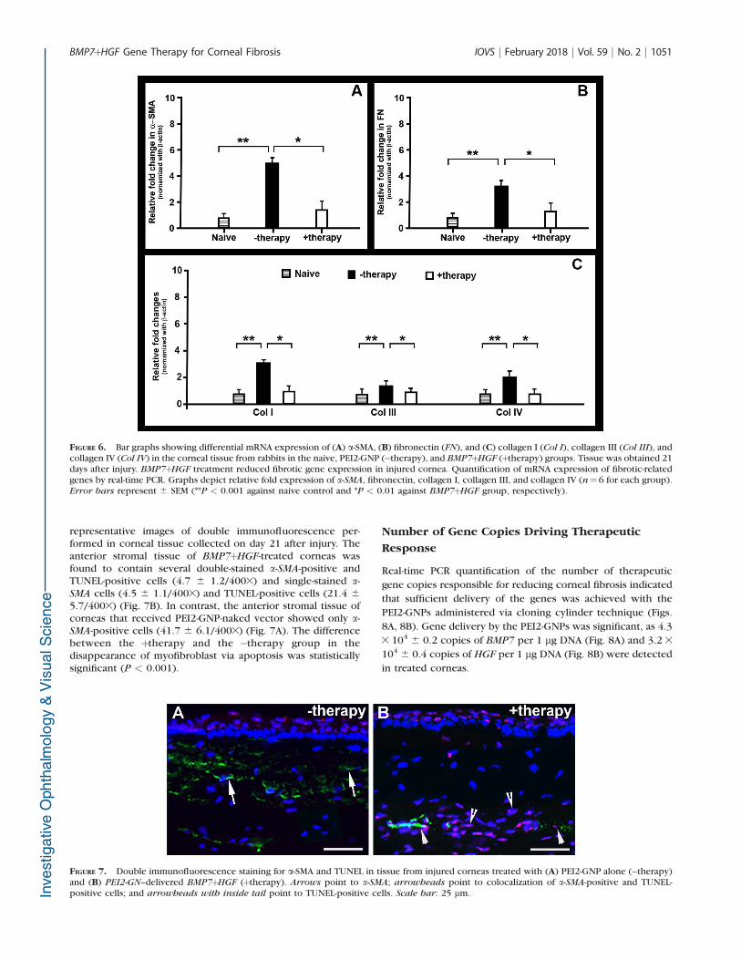
Fluorescein staining revealed that BMP7þHGF gene transferis not detrimental to re-epithelization of the injured corneas(Fig. 4). Fluorescence stereo biomicroscopy images showedcorneal ulceration and epithelial defect on day 1 after alkaliinjury (Figs. 4A, 4C), but by day 21 all of the treated rabbits
demonstrated complete corneal re-epithelialization and nega-tive fluorescein staining (Figs. 4B, 4D). The lacrimal lake, asobserved on tear strips, appeared similar in both PEI2-GNP-naked vector and BMP7þHGF groups, suggesting that combi-nation therapy with BMP7þHGF does not compromise tearproduction or invoke a dry eye condition.
Myofibroblast and Profibrotic Gene Expression
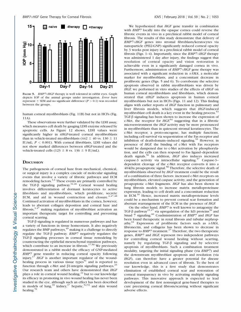
Determination of myofibroblast after alkali-induced cornealinjury indicates that BMP7þHGF gene therapy has aninhibitory effect on myofibroblast formation and profibroticgene expression (Fig. 5). Immunohistologic staining of a-SMA,a marker for myofibroblasts, demonstrated a clinically relevantand statistically significant reduction in a-SMA-positive cells incorneas that received BMP7þHGF genes (Fig. 5B) as comparedto corneas of the naive and PEI2-GNP-naked plasmids (Fig. 5A).The immunofluorescence quantification graph (Fig. 5C)showed an 83% reduction of a-SMA-positive cells (P < 0.001)in the þtherapy group as compared to the number of a-SMA-positive cells in the �therapy group (Fig. 5C).
Since BMP7þHGF gene delivery into fibrotic rabbit corneasled to a quantifiable improvement in wound healing after alkaliburn in our in vivo model, the expression of five prominentgenes (a-SMA, fibronectin, collagen I, collagen III, and collagenIV) involved in fibrosis pathways was also examined in thecorneas of rabbits that received BSS, PEI2-GNP-naked plasmidor BMP7þHGF genes (Fig. 6). As Figure 6 illustrates, alkaliinjury significantly increased levels of the five tested profi-brotic genes in the �therapy corneas compared to naivecorneas from 2.2- to 5.2-fold (P < 0.001), whereas BMP7þHGF
gene transfer significantly reduced profibrotic gene expressionof the a-SMA, a 3.2-fold reduction (Fig. 6A; P <0.01);fibronectin, a 2.3-fold reduction (Fig. 6B; P <0.01); collagenI, a 2.1-fold reduction (Fig. 6C; P <0.01); collagen III, a 1.6-foldreduction (Fig. 6C; P <0.01), and collagen IV, a 1.9-foldreduction (Fig. 6C; P <0.01).
BMP7þHGF Dissipates Myofibroblasts viaApoptosis
Double immunofluorescence staining of corneal tissue sectionsfor a-SMA (a myofibroblast marker) and TUNEL (an apoptosismarker) demonstrated that HGF delivery dissipates myofibro-blasts through apoptosis in the fibrotic cornea. Figure 7 shows
FIGURE 1. Biomicroscopy images before and after alkali-induced corneal injury of rabbit eyes that did not receive BMP7þHGF treatment (A–E) andthose that received BMP7þHGF treatment (F–J). Dense corneal opacity developed by day 1 after induction of alkali injury (B, G). On day 14 and day21 post injury, corneas that received PEI2-GNP–mediated BMP7þHGF eyes showed a significant reduction of opacification (H–J) as compared to thelevel of opacification in corneas that received naked vector (C–E). Scale bar: 2 mm.
FIGURE 2. Quantification of corneal opacity using the Fantes score inthe three groups of rabbits: no corneal injury group (naive; gray bar),PEI2-GNP-naked plasmid group (�therapy; black bar), and PEI2-GNP–BMP7þHGF group (þtherapy; white bar). The graph depicts compar-ative haze scores 24 hours post injury and at days 7, 14, and 21 postinjury (*P < 0.01 and **P < 0.001).
BMP7þHGF Gene Therapy for Corneal Fibrosis IOVS j February 2018 j Vol. 59 j No. 2 j 1049
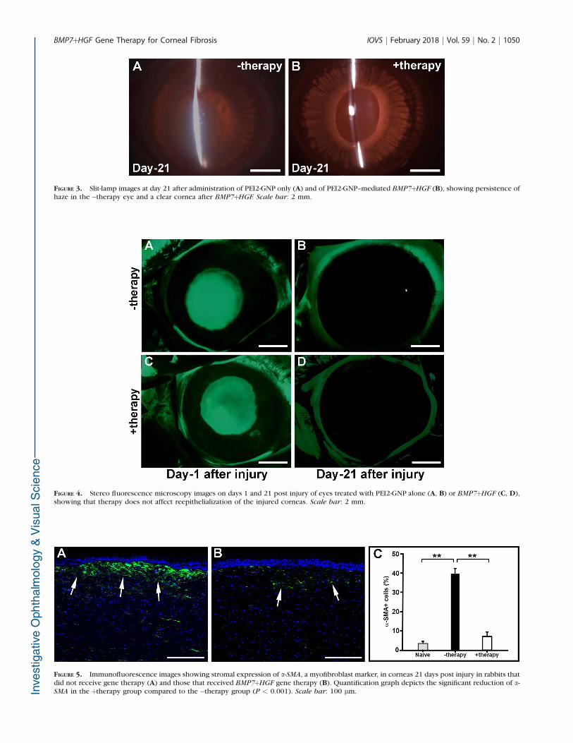
FIGURE 3. Slit-lamp images at day 21 after administration of PEI2-GNP only (A) and of PEI2-GNP–mediated BMP7þHGF (B), showing persistence ofhaze in the�therapy eye and a clear cornea after BMP7þHGF. Scale bar: 2 mm.
FIGURE 4. Stereo fluorescence microscopy images on days 1 and 21 post injury of eyes treated with PEI2-GNP alone (A, B) or BMP7þHGF (C, D),showing that therapy does not affect reepithelialization of the injured corneas. Scale bar: 2 mm.
FIGURE 5. Immunofluorescence images showing stromal expression of a-SMA, a myofibroblast marker, in corneas 21 days post injury in rabbits thatdid not receive gene therapy (A) and those that received BMP7þHGF gene therapy (B). Quantification graph depicts the significant reduction of a-
SMA in theþtherapy group compared to the�therapy group (P < 0.001). Scale bar: 100 lm.
BMP7þHGF Gene Therapy for Corneal Fibrosis IOVS j February 2018 j Vol. 59 j No. 2 j 1050
representative images of double immunofluorescence per-formed in corneal tissue collected on day 21 after injury. Theanterior stromal tissue of BMP7þHGF-treated corneas wasfound to contain several double-stained a-SMA-positive andTUNEL-positive cells (4.7 6 1.2/4003) and single-stained a-
SMA cells (4.5 6 1.1/4003) and TUNEL-positive cells (21.4 6
5.7/4003) (Fig. 7B). In contrast, the anterior stromal tissue ofcorneas that received PEI2-GNP-naked vector showed only a-
SMA-positive cells (41.7 6 6.1/4003) (Fig. 7A). The differencebetween the þtherapy and the �therapy group in thedisappearance of myofibroblast via apoptosis was statisticallysignificant (P < 0.001).
Number of Gene Copies Driving Therapeutic
Response
Real-time PCR quantification of the number of therapeutic
gene copies responsible for reducing corneal fibrosis indicated
that sufficient delivery of the genes was achieved with the
PEI2-GNPs administered via cloning cylinder technique (Figs.
8A, 8B). Gene delivery by the PEI2-GNPs was significant, as 4.3
3 104 6 0.2 copies of BMP7 per 1 lg DNA (Fig. 8A) and 3.2 3
104 6 0.4 copies of HGF per 1 lg DNA (Fig. 8B) were detected
in treated corneas.
FIGURE 6. Bar graphs showing differential mRNA expression of (A) a-SMA, (B) fibronectin (FN), and (C) collagen I (Col I), collagen III (Col III), andcollagen IV (Col IV) in the corneal tissue from rabbits in the naive, PEI2-GNP (�therapy), and BMP7þHGF (þtherapy) groups. Tissue was obtained 21days after injury. BMP7þHGF treatment reduced fibrotic gene expression in injured cornea. Quantification of mRNA expression of fibrotic-relatedgenes by real-time PCR. Graphs depict relative fold expression of a-SMA, fibronectin, collagen I, collagen III, and collagen IV (n¼ 6 for each group).Error bars represent 6 SEM (**P < 0.001 against naive control and *P < 0.01 against BMP7þHGF group, respectively).
FIGURE 7. Double immunofluorescence staining for a-SMA and TUNEL in tissue from injured corneas treated with (A) PEI2-GNP alone (�therapy)and (B) PEI2-GN–delivered BMP7þHGF (þtherapy). Arrows point to a-SMA; arrowheads point to colocalization of a-SMA-positive and TUNEL-positive cells; and arrowheads with inside tail point to TUNEL-positive cells. Scale bar: 25 lm.
BMP7þHGF Gene Therapy for Corneal Fibrosis IOVS j February 2018 j Vol. 59 j No. 2 j 1051

In Vivo Toxicity and Safety Studies
The results of time-dependent ocular irritation studies per-formed in live rabbits using the Draize and modifiedMacDonald-Shadduck scoring systems are highlighted in theTable. As expected, an alkali wound led to a significantlyincreased cumulative Draize score in the PEI2-GNP-nakedvector and PEI2–mediated BMP7þHGF group as compared tonaive corneas. On day 7, the average Draize score was 45.0 inthe�therapy group versus 0 in the naive group (P < 0.001); onday 14, 39.0 vs. 0 (P < 0.001); and on day 21, 29.8 vs. 0 (P <0.001). BMP7þHGF therapy was associated with a significant,time-dependent reduction in the cumulative Draize scorecompared to the score in the �therapy group as follows: day7, 29.1 with BMP7þHGF vs. 45.0 with�therapy (P < 0.01); day14, 18.9 vs. 39.0 (P < 0.01); and day 21, 5.1 vs. 29.8 (P < 0.01).The modified MacDonald-Shadduck test results showed asimilar pattern of scores following alkali wounding in PEI2-GNP-naked vector and BMP7þHFG groups. Injured eyes thatreceived BMP7þHGF therapy exhibited significantly lowermodified MacDonald-Shadduck scores than did the eyes ofrabbits that received PEI2-GNP-naked vector (day 7: 0.9 vs. 1.8,P < 0.05; day 14: 0.6 vs. 1.3, P < 0.01; day 21: 0.3 vs. 1.1, P <0.001). On the subjective clinical eye examinations performedindependently by three examiners, significantly improvedoverall ocular health was observed in the eyes of rabbits thatreceived gene therapy as compared to eyes of rabbits thatreceived the nanoparticles alone.
Evaluation of the effects of BMP7þHGF therapy on IOP andtear production revealed no significant differences in the IOP
(Fig. 9; P > 0.1) and tear levels (data not shown) in the eyes ofrabbits among the three groups.
H&E staining demonstrated noteworthy morphologic alter-ations in corneal epithelium and stroma at day 21 post alkaliinjury in the rabbits that received PEI2-GNP-naked vector (Fig.10A), corroborating the presence of the clinical opacificationvisualized with slit-lamp biomicroscopy. BMP7þHGF therapymarkedly mitigated the adverse impact of alkali wounding oncorneal epithelial and stromal tissues (Fig. 10B). In addition, nosignificant cellular inflammatory infiltrates were observed inthe corneas of either group, as quantified with CD11bimmunofluorescence (Figs. 10C, 10D; P > 0.1). The Masson’strichome staining demonstrated notably decreased collagendeposition in the rabbit corneas that received BMP7þHGF
therapy (Fig. 10F) compared to the �therapy naked vector–delivered corneas (Fig. 10E).
Role of HGF in Apoptosis of Corneal
Myofibroblasts and Fibroblasts
The molecular function of HGF on corneal fibroblasts andmyofibroblasts was studied in an established in vitro model ofcorneal fibrosis in humans. Primary stromal cultures obtainedfrom donor human corneas and grown in the absence of TGF-b1 provided corneal fibroblasts and grown in the presence ofTGF-b1 produced corneal myofibroblasts, as demonstrated bya-SMA staining and TUNEL staining. Recombinant HGF (rHGF)treatment of these cultures caused significant apoptosis, asindicated by the detection of several TUNEL-positive cells in
FIGURE 8. Bar graph of quantitative PCR analysis showing BMP7 (A) and HGF (B) gene copy numbers delivered by PEI2-GNPs to the rabbit cornea,indicating efficient delivery of genes by the customized nanoparticles. There were six samples in each group and error bars represent 6 SEM (*P <0.001 against naive control and�therapy group).
TABLE. Draize and Modified MacDonald-Shadduck Scoring Shows the Ocular Anomaly in �Therapy and þTherapy Groups
Groups
Draize Total Score MacDonald-Shadduck Score
Day 7 Day 14 Day 21 Day 7 Day 14 Day 21
Naive 0 0 0 0 0 0
�Therapy 45.0 6 0.31 39.0 6 0.23 29.8 6 0.18 1.8 6 0.32 1.3 6 0.21 1.1 6 0.19
þTherapy 29.1 6 0.29 18.9 6 0.19 5.1 6 0.14 0.9 6 0.18 0.6 6 0.12 0.3 6 0.12
The slit-lamp and stereo biomicroscopic images were scored by three independent resident observers. The naive group did not show any ocularanomaly during Draize and modified MacDonald-Shadduck scoring (P < 0.001 or P < 0.01 or P < 0.05 between the groups at different time points).
BMP7þHGF Gene Therapy for Corneal Fibrosis IOVS j February 2018 j Vol. 59 j No. 2 j 1052
human corneal myofibroblasts (Fig. 11B) but not in HCFs (Fig.11A).
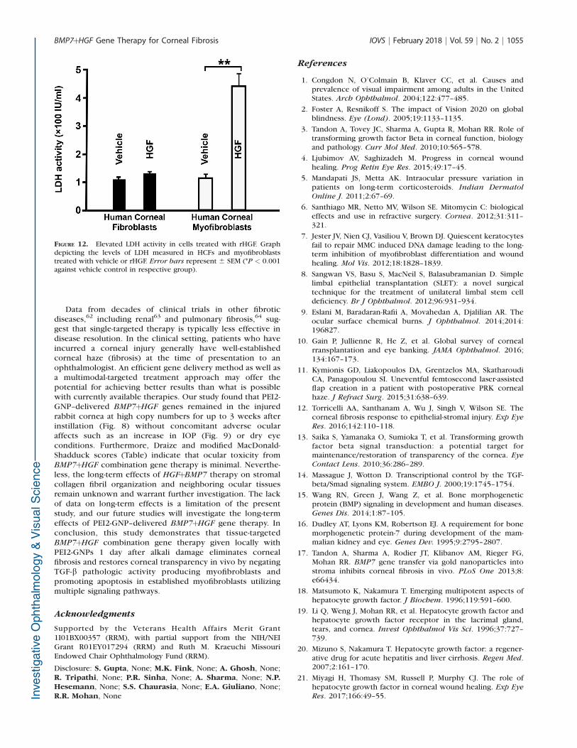
These observations were further validated by the LDH assay,which measures cell death by gauging LDH enzyme released byapoptotic cells. As Figure 12 shows, LDH values weresignificantly higher in rHGF-treated corneal myofibroblaststhan in vehicle-treated myofibroblasts (442 6 40 vs. 130 6 11IU/mL; P < 0.001). With corneal fibroblasts, LDH values didnot show marked differences between rHGF-treated and thevehicle-treated cells (123 6 8 vs. 136 6 9 IU/mL).
DISCUSSION
The pathogenesis of corneal haze from mechanical, chemical,or surgical injury is a complex cascade of molecular signalingevents that involve a variety of fibrotic pathways and ECMremodeling factors.3,4 These events are primarily controlled bythe TGF-b signaling pathway.34,46 Corneal wound healinginvolves differentiation of dormant keratocytes to activefibroblasts and myofibroblasts, which proliferate, secreteECM, and aid in wound contraction and closure.4,47,48
Continued activation of myofibroblasts in the cornea, however,leads to aberrant collagen deposition and corneal haze andfibrosis,3,47 making regulation of myofibroblast activation animportant therapeutic target for controlling and preventingcorneal scarring.
TGF-b signaling is regulated in numerous pathways and hasa variety of functions in tissue homeostasis. It also negativelyregulates the BMP pathways,49 making it a challenge to directlyregulate the TGF-b pathway. BMP7 negatively regulates theTGF-b signaling processes in corneal tissue remodeling bycounteracting the epithelial mesenchymal transition pathways,which contribute to an increase in fibrosis.17,50 We previouslydemonstrated in a rabbit model the efficacy of GNP-mediatedBMP7 gene transfer in reducing corneal opacity followinginjury.17
HGF is another important regulator of the wound-healing process in various tissue types51 and is reported tofunction through c-Met and b1-integrin signaling pathways.23
Our research team and others have demonstrated that HGF
plays a role in corneal wound healing,22 but to our knowledgeits efficacy in promoting in vivo wound healing has never beenstudied in the eye, although such an effect has been describedin models of lung,39 kidney,37 hepatic,52,53 and skin woundhealing.51
We hypothesized that HGF gene transfer in combinationwith BMP7 locally into the opaque cornea could reverse thefibrotic events in vivo in a preclinical rabbit model of cornealfibrosis. The results of this study demonstrate that delivery ofBMP7þHGF genes into stromal fibroblasts/keratocytes viananoparticle (PEI2-GNP) significantly reduced corneal opacityby 3 weeks post injury in a preclinical rabbit model of cornealfibrosis (Figs. 1–4). Importantly, since the BMP7þHGF therapywas administered 1 day after injury, the findings suggest thatresolution of corneal opacity and vision restoration isachievable even in a significantly damaged cornea in vivo.Furthermore, administration of BMP7þHGF gene therapy wasassociated with a significant reduction in a-SMA, a molecularmarker for myofibroblasts, and a concomitant decrease inprofibrotic genes (Figs. 5 and 6). To corroborate the selectiveapoptosis observed in rabbit myofibroblasts was driven byHGF, we performed in vitro studies of the effects of rHGF onhuman corneal myofibroblasts and fibroblasts, which demon-strated that rHGF induces apoptosis in human cornealmyofibroblasts but not in HCFs (Figs. 11 and 12). This findingaligns with earlier reports of HGF function in pulmonary andliver fibrosis models, which suggests that HGF-inducedmyofibroblast cell death is a key event in the healing process.53
TGF-b signaling has been shown to increase the expression ofc-Met, the receptor for HGF,54 suggesting that in a fibroticmicroenvironment the HGF activity may be more pronouncedin myofibroblasts than in quiescent stromal keratinocytes. Thec-Met receptor, a proto-oncogene, has multiple functions,including cell survival via sequestration of the Fas receptor andinhibition of death-domain–induced signaling.55 However, inpresence of HGF, the binding of c-Met with Fas receptorswould be dampened due to c-Met activation by phosphoryla-tion, and the cells can then respond to Fas ligand–dependentdeath signals.56 In addition, HGF also induces increasedcaspase-3 activity via intracellular signaling.57 Caspase-3–dependent cleavage of the c-Met receptor converts it into a40-kDa proapoptotic signal.58 Therefore, the selective death ofmyofibroblasts observed by HGF treatment could be the resultof a combination of three factors: increased c-Met receptors onmyofibroblasts, elevated caspase activity, and the generation ofproapoptotic c-Met fragments. HGF has also been shown inlung fibrosis models to increase matrix metalloproteinaseexpression, leading to cell death and a concomitant reductionin ECM.39 Hence, increased matrix metalloproteinase levelscould be a mechanism to prevent corneal scar formation andaberrant rearrangement of the ECM in the presence of HGF.
On the other hand, BMP7 is well known to antagonize theTGF-b pathway15,17 via upregulation of the Id3 proteins59 andSmad 7 signaling.60 Coadministration of BMP7 and HGF hasbeen found therapeutic in renal fibrosis and tubular nephrop-athy.61 Expression of profibrotic factors such as a-SMA,fibronectin, and collagens has been shown to decrease inresponse to BMP7 treatment.17 Therefore, the two therapeuticgenes, BMP7 and HGF, represent two independent pathwaysfor controlling corneal wound healing without scarring,namely by regulating TGF-b signaling and by selectiveapoptosis of myofibroblasts. Such a combination treatmentmodality, targeting the initial signaling phase (via BMP7) andthe downstream myofibroblast apoptosis and resolution (viaHGF), can therefore have a greater potential for diseaseresolution even in advanced cases of fibrosis. To the best ofour knowledge, this is a first study that demonstrateselimination of established corneal scar and restoration ofcorneal transparency in vivo by activating multiple signalingpathways. This innovative approach is expected to leaddevelopment of the first nonsurgical gene-based therapies tocure preexisting corneal fibrosis/scarring without significantadverse effects.
FIGURE 9. BMP7þHGF therapy is well tolerated in rabbit eyes. Graphdepicts IOP of the animal groups under investigation. Error bars
represent 6 SEM and no significant difference (P > 0.1) was recordedbetween the groups.
BMP7þHGF Gene Therapy for Corneal Fibrosis IOVS j February 2018 j Vol. 59 j No. 2 j 1053
FIGURE 10. Combination BMP7 and HGF gene therapy is safe and caused no adverse effects in rabbit eyes. H&E images show no alteration incorneal morphology with PEI2-GNPs alone (A) and with BMP7þHGF gene therapy (B). CD11B immunofluorescence images 21 days after cornealinjury show no infiltration of leukocytes and inflammatory markers in the stromal layer in corneal tissue from PEI2-GNP–treated rabbits (�therapy)and from BMP7þHGF-treated rabbits (þtherapy). Masson’s trichome–stained corneal tissue images show decreased collagen expression inBMP7þHGF-treated eyes (F) compared to the �therapy given eyes (E). Scale bar: 100 and 200 lm.
FIGURE 11. Immunocytochemistry images of a-SMA staining (green) and TUNEL staining (pink) in cell cultures of HCFs (A) and myofibroblasts (B)after rHGF treatment, demonstrating that rHGF selectively induces apoptosis of corneal myofibroblasts. Arrows point to TUNEL-positivemyofibroblast cells. Scale bar: 50 lm.
BMP7þHGF Gene Therapy for Corneal Fibrosis IOVS j February 2018 j Vol. 59 j No. 2 j 1054
Data from decades of clinical trials in other fibroticdiseases,62 including renal63 and pulmonary fibrosis,64 sug-gest that single-targeted therapy is typically less effective indisease resolution. In the clinical setting, patients who haveincurred a corneal injury generally have well-establishedcorneal haze (fibrosis) at the time of presentation to anophthalmologist. An efficient gene delivery method as well asa multimodal-targeted treatment approach may offer thepotential for achieving better results than what is possiblewith currently available therapies. Our study found that PEI2-GNP–delivered BMP7þHGF genes remained in the injuredrabbit cornea at high copy numbers for up to 3 weeks afterinstillation (Fig. 8) without concomitant adverse ocularaffects such as an increase in IOP (Fig. 9) or dry eyeconditions. Furthermore, Draize and modified MacDonald-Shadduck scores (Table) indicate that ocular toxicity fromBMP7þHGF combination gene therapy is minimal. Neverthe-less, the long-term effects of HGFþBMP7 therapy on stromalcollagen fibril organization and neighboring ocular tissuesremain unknown and warrant further investigation. The lackof data on long-term effects is a limitation of the presentstudy, and our future studies will investigate the long-termeffects of PEI2-GNP–delivered BMP7þHGF gene therapy. Inconclusion, this study demonstrates that tissue-targetedBMP7þHGF combination gene therapy given locally withPEI2-GNPs 1 day after alkali damage eliminates cornealfibrosis and restores corneal transparency in vivo by negatingTGF-b pathologic activity producing myofibroblasts andpromoting apoptosis in established myofibroblasts utilizingmultiple signaling pathways.
Acknowledgments
Supported by the Veterans Health Affairs Merit Grant1I01BX00357 (RRM), with partial support from the NIH/NEIGrant R01EY017294 (RRM) and Ruth M. Kraeuchi MissouriEndowed Chair Ophthalmology Fund (RRM).
Disclosure: S. Gupta, None; M.K. Fink, None; A. Ghosh, None;R. Tripathi, None; P.R. Sinha, None; A. Sharma, None; N.P.Hesemann, None; S.S. Chaurasia, None; E.A. Giuliano, None;R.R. Mohan, None
References
1. Congdon N, O’Colmain B, Klaver CC, et al. Causes andprevalence of visual impairment among adults in the UnitedStates. Arch Ophthalmol. 2004;122:477–485.
2. Foster A, Resnikoff S. The impact of Vision 2020 on globalblindness. Eye (Lond). 2005;19:1133–1135.
3. Tandon A, Tovey JC, Sharma A, Gupta R, Mohan RR. Role oftransforming growth factor Beta in corneal function, biologyand pathology. Curr Mol Med. 2010;10:565–578.
4. Ljubimov AV, Saghizadeh M. Progress in corneal woundhealing. Prog Retin Eye Res. 2015;49:17–45.
5. Mandapati JS, Metta AK. Intraocular pressure variation inpatients on long-term corticosteroids. Indian Dermatol
Online J. 2011;2:67–69.
6. Santhiago MR, Netto MV, Wilson SE. Mitomycin C: biologicaleffects and use in refractive surgery. Cornea. 2012;31:311–321.
7. Jester JV, Nien CJ, Vasiliou V, Brown DJ. Quiescent keratocytesfail to repair MMC induced DNA damage leading to the long-term inhibition of myofibroblast differentiation and woundhealing. Mol Vis. 2012;18:1828–1839.
8. Sangwan VS, Basu S, MacNeil S, Balasubramanian D. Simplelimbal epithelial transplantation (SLET): a novel surgicaltechnique for the treatment of unilateral limbal stem celldeficiency. Br J Ophthalmol. 2012;96:931–934.
9. Eslani M, Baradaran-Rafii A, Movahedan A, Djalilian AR. Theocular surface chemical burns. J Ophthalmol. 2014;2014:196827.
10. Gain P, Jullienne R, He Z, et al. Global survey of cornealrransplantation and eye banking. JAMA Ophthalmol. 2016;134:167–173.
11. Kymionis GD, Liakopoulos DA, Grentzelos MA, SkatharoudiCA, Panagopoulou SI. Uneventful femtosecond laser-assistedflap creation in a patient with postoperative PRK cornealhaze. J Refract Surg. 2015;31:638–639.
12. Torricelli AA, Santhanam A, Wu J, Singh V, Wilson SE. Thecorneal fibrosis response to epithelial-stromal injury. Exp Eye
Res. 2016;142:110–118.
13. Saika S, Yamanaka O, Sumioka T, et al. Transforming growthfactor beta signal transduction: a potential target formaintenance/restoration of transparency of the cornea. Eye
Contact Lens. 2010;36:286–289.
14. Massague J, Wotton D. Transcriptional control by the TGF-beta/Smad signaling system. EMBO J. 2000;19:1745–1754.
15. Wang RN, Green J, Wang Z, et al. Bone morphogeneticprotein (BMP) signaling in development and human diseases.Genes Dis. 2014;1:87–105.
16. Dudley AT, Lyons KM, Robertson EJ. A requirement for bonemorphogenetic protein-7 during development of the mam-malian kidney and eye. Genes Dev. 1995;9:2795–2807.
17. Tandon A, Sharma A, Rodier JT, Klibanov AM, Rieger FG,Mohan RR. BMP7 gene transfer via gold nanoparticles intostroma inhibits corneal fibrosis in vivo. PLoS One 2013;8:e66434.
18. Matsumoto K, Nakamura T. Emerging multipotent aspects ofhepatocyte growth factor. J Biochem. 1996;119:591–600.
19. Li Q, Weng J, Mohan RR, et al. Hepatocyte growth factor andhepatocyte growth factor receptor in the lacrimal gland,tears, and cornea. Invest Ophthalmol Vis Sci. 1996;37:727–739.
20. Mizuno S, Nakamura T. Hepatocyte growth factor: a regener-ative drug for acute hepatitis and liver cirrhosis. Regen Med.2007;2:161–170.
21. Miyagi H, Thomasy SM, Russell P, Murphy CJ. The role ofhepatocyte growth factor in corneal wound healing. Exp Eye
Res. 2017;166:49–55.
FIGURE 12. Elevated LDH activity in cells treated with rHGF. Graphdepicting the levels of LDH measured in HCFs and myofibroblaststreated with vehicle or rHGF. Error bars represent 6 SEM (*P < 0.001against vehicle control in respective group).
BMP7þHGF Gene Therapy for Corneal Fibrosis IOVS j February 2018 j Vol. 59 j No. 2 j 1055
22. Wilson SE, Chen L, Mohan RR, Liang Q, Liu J. Expression ofHGF, KGF, EGF and receptor messenger RNAs followingcorneal epithelial wounding. Exp Eye Res. 1999;68:377–397.
23. Li JF, Duan HF, Wu CT, et al. HGF accelerates wound healingby promoting the dedifferentiation of epidermal cells throughbeta1-integrin/ILK pathway. BioMed Res Int. 2013;2013:470418.
24. Matsumoto K, Funakoshi H, Takahashi H, Sakai K. HGF-Metpathway in regeneration and drug discovery. Biomedicines.2014;2:275–300.
25. Mohan RR, Rodier JT, Sharma A. Corneal gene therapy: basicscience and translational perspective. Ocul Surf. 2013;11:150–164.
26. Mohan RR, Tovey JC, Sharma A, Tandon A. Gene therapy inthe cornea: 2005–present. Prog Retin Eye Res. 2012;31:43–64.
27. Mohan RR, Tovey JC, Sharma A, Schultz GS, Cowden JW,Tandon A. Targeted decorin gene therapy delivered withadeno-associated virus effectively retards corneal neovascu-larization in vivo. PLoS One. 2011;6:e26432.
28. Mohan RR, Tandon A, Sharma A, Cowden JW, Tovey JC.Significant inhibition of corneal scarring in vivo with tissue-selective, targeted AAV5 decorin gene therapy. Invest
Ophthalmol Vis Sci. 2011;52:4833–4841.
29. Sharma A, Tandon A, Tovey JC, et al. Polyethylenimine-conjugated gold nanoparticles: gene transfer potential andlow toxicity in the cornea. Nanomedicine. 2011;7:505–513.
30. Sharma A, Rodier JT, Tandon A, Klibanov AM, Mohan RR.Attenuation of corneal myofibroblast development throughnanoparticle-mediated soluble transforming growth factor-beta type II receptor (sTGFbetaRII) gene transfer. Mol Vis.2012;18:2598–2607.
31. Donnelly KS, Giuliano EA, Sharma A, Tandon A, Rodier JT,Mohan RR. Decorin-PEI nanoconstruct attenuates equinecorneal fibroblast differentiation. Vet Ophthalmol. 2014;17:162–169.
32. Thomas M, Klibanov AM. Conjugation to gold nanoparticlesenhances polyethylenimine’s transfer of plasmid DNA intomammalian cells. Proc Natl Acad Sci U S A. 2003;100:9138–9143.
33. Umeda Y, Marui T, Matsuno Y, et al. Skeletal muscle targetingin vivo electroporation-mediated HGF gene therapy ofbleomycin-induced pulmonary fibrosis in mice. Lab Invest.2004;84:836–844.
34. Mizuno S, Matsumoto K, Kurosawa T, Mizuno-Horikawa Y,Nakamura T. Reciprocal balance of hepatocyte growth factorand transforming growth factor-beta 1 in renal fibrosis inmice. Kidney Int. 2000;57:937–948.
35. Cui S, Guo L, Li X, et al. Clinical safety and preliminaryefficacy of plasmid pUDK-HGF expressing human Hepatocytegrowth factor (HGF) in patients with critical limb ischemia.Eur J Vasc Endovasc Surg. 2015;50:494–501.
36. Powell RJ. Update on clinical trials evaluating the effect ofbiologic therapy in patients with critical limb ischemia. J Vasc
Surg. 2012;56:264–266.
37. Iekushi K, Taniyama Y, Azuma J, et al. Hepatocyte growthfactor attenuates renal fibrosis through TGF-beta1 suppres-sion by apoptosis of myofibroblasts. J Hypertens. 2010;28:2454–2461.
38. Mizuno S, Matsumoto K, Li MY, Nakamura T. HGF reducesadvancing lung fibrosis in mice: a potential role for MMP-dependent myofibroblast apoptosis. FASEB J. 2005;19:580–582.
39. Gazdhar A, Fachinger P, van Leer C, et al. Gene transfer ofhepatocyte growth factor by electroporation reduces bleo-mycin-induced lung fibrosis. Am J Physiol Lung Cell Mol
Physiol. 2007;292:L529–536.
40. Sharma A, Anumanthan G, Reyes M, et al. Epigeneticmodification prevents excessive wound healing and scarformation after glaucoma filtration surgery. Invest Ophthal-
mol Vis Sci. 2016;57:3381–3389.
41. Ormerod LD, Abelson MB, Kenyon KR. Standard models ofcorneal injury using alkali-immersed filter discs. Invest
Ophthalmol Vis Sci. 1989;30:2148–2153.
42. Sharma A, Mehan MM, Sinha S, Cowden JW, Mohan RR.Trichostatin a inhibits corneal haze in vitro and in vivo. Invest
Ophthalmol Vis Sci. 2009;50:2695–2701.
43. Wilhelmus KR. The Draize eye test. Surv Ophthalmol. 2001;45:493–515.
44. Altmann S, Emanuel A, Toomey M, et al. A quantitative rabbitmodel of vaccinia keratitis. Invest Ophthalmol Vis Sci. 2010;51:4531–4540.
45. Gronkiewicz KM, Giuliano EA, Kuroki K, et al. Developmentof a novel in vivo corneal fibrosis model in the dog. Exp Eye
Res. 2016;143:75–88.
46. Chaikuad A, Bullock AN. Structural basis of intracellular TGF-beta signaling: receptors and Smads. Cold Spring Harb
Perspect Biol. 2016;8:a022111.
47. Myrna KE, Pot SA, Murphy CJ. Meet the corneal myofibro-blast: the role of myofibroblast transformation in cornealwound healing and pathology. Vet Ophthalmol. 2009;12(suppl 1):25–27.
48. Jester JV, Petroll WM, Barry PA, Cavanagh HD. Expression ofalpha-smooth muscle (alpha-SM) actin during corneal stromalwound healing. Invest Ophthalmol Vis Sci. 1995;36:809–819.
49. Miyazono K. Positive and negative regulation of TGF-betasignaling. J Cell Sci. 2000;113(pt 7):1101–1109.
50. Zeisberg M, Hanai J, Sugimoto H, et al. BMP-7 counteractsTGF-beta1-induced epithelial-to-mesenchymal transition andreverses chronic renal injury. Nat Med. 2003;9:964–968.
51. Conway K, Price P, Harding KG, Jiang WG. The molecular andclinical impact of hepatocyte growth factor, its receptor,activators, and inhibitors in wound healing. Wound Repair
Regen. 2006;14:2–10.
52. Ueki T, Kaneda Y, Tsutsui H, et al. Hepatocyte growth factorgene therapy of liver cirrhosis in rats. Nat Med. 1999;5:226–230.
53. Kim WH, Matsumoto K, Bessho K, Nakamura T. Growthinhibition and apoptosis in liver myofibroblasts promoted byhepatocyte growth factor leads to resolution from livercirrhosis. Am J Pathol. 2005;166:1017–1028.
54. Ghatak S, Bogatkevich GS, Atnelishvili I, et al. Overexpressionof c-Met and CD44v6 receptors contributes to autocrine TGF-beta1 signaling in interstitial lung disease. J Biol Chem. 2014;289:7856–7872.
55. Smyth LA, Brady HJ. cMet and Fas receptor interactioninhibits death-inducing signaling complex formation inendothelial cells. Hypertension. 2005;46:100–106.
56. Tulasne D, Foveau B. The shadow of death on the METtyrosine kinase receptor. Cell Death Differ. 2008;15:427–434.
57. Arakaki N, Kazi JA, Kazihara T, Ohnishi T, Daikuhara Y.Hepatocyte growth factor/scatter factor activates the apopto-sis signaling pathway by increasing caspase-3 activity insarcoma 180 cells. Biochem Biophys Res Commun. 1998;245:211–215.
58. Tulasne D, Deheuninck J, Lourenco FC, et al. Proapoptoticfunction of the MET tyrosine kinase receptor through caspasecleavage. Mol Cell Biol. 2004;24:10328–10339.
59. Lim RR, Tan A, Liu YC, et al. ITF2357 transactivates Id3 andregulate TGFbeta/BMP7 signaling pathways to attenuatecorneal fibrosis. Sci Rep. 2016;6:20841.
60. Gupta S, Rodier JT, Sharma A, et al. Targeted AAV5-Smad7gene therapy inhibits corneal scarring in vivo. PLoS One.2017;12:e0172928.
BMP7þHGF Gene Therapy for Corneal Fibrosis IOVS j February 2018 j Vol. 59 j No. 2 j 1056

61. Klahr S, Morrissey J. Obstructive nephropathy and renalfibrosis: the role of bone morphogenic protein-7 andhepatocyte growth factor. Kidney Int Suppl. 2003;S105–112.
62. Akhurst RJ, Hata A. Targeting the TGFbeta signalling pathwayin disease. Nat Rev Drug Discov. 2012;11:790–811.
63. Munoz-Felix JM, Gonzalez-Nunez M, Martinez-Salgado C,Lopez-Novoa JM. TGF-beta/BMP proteins as therapeutic
targets in renal fibrosis. Where have we arrived after 25
years of trials and tribulations? Pharmacol Ther. 2015;156:
44–58.
64. King TE Jr., Bradford WZ, Castro-Bernardini S, et al. A phase 3
trial of pirfenidone in patients with idiopathic pulmonary
fibrosis. N Engl J Med. 2014;370:2083–2092.
BMP7þHGF Gene Therapy for Corneal Fibrosis IOVS j February 2018 j Vol. 59 j No. 2 j 1057


![k'j{ tof/L tyf k|ltsfo{ of]hgf](https://static.fdocuments.us/doc/165x107/6279b13856ec0c7ec42ce65b/kj-tofl-tyf-kltsfo-ofhgf.jpg)


![kl/of]hgf ;Demf}tf ;DkGg - Swc-Social Welfare Council … kl/of]hgf ;+rfng ug]{ p2]Zon] 24 Aug, 2011 df # jif{ cjlwsf] kl/of]hgf ;Demf}tf ;DkGg eof] . ;+emf}tf kqdf ;dfh sNof0f kl/ifb\sf](https://static.fdocuments.us/doc/165x107/5afdb9ff7f8b9a8b4d8dfbf3/klofhgf-demftf-dkgg-swc-social-welfare-council-klofhgf-rfng-ug.jpg)
![Invenio@HGF – status and perspectivesjuser.fz-juelich.de/record/139421/files/FZJ-2013-05410.pdf · Helmholtz-Gemeinschaft Invenio@HGF – status and perspectives [sic!]Jülich –](https://static.fdocuments.us/doc/165x107/5ed39e1a18dc2351871e3c70/inveniohgf-a-status-and-helmholtz-gemeinschaft-inveniohgf-a-status-and-perspectives.jpg)