Novel approach of treating Gorham-Stout disease in the ... · pulled by umbilical tape. B, The...
7
426 Abstract. – Gorham-Stout disease or the so- called vanishing bone syndrome is a rare disorder characterized by intra-osseous proliferation of vascular channels resulting in destruction and re- sorption of the osseous matrix. The exact patholo- gy of this disease showed no evidence of malig- nant, neuropathic, or infectious components in- volved in the causation of this disorder except for the culprit of lympho-vascular malformations in the bone. The mechanism of bone resorption is yet to be clarified. The clinical presentation of Gorham’s disease varies according to the organ of involvement. Patients diagnosed with Gorham’s disease in the bone may initially present with in- sidious onset of dull aching pain, progressive weakness, or pathologic fractures as the initial presentation. Gorham’s disease is progressive in most patients; yet it can be self-limiting in a few reported cases. The axes of treating this disease as reported in the literature include the use of medical treatment, surgical intervention, radio- therapy and/or the combination of any them. How- ever, there is no consensus about the most effec- tive approach for treating this rare disease. The challenge in this disease lies in both: how to diag- nose and how to treat. Our novel approach com- bined surgical intervention, medication and radio- therapy as a treatment of Graham-Stout disease in the humerus, and showed no progression of the disease our case. Key words: Gorham-Stout disease, Bone resorption, Progres- sive weakness, Pathologic fractures Case Report An 18 year old gentleman was referred to our quaternary cancer center from private practice, for a non-healing mid-shaft humeral fracture af- ter a sequence of conservative and operative management. The patient presented with a left arm pain after falling down. The physical exam Corresponding Author: Ahmed Shehadeh, MD, FRCS; e-mail: [email protected] European Review for Medical and Pharmacological Sciences Novel approach of treating Gorham-Stout disease in the humerus – Case report and review of literature R. ELLATI 1 , A. ATTILI 1 , H. HADDAD 2 , M. AL-HUSSAINI 2 , A. SHEHADEH 3 1 Department of Surgery, King Hussein Cancer Center, Amman, Jordan 2 Department of Pathology and Laboratory Medicine, King Hussein Cancer Center, Amman, Jordan 3 Orthopedic Oncology Unit, Department of Surgery, King Hussein Cancer Center, Amman, Jordan 2016; 20: 426-432 then, showed swelling, bruises, deformity, ten- derness and a decrease in the range of movement of the affected arm. Antero-posterior and lateral left humerus X-rays showed a mid shaft trans- verse non-displaced left humeral fracture (Figure 1-A). Initially, the patient was treated conserva- tively using a back- slab and arm sling. Six weeks later, the X-ray of his left arm showed per- sistence of the fracture (Figure 1-B), besides a noticeable bone resorption at the fracture site, and tapering of the bone ends. Twelve weeks af- ter treatment a new x-ray was obtained and showed more resorption of bone and very clear tapering bone ends (Figure 1-C). Consecutively, the patient underwent an open-reduction, and a rush nail-fixation with iliac-bone grafting outside our facility. Eight weeks after surgery, X-ray showed surprisingly even more resorption at the fracture site and tapering of the bone edges (Fig- ure1-D, E). Accordingly, the patient was referred to the Orthopedic Oncology Unit at King Hus- sein Cancer Center (KHCC). Detailed evaluation at our institution included a new X-ray which showed further bone loss at mid humerus, tapering ends, yet no periosteal re- action or matrix formation were noted (Figure 1- F). Moreover, extensive blood work-up excluded secondary causes of bone resorption (secondary hyperparathyroidism, renal osteodystrophy, etc.). The next step was to obtain an open biopsy for histopathology, which later showed fibrous tis- sue, inflammatory cells but no malignant cells, further immune-staining with D2-40 (ABCAM ® , Cambridge, UK) showed the presence of lym- phatic vessels in the bone (Figure 2- A,B and C). Eventually, the diagnosis of Gorham’s disease was reached. The basis for the diagnosis was the non-healing nature of the fracture despite an ade- quate fixation, the classical radiological appearance of Gorham’s disease (tapering bone ends or mouse
Transcript of Novel approach of treating Gorham-Stout disease in the ... · pulled by umbilical tape. B, The...

426
Abstract. – Gorham-Stout disease or the so-called vanishing bone syndrome is a rare disordercharacterized by intra-osseous proliferation ofvascular channels resulting in destruction and re-sorption of the osseous matrix. The exact patholo-gy of this disease showed no evidence of malig-nant, neuropathic, or infectious components in-volved in the causation of this disorder except forthe culprit of lympho-vascular malformations inthe bone. The mechanism of bone resorption isyet to be clarified. The clinical presentation ofGorham’s disease varies according to the organof involvement. Patients diagnosed with Gorham’sdisease in the bone may initially present with in-sidious onset of dull aching pain, progressiveweakness, or pathologic fractures as the initialpresentation. Gorham’s disease is progressive inmost patients; yet it can be self-limiting in a fewreported cases. The axes of treating this diseaseas reported in the literature include the use ofmedical treatment, surgical intervention, radio-therapy and/or the combination of any them. How-ever, there is no consensus about the most effec-tive approach for treating this rare disease. Thechallenge in this disease lies in both: how to diag-nose and how to treat. Our novel approach com-bined surgical intervention, medication and radio-therapy as a treatment of Graham-Stout disease inthe humerus, and showed no progression of thedisease our case.
Key words:Gorham-Stout disease, Bone resorption, Progres-
sive weakness, Pathologic fractures
Case Report
An 18 year old gentleman was referred to ourquaternary cancer center from private practice,for a non-healing mid-shaft humeral fracture af-ter a sequence of conservative and operativemanagement. The patient presented with a leftarm pain after falling down. The physical exam
Corresponding Author: Ahmed Shehadeh, MD, FRCS; e-mail: [email protected]
European Review for Medical and Pharmacological Sciences
Novel approach of treating Gorham-Stout disease inthe humerus – Case report and review of literature
R. ELLATI1, A. ATTILI1, H. HADDAD2, M. AL-HUSSAINI2, A. SHEHADEH3
1Department of Surgery, King Hussein Cancer Center, Amman, Jordan2Department of Pathology and Laboratory Medicine, King Hussein Cancer Center, Amman, Jordan3Orthopedic Oncology Unit, Department of Surgery, King Hussein Cancer Center, Amman, Jordan
2016; 20: 426-432
then, showed swelling, bruises, deformity, ten-derness and a decrease in the range of movementof the affected arm. Antero-posterior and lateralleft humerus X-rays showed a mid shaft trans-verse non-displaced left humeral fracture (Figure1-A). Initially, the patient was treated conserva-tively using a back- slab and arm sling. Sixweeks later, the X-ray of his left arm showed per-sistence of the fracture (Figure 1-B), besides anoticeable bone resorption at the fracture site,and tapering of the bone ends. Twelve weeks af-ter treatment a new x-ray was obtained andshowed more resorption of bone and very cleartapering bone ends (Figure 1-C). Consecutively,the patient underwent an open-reduction, and arush nail-fixation with iliac-bone grafting outsideour facility. Eight weeks after surgery, X-rayshowed surprisingly even more resorption at thefracture site and tapering of the bone edges (Fig-ure1-D, E). Accordingly, the patient was referredto the Orthopedic Oncology Unit at King Hus-sein Cancer Center (KHCC). Detailed evaluation at our institution included
a new X-ray which showed further bone loss atmid humerus, tapering ends, yet no periosteal re-action or matrix formation were noted (Figure 1-F). Moreover, extensive blood work-up excludedsecondary causes of bone resorption (secondaryhyperparathyroidism, renal osteodystrophy, etc.).The next step was to obtain an open biopsy forhistopathology, which later showed fibrous tis-sue, inflammatory cells but no malignant cells,further immune-staining with D2-40 (ABCAM®,Cambridge, UK) showed the presence of lym-phatic vessels in the bone (Figure 2- A,B and C). Eventually, the diagnosis of Gorham’s disease
was reached. The basis for the diagnosis was thenon-healing nature of the fracture despite an ade-quate fixation, the classical radiological appearanceof Gorham’s disease (tapering bone ends or mouse

427
Novel approach of treating Gorham-Stout disease in the humerus
tail appearance), exclusion of secondary causes ofbone resorption, and the pathological findings oflymph-vascular tissue in the bone as seen in biopsywith positive D2-40 immunostain for lymph vesselssensitivity is 92.6% and specificity is 98.8%1.The treatment plan was discussed and con-
firmed in the meeting of the multidisciplinaryclinic (doctors of different specialties: orthope-dic oncology, pathology, medical oncology, radi-ation oncology and radiology). Since the patientis left-handed, and the limb was already immo-bilized for more than 5 months; therefore, therewas a clear need for intervention which will al-low early resumption of his left upper limb
movement. Hence, the plan of treatment was toproceed with a reconstructive surgery and towithhold the treatment with bisphosphonatesand radio therapy.
Surgical procedure
• Under general anesthesia, endotracheal intu-bation, supine position, painting, toweling anddraping.• Anterolateral approach to the mid humeruswith ellipse skin incision around the old surgi-cal scar.
Figure 1. An antero-posterior X-ray of theleft humerus over the course of treatment.A, At time of first presentation at an outsidefacility. A transverse fracture at the mid-shaft humerus, post conservative manage-ment with U-slab immobilization. B, Sixweeks after fracture, no signs of healing, in-creased bone resorption and tapering of thebone ends. C, 12 weeks after conservativetreatment. D, Four weeks post-operativenailing done by other facility. E, Eightweeks after nailing, more bone loss, andmore tapering ends of the fracture. F, Attime of presentation to our facility.

428
R. Ellati, A. Attili, H. Haddad, M. Al-Hussaini, A. Shehadeh
• Almost 15 cm of the mid-shaft humeral bonewas replaced by highly vascular fibrous tis-sue. Identification, isolation and protection ofthe radial nerve. • Resection of the middle 18 cm of the humerusand removal of the old rush nail.
Figure 2. Histopathology of left mid-shaft humerus biop-sy; showing lymphatic vessels in the bone. A, Bone trabecu-lae (BT) separated by vascular spaces (white arrow) stainedby Hematoxylin and Eosin, magnified 20 times (X20). B,CD31 immunostain further highlighted the vascular spacesseparating the bone trabeculae (X20). C, D2-40 immunos-tain labeled some of the vascular spaces confirming thelymphatic nature of some of the vessels (Solid black arrow),and unlabelled blood vessels (white arrow) (X40).
Figure 3. A, An intra-operative photo showsthe left shoulder and proximal humerus. Mark-er shows the planned surgical approach withexcision of the previous biopsy tract in thenew incision. B, The frozen structural bone al-lograft that we used, after heating for 20 min-utes in 40° C normal saline solution.
Figure 4. A, Intra operative photo showing the bone is de-structed, and the fixation device (rush nail) is exposed withalmost complete bone resorption from all directions and re-placed by fibrous tissue. The radial nerve is identified andpulled by umbilical tape. B, The proximal humerus LCPplate is fixed to the host bone first. C. Introduction of thebone allograft at the gap, and fixing it to the proximalhumerus LCP plate.

429
Novel approach of treating Gorham-Stout disease in the humerus
• Fixation of the host proximal humerus with3.5 mm Titanium LCP proximal humerusplate (Synthes®, Warsaw, IN, USA) and fixa-tion of the distal part with anatomical 3.5 mmTitanium LCP medial distal humerus plate(Synthes®, Warsaw, IN, USA).• Introduction of structural bone allograft withmatching size and laterality into the gap(18cm). The allograft was brought frozen andheated at 35oC in a sterile saline for 30 min-utes. The graft was then fixed to the proximaland distal parts of the host bone using theproximal humerus plate to fix the graft to theproximal part of the humerus, and anatomicaldistal humerus plate to fix the graft to the dis-tal part of the host humerus.• Re-attachment of the deltoid muscle to the al-lograft using anchor suture techniques.• Drain inserted and closure in layers.The multidisciplinary clinic was held again in
regards to this case and the decision was made tostart radiotherapy as the next step followed bybisphosphonates (Fosamax 70 mg once weeklyfor 2 years). The patient received the followingregimen of radiotherapy (Total number of frac-tions: 20. Regional dose cGY: 4000. Type of ra-diation: radical).The patient felt progressive pain and a drain-
ing sinus was formed from the lateral aspect of
the arm. Cultures were obtained and were posi-tive for infection. He was diagnosed with chronicosteomyelitis, and received suppressive antibiotictreatment, with drainage of the local abscesses in2 occasions in the six-year course post operative-ly. His infection was well controlled using thesuppressive antibiotics and only minor drainagewas needed. Despite the infection, our team de-cided to avoid the risk of plate removal, and thelikelihood of no healing at the interface of thehost allograft proximally and distally. Since stableinfected non union is better than an unstable in-fected non union.At the last follow-up seven years after surgery,
the patient continued to have an active movementat the left elbow. No recurrence of the conditionwas found i.e. no resorption of the structural boneallograft. However, signs of non-union at the dis-tal host-allograft junction were still evident. Henevertheless continues to follow the infectiousdisease clinic for chronic osteomyelitis and treat-ment with antibiotics.
Discussion
Gorham-Stout disease or the so-called vanish-ing bone syndrome is a rare disorder character-ized by intra-osseous proliferation of vascular
Figure 5. X-ray of the left humerussame day postoperatively showing ef-fective sufficient fixation of the frac-ture A, Antero-posterior view. B, Lat-eral view.
430
R. Ellati, A. Attili, H. Haddad, M. Al-Hussaini, A. Shehadeh
channels resulting in destruction and resorptionof the osseous matrix2. The exact pathology ofthis disease showed no evidence of malignant,neuropathic, or infectious components involvedin the causation of his disorder except for theculprit of lympho-vascular malformations in thebone3. The mechanism of bone resorption is yetto be clarified. Gorham’s disease is progressivein most patients; yet it can be self-limiting in afew reported cases4. The axes of treating this dis-ease as reported in the literature include the use
of medical treatment, surgical intervention, ra-diotherapy and/or the combination of any them5.However, there is no consensus about the mosteffective approach for treating this rare disease.The challenge in this disease lies in both; its di-agnosis and treatment.A thorough literature review revealed there
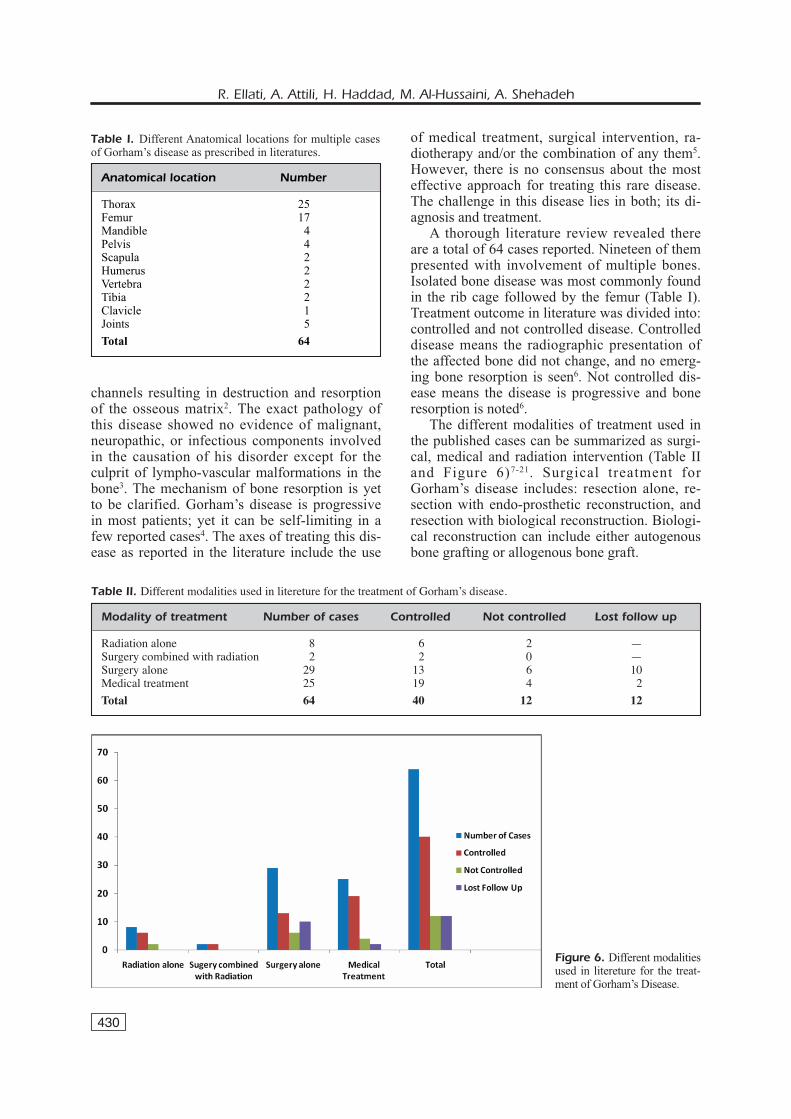
are a total of 64 cases reported. Nineteen of thempresented with involvement of multiple bones.Isolated bone disease was most commonly foundin the rib cage followed by the femur (Table I).Treatment outcome in literature was divided into:controlled and not controlled disease. Controlleddisease means the radiographic presentation ofthe affected bone did not change, and no emerg-ing bone resorption is seen6. Not controlled dis-ease means the disease is progressive and boneresorption is noted6.The different modalities of treatment used in
the published cases can be summarized as surgi-cal, medical and radiation intervention (Table IIand Figure 6)7-21. Surgical treatment forGorham’s disease includes: resection alone, re-section with endo-prosthetic reconstruction, andresection with biological reconstruction. Biologi-cal reconstruction can include either autogenousbone grafting or allogenous bone graft.
Anatomical location Number
Thorax 25Femur 17Mandible 4Pelvis 4Scapula 2Humerus 2Vertebra 2Tibia 2Clavicle 1Joints 5Total 64
Table I. Different Anatomical locations for multiple casesof Gorham’s disease as prescribed in literatures.
Modality of treatment Number of cases Controlled Not controlled Lost follow up
Radiation alone 8 6 2 —Surgery combined with radiation 2 2 0 —Surgery alone 29 13 6 10Medical treatment 25 19 4 2Total 64 40 12 12
Table II. Different modalities used in litereture for the treatment of Gorham’s disease.
Figure 6. Different modalitiesused in litereture for the treat-ment of Gorham’s Disease.

431
Novel approach of treating Gorham-Stout disease in the humerus
Resection alone is more suitable for expend-able bone like: fibula, iliac bone, pubic bone,and clavicle. While for weight bearing boneslike femur, tibia in the lower limb, and longbones in the upper limbs, reconstruction ismandatory (Table III). None of the previouslymentioned surgical techniques showed high suc-cess rate in controlling the progression of thedisease in long bones. The disease was controlled in only 13 cases
out of 29 cases that received surgical treatment,six were progressive and not controlled and tenwere lost from follow up. Regarding humerusbone, two cases were reported. In the first case: asegment of proximal 25 centimeters (9 inches) ofthe right humerus was resected by extra-pe-riosteal dissection and replaced by a hollow Tita-nium 160 prosthesis that was sealed by weldingand no recurrence for more than 3 years15. Whilein the second case the patient was an adolescentmale of Noonan syndrome presented with a non-healing fracture of the proximal right humerus.He was treated with radiotherapy which showedrecurrence after 7 years of follow up10.Medical treatment includes: Interferon, bispho-
phonates, calcium salts and vitamin D, and cy-clophosphamides (Table IV). Bisphosphanatesgained a high success rate, Zoledronic acid was ef-
fective in the treatment with no recurrence in a 9year old boy6 and a 24 year old lady with mandibleaffection11.Radiation therapy has been reported also in lit-
erature, definitive radiation therapy in moderatedoses (40-45 Gcy in 2 Gcy fractions) appears tohave a good outcome as three out of four survivedwith no recurrence except for few long-term com-plications9. Taking into consideration the abovementioned tables of treatment options to controlthis rare disease, we chose to combine surgicalmodalities with the other modalities.In our case the intercalary segment was involved,
we decided to keep both ends of the humerus, resectthe diaphysis, and replace it with structural bone allo-graft matching laterality and size of the resectedbone. Surgery was followed by radiotherapy basedon its added benefit from the literature and the use oforal bisphosphantaes for two years. At seven-yearfollow up, the disease was controlled, and no fatherresorption of bone was noted.Based on the outcomes of the different modali-
ties of treatment and exemplified by our case, thecombination of surgery, medication, and radiother-apy gave an added benefit to control this rare dis-ease. This novel approach was adopted and gaveresults of well-controlling the disease and no pro-gression.
Surgery Number of Controlled Uncontrolled Lost to treated cases follow up
Resection of the lesion 13 3 2 8Reconstruction using prosthesis 10 8 0 2Reconstruction with bone graft 3 0 3 0Thoracic duct ligation with chest drainage 1 1 0 0Thoracic duct ligation with pleurodesis 1 1 0 0Pleurodesis with chest drainage 1 0 1 0Total 29 13 6 10
Table III. Different surgical modalities in treating patients with Gorham’s disease.
Medical treatment Number of Lost to Treated cases Controlled Uncontrolled follow up
Interferon 11 8 2 1Bisphosphonate 6 6 0 0Interferon + Zolendronic acid 2 2 0 0Bevacizumab 2 1 1 0Calcium Salts + Vitamin D 1 1 0 0Cyclophosphamide +Fluorouracil 2 0 1 1Calcitonin Salmon + Alendronate Sodium 1 1 0 0Total 25 19 4 2
Table IV. Different medical treatments used in treating patients with Gorham’s disease.

432
R. Ellati, A. Attili, H. Haddad, M. Al-Hussaini, A. Shehadeh
Conclusions
This case is a successful story of multimodali-ty treatment of Gorham’s disease of the longbone. Surgical resection plus adjuvant radiothera-py followed by oral bisphosphonates for 2 years.This novel combination was not previously re-ported in literature for the treatment of Gorham’sdisease, and showed a success at seven-year fol-low up.
–––––––––––––––––AcknowledgmentWe extend our thanks to our beloved patient who chose tobe anonymous.
–––––––––––––––––––Conflict of InterestsThe Authors declare that they have no conflict of interests.
References
1. EVANGELOU E, KYZAS PA, TRIKALINOS TA. Comparisonof the diagnostic accuracy of lymphatic endotheli-um markers: Bayesian approach. Mod Pathol2005; 18: 1490-1497.
2. PATEL DV. Gorham's disease or massive osteoly-sis. Clin Med Res 2005; 3: 65-74.
3. ELLURU RG, BALAKRISHNAN K, PADUA HM. Lymphaticmalformations: diagnosis and management.Semin Pediatr Surg 2014; 23: 178-185.
4. DELLINGER MT, GARG N, OLSEN BR. Viewpoints onvessels and vanishing bones in Gorham-Stoutdisease. Bone 2014; 63: 47-52.
5. LIU Y, ZHONG DR, ZHOU PR, LV F, MA DD, XIA WB,JIANG Y, WANG O, XING XP, LI M. Gorham-Stoutdisease: radiological, histological, and clinicalfeatures of 12 cases and review of literature. ClinRheumatol 2014 Sep 18. [Epub ahead of print].
6. HU P, YUAN XG, HU XY, SHEN FR, WANG JA.Gorham-Stout syndrome in mainland China: acase series of 67 patients and review of the litera-ture. J Zhejiang Univ Sci B 2013; 14: 729-735.
7. AVELAR RL, MARTINS VB, ANTUNES AA, DE OLIVEIRA NE-TO PJ, ANDRADE ES. Use of zoledronic acid in thetreatment of Gorham's disease. Int J PediatrOtorhinolaryngol 2010; 74: 319-322.
8. GRUNEWALD TG, DAMKE L, MASCHAN M, PETROVA U,SURIANINOVA O, ESIPENKO A, KONOVALOV D, BEHRENDSU, SCHIESSL J, WÖRTLER K, BURDACH S, VON LUETTICHAUI. First report of effective and feasible treatment ofmultifocal lymphangiomatosis (Gorham-Stout)
with bevacizumab in a child. Ann Oncol 2010; 21:1733-1734.
9. DUNBAR SF, ROSENBERG A, MANKIN H, ROSENTHAL D,SUIT HD. Gorham's massive osteolysis: the role ofradiation therapy and a review of the literature. IntJ Radiat Oncol Biol Phys 1993; 26: 491-497.
10. FONTANESI J. Radiation therapy in the treatment ofGorham disease. J Pediatr Hematol Oncol. 2003;25: 816-817.
11. MIGNOGNA MD, FEDELE S, LO RUSSO L, CICCARELLI R.Treatment of Gorham's disease with zoledronicacid. Oral Oncol 2005; 41: 747-750.
12. VENKATRAMANI R, MA NS, PITUKCHEEWANONT P, MALO-GOLOWKIN MH, MASCARENHAS L. Gorham's diseaseand diffuse lymphangiomatosis in children andadolescents. Pediatr Blood Cancer 2011; 56:667-670.
13. HAMMER F, KENN W, WESSELMANN U, HOFBAUER LC,DELLING G, ALLOLIO B, ARLT W. Gorham-Stout dis-ease--stabilization during bisphosphonate treat-ment. J Bone Miner Res 2005; 20: 350-353.
14. ZHENG MW, YANG M, QIU JX, NAN XP, HUANG LY,ZHANG WD, GONG L, HUANG ZZ. Gorham-Stoutsyndrome presenting in a 5-year-old girl with asuccessful bisphosphonate therapeutic effect.Exp Ther Med 2012; 4: 449-451.
15. POIRIER H. Massive osteolysis of the humerustreated by resection and prosthetic replacement.J Bone Joint Surg Br 1968; 50: 158-160.
16. VITALI M. The prosthetic management of a case ofessential osteolysis. J Bone Joint Surg Br 1962;44-B: 652-654.
17. KAKUTA Y, IIZUKA H, KOBAYASHI R, IIZUKA Y, TAKAHASHIT, MOHARA J, TAKAGISHI K. Gorham disease of thelumbar spine with an abdominal aortic aneurysm:a case report. Spine J 2014; 14: e5-9.
18. HEYD R, RABENECK D, DORNENBURG O, TSELIS N, ZAM-BOGLOU N. Gorham-Stout syndrome of the pelvicgirdle treated by radiation therapy: a case report.Strahlenther Onkol 2011; 187: 140-143.
19. KURIYAMA DK, MCELLIGOTT SC, GLASER DW, THOMPSONKS. Treatment of Gorham-Stout disease with zole-dronic acid and interferon-alpha: a case reportand literature review. J Pediatr Hematol Oncol2010; 32: 579-584.
20. KOSE M, PEKCAN S, DOGRU D, AKYUZ C, OZCELIK U,OZSUREKCI Y, GULHAN B, DEMIRCIN M, KIPER N.Gorham-Stout Syndrome with chylothorax: suc-cessful remission by interferon alpha-2b. PediatrPulmonol 2009; 44: 613-615.
21. TAKAHASHI A, OGAWA C, KANAZAWA T, WATANABE H,SUZUKI M, SUZUKI N, TSUCHIDA Y, MORIKAWA A, KUWANOH. Remission induced by interferon alfa in a patientwith massive osteolysis and extension of lymph-he-mangiomatosis: a severe case of Gorham-Stoutsyndrome. J Pediatr Surg 2005; 40: E47-50.