Novak 2003 Hydatidiform Mole Persistent Gestational Trophoblastic Tumor Chemotherapy.
62
-
Upload
hollie-griffin -
Category
Documents
-
view
221 -
download
4
Transcript of Novak 2003 Hydatidiform Mole Persistent Gestational Trophoblastic Tumor Chemotherapy.


GESTATIONAL TROPHOBLASTIC DISEASENovak 2003

Hydatidiform Mole Persistent Gestational Trophoblastic Tumor
Chemotherapy

HYDATIDIFORM MOLE

Epidemiology Complete versus partial
mole Clinical picture Natural history Diagnosis Treatment Follow up

INTRODUCTION
GTD is among the rare tumors that can be cured even if metastasizedTypes:
Complete mole Partial mole Placental site mole Choriocarcinoma
Persistent GTT:Most commonly follow molar pregnancyMay also follow: abortion, ectopic or term
pregnancy

EPIDEMIOLOGY
%varies in different sites:Japan = 2 : 1000 pregnanciesUSA = 0.6 – 1.1 : 1000 pregnanciesIn pathological studies:
Complete mole 1 : 945 Partial mole 1 : 695

Risk factors in complete mole: 1 – nutritional :
↓carotene ↓vit A
2 – Age: > 35 years = X 2 > 40 years = X 7.5
Risk factors in partial mole: 1 - OCCP
2 - H/O irregular menstruation

COMPLETE VERSUS PARTIAL MOLE
Complete molePathology:
No fetal or embryonic tissue Villi show:
Diffuse hydropic swelling Diffuse trophoblastic hyperplasia
Chromosome: 90% 46XX
10% 46XY

Chromosomes are entirely paternal Mitochondria DNA is maternal in origin
1 - Absent or inactivated ovum nucleus + 1 haploid sperm
endoredublication homozygous mole
2 –Absent or inactivated ovum nucleus + 2 haploid sperms
heterozygous mole

Partial moleVilli vary in size and show:
Focal hydropic swelling Focal trophoblastic hyperplasia Focal cavitation Stromal trophoblastic inclusion Scalloping
Fetal or embryonic tissues
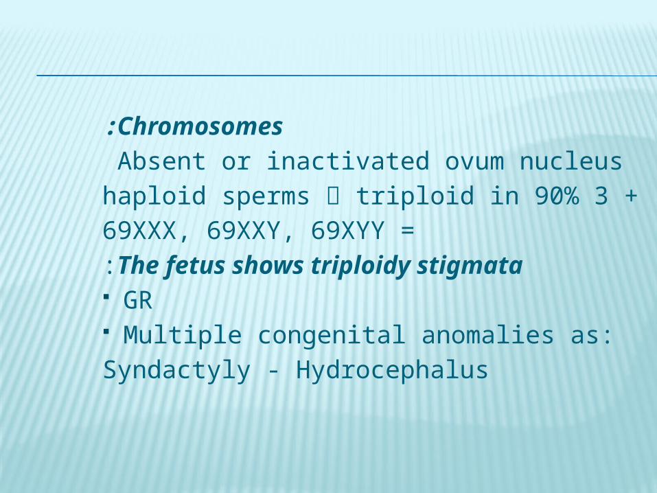
Chromosomes:Absent or inactivated ovum nucleus
+3 haploid sperms triploid in 90% =69XXX, 69XXY, 69XYY
The fetus shows triploidy stigmata:
GR Multiple congenital anomalies as:
Syndactyly - Hydrocephalus
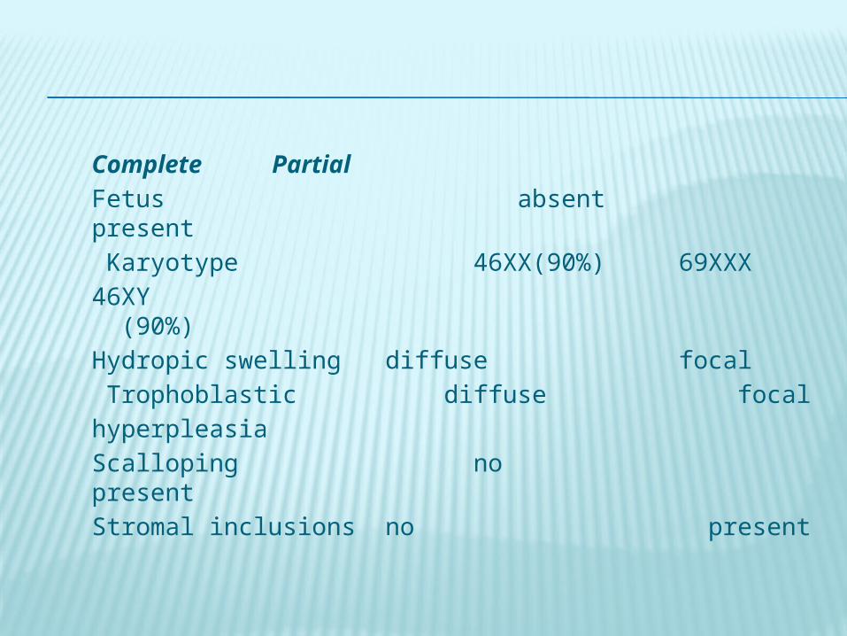
Complete PartialFetus absent presentKaryotype 46XX(90%) 69XXX
46XY (90%)Hydropic swelling diffuse focalTrophoblastic diffuse focal hyperpleasiaScalloping no presentStromal inclusions no present

CLINICAL PICTURE
Complete Partial
past nowVaginal bleeding 97% 84% 74%Anemia 50% 5%Excessive uterine size 50% 28% 4%
Preeclampsia 50% 1.5% Hyperemesis 27% 8%
Hyperthyroidism 7% 0% Trophoblastic embolism 2% 0%Theca lutein cysts 50%HCG > 100,000mIU/mL 6%

Excessive uterine size: = ↑trophoblastic tissue
↑ hCG ↑ preeclampsia
↑ hyperthyroidism ↑ hyperemesis gravidarum
↑ trophoblastic embolization ↑ theca lutein cyst size
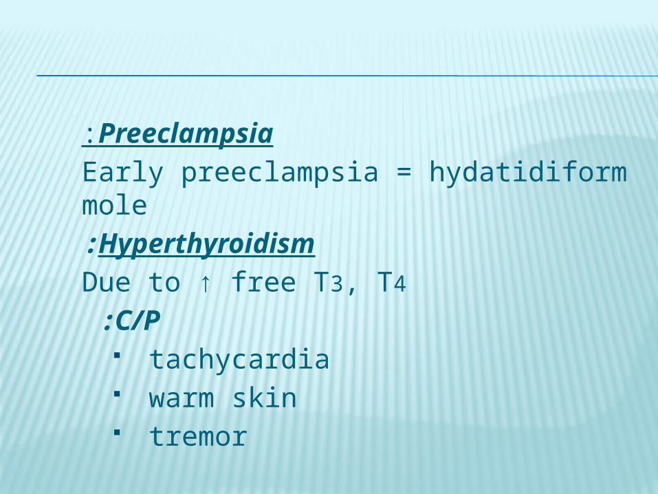
Preeclampsia:Early preeclampsia = hydatidiform moleHyperthyroidism:
Due to ↑ free T3, T4
C/P : tachycardia warm skin tremor
Thyroid storms: Give β–blockers before anesthesia
to avoid thyroid stormsC/P:
↑pulse, ↑ temp, ↑ COP + delirium + convulsions
may HF
Trophoblastic embolization:C/P :
dyspnea cough tachypnea ↑ P chest pain asymptomatic
Chest examination diffuse ralesChest X ray bilateral infiltratesCauses of respiratory distress: Trophoblastion embolization Complications of: • preeclampsia • thyroid storm • excessive fluid intake

Theca lutein ovarian cysts Due to ovarian overstimulation by ↑
hCG May not be felt with oversized uterus May pressure symptoms treated by decompression by laparoscopic or U/S guided aspiration If ruptured or torsion occur acute pain laparoscope

NATURAL HISTORY
Complete mole Invasive = 15% Metastatic = 4%
Risk factors: hCG > 100,000 mIU/mL Excessive uterine size Theca lutein cysts = 6 cm

Low risk = 60% 3.4% persistent mole
0.6% metastaticHigh risk = 40%
31% persistent mole 9% metastatic
Age: > 40 years = 37%
> 50 years = 56%

DIAGNOSIS
Complete moleU/S vesicular patternPartial moleU/S focal cystic spaces in placenta
+ ↑transverse diameter of GSBoth together 90% +ve predictive
value

TREATMENT
I – Hystrectomy + aspiration of CL cyst
+ follow up as usual2 - Suction evacuation
Preferred ttt for hydatidiform mole Give oxytocine before anesthesia
Use 12 canula If > 14 weeks support the fundus
+ do fundal massage

Dilatation ↑ bleeding Suction evacuation ↓ bleeding
If RH –ve give Anti RH Ig3 - Prophylactic chemotherapy
↓invasive mole to 4% after 1st course ”””””””””””””””””“ ↓0% after 2nd
course Controversial : Why to expose all
patients to chemotherapy while only 20% will need it ?
Useful if follow up is: Unreliable
Unavailable Study:
Prophylactic chemotherapy in high risk patients ↓ persistent
mole from 47% to 14%

FOLLOW UP
1 - HCG Average time needed to return to
normal values = 9 weeks Measure hCG/week
3 consecutive normal results /month 6 consecutive normal R
2 - Contraception: OCCP or barrier methods
IUD is C/I perforation
PERSISTENT GESTATIONAL TROPHOBLASTIC TUMOR

Nonmetastatic disease Placental-site TT Metastatic D Staging Prognostic scoring systems Diagnostic evaluation Management
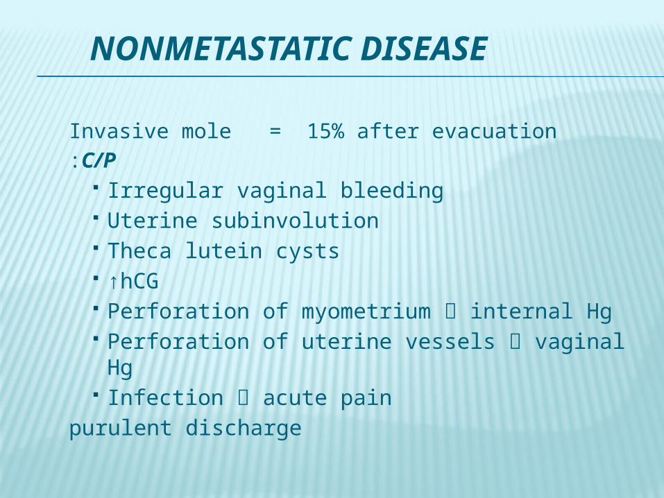
NONMETASTATIC DISEASE
Invasive mole = 15% after evacuationC/P:
Irregular vaginal bleeding Uterine subinvolution Theca lutein cysts ↑hCG Perforation of myometrium internal Hg Perforation of uterine vessels vaginal Hg Infection acute pain purulent discharge

Histology : After molar pregnancy hydatidiform mole or choriocarcinoma After nonmolar pregnancy choriocarcinoma = sheets of anaplastic cytotrophoblast and syncytiotrophoblast + no villi
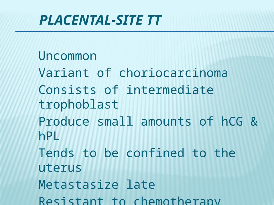
PLACENTAL-SITE TT
UncommonVariant of choriocarcinomaConsists of intermediate trophoblastProduce small amounts of hCG & hPLTends to be confined to the uterusMetastasize lateResistant to chemotherapy

METASTATIC DISEASE
=4% after molar pregnancyMore often after nonmolar pregnancyUsually associated with choriocarcinomaHighly vascular spontaneous bleedingEarly vascular spreading Sites:
Pulmonary 80% Hepatic 10% Vaginal 30% Brain 10%
Pelvic 20%
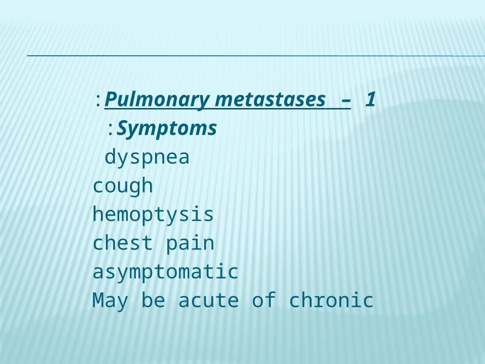
1 –Pulmonary metastases: Symptoms :
dyspnea cough
hemoptysis chest pain
asymptomatic May be acute of chronic

Chest X ray: Snowstorm pattern Discrete rounded densities Pleural effusion Pulmonary artery embolism
May be misdiagnosed as 1ry pulmonarydisease and only recognized as GTD after thoracotomy

Pulmonary embolism may pulmonary HTN
Early RF + intubation = bad prognosis 2 – Vaginal metastasis
highly vascular biopsy may excessive bleeding
Symptoms: Vaginal bleeding Purulent discharge Site: fornices/suburethral

3 – Hepatic metastasis Usually in advanced cases
Symptoms: Epigastric or upper RT ¼ pain due to
stretching subcapsular hematoma Rupture internal Hg4 – Brain metastasis
Usually in advanced cases Spontaneous bleeding acute focal
neurological defects
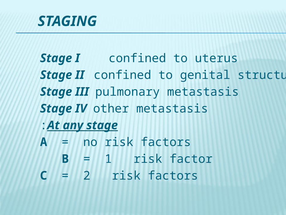
STAGING
Stage I confined to uterusStage II confined to genital structuresStage III pulmonary metastasisStage IV other metastasisAt any stage:
A = no risk factors B = 1 risk factor
C = 2 risk factors

PROGNOSTIC SCORING SYSTEMS
0 1 2 4Age ≤39 >39Pregnancy mole abortion termDuration <4m 4-6 7-12 >12
hCG <1000 <10000 <100000> Largest size <3cm 3-5 >5Site of met 0 kidney/spleen GIT/liver brainNumber <3 1-3 4-8 >8ABO group 0 A/O B/ABChemotherapy 1 ≥2

DIAGNOSTIC EVALUATION
H/O Examination hCG Liver function tests Kidney function tests Thyroid function tests WBCs Platelet count
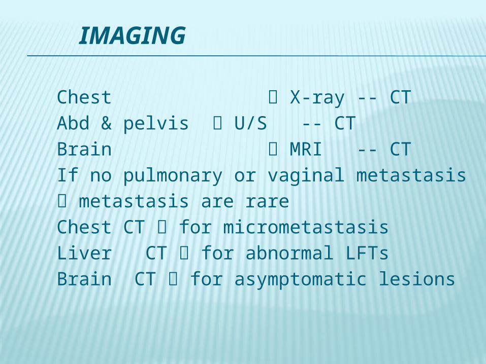
IMAGING
Chest X-ray -- CTAbd & pelvis U/S -- CTBrain MRI -- CTIf no pulmonary or vaginal metastasis metastasis are rareChest CT for micrometastasisLiver CT for abnormal LFTsBrain CT for asymptomatic lesions

If brain CT is normalmeasure CSF hCGIf serum hCG/CSF hCG = < 60% then there is brain metastasisPelvic U/S for:
Extent of uterine lesion Localization of resistant lesions Identifying patients who will benefit from hystrectomy

MANAGEMENT
Stage I Stage II & III Stage IV

STAGE I
If the patient does not wish to preserve fertility Hystrectomy + Chemotherapy to:
↓ dissemination of GTD Treat dissemination of GTD Treat occult metastasis ↓ bleeding ↓ sepsis

If the patient wish to preserve fertility:Low risk Single agent High risk Combined chemotherapyResistant Local uterine resection
after localization of resistant sites by U/S, MRI, or arteriography

Placental site GTD: -Only curative ttt for nonmetastatic
cases is hystrectomy -Resistant to chemotherapy few
metastatic cases reported complete remission after chemotherapy

STAGE II & III
Pulmonary metastasisLow risk single agent 82% CRHigh risk combined chemotherapyResistant thoracotomy after
localization of and exclusion of other
resistant sites
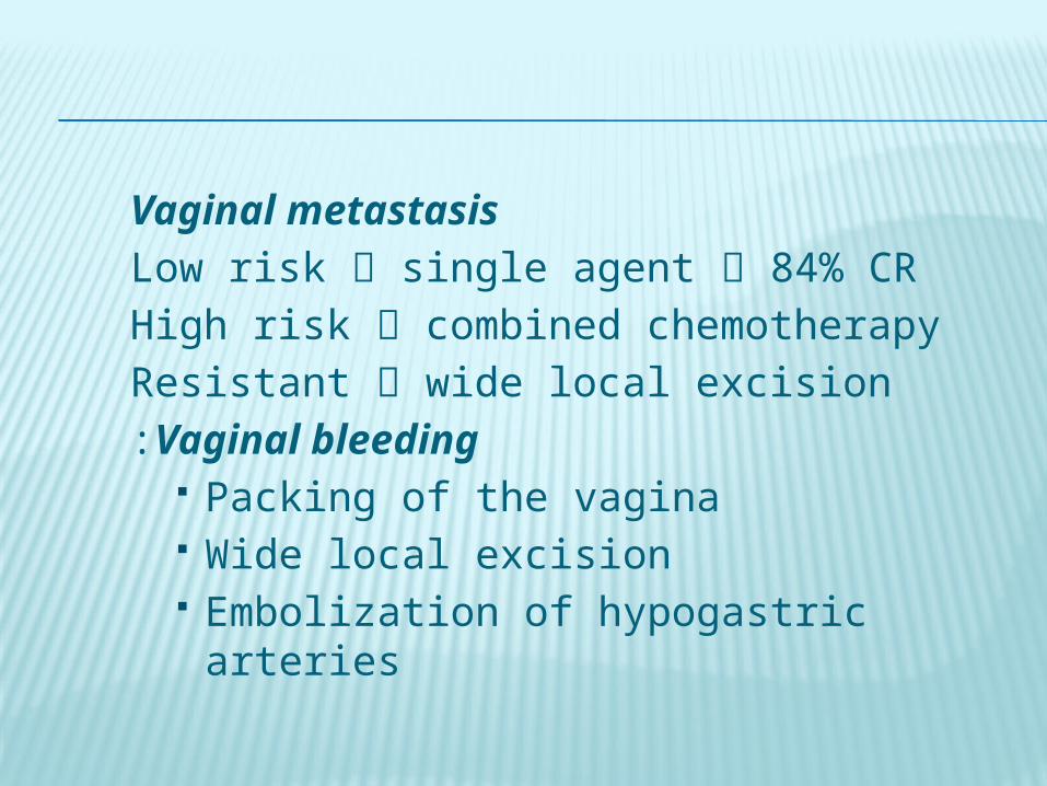
Vaginal metastasisLow risk single agent 84% CRHigh risk combined chemotherapyResistant wide local excisionVaginal bleeding:
Packing of the vagina Wide local excision Embolization of hypogastric arteries

Hystrectomy -In metastatic disease
- to control Hg - to control sepsis
-In extensive uterine disease - to ↓ GTT burden
- to ↓ chemotherapy courses

Follow up of stage I, II, III:hCG/week
3 consecutive normal results hCG/month
12 consecutive normal results +effective contraception

STAGE IV
Should be referred to specialized centersMay be unresponsive or rapidly progress All should receive intensive combined chemotherapy ± irradiation / surgeryHepatic metastasis:
Resistant cases intrahepatic infusion of chemotherapy
Hemorrhage local excision or arterial embolization
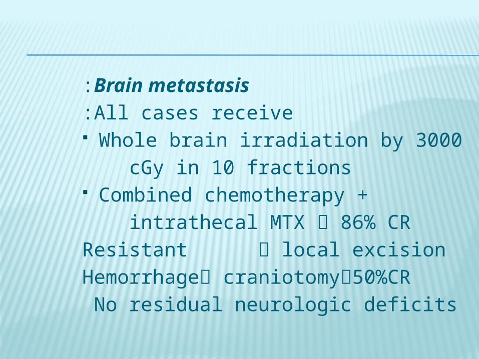
Brain metastasis:All cases receive:
Whole brain irradiation by 3000 cGy in 10 fractions Combined chemotherapy + intrathecal MTX 86% CRResistant local excisionHemorrhage craniotomy50%CRNo residual neurologic deficits

CHEMOTHERAPY

SINGLE AGENT CHEMOTHERAPY
Used in nonmetastatic and low risk mmMTX&Act-D are used/other week X5days
1964 :MTX-FA well tolerated ↓ toxicity
MTX-FA the preferred ttt for GTDMTX-FA 88% CR
81% by single course 90% CR in stage I 68% CR in stage II

Complications: Thrombocytopenia 1.6% Agranulocytopenia 6% Hepatotoxicity 14%
Resistant cases: Choriocarcinoma Metastasis Initial hCG > 50,000 mIU/mL

Technique: Measure hCG after each course Draw a curve Stop MTX if the curve is progressively ↓ Do not give MTX at any predetermined or fixed interval Give another course if:
hCG is ↑ or plateaus for > 3 weeks hCG ↓ < 1 log at day 18 post ttt
If the response to the 1st course is adequate give the same dose If the response to the 1st course is inadequate ↑ the dose to 1.5 mg/Kg body weight/day X 4 days Adequate response = ↓ hCG by 1 log If the response to the 2nd & 3rd courses is inadequate give ACT-D If the response to ACT-D is inadequate give combined chemotherapy
COMBINED CHEMOTHERAPY
Triple therapy ( MTX + ACT-D + cyclophosphamide ) is inadequate in ttt of high risk cases 50% CR only Etoposide 95% CR in nonmetastatic and low risk metastatic cases 1984: triple therapy + Etoposide + Vincristine ( EMA-CO )
83% CR in high risk patients

EMA-CO is well tolerated and is the preferred 1ry ttt for patients with metastasis and high risk score
76% CR when used as 1ry ttt 86% CR in brain metastasis If resistant to EMA-CO give EMA-EP (cisplatin) on day
8 76% CR in resistant patients
Duration of Therapy: Give combined chemotherapy 3 consecutive normal results Add at least 2 additional courses to ↓ risk of relapse

SUBSEQUENT PREGNANCIES
Complete/Partial mole Persistent GTTTerm 70% 70%PTL 7% 6%Ectopic 1% 1%SB ½% 11/2%
Recurrence 11/2% 1%1st T abortion 16% 15%2nd T abortion 1.6% 1.5%Congenital anom 4% 2.5%CS 16% 19%

Recurrence rate: 1 mole = 1% 2 mole = 20%
In the next pregnancy: Do U/S < 14 weeks Measure hCG 6 weeks after termination/labour Send placenta or product of
conception to pathology