Plantes Arbres Arbustes Toxiques Perrin Julia Et Giraud Melanie 1 D

NOTE TO USERS
This reproduction is the best kopy available

CHARACTERJZATION AND USE OF GENETICALLY-PROGRAMMED
RESPONSES TO CHLORDANE, DINOSEB, BROMACIL, AND ARSENIC
OXYANIONS
by Michael A. Costanzo
Department of Microbiology and lmmunology
McGill University
Montréal, Québec, Canada
A thesis submitted to the Facuity of Graduate Studies and Research in partial fulfillrnent of the requirements for the degree of Master of Science
O Michael A. Costanzo May 1997
National Library B+B ,,,,da Bibliothèque nationale du Canada
Acquisitions and Acquisitions et Bibliographic Services services bibfiograp hiques
395 Wellington Street 395. rue Wellington Ottawa ON K1A ON4 OttawaON KtAON4 Canada canada
Your f i k Voire réference
Our Ne Noire r e f d r e ~ c e
The author has granted a non- L'auteur a accorde une licence non exclusive licence allowing the exclusive permettant à la National Library of Canada to Bibliothèque nationale du Canada de reproduce, loan, distribute or sell reproduire, prêter, distribuer ou copies of this thesis in rnicroform, vendre des copies de cette thèse sous paper or electronic formats. la fome de microfiche/^ de
reproduction sur papier ou sur format électronique.
The author retains ownership of the L'auteur conserve la propriété du copyright in this thesis. Neither the droit d'auteur qui protège cette thèse. thesis nor substantial extracts f?om it Ni la thèse ni des extraits substantiels may be printed or otherwise de celle-ci ne doivent être imprimés reproduced without the author's ou autrement reproduits sans son permission. autorisation,

TABLE OF CONTENTS
CONTENTS PAGE
Abstract iii
Abrégé
Acknowledgrnents vii
List of Tables and Figures viii
List of Ab breviations
Chapter 1 - Literature Review 1 .O. Introduction
2.0. S hort-term Toxicity Assays
2.1 . Enzymatic Assays
2.2. ATP-based Assays
2.3. Growth Inhibition Assays
2.4. Ecological Effect Assays
2.5. Commercially Available S hort-term Toxicity Assays
2.6. The Future of Short-term Toxicity Assays
3.0. Gene Fusion Biosensors
3.1. Genetically-programmed Responses as Tools for
Creating Biosensors
3.2. Reporter Genes for Biosensor Applications
3.3. Bacteriophages as Biosensors
3.4. Heat Shock Gene Fusions
3.5. Luminescence-based Mercury Biosensor
3.6. Luminescence-based Naphthalene Biosensor
3.7. Luminescence-based Biosensor for the Detection
of Senzene Derivatives
3.8. Luminescence-based Heavy Metal Biosensor
3.9- Luminescence-based Arsenic Biosensor
3.1 0. Additional Luminescence-based Biosensors
4-0- The Future of Gene Fusion Biosensors
Outline of the Thesis
Chapter II - Characterization of Genetically-programmed Responses in Eschenchia coli to Bromacil, Chlordane, and Dinoseb 25
Chapter III - Use of an Arsenic Oxyanion-responsive Escherichia coli Luciferase Clone for the Detection of Arsenic Compounds in an Environmental Aquatic Sample 78
Chapter IV - Conclusions and Directions for Future Research 92
References 97

ABSTRACT
Soil and aquatic ecosystems are constantly being exposed to chemical
contaminants from industrial, agricultural, and municipal sources. For
example, pesticides and herbicides are used on a world-wide scale to control
a wide variety of harmful insects and competing weeds, to increase the
production of food and fiber, and to facilitate modern agricultural production
methods. Although these chemicals are designed to be selectively toxic
towards target organisms, this iç not absolute, and in most cases pesticides
and herbicides are toxic toward non-target organisms, including humans. To
identify genes which are transcriptionally regulated by cellular exposure to
environmental pollutants, such as pesticides and herbicides, a 3000
Eshedchia coli single-copy luxAB gene fusion library was previously screened
in the presence of a pesticide or two herbicides (Costanzo, 1995). Five
different clones whose luminescence is induced by either the herbicides,
dinoseb and bromacil, or the pesticide, chlordane, were identified (Costanzo,
1995). Following partial characterization of the pesticide- and herbicide-
responsive genes, luminescence experiments were repeated using new lots of
brornacil, chlordane, and dinoseb. Despite numerous attempts to reproduce
the dose-dependent iight ernissions, the original patterns of luminescence
could not be duplicated. Experiments were therefore performed to determine
the basis for the observed fluctuations in luciferase expression. Aithough
difficulties were experienced when screening in the presence of chernically
complex organic cornpounds, bacterial systems, such as the E. coli luxAB
gene fusion clones, represent effective tools for studying the effects of toxic
substances. Several common approaches are currently used to monitor and
control environmental contamination. Direct analytical methods allow the
determination of specific contaminant concentrations while biological toxicity
assays rneasure harmful effects on living organisms, such as fish or Daphnia
spp. In recent years, rnicrobial bioassays have gained popularity because they
are sensitive, rapid, and cost-effective methods for measuring toxicity. The
possibility of utilizing one E. coli chromosomal IuxAB gene fusion clone, the
arsB::luxAB clone (Cai and DuBow, 1996), to detemine the presence of
arsenic in a water effluent (Goodfellow effluent, Quebec) was investigated.
The arsS::luxAB gene fusion clone detected trace amounts of arsenic-
containing compounds in the water sample and may therefore be used as an
environmental biosensor for arsenic detection.
Les écosystèmes terrestres et aquatiques sont constamment exposés à des
contaminants chimiques de provenances industrielle, agricole et municipale.
Des pesticides et des herbicides, par exemple, sont utilisés à l'échelle
mondiale pour contrôler grands nombres d'insectes néfastes et de mauvaises
herbes, pour augmenter la production de nourriture et de fibre, et pour faciliter
les méthodes modernes de production agricole. Malgré le fait que ces
produits chimiques soient dirigés specifiquement contre des organismes
cibles, cela n'est pas absolu; dans la plupart des cas, les pesticides et les
herbicides sont toxiques pour des organismes non-cibles dont les humains.
Afin d'identifier des gènes qui sont réglés, au niveau de la transcription, par
l'exposition cellulaire a des polluants environnementaux tels des pesticides et
des herbicides, une librairie de fusions géniques uniques de IuxAB dans E.
coli fut mise en présence d'un pesticide et de deux herbicides (Costanzo,
1995). Cinq clones différents furent identifiés, dont la luminescence est induite
par les herbicides dinoseb et bromacil, ou par le pesticide chlorda~e
(Costanzo, 1995). Suivant la characterization partielle des ces genes
sensibles aux herbicides et au pesticide, les expériences de luminescence
furent répétées en utilisant de nouveaux lots de bromacil, chlordane et
dinoseb. Malgré maintes tentatives de reproduire les émissions lumineuses
montrant une dépendance de dose, les modèles de luminescence originaux
ne purent pas être reproduits. Des expériences furent donc effectuées afin de
déterminer les causes des fluctuations observées dans l'expression de la
luciférase. Bien que nous avons eu des problèmes en utilisant des composés
organiques complexes, des systèmes bactériens, tels que les clones de
fusions géniques IuxAB d' E. coli, représentent des outils efficaces pour
étudier les effets de substances toxiques. Plusieurs approches communes
sont présentement utilisées pour évaluer et contrôler la contamination
environnementale. Les méthodes directes d'analyse permettent de déterminer
spécifiquement la concentration d'un agent polluant, tandis que les analyses
de toxicité biologique measurent les effets néfastes sur des organismes
vivants, tels les poissons ou Daphnia spp. Dans les dernières années, les
"bio-analyses" microbiennes ont gagné de la popularité car elles sont des
méthodes permettant de mesurer la toxicité de manière sensible, rapide et
non-dispendieuse. Nous avons examiné la possibilité d'utiliser un clone
dlEscheichia coli ayant une fusion chromosomale du gène iuxAB, le clone
arsB::luxAB (Cai and DuBow, l996), pour déterminer la présence d'arsenic
dans un effluant (effiuant Goodfellow, Québec). Le clone arsBrrluxAB détecta
dans l'échantillon des traces de composés contenant de l'arsenic et pourrait
donc être utilisé comme "biosensor" environnemental pour la détection
d'arsenic.
ACKNOWLEDGEMENTS
1 would like to thank my supervisor, Dr. Michael S. DuBow, for his
support throughout my graduate studies. I would also like to express my
sincere gratitude to Dr. Nicholas H. Acheson for his help and advice prior to
submission of this thesis.
I am especially grateful to Caroline Diorio, Jie Cai, Dr. Angelina Guzzo,
and Dr. Georgina Maclntyre for their guidance and seemingly endless
expanse of ideas throughout the course of rny research. I would also like to
thank Felix Sieder for his cornputer expertise, David Alexander for his help
with gene analysis and homology searches, and Madani Thiam for translating
the abstract of this thesis. A special thank you goes to Julie G u u o for her
continuous love and support over the last three years. 1 am also grateful to
Christian Blaise and Brian Walker, of Environment Canada, for providing us
with the water effiuent sarnples used in the arsenic biosensor experirnents.
Finally, I would like to express my deepest gratitude to my parents,
Salvatore and Angela, for their love, support, and guidance throughout rny life
and for instilling the confidence in me to believe that there is no goal that
cannot be achieved. This thank you extends to my whole family, without
whose love and support I would not have been able to accornplish this.
vii

LIST OF TABLES AND FIGURES
PAGE
Table 1:
Table 2:
Table 1 :
Table 2:
Figure 1:
Figure 2:
Figure 3:
Figure 4:
Figure 5:
Figure 6:
Figure 7:
Figure 8:
CHAPTER I
Commercially available short-terni toxicity assays
Heavy rnetal-responsive luciferase gene fusions
CHAFTER II
Bacterial strains and plasmids
Types of luminescence assays perfonned
Light emission from strains LF20204, LF20205, LF20206, LF20207, and LF20208 in the presence of bromacil, dinoseb, and chlordane
Southern blot analysis of strains LF20204, LF20205, LF20206, LF20207, and LF20208
Schematic diagram for the cloning and sequencing of the herbicide- and pesticide-responsive genes
Schematic illustrating Tn5-luxAB insertion sites for strains LF20204, LF20205, LF20206, LF20203, and LF20208
Nucleotide sequence of the region adjacent to the Tn5-IuxAB insertion of strain LF20206
Light ernission from strains LF20204, LF20205, LF20206, LF20207, and LF20208 in the presence of bromacil, dinoseb, and chlordane
Light emission from strains LF20204 and LF20205 in the presence of dinoseb
Ligt-rt ernission frorn strains LF20206 and LF20207 in the presence of chlordane.
Figure 9:
Figure 10:
Figure 11:
Figure 12:
Figure 1 3:
Table 1 :
Figure 1:
Figure 2:
Light emission from strain LF20208 in the presence of brornacil
Southem blot analysis of strains LF20204, LF20206, andLF20207
Lig ht emission from strains LF2WO9, LF20210, and LF2021-l in the presence of dinoseb or chlordane
Light emission from strain LF20209 in the presence of dinoseb
Light emission from strains LF2021û and LF20211 in the presence of chlordane
CHAPTER 111
Bacterial strâins
Light emission of strain LF20012 in the presence of arsenic-contaminated Goodfellow effluent
B-galactosidase activity of strain LF20001 in the presence of arsenic-contaminated Goodfellow effluent
LIST OF ABBREVfATlONS
A
C
DNA
dNTP
DTT
G
9
IS
kb
kDa
P!2
PI
PM
mg
ml
mM
ORF
PPM
PPB
adenine
cytosine
deoxyribonucleic acid
dinucleotide triphosphate
dithiothreitol
guanosine
gram
insertion sequence
kilo base-pair
kiiodalton
microgram
microliter
millimolar
milligram
milliliter
millirnolar
open reading frame
parts per million
parts per billion
RLU
T
Tn
v/v
w/v
resistancehesistant
relative light unit
thymidine
transposon
volume per volume
weig ht per volume

CHAPTER I
Literature Review

1 .O. INTRODUCTION
The environment is constantly being exposed to a wide variety of
contaminants from industrial, agricultural, and municipal sources.
Consequently, many countries are now facing serious ecological and
toxicological problems resulting from the reiease of toxic effluents into soi1 and
aquatic ecosystems (Coleman and Qureshi, 1985; Bitton and Dutka, 1986).
Important goals in addressing these problems are the detection and
elimination of poliutants which include living organisms, such as bacterial and
parasitic pathogens, toxic organic chemicals, such as aromatics and
hydrocarbons, inorganic chemicals, which include toxic heavy metal
compounds, and chemical mutagens. Pollution caused by the release of these
toxic pollutants has received a great deal of attention due to a high demand
for chemicals, as well as the problem of ensuring the safety of food and water
(Codina et al., 1993). Once released into ecosystems, effects of these
chemicals may be observed up to several seasonal cycles, after which they
can become biologically unavailable through incorporation into organic and
inorganic particles in sediments (Peterson et a/., 1996). These biological and
chemical pollutants must therefore be continuously monitored and their levels
controlled in order to conserve and protect ecosysterns, wildlife, and human
welfare.
Concentrations of chemical contaminants in soi1 and aquatic
environments can be assessed via numerous methods. Detection of
environmental pollutants has traditionally relied on highly sensitive and
specific analytical chemistry methods (Van Dyk et ai., 1994). Older,
conventional methods include atomic absorption spectroscopy (AAS) and
chrornatographic analysis tools, such as high performance liquid
chromatography (HPLC) and gas chromatography (GC), which measure
specific chemical components in water samples (Gu et aL, 1996). Fiber-optic
research has also expanded in this area (Coulet and Blum, 1992; Blum et ai.,

1993). For example, one assay has been developed to measure
benzo(a)pyrene concentrations based on the interaction of benzo(a)pyrene
with antibody-coated optical fibers. Antibody-bound benzo(a)pyrene is
detected Dy measuring laser-induced fiuorescence (Tromberg et al. 1988; Vo-
Dinh et al., 1987). Continued research in this area will undoubtedly provide
biosensors for the detection of a wide array of other chemical compounds.
Although effective, the analytical methods described above may have several
shortcomings. First, they can be costly and labour-intensive (Kahru et al.,
1996). Second. they may not be able to distinguish pollutants that are
available to biological systems from those that exist in the environment as
inert, unavailable foms (Van Dyk et al., 1994). This is a particular concern
with respect to toxic metals. Bioavailability is therefore a critical issue in
determining metal toxicity (Van Dyk et al., 1994). Finally, analytical methods
may fail to account for possible synergistic or antagonistic effects on biological
organisms caused by chernicals present in complex mixtures (Kahru et al.,
1996).
As a result of these shortcomings, a trend has developed toward the
use of living organisms to screen for toxic substances. The deleterious effects
of toxic contaminants released into the environment have been assessed via
acute and chronic toxicity tests, using mostly fis h and invertebrate species
(Peltier and Weber, 1985). However, these assays may be extremely labour-
intensive and expensive. Therefore, toxicologists and environmental scientiçts
have shifted their focus to short-term toxicity assays. The remainder of this
chapter will explore the biological principles and applications currently in use
to confront the problems of environmental toxicity and pollution. The
development of short-term biological toxicity assays and biosensors will be
discussed. along with the potential for these biosensors to complement
conventional analytical monitoring methods.

2.0. SHORT-TERM TOXiCITY ASSAYS
Short-term toxicity assays are based on microbial characteristics such
as enzyme activity and biosynthrsis, bioluminescence, motility, growth
viability, ATP production, oxygen uptake, nitrification, and heat production.
Therefore, by exploiting these characten'stics, bacteria rnay be employed as
environmental biosensors (Bitton and Koopman, 1992). A variety of short-term
toxicity açsays exist and have been utilized in the maasurement of toxicity,
genotoxicity, bioavailability, and biodegradability of numerous environmental
pollutants.
2.1. Enzymafic Assays
Several microbial biosensors have been developed based on
relationships between bacterial populations, present in soi1 or aquatic
environrnents, and the effects of toxic substances on enzyme activity. Almost
two decades ago, environmental scientists hypothesized that microbial
enzyme activity could be a powerful indicator of the adverse effects caused by
toxic environmental contaminants (Burns et al., 1978). Environmental sarnples
containing a particular contaminant can now be assayed for specific enzyme
activities, based on colourimetric or luminescent tests (Bitton and Koopman,
1986). The effect of the toxic contaminant on bacterial enzyme activity can
then be quantified using spectrophotometers, microtitre plate readers,
fluorometers, or liquid scintillation counters. The degree of inhibition of
enzyme activity, in the presence of the pollutant, can then be correlated the
toxicity of the contaminant (Bitton and Koopman, 1992). Since the initial
discovery of this relationship, many bacterial enzymes have been tested for
their potential as environmental biosensors (Christensen et al., 1982; Bitton
and Kooprnan, 1986; Obst et al., 1988).
Dehydrogenases are commonly used for this purpose in toxicity
assays. Lehnard (1963) first proposed that dehydrogenase activity could be a
useful indicator of the toxic effects of inorganic chemicals, such as mercury,
silver, and chromium, and organic chemicals, such as fomaldehyde and
phenol. Dehydrogenase activity was later found to correlate well with microbial
Rora present in aquatic ecosystems (Dermer et al., 1980). Dehydrogenase
enzyme inhibition assays involve the use of specific oxidoreduction dyes, such
as triphenyl tetrazolium chloride (TTC), which become reduced in the
presence of the dehydrogenase enzyme (Bittor! and Kooprnan, 1986). In the
case of TTC, dehydrogenase catalyzes a reduction reaction which results in
the transformation of TTC, which is colourless, to triphenyl fomazan, a red
insoluble precipitate (Altman, 1976). Hence, a toxic contaminant present in an
environmental sample will lead to inhibition of bacterial dehydrogenase
activity, resulting in decreased reduction of TTC, and decreased formation of
the red precipitate. Toxicity is then assessed by the degree of colour change
obsenred (Bitton and Koopman, 1986). Toxicity tests based on the inhibition of
enzyme activity, other than dehydrogenases, have also been investigated and
these include ATPases (Riedel and Christensen, 1979), esterases (Guilbault
and Krarner, 1964; Holland et al., 1967), and phosphatases (Tyler, 1976).
Inhibition of bacterial enzyme biosynthesis, rnay also be representative
of environrnental toxicity (Cenci et al., 1985; Reinhartz et ai-, 1987). For
example, P-galactosidase is an enzyme responsible for the breakdown of
lactose. In Escherichia coli, the P-galactosidaçe enzyme is encoded by the
lacZ gene. The lac2 gene belongs to a cluster of genes, collectively referred
to as the lac operon (Jacob and Monod, 1961). The lac operon encodes
proteins involved in the transport of lactose into the cell, regulation of the
operon itself, and biosynthesis of the B-galactosidase enzyme (Jacob and
Monod, 1 96 1 ). De novo biosynthesis of P-galactosidase in Escherichia coli
has been shown to be more sensitive to toxic chernical exposure than enzyme
activity (Dutton et al., 1988; Reinhartz et al., 1987). Based on this discovery, a
commercial toxicity assay, known as the Toxi-Chromotes", was developed
(Reinhartz et al., 1987). This assay exploits the fact that toxic chemicals exert
an inhibitory effect on de novo biosynthesis of the P-galactosidase enzyme
(Reinhartz et al-, 1987). E. coli P-galactosidase, in addition to lactose, also
breaks down synthetic lactose analogues (Miller, 1992). Breakdown of these
compounds by P-galactosidase causes a colour change when E. coli is grown
in media containing the lactose analogue (Miller, 1992). Hence, the toxicity of
environmental pollutants rnay be assessed by the absence of colour change,
which is indicative of enzyme biosynthesis in hibition (Reinhartz et al., 1987).
The effect of toxic chemicals on the de novo biosynthesis of other
inducible enzymes, such as E. coli tryptophanase and Bacillus lichenifomis a-
glucosidase, has also been explored (Dutton et al., 1990). For example,
biosynthesis of a-glucosidase in B. licheniformis has been shown to be very
sensitive to environmental contamination, especially by hydrophobic
compounds and detergents (Dutton et al., 7990). Furthermore, B. licheniformis
a-glucosidase synthesis is more sensitive to hydrophobic compounds than E.
coli tryptophanase. This may be explained by the lack of an outer membrane
in gram positive bacteria (Dutton et al., 1990). Unlike the Bacillus spp., gram
negative bacteria, such as E. coli, have an outer membrane which inhibits
hydrophobic compounds from entering the bacterial cell (Neidhart et al.,
1 990).
2.2. A TP-based Assays
Early work by Brezonik and Patterson (1972) introduced the use of
ATP production in the screening of contaminanls in activated sludge systems.
Further studies have confirmed the utility of ATP-based toxicity assays for
water and waste water (Parker and Pribyl, 1984). A more recent approach is
the ATP-TOX" assay developed by Xu and Dutka (1987). This test is based
on both growth inhibition, via ATP measurement of bacteria, and luciferase
activity. Inhibition of this enzyme is determined by adding a standard ATP
solution, as an enzyme substrate, followed by the rneasurement of light
ernission. Similar assays have also been based on the inhibition of ATP
production in algae. One example is the Algae-Tox@ assay which measures
the inhibition of ATP production in Selenastmm capricomutum (Blaise ef al.,
1986).
2.3. Growfh Inhibition Assays
Analysis of growth rates of pure or mixed bacterial cultures has also
proven useful in the detection of environmental contaminants (Alsop ef al,.
1980; Trevors, 1986). These assays determine toxicity by the ability of
environmental pollutants to inhibit bacterial growth. lnhibition of bacterial
growth is detected simply by measuring changes in absorbance of the
bacterial cultures, which is indicative of bacterial ceIl densities. Decreases in
ceIl density therefore suggest increased toxicity by contam inated samples
(Alsop et al,. 1980; Trevors, 1986).
2.4. Ecological Effect Assays
Cycling of nutrients by microorganisms in aquatic and soi1
environments rnay also be adversely affected by toxic chemicals. Carbon,
nitrogen, phosphorus, and sulfur cycling are carried out by various groups of
microorganisms and these processes may be subjected to inhibition by toxic
chemicals (Bitton et al., 1989). The carbon cycle rnay be affected by toxic
chemicals and toxicity can be determined through measurement of microbial
respiration. Several methods are currently available to measure respiration
and these include oxygen electrodes, manometers, and electrolytic
respirometers (King and Dutka, 1986). Furthemore, some of these assays are
capable of measuring the inhibition of biodegradation of specific organic
compounds, such as cellulose (Martin et a/., 1982; Wainwright, 1978).

Nitrogen fixation, ammonification, nitrification, and denitrification are al1
processes invoived in the nitrogen cycle. Among these processes, nitrification
is probabiy the most sensitive to the effects of toxic chemicals and
environmental pollutants (Bitton and Kooprnan, 1992). Nitrification is carried
out by two gro ups of chernoautotrop hic bacteria; Nitrosomonas spp. oxidize
ammonium to nitrite and Nitrobacter spp., sometimes referred to as nitrifiers,
convert nitrite to nitrate (Bitton and Koopman, 1992). A test based on the
inhibition of respiration of Nitrosomonas spp. has been developed and may be
used for rapid measurement of waste water toxicity (Alleman, 1988). Another
test, based on the reduction of nitrite in Nitrobacter spp . in the presence of
waste water, has also been developed and has been shown to be sensitive to
the action of pesticides, heavy metals, and industrial effluents (Carlisle and
Trevors, 1986; Bewley and Stotzky, 1983; Williamson and Johnson, 1981).
Anaerobic microorganisms, particularly methanogens, have also been
shown to be very sensitive to environmental contamination. Owen et al-
(1 979) initially developed the Anaerobic Toxicity Assay (ATA) to determine the
impact of toxic chemicals on gas production using easily degradable
substrates, such as acetate or propionate. This assay has been used for
testing the effect of industrial chernicals on methanogenesis (Benjamin et al.,
1984).
2.5. Comrnercially A vailable Short-terrn Toxicity Assays
Several short-term assays are currently commercially available. These
toxicity tests include MetPadm, PolyTox, the ECHA Biocide Monitor,
Microtox", Biotox", and Lumistox" (Table 1). MetPadB is designed for the
detection of heavy metal toxicity. The test is based on inhibition of enzyme
activity in a mutant strain of E. co/i caused by bioavaiiable heavy metals in
aqueous samples (Bitton et al., 1992). A variation on this assay is the
Metplate" assay which is performed in micro-titer plates (Bitton et al., 1994).

The PolyTox assay utilizes a mixture of bacterial cultures isolated from waste
water. The assay is based on the reduction of respiratory activity of
rehydrated bacterial cultures in the presence of contaminants (Elnabarawy et
al., 1988). The ECHA Biocide Monitor is a dipstick test for monitoring toxicity.
It incorporates a test microorganism, present on the dipstick, and an
oxidoreduction dye, tetrazolium salt, which is used to indicate the growth of
the test bacterium. Toxicity detection is based on the inability of the bacteria to
grow in the presence of contarninants (Dutka and Gorrie, 1989).
Numerous bacterial biosensors, based on bacterial characteristics
other than enzyme inhibition and biosynthesis, have also been developed for
assessing the toxicity of environmental samples. For example, bioluminescent
organisms are widely distributed in nature and comprise a remarkably diverse
set of species. Bacteria, fungi, fish, insects, shfimp, and squid al1 have
identifiable light-emitting species (Meighen, 1991). Luminous bacteria are the
most abundant and widely distributed of the light-emitting organisms and they
are found in marine, freshwater, and terrestrial environments (Meighen, 1991).
Bioluminescence in marine bacteria is driven by specific enzymes in a
branched pathway of the electron transport system (Hastings et al., 1985). It
has long been known that toxic chemicals c m adversely affect the light output
of these bacteria and may therefore be used as an indicator of ceIl viability
(Bulich, 1979). Various monitoring procedures have therefore been developed
for the assessment of chernical pollutants on luminescent freshwater and
marine biota (Bulich 1979; Bulich 1986). The most widely used assay,
Microtox@, incorporates freeze-dried cultures of the constitutively
bioluminescent bacterium, Photobactefium phosphoreum (Bulich, 1979;
Bulich 1986). In the Microtox" assay, P. phosphoreum cells are added to a
water sample, its luminescence is measured, and these numbers are
cornpared to the light output of a control sample of untreated water (Bulich,
1979; 1986). This assay has been adopted by many environmental
laboratories and agencies and is extensively used in the assessrnent of
sewage effluents, complex industrial wastes, fossil fuel process water,
sediment extracts, sanitary landfills, and hazardous waste leachates
(Munkittrick et al., i 991).
Several other bacterial toxicity assays, based on the inhibition of light
emission in bioluminescent bacteria. include BioToxa (Kahru, 1993) and
Lumistoxm (Fernandez et al., 1995). These toxicity assays, which also
incorporate freeze-dried cultures of P. phosporeum, are variations of the
Microtox" assay. Moreover, these toxicity assays have been used extensively
in field experirnents. For example, Lumistox@ was used to evaluate the water
quality of the Tormas river in Spain (Fernandez et al., 1995). The BioToxB
assay was also used by Kahru et al. (1996) to deterrnine the toxicity of
numerous pesticides. The use of Microtoxm for continuous on-line monitoring
of river water entering a water treatment plant in France was also investigated
(Levi et al., 1989). In this study, samples were monitored continuously and a
computer triggered an alarm when samples were found to be toxic (Levi et al.,
1989). These detection systems, along with on-line monitoring of waste water
and drinking water treatment plants, have proven to be extremely useful
(Bitton and Koopman, 1992).

Table 1 : Commercially Available Short-tenn Toxicity Assays
Assay Toxicity Measured Reference
MetPadm heavy rnetal toxicity Bitton et al., 1992
Metplate@ heavy rnetal toxicity Bitton et al-, 1994
PolyTox overall toxicity Elnabarawy et al., 1988
ECHA Siocide Monitor overall toxicity Dutka & Gome, t 989
Microtop overall toxicity Buiich, 1979
BiotoP overall toxicity Kahru, 1993
Lumistox@ overall toxicity Fernandez et al., 1985
SOS Chromotest rnutagenicity Quillardet et al., 1982
Toxi-Ch rornotesp mutagenicity Reinhartz et al., 1987
h. Inductest mutagenicity Moreau et al., 1976
2.6. The Future of Short-term Toxicity Assays
Unlike conventional methods of pollution detection, the microbial
biosensor assays described above are extremely cost-efficient. They also
provide rapid responses to toxicants and require only modest laboratory
equiprnent and space (Bitton and Koopman, 1992). A major disadvantage of
these assays is that they only measure overall toxicity or genotoxicity to living
organisms. To circumvent this problem, gene fusion biosensors, which detect
environmentally-relevant concentrations and bioavailability of specific
environmental pollutants, have been developed (Bitton and Koopman, 1992).
3.0. GENE FUSION BIOSENSORS
3.1. Genetically-programmed Responses as Tools for Creating Biosensors
The genorne of al1 cells is programmed to respond to changes in the
environment. These responses occur whether the changes are beneficial, as in
the presence of a nutrient, or harmhl, as in the presence of a toxic agent, and
usually involve the tuming on (or off) of seiected genes. For example,
Eschenchia coii can express new and specific proteins when exposed to
environmental stress. These proteins may be involved in detoxiwing harrnful
compounds or they may be involved in the activation or repression of reactions
which counteract or prevent further damage to the cell. One example of a
cellular response to stress is the expression of a set of proteins when a growing
cell is rapidly shifted to a higher temperature, termed the "heat shock" response.
When the growth temperature of E. coli is rapidly shifted from 28°C to 42OC, at
least seventeen new proteins are expressed. This is due to the activation of an
alternate sigma factor, sigma-32, which replaces the normal sigma factor,
sigma-70, on RNA polymerase. Sigma32 alters the specificity of promoter
recognition by RNA polymerase, resulting in the expression of a new set of
proteins, the heat shock proteins (Neidhardt and VanBogelen, 1987). Many
laboratories have used these types of genetically-programmed responses to
detemine the presence of toxic chernicals in soi1 and aquatic ecosystems.
3 -2. Reporfer Genes for Biosensor Applications
"Reporter genesn encode gene products (Le. enzymes) which are easily
rneasurable and are not normally expressed in the cells under study. Reporter
genes are void of any regulatory or promoter elements and are therefore used
to "report" or examine the expression of normal cellular genes. Ideally, reporter
genes should detect environrnentally-relevant concentrations of contarninants
and these measurements should be proportional to gene expression. A reporter
gene that has been extensively used for these purposes is the gene encoding
luciferase. For exarnple, luciferase is the enzyme expressed by P.
phosphoreum in the Microtoxm assay (Bulich, 1979; 1986). Moreover, luciferase
from the marine bacterium, Vibno harveyi, has also been utilized (Meighen,
1991). Luciferase is a mixed-function oxidase which catalyzes a reaction
requiring an aldehyde and a diflavin rnononucleotide in the presence of oxygen
and results in the production of a carboxylic acid, a Ravin rnononucleotide, a
water molecule, and a photon of light at 490 nm (Meighen, 1991). The light
produced from this reaction can be easily measured with photographic film, a
scintillation counter, a luminometer, or the naked eye. Luciferase genes, when
coupled to the expression of individual genes, can therefore be used as reporter
genes to convert cellular gene expression into easily assayable light emission.
Furthemore, measurernent of luciferase expression is ideal because it is a
more sensitive indicator of gene expression than most other reporter enzymes
(Meighen, 1991).
One of the first reporter gene fusion assays exploited knowledge of the
"SOS response" of E. coli (Quillardet et al., 1982). The SOS response occurs
when E. coli is exposed to rnutagens (e-g. UV light) and results in the
expression of proteins that repair DNA damage caused by rnutagenic agents
Under normal conditions, E. coli DNA repair enzymes are not expressed
because their promoters are blocked by the LexA repressor. In the presence of
excess single-stranded DNA (caused by DNA darnage andlor stalled replication
forks), the RecA protein, a protease, becornes activated and cleaves LexA,
rendering it incapable of binding and repressing transcription. RNA polymerase
is thus able to transcribe genes encoding repair enzymes, ultimately resulting in
their expression and repair of the damaged DNA (Walker, 1987). The SOS
Chromotest represents one of the first examples of a bacterial gene fusion
biosensor for the detection of mutagens. In the SOS Chromotest, sulA, a gene
whose expression is induced during the SOS response, is fused to the lac2

gene such that expression of the l a d gene and its gene product, p-
galactosidase, is now under control of the bacterial SOS response. Upon
exposure to a mutagen, P-galactosidase is therefore expressed and can be
measured using colourirnetric substrates (Quillardet et al.. 1982).
Similar techniques have been adopted by other laboratories (Guuo and
DuBow, 1991 ; Van Dyk et al., 1994; Heitzer et al., 1994). Through the creation
of reporter gene fusions, investigators have succeeded in exploiting the
genetically-programmed responses of bacteria to toxic environmental pollutants.
3 -3. Bacteriophages as Biosensors
A series of assays have been developed to assess the mutagenic
potential of chemicals. One exarnple is the A Inductest which involves the use
of bacterial prophages. Lysogenic E. coli, containing a DNA damage-inducible
A prophage, are grown in the presence of a particular contaminant. If DNA
damage occurs, the SOS response for DNA repair is triggered and the
prop hage lytic cycle is induced. PIaque formation therefore indicates the
presence of mutagens in the sample (Moreau et al., 1976). A sirnilar
technique, which combines bacteriophage-induction and luciferase gene
fusions, has also been developed (Maillard et aL, 1996). In this assay,
bacteriophages contain a promoterless /uxAB reporter gene fused randomly to
different locations in the phage genome. The recombinant phages are then
used to lysogenize specific bacterial species, and one lysogen is chosen
which exhibits increased luminescence in the presence of mutagens. This
bacterial lysogen can therefore be grown in the prssence of a contaminant. If
the contaminant is mutagenic, induction of the prophage lytic cycle is
triggered, the luciferase genes are expressed, and light is emitted, thus
providing for sensitive detection of mutagens (Maillard et al., 1996).
Similar luminescent bacteriophages have also been designed to detect
food-borne pathogenic organisrns (Loessner et a/., 1996; Bloom et a/., 1993;
Jacobs et al., 1993). Many conventional tests are currently available to
monitor microbial contamination. However, due to the slow growth of many
pathogenic microbes, conventional methodology does not allow for rapid
identification of such contarninants. which is necessary for the prevention of
disease and maintenance of food supplies. Luciferase is therefore being used
to develop biosensors which detect ihese pathogens more sensitively and
expediently. In these assays, bacteriophages contain the luciferase-encoding
lux (prokaryotic origin) or luc (eukaryotic origin) genes. This bacterial detection
system was first described in 1987, when Ulitzur and Kuhn cloned the
bacterial luciferase (lux) genes into the bacteriophage lambda genome (Ulitzur
and Kuhn, 1987). More recently. two groups have used this system to identify
drug resistant strains of Mycobactenum tuberculosis or Listena spp. from
hurnan sputum and contaminated food, respectively (Jacobs e t al. 1993;
Bloorn et al., 1993; Loessner et al., 1996). These species-specific,
recombinant lux- or lm-containing phages work by infecting specific bacterial
pathogens present in food or hurnan sputurn. Upon analysis via lurninornetry,
the presence of viable pathogens is detected through infected phage-based
luciferase expression and consequent increases in light emission. This system
is extrernely sensitive, rapid, and relatively inexpensive. Efforts are now under
way to create and optimize luminescent bacteriophages for the detection of
both common and disease causing bacteria in water (DuBow, 1993).
3.4. Heat Shock Gene Fusions
The "heat shock response, a process that takes place in al1 organisms,
is a response to abrupt increases in temperature whereby synthesis of large
numbers of proteins is induced (Van Dyk et al., 1994). In particular, the heat
shock response is thought to allow cells to reçpond to protein unfolding (Craig
et al., 1991; LaRossa and Van Dyk, 1991). Proteins produced during the heat
shock response include Hsp6O and Hsp70, which are rnolecular chaperones
having important cellular functions in protein folding and renaturation during
both steady-state growth as well as during heat shock (MoRmoto et al., 1990).
In E. coli, Hsp6O and Hsp70 are encoded by the groEL and dnaK heat shock-
inducible genes, respectively. Another important heat-shock inducible gene,
grpE, encodes an essential protein known to interact with the dnaK and groEL
gene products (Ang and Georgopoulos, 1989; Langer et al., 1992; Liberek et
al., 1991).
In addition to heat shock, several other stress conditions have also
been shown to induce the synthesis of heat shock proteins in rnany
organismç. Viral infection, a change from anaerobic to aerobic conditions, the
presence of abnomal proteins, and exposure to various chemicals including
ethanol, 2,4-dinitrophenol, sodium azide, hydrogen peroxide, heavy metals,
and amino acid analogues are al1 known inducers of the heat shock response
(Welch, 1990). In E. coli, a subset of proteins induced by heat shock can also
be induced by starvation conditions or by the presence of toxic chemicals
(Van Dyk et al., 1994). Induction of the heat shock response is therefore
thought to play an important role for survival in the presence of environmental
contaminants (Van Dyk et al., 1994). Consequently, investigators have
proposed that monitoring of the heat shock response may also be a sensitive
method for detecting environmental contaminants (Van Dyk et al., 1994). To
address the utility of the heat shock response for pollutant detection, Van Dyk
et al. (1994) fused two known heat shock promoter elements, dnaK and grpE,
to the promoterless luciferase reporter gene operon, IuxCDABE, of the marine
bacterium, Vibno fisheri These gene fusions were then cloned into multi-copy
plasmids and transfomed into specific E. coli bacterial strains. Metals,
solvents, crop protection chemicals, and other organic molecules were found
to rapidly induce light emission from E- coli strains containing these plasmid-
borne fusions (Van Dyk et al., 1994).

An important consideration when utilizing bacterial biosensors in
toxicity assays is the permeability of bacterial cells to environmental
contaminants. For example, the cell wall of gram negative bacteria is
extremely complex, comprised of an outer membrane, a layer of
peptidoglycan, and a cytoplasrnic membrane. Moreover, this cellular envelope
is rich in protein, phospholipids, and lipopolysaccharides (Neidhardt et al.,
1990). Due to its composition, the outer membrane of gram negative bacteria
serves as an impermeable barrier preventing the escape of cellular
components, such as enzymes. More importantly, however, with respect to
biosensor applications, the outer membrane also serves as a strong barrier to
numerous chemicals, including many large, hydrophobic compounds which
could potentially damage the bacterial cell membrane (Neidhardt et a1.J 990).
Conversely, smaller, hydrophilic molecules can permeate the bacterial ce11
wall via specific protein channels, known as porins. Although the permeability
of the outer membrane of gram negaiive bacteria to antibiotics has been well
studied (Nikaido and Vaara, 1985), less is known about the effect of this
permeability on the sensitivity of bacteria in short-term toxicity assays (Bitton
et al., 1988). Therefore, the chemical nature of the toxic compound rnay play
an important role in determining the efficacy of the microbial biosensor. Since
induction of the heat shock response most likely requires intracellular
localization of toxic compounds, investigators also introduced a mutation at
the E. coli tolC locus, which encodes a minor outer membrane protein (Van
Dyk et al., 1994). The new phenotype was shown to enhance detection of
toxic hydrophobic molecules, such as pentachlorophenol, through alteration of
the route of access across the outer membrane (Van Dyk et al., 1994).
Induction of stress promoters that respond to a wide variety of hostile
environments therefore repesent useful biosensors to detect the presence of
numerous environmental pollutants (Van Dyk et al., 1994).
3.5. Luminescence-based Mercury Biosensors
Selifonova et al. (1993) have constructed gene fusion biosensors for
the detection of rnercury II compounds. These biosensors were constructed
by cloning the mercury resistance operon (including the regulatory elements)
of transposon 21 (Tn21 mer) upstrearn of the promoterless lux operon
(luxCDABE) from Vibrio fishen Plasmids harbouring these mer-lux genes
fusions were then transformed into a particular strain of E. coli (Selifonova et
a1.,1993). The mer operon is the most well understood genetic system for
bacterial detoxification of a heavy metal, and much of this knowledge was
based on studies of Tn27 mer (Barrineau et al., 1984)- Tn21 mer is a narrow-
spectrurn mercury operon specifying resistance to ana reduction of inorganic
mercury, but not of organomercury compounds. It consists of six functional
genes, merRTPCAD, and is tightly regulated by Hg(ll) (Selifonova et a/., 1993).
Furthermore, the sensitivity of the mer-lux biosensors was increased through
introduction of the merTPC transport functions of the mer operon (Selifonova
et a1.,1993). In doing so, the investigators demonstrated that the mercury
biosensors could detect Hg(l1) in natural waters at the 0.5 to 1000 nM
concentration range. At the 0.5 to 50 nM concentration range in defined
medium, the biosensors responded to Hg(l1) in a linear manner. Moreover,
these ranges of concentrations encompass levels found in natural waters, as
well as those commonly encountered in contaminated environments
(Selifonova et a1.J 993). Selifonova et a/. (1 993) have therefore demonstrated
that the mercury biosensors provide a simple bioassay for the semi-
quantitative detection of Hg(ll) in contaminated waters and industrial waste
waters (Selifonova et al., 1993).
3.6. Luminescence-Based Naphthalene Biosensor
Burlage et al. (1 990) and King et al. (1 990) have genetically engineered
biosensors for monitoring the bioavailability and catabolism of naphthalene
and its degradation intermediate, salicylate. The bacterium utilized,
Pseudomonas flourescens HK44, carries a plasmid, pUT21, which encodes a
naphthalene degradation gene and a bioluminescent reporter gene (Burlage
et al., 1990; King et ab, 1990). Plasmid pUT21 contains a transcriptional
fusion between the IuxCDABE operon from Vibno fisheri and the nahG gene
of the naphthalene/salicylate degradation operon. Exposure of P. ffourescens
HK44 to both naphthalene and salicylate resulted in increased gene
expression and consequently, increased bioluminescence (Burlage et al.,
1990; King et al., 1990). Investigators subsequently illustrated a linear
relationship between the naphthalene/salicylate concentration and overall
bioluminescence. This assay therefore represents a specific, quantitative
bioassay for the detection of these pollutants (Burlage et al., 1990; King et al.,
1990).
Heitzer et al. (1994) also recently developed an optical whole-cell
assay, incorporating the naphthalene biosensor, for the on-line monitoring of
naphthalene and salicylate in waste water effluents. The luminescent P.
flourescens HK44 biosensor was immobilized onto the surface of an optical
light guide using a translucent matrix, strontium alginate. The biosensor probe
was then inserted into a rneasurernent ce11 which simu1tar;eously received the
waste water effluent (Heitzer et a/., 1994). Exposure to both naphthalene and
salicylate resulted in rapid increases in bioluminescence. The bacterial
biosensor was then tested using contaminated environmental samples
(Heitzer et al., 1994). Bioluminescent responses were obtained upon
exposure to either an aqueous solution of JP-4 jet fuel or an aqueous
leachate from a gas plant, which were known to contain naphthalene (Heitzer
et al., 1 994).
3.7. Luminescence-based Biosensor for the Detection of Benzene Denvatives
A gene fusion biosensor has also been developed for the detection of
benzene derivatives (Kobatake et al., 1995). This biosensor was constructed
through fusion of firefly luciferase (luc) to specific genes of the TOL plasmid
(Kobatake et al., 1995). The TOL plasmid of Pseudomonas putida encodes a
series of enzymes involved in the degradation of benzene and its derivatives.
Expression of these enzymes is controlled by the XylR and XylS regulatory
proteins, whose promoters are activated in the presence of aromatic
compounds (Kobatake et al., 1995). The gene encoding firefly luciferase was
inserted under the control of the xylS promoter, and the resulting gene fusion
plasmid, pTSN316, was transformed into a strain of E. coli (Kobatake et al.,
1995). Expression of luciferase in this biosensor was induced in the presence
of aromatic compounds, the lower detection limit for m-xylene being 5 pM
(Kobatake et al., 1995). This biosensor therefore offers a rapid and sensitive
detection method for some potentially harmful by-products of industrial
pollutants (Kobatake et al., 1995).
3.8. Luminescence-based Heavy Metal Biosensor
Guuo and DuBow (1991) constructed a library of over 3000 gene E.
coii clones, each of which contained a single insertion of the promoterless
luciferase genes, IuxAB, from the marine bacterium, Vibno harveyi. Upon
screening this collection of clones with a variety of dîfferent compounds, a
clone was isolated whose luminescence is induced in the presence of various
heavy metals, namely, aluminum, copper, iron, and nickel (Guzzo et al.,
1991). Analysis of this clone revealed that this heavy metal-inducible gene
was flic, a gene invoived in flagella biosynthesis (Guzzo et al., 1991). Hence,
this IuxAB-flic gene fusion clone may potentially be used as an environmental
biosensor for the detection of bioavailable heavy metals.

3.9. Luminescence-based Arsenic Biosensor
A gene fusion biosensor was also constructed for the detection of
arsenic- and antimony-containing cornpounds present in the environment
(Diorio et ai-, 1995; Cai and DuBow, 1996). Unlike the plasmid-encoded gene
fusions. this biosensor consisted of the luxAB genes, derived from Vibno
harveyi. fused to the chromosomally-located arsB gene of E. coli (Cai and
DuBow, 1996). The a r a gene belongs to an operon responsible for arsenic
detoxification (Diorio et al., 1995). This gene encodes a membrane-localized
arsenite ion-specific export pump (Diorio et al., 1995; Cai and DuBow, 1996).
3.1 0. Additionai Luminescence-based Biosensors
Several other luminescence-based biosensors have been developed
for the detection of various heavy metals and organic compounds such as
copper, cadmium, cobalt, chromium, zinc, nickel, tributyl tin, and
dimethylsulfoxide (Table 2) (Collard et al., 1994; Corbisier et al., i 993; Rouch
et ai., 1995; Guzzo and DuBow, 1994a; Briscoe et al., 1996).
4.0. THE FUTURE OF GENE FUSION BIOSENSORS
The development of gene fusion biosensors represents an exciting
alternative to toxicity testing. Although biosensors are still in the early stages
of development, reporter bacteriophages and luciferase gene fusion
biosensors allow investigators to determine both the bioavailability and the
physiological effects caused by exposure to environmentally-relevant
concentrations of specific contaminants. One problem that has still not been
overcome by these rnicrobial bioassays is that no single rnicrobial bioassay
can detect all categories of environmental toxicants with equal sensitivity
(Bitton and Koopman, 1992). A "battery of tests" approach, incorporating
several microbial assays, has therefore been recommended (Blaise et
al., 1991).

Table 2: Heavy Metal-responsive Luciferase Gene Fusions.
Biosensor Gene Fusion Compound Oetected Reference Heat Shock dnaK, grpE genes from metals, solvents, Van Dyk et al., 1994
E- coli pesticides. and other organic molecules
Mercury merT, merTCP, merTPCAD genes from transposon Tn21
Selifonova et al-. 1993
Naphthalene
Benzene derivatives
nahG gene from P. fluorescens
Heitzer et al-, 1994
xylS gene from P- putida arornatic wmpounds e-g. m-xylene
Kobatake et al., 1995
Copper pcoE gene from plasmid pR J 1 004 of E. coli
Rouch et al.. 1995
Cadmium
Cobalt
Zinc
cadA gene from S. aureus
Corbisier et al., 1993
cnr and czc operons from A. eutrophus
Collard et al., 1994
czc operon from A. e utroph us
Collard et al.. 1994
Arsenic
Heavy metals
Nickel
arsB gene from E- col; Diorio et al., 1995
Guuo et al., 1991
Guuo & DuBow, 1994
flic gene from E. coli
celF gene from E. coli
OUTLINE OF THE THESIS
AI1 living cells have evolved the capacity to adapt or cope with
environmental stress. This ability to adapt to new environments may be
attributed to alterations in gene expression occurring within the cell, such as
the induction or repression of one or several genes. Through use of a 3000
Eschenchia coli IuxAB gene fusion library, Our laboratory has identified several
genetically-programmed responses to toxic inorganic compounds (Guuo et
al., 1991; Guuo and DuBow, 1994a; Briscoe et al., 1996). As part of my
undergraduate research project, genetically-programmed responses to toxic
organic compounds, such as pesticides and herbicides, were identified
(Costanzo, 1995). However, despite numerous attempts, dose-dependent
expression of luciferase and subsequent light emissions, observed in the
original study (Costanzo, 1 995), could not be reproduced. Experirnents were
therefore designed to determine the basis of these inconsistencies.
The developrnent of bacterial gene fusion clones represents a powerful
tool for monitoring toxic environmental substances. Environmental toxicity
tests, employing microorganisms, have gained rnuch popularity due to their
ability to provide data rapidly and sensitively, as well as their cost-
effectiveness. We therefore investigated the possibility of utilizing a well
characterized arsenic-responsive Escherichia co i gene fusion clone as an
environmental biosensor.
This thesis reports the results obtained while conducting this research.
Chapter II includes a synopsis of studies conducted as an
undergraduate where genetically-programmed responses in E. coli to the
herbicides, bromacil and dinoseb, and the pesticide, chlordane, were
identified. The remainder of the chapter reports experiments perfomed to
determine the basis for variability in luciferase expression and Iight emission.
Chapter II! describes the use of a previously characterized E. coli
clone, the arsB::luxAB chromosornal gene fusion clone, as a biosensor for the
detection of arsenic-containing compounds present in an aquatic sample
taken from the St. Lawrence River, Québec.
The experimental procedures utilized throughout these analyses are
reported in their respective chapters.
Chapter IV summarizes the results obtained. Concluding remarks and
directions for future research are afso discussed.
CHAPTER II
Characterization of Genetically-programmed Respones in Escherichia coli to Chlordane, Dinoseb, and Bromacil
INTRODUCTION
Pesticides and herbicides have been developed to control a wide
variety of harmful insects and competing weeds, respectively. By the very
nature of their use in weed and pest control, they are common contaminants
of food, water, and domestic products. Although pesticides and herbicides are
designed to be selectively toxic towards target organisms, this is not absolute,
and pesticides and herbicides can be toxic, to varying extents, towards non-
target organisms (Hodgson and Levi, 1987; McEwen and Stephenson, 1979).
In previous experiments, the 3000 clone IuxAB (single-copy) gene fusion
iibrary (Guuo and DuBow, 1991) was screened separately in the presence of
each of two herbicide mixtures, brornacil and dinoseb, and one pesticide
mixture, chlordane (Costanzo, 1995).
Bromacil works by interfering with photosynthesis (Gosselin, 1984;
Meister, 1992) and is cornmonly used for brush control on non-cropland
areas. It is also used to selectively control weeds in citws and pineapple crops
(Menzie, 1974). Once released into the environment, bromacil binds loosely to
soi1 particles, remains water soluble, and has a very long half-life. Moreover,
bromacil readily leaches through soil, eventually contaminating ground water
(Cohen, 1984). Dinoseb is a phenolic herbicide used in soybean, vegetable,
and fruit crops for the selective control of grass and broadleaf weeds (Cohen,
1984). Evidence has been found which suggests that dinoseb binds to certain
organic and clay soils (Cohen, 1984). Dinoseb is ais0 very stable in surface
water and has been found in streams at concentrations of approximately five
parts per billion (PPB) (Cohen, i 984).
Bromacil and dinoseb are both highly toxic to mammals, birds, and fish,
and prolonged exposure rnay lead to inhibition of cellular division (Sarkar et
al., 1993; Verschueren, 1983). Extensive exposure to brornacil or dinoseb has
been shown to have teratogenic effects, causing developmental abnomalities
in rat fetuses (Walker and Keith, 1991). Bromacil and dinoseb are also

considered possible hurnan carcinogens (Hall et al., 1978). Gastroententis,
swollen lymph nodeç, bleeding heart, and abnormal adrenal glands were
observed in sheep following long-term exposure to bromacil (Gosselin, 1 984).
Chronic exposure to elevated concentrations of dinoseb has been found to
interfere with cellular rnetabolism and ATP production. This interference is the
basis for most toxic effects related to the compound (Hall, 1978; Walker and
Keith, 1991).
Another widely used pesticide, chlordane, is an organochlorine
insecticide used for the control of ant, grasshopper, subterranean termites,
and other insects (Verschueren, 1983). In soils, chlordane is very persistent,
having a half-life of approximately four years (Agency for Toxic Substances
and Disease Registry, 1989). However, chlordane has also been detected in
surface waters, suspended solids, sediments, drinking water, sewage sludge,
and urban run-off (US. E.P.A., 1990; Toxnet, 1995).
Chlordane is extremely toxic to birds, fresh water invertebrates, and
bacteria (Meister, 1992). It exerts a wide array of toxicological effects with
respect to chronic exposure . It is extrernely lipophilic and can therefore
accumulate in body fat and lipid-containing organs such as adipose tissue,
kidneys, muscle tissue, liver, and brain (Hardell et ai., 1996; Hayes and Laws,
1990). Extensive mammalian exposure may cause serious chronic and
cumulative toxicity, including central nervous system disorders, liver and
kidney darnage, and blood diseases (Hartely and Kidd, 1983). Excessive
exposure to chlordane has atso been shown to cause severe alterations in the
human immune systern including aberrant peripheral T and B cell regulation
and autoimmune activation (McConnachie and Zahalsky, 1992; Theus et al.,
1992). Finally, chlordane has also been shown to induce single-stranded
breaks in DNA (Venkat et ai., 1995).
Considering the universal
pesticides and herbicides, very
exposure to toxic chernicals such as
little is known regarding the biological
responses to environmental stress caused by low or intermediate levels of
exposure. The genome of al1 cells is programmed to respond to changes in
the environment. These responses occur whether the changes are beneficial,
as in the presence of a nutrient, or harmful, as in the presence of a toxic
agent, and usually involve the turning on (or off) of selected genes. Many
genes in Escherichia coli whose products are involved in the metabolisrn of
toxic agents are currently unknown. To identify genes that are transcriptionally
regulated by cellular exposure to environmental pollutants, a "library" was
constructed in which chromosomal genes in E. coli were fused to a
transcriptional "reporter gene". Promoterless reporter gene fusions can
facilitate the measurement of transcription and can be used in vivo to study
transcriptional control and to elucidate gene function (Guzzo and DuBow,
1991). The prornoterless reporter gene employed in the construction of this
library was the IuxAB operon isolated from the marine bacterium, Vibrio
harveyi The IuxAB operon is very useful because it encodes the enzyme
luciferase which catalyzes a quantifiable luminescent reaction. Furtherrnore,
transcription of a gene, fused to the IuxAB reporter gene, is proportional to the
amount of light produced (Meighen, 1991).
To create chromosomal IuxAB gene fusions, the reporter gene was
inserted between the two inverted repeats (IS50L and IS50R) of a modified
Tn5 transposon. This transposable element was altered such that its lefi
inverted repeat consisted of only the outer 23 base pairs. This modification
was made to ensure that the left end of the transposable element did not
contain any transcriptional start or stop sequences. As a result, expression of
the integrated IuxAB operon, when fused to a chromosomal gene, was now
under the control of an upstream promoter. A tetracycline resistance gene
( te0 was also placed between the inverted repeats of the transposable
element, serving as a selectable marker. The Tn5-IuxAB element was then
inserted into a ColEl-based plasrnid and named pFUSLUX (Guuo and

DuBow, 1991). ColEl plasrnids contain an origin of replication which, when
introduced within a bacterial host, allows the plasmid to replicate
autonomously. ColEl replicons initiate replication with an RNA primer, called
RNA 2. To isolate and select E. coli clones containing the Tn5-luxAB
transposable element inserted randomly, and in single copy, into their
genome, plasmid pFUSLUX was transformed into an E. coli strain which
contained the ampicillin resistance-encoding p l 5A-based plasmid, pTF421
(Guuo and DuBow, 1991). Plasmid pTF421 overproduces RNAI. Since
RNAI is complementary to RNA2, it hybridizes to RNA2 and inhibits the
initiation of ColEl replication (Fitzwater et al., 2984). Thus, replication of
pFUSLUX is inhibited upon transformation into a strain containing pTF421.
Theoretically, only clones containing the Tn5-iuxAB elernent transposed from
pFUSLUX randomly into the E. coli chromosome will grow after transformation
and selection on the basis of tetracycline resistance (tet R), from the Tn5-
IuxAB insert, and ampicillin resistance (amp R), from plasmid pTF421.
Through use of this 3000 clone IuxAB (single-copy) library, our
laboratory has successfulIy identified genes in E. co!i which are induced in the
presence of toxic rnetals (Guuo and DuBow, 1994a; Guuo and DuBow,
1994b; Cai and DuBow, 1996; Briscoe et al., 1996). My undergraduate
research project invoived screening the E. coli iuxAB gene fusion library in
the presence of cornplex, organic chemicak, narnely, the herbicides, bromacil
and dinoseb, and the pesticide, chlordane (Costanzo, 1995). Three
concentrations (O pgfml, 1 pgfml, and 10 pgfml) of each herbicide or pesticide
were used in the initial screening of the E- coli gene fusion library to identify
genes whose level of transcription (Le. luciferase expression) was altered by
these organic chemicals. Five different clones were identified whose
luminescence was induced by either one of the herbicides or by the pesticide
in a dose-dependent manner (Figure 1). Two gene fusion clones, LF20206
and LF20207, were found whose luminescence increased with augmenting
concentrations of chlordane. These clones were designated CHL1 and CHL2,
respectively. Two additional gene fusion clones, LF20204 and LF20205,
showed a dose-dependent increase in luminescence following exposure to
dinoseb. These clones were designated DlNl and DIN2, respectively. The
remaining E. coli gene fusion clone, LF20208, designated BRO, also showed
concentration-dependent increases in light emission following exposure to
bromacil (Costanzo, 1995).
Following identification and isolation of these pesticide- and herbicide-
responsive clones, experiments were performed to determine the Tn5-luxAB
copy number within the genomes of these luminescent clones. Southern
blotting analysis of the E. colistrains BR0 (LF20208), CHLl (LF20206), CHLP
(LF20207), DlNl (LF20205), and DIN2 (LF20206) revealed that the Tn5-
luxAB eiement was present in single copy (Figure 2A and 28) (Costanzo,
1995). In al1 cases, a single band was obsewed in the sarnpies digested with
the restriction endonuclease, San, and two bands where observed upon
EcoRl digestion (Figure 2A). Of the two bands obsetved following EcoRl
hydrolysis, one band was constant in size for each clone, representing the
single interna1 EcoRl fragment located within the Tn5-luxAl3 element (Figure
2A and 28). The second band varied in size, representing the DNA segment
spanning the EcoRl site in the luxAB geneç to the next EcoRl site located in
the chromosome (Figure 2A and 2B). Faint bands obse~ed at approximately
7 kilobase (Kb) represent non-specific cross-hybridization of the non-
radioactively labeled luxAB probe to the multi-copy plasmid, pTF421,
harboured by the E. coli clones (Guzzo and DuBow, 1991) (Figure 2A)
(Costanzo, 1995).
Subsequent experiments involved cloning the pesticide- and herbicide-
responsive genes (Figure 3) (Costanzo, 1995). The upstream regions of the
ch17 and dinl gene fusions were cloned using the tet gene in the Tn5-luxAB
cassette as a selectable marker. Cloning was performed by enzymatically

digesting genomic DNA from strains LF20206 and LF20204 with Hindlll and
Iigating the DNA to a suitable plasrnid vector, such as plasmid pUCI 19 (Vieira
and Messing, 1982). hydrolyzed with the sarne restriction endonuclease
(Figure 3). Since no Hindlll restriction sites exist in either the fef or IuxAB
genes , the only clones which survived selection were those which contained
the intact tep gene, the IuxAB genes, the truncated IS50L1 and the
chromosornal sequence located between IS50L and the adjacent
chrornosornal Hindlll site (Figure 3). Plasmids pCHLl and pDlNl were
obtained using this procedure (Costanzo, 1995). Following restriction
rnapping, the DNA fragments adjacent to the IuxAB genes in plasmid pDlNl
was determined to be approximately 1.35 Kb. Conversely, plasmid pCHLl
was found to contain approximately 2.4 Kb of chromosornal DNA. The
upstrearn region of the ch12 gene fusion was also cloned in one step
(Costanzo, 1995). In this case, chromosornal DNA was hydrolyzed with the
restriction endonuclease, San, and the IuxAB genes were used as a
selectable marker. Since there are no San restriction sites in the IuxAB genes,
desired clones contained the IuxAB genes, the truncated IS50L element, and
the adjacent chromosomal San restriction site. Plasrnid pCHL2, containing
approximately 5.5 Kb of chromosornal DNA, was obtained using this
procedure. Similar attempts to clone the second dinoseb-responçive gene,
din2, and the bromacil-responsive gene, bro, were unsuccessful (Costanzo,
1995).
To precisely identify the pesticide- and herbicide-responsive genes, the
junctions between IS50L and the sites of the Tn5-IuxAB insertions were
sequenced from plasmids pCHL1, pCHL2, and pDIN1. The çequences were
then analyzed by cornputer homology searches against the GENBANK
database. It was determined that the Tn5-IuxAB element of the CHL? clone
(LF20206) was inserted at 76.9 minutes on the E. coli chromosome (Figure
4A). This site of insertion corresponds to a previously sequenced region
containing a putative open reading frarne whose gene product has yet to be
elucidated (Sophia et al., 1994). The nucleotide and amino acid sequences of
this uncharacterized protein are illustrated in Figure 5. Cornputer analysis also
revealed that this unknown open reading frarne may encode a protein
consisting of 130 amino acids. This putative protein is hydrophobie in nature
and has a predicted isoelectric point of 10.9. Sirnilar to LF20206, the Tn5-
IuxAB element of the CHL2 clone (LF20207) also randomly inserted within a
previously sequenced open reading frame, encoding an uncharacterized
protein (Sophia et al., 1994). This site of insertion was located at 78.8 minutes
on the E. coli chromosome (Figure 48). Finally, the DlNl clone (LF20204)
contained the Tn5-IuxAB element inserted at 35.3 minutes on the E. coli
chromosome (Figure 4C). This insertion site lies within a region downstream
of the cspB gene, which encodes a major cold shock protein (Lee et al.,
1994). Since this chromosornal region contains no open reading frames, it is
likely that transcription of the prornoterless luxAB genes is controlled by the
reg ulatory region of the cspB gene (Costanzo, 1995).
The work presented in this chapter focuses on attempts to reproduce
the dose-dependent light emission in the presence of chlordane, bromacil,
and dinoseb. Analyses were petformed to determine the nature of the
experimental inconsistencies observed when luminescence assays were
repeated under varying conditions. Experiments are presented which rule out
a genetic basis for the lack of reproducibility of the luminescence assays.
Based on experiments presented in this chapter, hypotheses concerning the
complex chemical nature of the compounds tested, the sensitivity of the
reporter gene used in the assays, and the limitations of the E. coli gene fusion
library are discussed.
MATERIALS AND METHODS
11.0. Bacterial Strains and Growth Media
Table 1 describes bacterial strains and plasmids used. Media used in
the luminescence assays include Luria Bertani Broth (LB) 11% (wlv) tryptone,
1 % (w/v) NaCI, 0.5% (wlv) Bacto yeast extract, 2 N NaOH], M9 Minimal Media
10.6% (wlv) Na,HPO,, 0.3% (wlv) KH,PO,, 0.05% (wh) NaCI, 0.1% (wlv)
NH,CI] (Miller, 1992), and TCMG media [1% (wlv) BBL trypticase, 0.5% (wlv)
NaCI, 1 mM MgSO,] (Baker et al., 1983). Antibiotics were used at the
following concentrations: ampicillin (0.04 mgfml) and tetracycline (0.01 mglml).
2.0. Assay for Light Production
This technique is taken from Guuo and DuBow (1 994a). Briefly, 1-cm2
patches of cells were grown overnight on petri dishes containing 12.5 ml LB
agar [LB broth, 1.5% (wlv) agar], M9 agar [Mg minimal media, 1.5% (wlv)
agar] or TCMG agar [TCMG broth, 1.5% (wh) agar] along with O, 1, or 10
pglml (final concentration) (unless otherwise specified) of bromacil, chlordane,
or dinoseb (Accustandard, New Haven, USA). E. coli strain LF20208 was
grown in the presence of bromacil; E. coli strains LF20206 and LF20207 were
grown in the presence of chlordane; and E coli strains LF20204 and LF20205
were grown in the presence of dinoseb. Plates were then placed upside down
and exposed to Agfa Curïx RP-1 X-ray film at 23OC (unless otherwise
specified) after the addition of 100 pl decyl aldehyde (Aldrich, Milwaukee.
USA) to the covers of the petri dishes. X-ray films were developed at various
times. Upon developing, light emission was quantified by scanning the X-ray
film on a Ratbed scanner linked to a phosphorimager SF apparatus (Molecular
Dynamics, Sunnyvale, USA). Light ernission from each patch of cells was then
analyzed using the IrnageQuant software program (Molecular Dynamics,
Sunnyvale, USA).

3.0. Isolation of Total Cellular DNA
Ten ml of cells (E. coli strains LF20204, LF20206, and LF20207) were
grown for 18 hours in the presence of tetracycline and ampicillin. The cells
were then pelleted by centrifugation (IO minutes, 4OC, 5000 xg) and
resuspended in 1.4 ml 10X TE buffer [IO0 mM Tris-HCI (pH 8.0), 10 mM
EDTA]. Sodium dodecyl sulfate [IO% (w/v)] and RNase A [1 mg/ml in 10 mM
Tris-HC1 (pH 7.6)J were added to a final concentration of 0.53% (w/v) and 0.21
mg/rnl, respectively, and the mixture was incubated at 37% for 2 hours.
Pronase [20 mglml in I O mM Tris-HCI (pH 7.6)J was then added to a final
concentration of 1.9 mg/ml and the mixture was incubated at 37'C for 2 hours.
The DNA was then purified by perfoming 3 phenol extractions [pheno!
saturated in 1X TE buffer [ I O mM Tris-HCI (pH 8.0), 1 mM EDTAJ and 4 ether
extractions and precipitated with 70% ethanol. FIocculent DNA fibers were
immediately isolated with a micropipet, dried by vacuum dessication, and
resuspended in 50 pl I X TE buffer [ I O mM Tris-HCI (pH 8.0), 1 mM EDTA].
4.0. Conditions for Enzymatic Hydrolysis of DNA
Total cellular DNA was subjected to hydrolysis with 3 units per pg DNA
of restriction endonuclease, in Digestion Buffer [6.5 mM Tris-HCI (pH 7.5), 6
mM MgCI,, 75 mM NaCI, 12 mM P-rnercaptoethanol, 0.25 mglml BSA] for 4
hours at 37OC and subsequently inactivated at 65'C for 10 minutes. All
enzymes used in this study were purchased from Pharmacia (Baie dlUrfe,
Canada).
5.0. Southern Blotting and Hybridization of Probe
The luxAl3 hybridization probe was obtained by hydrolysis of plasmid
pFUSLUX with BamHI. The 3.2 kilobase (Kb) IuxAB genes were isolated from
a 0.75% horizontal agarose gel and purified using the Gene Clean II kit (Bio
101, Vista, USA). Approximately 200 ng IuxAB DNA was unifonnly labeled

with [a-32P] dGTP or dCTP (3000 Ci/rnrnole, Amersham) using oligonucleotide
primers from the Regional DNA Synthesis Laboratory (Calgary. Canada),
according to the random priming method (Sambrook et al., 1989). For the
Southern blot, IO pg of total cellular DNA was isolated (see Isolation of Total
Cellular DNA) and hydrolyzed with the restriction endonucleases, San or
EcoRI. The DNA was subjected to electrophoresis through a 0.75% agarose
gel at 20 volts for approximately 16 hours and transferred to a Hybond-N
membrane (Arnersham Ltd., Oakville, Canada). Approximately 1 x I O 8 to 1 x
IO9 cpmlpg of the labeled IoxAB probe was added to the membrane.
Pretreatment of the membrane, hybridization with the iabeled probe, and
washing were al1 performed according to Sambrook et al. (1989).
6.0. P I Transductions
This protocoi is taken from Miller (1 992).
6.1. Preparation of P l vir Lysate
Saturated cultures of E. colr' strains LF20204, LF20206, and LF20207
were diluted 1:50 into LB containing 5mM CaCI, and then incubated at 37OC
for 1 hour. The bacterial cells (108 cells) were then absorbed with l o 7 P l vir
bacteriophage for 20 minutes at 37'C. Molten R-Top agar [1% (wlv) bacto
tryptone, 0.1% (w/v) bacto yeast extract, 0.8% (w/v) NaCI, 0.8% (wh) agar, 2
mM CaCI,, 0.5% (w/v) glucose] was added to the Plvir-absorbed cells and
this mixture was then plated on R-media [differs frorn R-Top agar because
media used for R-plates contains 1.2% (wlv) agar rather than 0.8% (wlv)
agar]. The plates were then incubated at 37% for 8 hours. The soft R-Top
agar layer was then scraped off the plates and transferred to a centrifuge
tube. The R-plates were subsequently washed with 1 ml of LB which was also
transferred to the centrifuge tube. Five drops of chioroform were then added
to the tubes containing the R-Top agar and LB wash. This mixture was

subjected to centrifugation (10 minutes, 4OC, 5000 xg) to remove ceIl debris.
The resulting supernatant, containing the Plvir phage, was subsequently
used in the transduction experiment.
6.2. Transduction with P l vir Lysate
A fresh 5 ml saturated culture of E. coli MG1655 was pelleted by
centrifugation (10 minutes. 4OC, 5000 xg) and resuspended in an equal
volume of MC buffer (0.1 M MgSO,, 5 mM CaCI,). MG1655 cells (108 cells)
were then absorbed with 107 Plvir bacteriophage (0.1 ml lysate) for 20
minutes at 37OC. Sodium citrate (0.5 M, final concentration) was added to the
ceIl-lysate mixtures and 0.1 ml samples were plated on LB [LB broth, 1.5%
(w/v) agar] containing tetracycline (0.01 mglml).
RESULTS
Inconsistent Results Observed in Repeated Luminescence Assays
In previous experiments, five clones were identified and isolated from a
luxAB gene fusion library which demonstrated dose-dependent increases in
light emission following exposure to the herbicide mixtures. bromacil and
dinoseb, or the pesticide mixture, chlordane (Figure 1) (Costanzo, 1995).
Three different trials of luminescence assays yielded consistent dose-
dependent increases (Costanzo, 1995).
Following cloning and partial characterization of the pesticide- and
herbicide-responsive genes, luminescence assays were repeated using new,
lots of chlordane, dinoseb, and bromacil. However, subsequent luminescence
assays did not yield results consistent with those previously observed
employing the original lots of chemicals. Figure 6 illustrates one of the
numerous luminescence assays performed on solid LB agar media. It is
evident that none of the original E. coli clones, DlNl (LF20204), DIN2
(LF20205), CHLl (LF20206), CHL2 (LF20207), or BR0 (LF20208)
demonstrated the previously observed, two- to five-fold, dose-dependent
increases following exposure to the appropriate organic compound (compare
Figure 1 and Figure 6). In fact, in the particular assay depicted in Figure 6,
DINI (LF20204) and BR0 (LF20208) exhibited a relatively constant level of
light emission regardless of the amount of dinoseb or bromacil present in the
media (Figure 6). Moreover, luciferase expression seemed to decrease in the
DlN2 (LF20205), CHLl (LF20206). and CHL2 (LF20207) clones with
increasing concentrations of dinoseb and chlordane (Figure 6). It should be
noted that the patterns of light emission varied with each different assay
perforrned.
Luminescence assays were performed over thirty times (Table 2). Cells
derived from 50% (vlv) glycerol or LB stab stocks (three to six months old)
were assayed. Colonies which seemed to show dose-dependent increases in
light emission were chosen and streaked ont0 fresh LB agar plates to ensure
purity of the strains. Since al1 progeny cells were derived from the same
parental clone, they should exhibit the same levels of luciferase expression
and hence yield the same luminescence pattern when exposed to the
appropriate compound. However, light emission patterns fluctuated with each
cell generation. One of these luminescence assays was randomly chosen and
analyzed quantitatively using a phosphorimager (Figures 7, 8, and 9). The
DlNl clone (LF20204) demonstrated a constant level of luciferase expression,
emitting approximately 1.8 X I O 6 relative light units (RLUs) (Figure 7A),
whereas the DIN2 clone (LF20205) showed a decrease in light ernission in the
presence of increasing concentrations of dinoseb (Figure 78). Similar to the
DIN2 clone, decreased luciferase expression and luminescence was also
observed with the CHLl (LF20206) and CHL2 (LF20207) clones for this
particular trial (Figure 8A and 88). Finally, the bromacil-responsive clone also
exhibited an inconsistent pattern of light emission following exposure to

increasing concentrations of the herbicide (Figure 9). It should be noted that
patterns of luminescence varied each time the experiments were repeated.
For example, decreases in light emission were also observed between O and
1 pglml followed by slight increases in luminescence noted between 1 and 10
pglrnl (data not shown).
Luminescence assays described above were al1 performed exactly the
same way as those used to originally identiv and isolate the five clones. They
were ali performed using LB agar media, in the presence of O, 1, and 10 pglrnl
of bromacil, dinoseb, or chlordane. Furthermore, al1 clones were exposed to
the appropriate pesticide or herbicide at room temperature (23°C).
Luminescence assays were then perfomed under varying conditions. For
example, although LB-agar is optimal for ce11 growth, it is a chemically-
undefined mixture composed of complex components, such as whole ceIl
yeast extracts. It was hypothesized that certain factors or ligands present in
the media may bind the chemicals, rendering them inaccessible to the cells.
Luminescence assays were therefore repeated incorporating defined media,
such as TCMG (Baker et al., 1983) and M9 minimal media (Miller, 1992).
Furthermore, light emission assays were performed using more chemically
pure Noble agar as opposed to technical grade agar. The effect of exposure
temperature was also investigated. Luminescence assays were conducted
where the clones were exposed to the pesticide or herbicides at 15OC, 23°C,or
37OC. The range of concentrations of chemicals was also tested. Cells were
exposed to concentrations of brornacil, dinoseb, and chlordane ranging from O
to 1 pg/rnl as well as from O to 40 pglml. The inconsistent results obtained
frorn the luminescence assay were unaffected by these changes (Table 2).
Since the two chlordane- and the first dinoseb-responsive genes had
been previously cloned and partially characterized (Costanzo, 1995), future
experiments focused on these three clones.
Determination of Whether lnconsistencies Observed in Luminescence
Assays Are Attributable to Transposition or Genomic Rearrangements
It was hypothesized that the difficulties encountered in reproducing the
original luminescence assays may be of a chemical or genetic nature. Over a
year had passed since the original Southern blot experiment (Figure 2), which
illustrated that our clones contained single-copy chromosomal insertions of
the luxAB genes. Since the Tn5-IuxAB element is functional in transposition, it
is possible that secondary transposition occurred within the genomes of Our
clones, potentially cornplicating the luminescence readings. To determine
whether a genetic phenornenon, such as secondary transposition of the Tn5-
luxAB genes, or other genomic rearrangements, was responsible for the
inconsistent light emissions, Southern blotting experiments were repeated.
However, unlike the original analysis (Figure 24, the luxAB probe used in this
experiment was radioactively labeled.
Southern blot analysis of E. coli strains CH11 (LF20206), CHLP
(LF20207), and DlNl (LF20208) revealed that the Tn5-iuxAB cassette was
still present in single copy (Figure 10). In al1 cases, a single band, and two
bands were observed in the samples hydrolyzed with SaIl and EcoRI,
respectively (Figure 10A). Of the two bands observed following EcoRl
hydrolysis, one band was constant in size for each cione, representing the
single internai EcoRl fragment located within the Tn5-luxAl3 element (Figure
10A and 105). The second band varied in size, representing the DNA
segment spanning the EcoRl site in the IuxAB genes to the next EcoRl site
located in the chromosome (Figure 10A and 1 OB). More importantly, however,
the migration patterns of the bands, to which the radiolabeled IuxAB probe
hybridized, were identical to the migration patterns observed in the original
analysis (compare Figure 2A and Figure 10A) thus confirming, within a limit of
detection of 100 base pairs, that transposition of the IuxAB reporter gene or

other foms of genomic rearrangements had not occurred within the
chromosomes of the chlordane- and dinoseb-responsive clones.
P l Transductions
The 3000 clone IuxAB gene fusion library was originaliy constructed
using E. coli DH1 . E. coli DH 1 has a recA- genotype, thus making these cells
more susceptible to spontaneous mutagenesis as a result of their inability to
perform in recombinational DNA repair or to express genes under the control
of the "SOS" regulon (Friedberg et al., 1995). Hence, it was possible that Our
original clones had accumulated random point mutations within the pesticide-
or herbicide-responsive promoters or within genes encoding proteins involved
in the regulation of these promoters thereby altering luciferase expression.
Since the Tn5-luxAi3 transposon was shown to be intact (Figure IO), it was
possible to transduce the reporter gene element into a wild-type, recA' E. coli
strain to determine whether the observed inconsistent light emissions were
due to spontaneous mutations in the pesticide- or herbicide-responsive genes.
P l transduction experiments were therefore performed using bacteriophage
P l vir to infect the parental E. coli strain (E. col; DH1) of the CHU, CHL2, and
DlNl clones. The resulting lysates were then used to infect a wild-type E. col;
strain, E. coli MG1655. E. coli MG1655 transductants, containing the Tn5-
luxAB element inserted upstream of the chlordane- or dinoseb-responsive
genes, were selected on the basis of tetracycline resistance. The resulting
transductants (LF20209, LF20210, and LF20211) were then screened for
dose-dependent increases in light emission following exposure to dinoseb or
chlordane. Dose-dependent increases in light emission frorn these
transductants were not observed (Figures 11, 12, and 13). The dinoseb
transductant, LF20209, containing the IuxAB reporter downstrearn of the
dinoseb-responsive gene, exhibited inconsistent fiuctuations in light emission
following exposure to the herbicide (Figure 11). The two chlordane

transductants, LF20210 and LF20211, demonstrated a slight decrease in
luminescence and a relatively constant level of lig ht emission, respectively
(Figure 1 1). Luminescence assays utilizing these P l vir transductants were
repeated numerous times and the results were analyzed via densitometry
using a phosphorimager (Figures 12 and 13). In one particular assay, the
dinoseb transductant (LF20209) showed a slight decrease in luminescence. In
the absence of dinoseb, 1.5 x 10⁶ RLUs were ernitted from the colonies
(Figure 12). However, in the presence of 10 pg/ml dinoseb, light emission
decreased to less than 1.0 x I O 6 RLUs (Figure 12). The first chlordane
transductant (LF20210) showed a drastic decreaçe in light emission, falling
from 2.2 x I O 6 RLUs in the absence of chlordane to 0.8 x I O 6 RLUs in the
presence of 1 pglrnl chlordane (Figure 13A). Finally, the amount of light
ernitted from the second chlordane transductant (LF20211) remained
relatively constant in the presence of O, 1, and 10 pglml chlordane (Figure
138). Since the IuxAB reporter genes were transduced into a wild-type strain
(E. coli MG1655), these results suggest that the inconsistencies observed in
the luminescence assays may not be attributable to spontaneous mutations in
the host pesticide- or herbicide-responsive genes.
DISCUSSION
Previous studies (Costanzo, 1995) involved identification of five clones
containing the IuxAB genes fused to chromosomal genes in E. coli.
Transcription of these genes was induced in the presence of the herbicides,
bromacil and dinoseb, or the pesticide, chlordane. Two clones, CHLl
(LF20206) and CHL2 (LF20207), exhibited increased luminescence in the
presence of chlordane (Figure 1). Two clones, DlNl (LF20204) and DIN2
(LF20205), demonstrated increased light ernission in the presence of dinoseb
(Figure 1) and a final clone, BR0 (LF20208), was induced following exposure
to bromacil (Figure 1). Light emission increased two- to five-fold in al1 cases
(Costanzo, 1995)- Southern blotting analysis confirmed that the these five
clones contained a single copy of IuxAB genes inserted randomly wÏthin the
host chromosome (Figure 2A and 2B).
Following initial characterization of the pesticide- and herbicide-
responsive genes, numerous attempts were made to reproduce the dose-
dependent luminescence assays originally obsewed. It should be noted that
these luminescence assays were perforrned using new lots of compounds,
different from those used to isolate the five responsive clones in the original
assays. Despite repeated attempts, the dose-dependent results could not be
reproduced with any consistency (Figures 6, 7, 8, and 9). Colonies which
showed a dose-dependent increase in luminescence, following exposure to
either bromacil, chlordane, or dinoseb, were isolated and grown on fresh
media to ensure genetic purity. However, subsequent assays, employing
these progeny cells, yielded fl uctuating light emissions in the presence of O, 1,
and 10 pg/ml of pesticide or herbicide. These results were surprising since the
progeny cells were genetically identical to the parental colony which exhibited
dose-dependent light emission and should therefore exhibit similar patterns of
luciferase expression. Furthermore, inconsistencies and fluctuations were
obtained regardless of the experimental conditions. For example, changes in
the type of media used, temperature, or concentration range of the
compounds did not to affect the outcome of the luminescence assays (Table
2). It was therefore hypothesized that one or more genetic alterations, having
accurnulated within the pesticide- and herbicide-responsive clones over the
course of a year, may be responsible for the lack of reproducibility of the once
consistent Iight emission experiments. Another explanation for the observed
fluctuations and variable Iight emissions rnay lie within the complex nature and
composition of the pesticides and herbicides thernseives.
Experiments were therefore designed to determine whether the cause
of the experirnental inconsistencies was genetic in nature. The 3000 clone
luciferase gene fusion library was constructed by inserting the luxA5 reporter
genes between the inverted repeats of a modified Tn5 transposon (Guuo and
DuBow, 1991). Tn5 is a composite transposon in which antibiotic resistance
genes are fianked by two nearly identical insertion sequences, IS50R and
IS50L (Reznikoff, 1993). The transposable elernent used in the construction of
the library was altered such that its left inverted repeat (IS50L) was truncated
to consist of only the outer 23 base pairs. This modification was made to
ensure that the left end of the transposable elernent did not contain any
transcriptional start or stop sequences (Guzzo and DuBow, 1991). However,
the right inverted repeat (IS50R) is a fully functional transposable elernent,
encoding an enzyme, known as TnP (Reznikoff, 1993). TnP is a transposase
which catalyzes transposition of the Tn5 or IS50 sequences from which it is
encoded (Reznikoff, 1993). In addition to transposition, transposable
elements, such as Tn5, are associated with other types of genetic
rearrangements such as deletions, inversions, and chromosomal fusions
(Reznikoff, 1993).
Southern blotting experiments (Figure 1 0) were therefore perforrned to
determine whether the variability of luciferase expression and light emission
could be attributed to secondary transposition events or to other forms of
genomic rearrangements having occurred within the responsive clones.
However, hybridization of the IuxAB probe to genomic DNA isolated from E.
coli strains CHLl (LF20206), C H U (LF20207), and Dl N l (LF20204) revealed
that the Tn5-iuxAB element was still present in single copy (Figure 10A and
10B). In addition, based on extensive similarities observed in band migration
patterns between the Southern blots, it was concluded that transposition or
other forms of genomic rearrangements had not occurred within the genornes
the CHLl (LF20206), CHL2 (LF20207), and DlNl (LF20204) clones (compare
Figure 2.A and IOA).
To further investigate the possibility of genetic alterations within the
chromosomes of Our clones, P l vir transduction experirnents were performed.
In addition to employing a rnodified Tn5 transposon, the 3000 clone luciferase
Iibrary was constructed using E. coli strain DHI. E. coli DHI contains a recA-
genotype. The product of the recA gene playç a central role in cellular
responses to DNA damage (Walker, 1995). The RecA protein is involved in
repair of daughter-strand gaps generated when cells attempt to replicate a
damaged DNA template. It is also implicated in the repair of double-stranded
breaks along with translesion replicative bypass (Friedberg et al., 1995). As a
result of this vital and integral role in DNA repair, DHI cells which do not
produce a functional RecA protein may be more susceptible to spontaneous
mutagenesis. Hence, it was postulated that Our original pesticide- and
herbicide-responsive clones may have accumulated random point mutations
within the responsive prornoters or within genes encoding proteins involved in
the regulation of these prornoters. Such a phenornenon rnay explain the
erratic patterns of luciferase expression and Iight emission. To test this
hypothesis, the Tn5-IuxAB element was transducea from the DHI parental
strain to a wild-type, recA' E. coli strain, E-coli MG1655. Following
transduction, luminescence experiments were repeated. However, luciferase
expression rernained inconsistent and light emissions varied with each trial of
the assay (Figures 11, 12, and 13). It was thersfore concluded that
fluctuations in light emissions following exposure of the clones to the
appropriate compound, were not attributable to mutagenesis. The data
obtained from the Southern blot analysis and the Plvir transduction
experiments ruled out the possibilities of transposition, genetic
rearrangernents, or accumulation of spontaneous point mutations. These
results suggest that the inconsistency and variability of luciferase expression
and light emission, following exposure to increasing concentrations of either
bromacil, chlordane, or dinoseb, may not be of a genetic nature.
Another possible explanation may be based on the chemical nature of
the media used. The majority of luminescence assays were perfoned in LB-
agar which is a complex, nutrient-rich media. Due to its undefined nature, it
was postulated that various unknown components, present in LB-agar, could
act as ligands and bind the pesticides and herbicides rendering them
inaccessible to the cells. To test this hypothesis, other types of media, such as
TCMG (Baker et al., 1983) and M9 minimal media (Miller, 1992), were used in
the luminescence assays. Furthemore, technical grade agar, norrnally used in
the preparation of solid media, was substituted with a more pure form of agar,
Noble agar (Table 2). The use of minimal media and Noble agar enabled the
performance of the luminescence assay under more pure, chemically-defined
parameters. However, use of these minimal media did not restore the
consistent, dose-dependent light emissions originally observed.
The variability in luminescence may be also attributed to the complex
nature and formulations of the pesticides and herbicide mixtures themselves.
For example, technical grade chlordane is cornposed of up to fi@ different
chernicals including various chlorinated hydrocarbons and their by-products
(Hartley and Kidd, 1983). These individual components are purchased
separately and subsequently mixed by the supplier. According to the supplier,
the lots of individual chemical components Vary constantly and are commonly
contaminated with impurities (personal communication, Accustandard).
Moreover, the exact amounts and the ratios of the components which make
up these pesticides and herbicides are not well-defined (Hartley and Kidd,
1983). Hence, it is possible that due to variations in chemical production, the
luminescence assays performed with the initial lots of chemicals yielded
consistent, reproducible results whereas subsequent assays, using new lots
of compounds, were not reproducible.
The varying composition of these chemicals may also have affected
their ability to pemeate the bacterial ce11 membrane. Gram negative bacteria,
such as E. coli, possess an extremely complex ceIl wall cornprising an outer
membrane, a layer of peptidoglycan, and a cytoplasmic membrane. As a
result, the cell wall acts as an effective barrier against large hydrophobie
molecules, such as bromacil, chlordane, and dinoseb (Neidhardt et al-, 1990).
Therefore, it may be possible to introduce mutations into our clones, to
destabilize the ceil wall and hence, increase permeability. For example,
VanDyk et al. (1994) created luminescent biosensors using a tolC- bacterial
mutant. The tolC gene encodes an outer membrane protein which serves as
an ion transport channel (Neidhardt et al., 1990). Such mutations therefore
increase bacterial ce11 wall permeability allowing organic cornpounds, such as
pesticides and herbicides, greater accessibility to the cell, ultimately allowing
them to interact with specific genes or proteins located within the bacterial
host.
Another expianation for the variability observed in the luminescence
assays may involve the hxAB genes themselves. Luciferase activity is easily
asçayable and extremely sensitive. The luxAB reporter gene system has been
successfully used by Our laboratory to identify toxic metal-inducible genes
(Guzzo et al., 1991; Guzzo and DuBow 1994a; Cai and DuBow, 1996).
Moreover, the next chapter of this thesis will illustrate the effectiveness of the
IoxAB reporter gene system to detect arsenic-containing compounds in
environmental samples. However, the compounds used in these studies were
al[ inorganic, pure chemicals. Due to the extreme sensitivity of the luciferase
enzyme and the different variables involved in the bioluminesence pathway, it
is possible that reproducibility of the two- to five-fold increase in light emission,
observed following exposure to brornacil, chlordane, or dinoseb may have
been affected by various factors associated with the bioluminescent reaction.
Luciferase is a heterodimeric, mixed-function oxidase which catalyzes
the following quantifiable bioluminescence reaction (Meighen, 1991):
FMNH, + RCHO + O, o FMN + H,O + RCOOH + hv,,,,,,,
This light emitting reaction involves an intracellular, luciferase-catalyzed
oxidation of the reduced flavin mononucleotide (FMNH,) and a long-chained
aliphatic aldehyde by molecular oxygen (Stewart and Williams, 1992). From a
biochemical viewpoint, bacterial bioluminescence requires oxygen, a source of
energy, a luciferase enzyme, and a long chain fatty aldehyde (Stewart and
Williams, 1992). The FMNH, required for the luminescence reaction iç
supplied by the bacterial cell, whereas the long-chain aldehyde rnay be added
exogenously. The above reaction is highly specific for FMNH, and
modification of the flavin ring or removal of the phosphate group decreases
luciferase activity sig nificantly (Meig hen, 1 99 1). Since the reaction catalyzed
by luciferase consists of nurnerous variables such as oxygen, FMNH,, and
aldehyde, great care must be taken to ensure that these do not becorne rate-
limiting (Slauch and Silhavy, 1991). It is possible that the new lots of
chernicals, in addition to the component(s) responsible for the two- to five-fold
increase in light emission, rnay have also contained different ratios of other
components along with unidentified impurities. The presence of these other
compounds rnay have elicited an inhibitory effect on luciferase expression and
the bioluminescence reaction. These impurities rnay have altered oxygen or
FMNH, levels in the cell or affected the reducing potential of the five clones.
The occurrence of such phenomena rnay explain the variability observed in
the luminescence assays. Future experiments rnay therefore include
conducting luminescence assays following exposure of the clones to the
individual, pure cornponents which make up bromacil, chlordane, and dinoseb
to attempt to determine which component(s) is responsible for the increases in
light ernission.
As a result of the sensitivity and the variables involved in luciferase
expression and the bioluminescence pathway, future experiments should also
focus on fusing the regulatory regions of Our pesticide- and herbicide-
responsive clones to other reporter genes, such as the lacZ gene. The lac2
gene, encoding P-galactosidase, has been widely used in genetic fusion
studies. Although not as sensitive as IuxAB, the lac operon is one of the rnost
understood reporter systems in which numerous genetic and biochemical
aspects have been characterized and defined (Silhavy and Beckwith, 1985).
Furthermore, since lactose, the substrate of the systern, is a sugar which can
be utilized by E. coli as a sole carbon source, there are several indicator
media available for detecting p-galactosidase sugar metabolism (Silhavy and
Beckwith, 1985). Furtherrnore, the cornpound O-nitrophenyl-P-d-galactosidase
(ONPG) is also hydrolyzed by B-galactosidase, yielding O-nitrop henol, which is
yellow in solution (Miller, 1992). Miller (1992) described an assay for P- galactosidase activity using this compound. This assay has been standardized
such that lac2 reporter gene expression can be rneasured in defined units
(Miller Units) (Miller, 1992). Construction of lacZ fusion clofies which respond
to brornacil, chlordane, and dinoseb rnay therefore be necessary to minimize
luxAB artifacts (Higgins et al., 1994). Other reporter genes which may be
utilized in the study of our pesticide- and herbicide-responsive genes include
the galK gene, which encodes galactokinase, the cat gene, encoding
chloramphenicol acetyl transferase, and the green Ruorescent protein (GFP)
isolated from yeast (Slauch and Silhavy, 1991).
It should be noted that other laboratories have successfully used the
IuxAB reporter system to identify genes whose levels of transcription are
altered by exposure to organic compounds (VanDyk et al., 1994; Kobatake et
ai-, 1995; Burlage et al., 1990; King et al., 1990). These compounds, which
include benzene, naphthalene, and toluene. are comrnon organic solvents,
easily obtainable in large quantities. Moreover, in these studies, the IuxAB
gene fusions were cloned onto multi-copy number plasrnids within the host
ceil. Therefore, future experirnents rnay also include cloning the pesticide- and
herbicide-responsive gene fusions onto multi-copy nurnber plasmids prior to
conducting luminescence assays in the presence of bromacil, chlordane. and
dinoseb. Once these possibilities have been investigated and consistent,
reproducible gene expression is observed upon exposure to the appropriate
pesticide or herbicide, experiments should be designed to further characterize
these genes and their products. Such experiments rnay provide valuable
insight into the molecular mechanisms of toxicity of these pesticides and
herbicides. Finally. following characterization, assays such as those described
in the next chapter of this thesis, should be developed to test the potential
applications of these gene fusions as environmental biosensors.
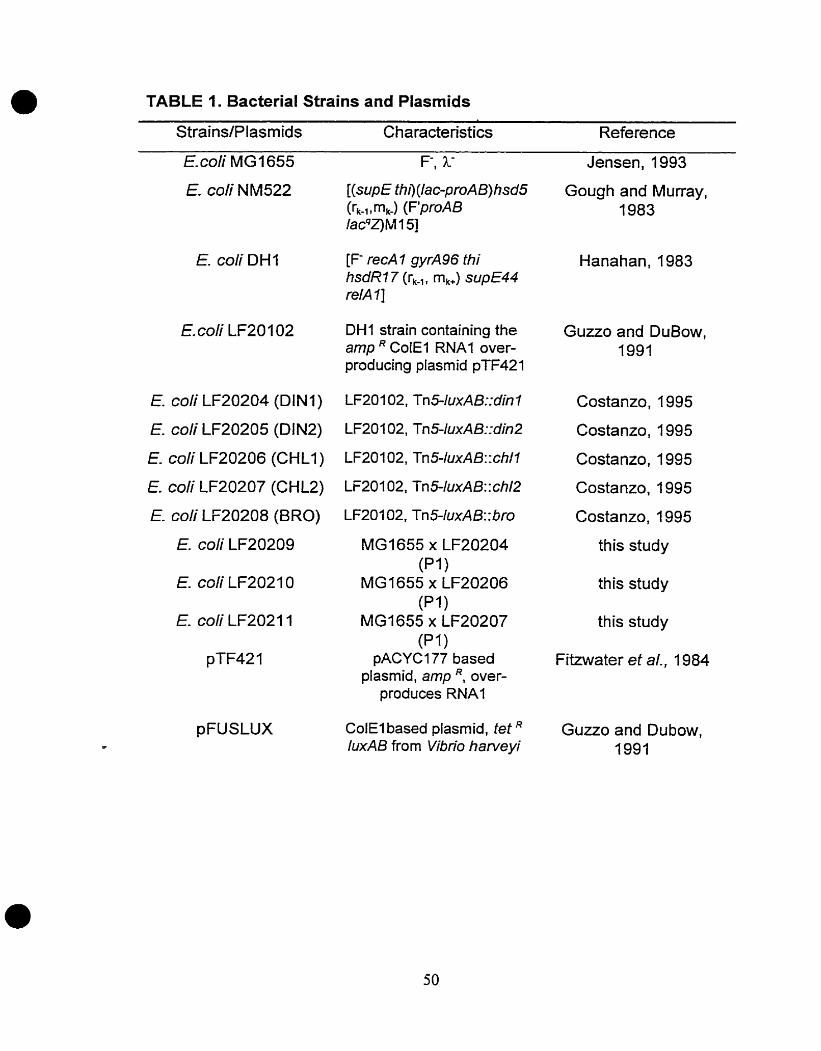
TABLE 1. Bacterial Strains and Plasmids
Strains/Plasmids Characteristics Reference
Jensen, 1993
E. coli DH 1
E. coli LF20204 (DI N 1)
E. coli LF20205 (DIN2)
E coli LF20206 (CHLI )
E. coli LF20207 (CHL2)
E. coli LF20208 (BRO)
E. coli LF20209
E. coli LF20211
[(sup E thil (lac-proA B) hsd5 (rk-, , m,) (F'proA B /acqZ) M 1 51
[F- recA 1 gyrA96 thi hsdR7 7 (rk-1, mk+) s~ipE44 relA 11
DH1 strain containing the amp * CoiEl RNA1 over- producing plasrnid pTF421
LF20102, Tn5-IuxAB:: bro
MG1655 x LF20204 (Pi )
MG1655 x LF20206 (P i 1
MG1655 x LF20207 (Pl
pACYCI77 based plasrnid, amp over-
produces RNA1
ColEl based plasmid. tet IuxAB from Vibrio harvevi
Gough and Murray, 1983
Guzzo and DuBow, 1991
Costanzo, 1995
Costanzo, 1995
Costanzo, 1995
Costanzo, 1995
Costanzo, 1995
this study
this study
this study
Fitzwater et al., 1984
Guzzo and Dubow, 1991
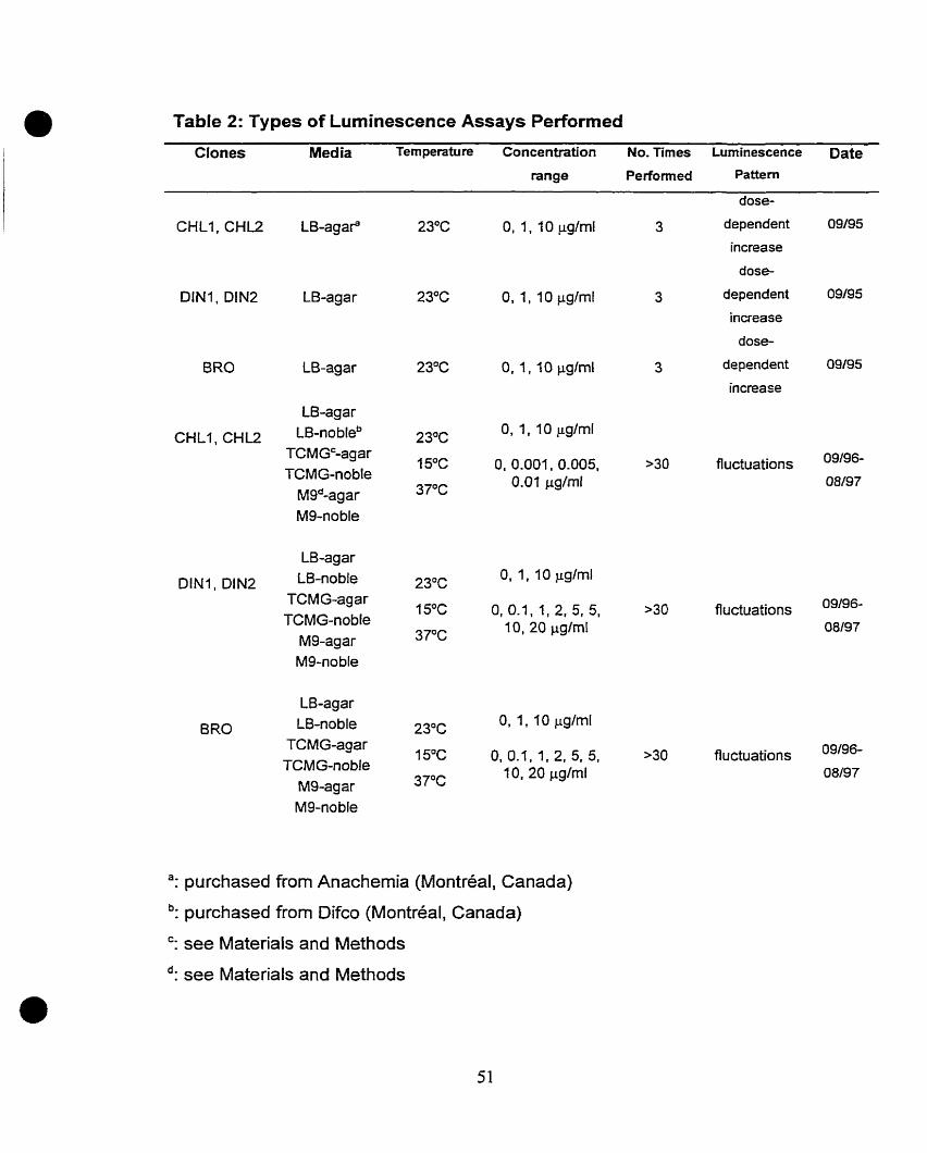
Table 2: Types of Luminescence Assays Performed
Clones Media Temperature Concentration No. Times Luminescence Date range Performed Pattern
-
dose-
CHL1, C H U LB-agar" 23OC 0, 1, 1 O p g h l 3 dependent 09/95
increase
dose-
DIN1, DIN2 LB-agar 23°C 0, 1, 10 pgfml 3 dependent 09/95
incrsase
dose-
dependent 09/95
increase
LB-agar
CHLI, C H U LB-nobleb TCMGc-agar TCMG-noble
Mgd-agar Mg-noble
O, 0.001, 0.005, >30 fluctuations 09196-
5.01 pg/ml 08/97
23°C O, 1, 10 pglml
15OC 0,0.1,1,2,5,5, >30 fluctuations
15°C 0,0.1,1,2,5,5, >30 fluctuations
37°C 1 O, 20 pgfrnl
": purchased from Anachernia (Montréal, Canada)
b: purchased from Difco (Montréal, Canada)
": see Materials and Methods
d: see Materials and Methods
Figure 1. Light ernission of 1-cm2 patches of strains LF20204 (DINI), LF20205
(DINZ), LF20206 (CHLI), LF20207 (CHL2), and LF20208 (BRO), grown in the
presence of O, 1, and 10 pprn (pg/ml) of the herbicides, bromacil and dinoseb,
and the pesticide, chlordane. See Materials and Methods for experimental
details. Taken from Costanzo, 1995.
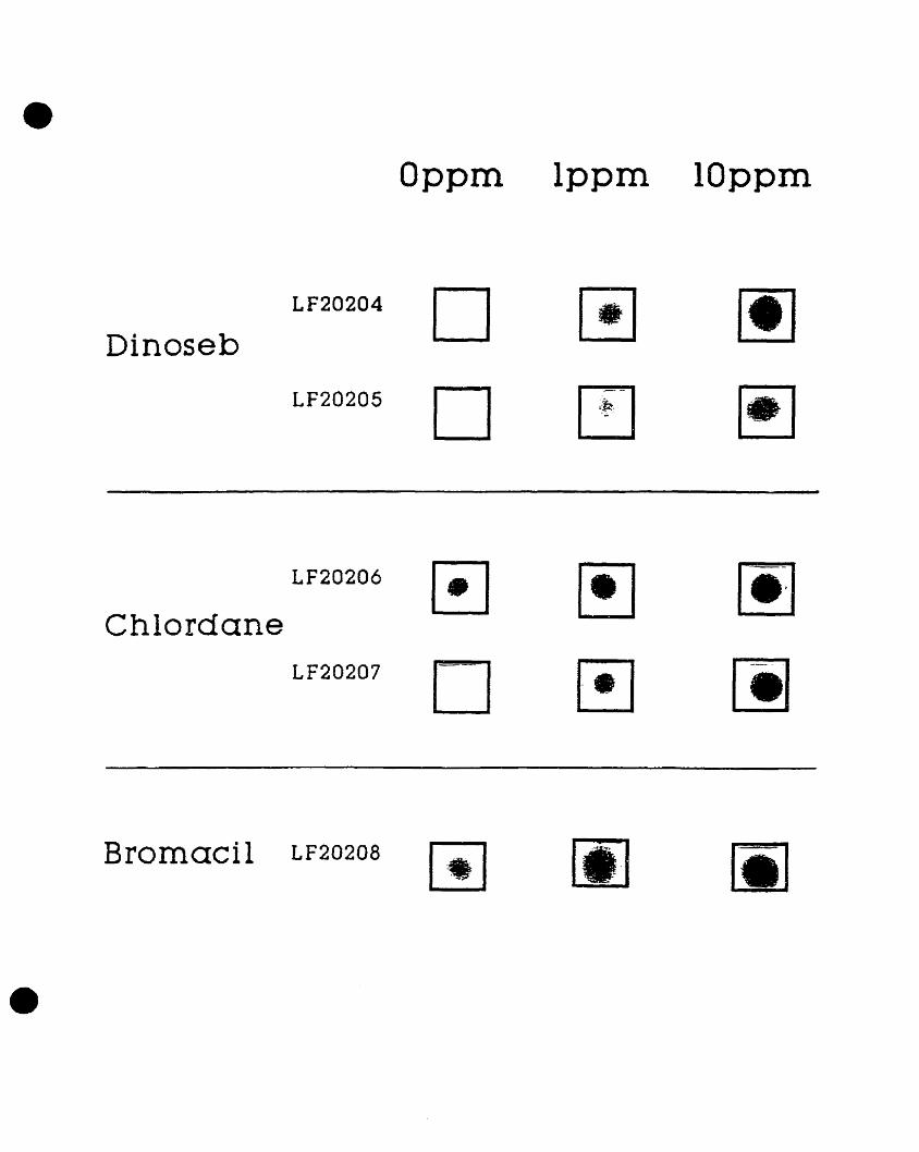
Dinoseb
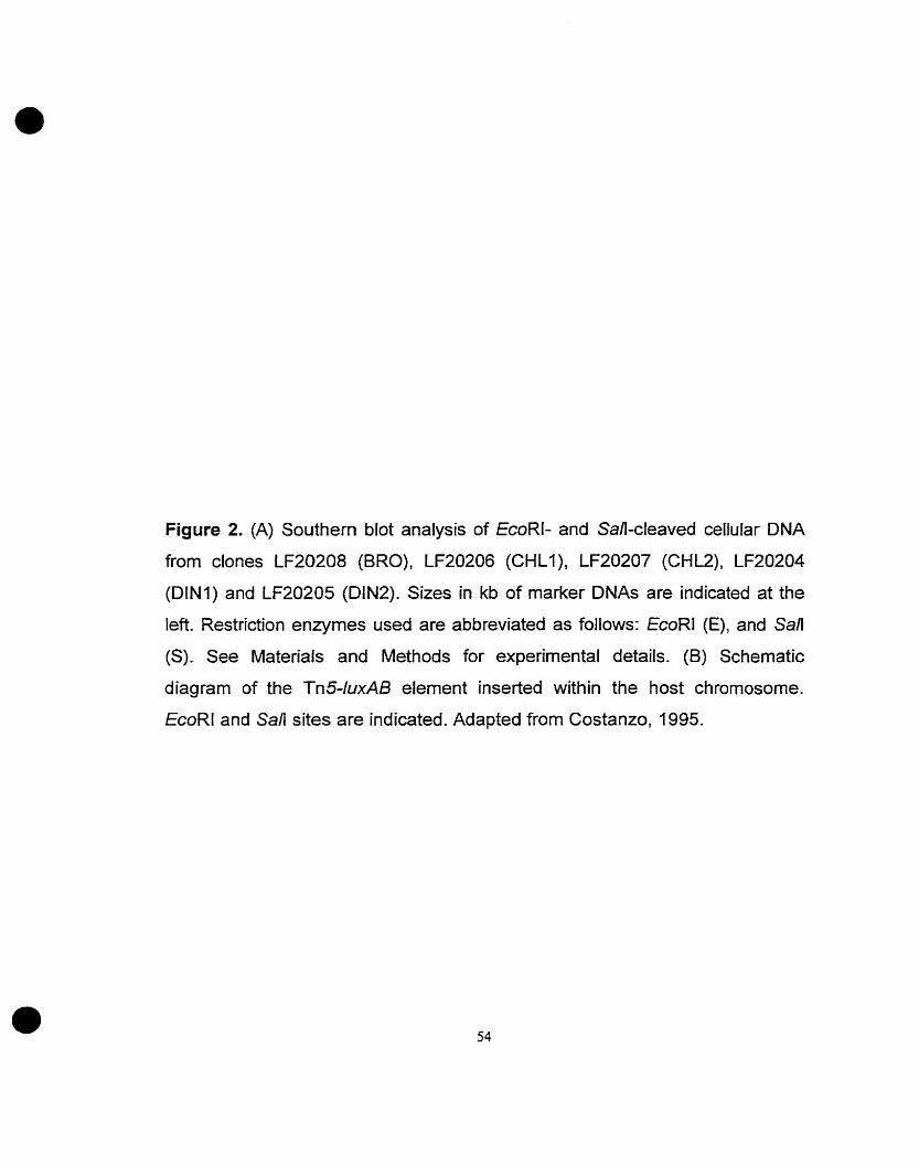
Figure 2. (A) Southern blot analysis of EcoRI- and San-cleaved cellular DNA
from clones LF20208 (BRO), LF20206 (CHLI), LF20207 (CHE), LF20204
(DINI) and LF20205 (DlN2). Sizes in kb of marker DNAs are indicated at the
left. Restriction enzymes used are abbreviated as follows: EcoRl (E), and San
(S). See Materials and Methods for experimental details. (B) Schematic
diagram of the Tn5-luxAl3 elernent inserted within the host chromosome.
EcoRl and San sites are indicated. Adapted from Costanzo, 1995.

Bromacil Chlordane Dinoseb
Figure 3. Schematic diagram for the cloning and sequencing of the herbicide-
and pesticide-responsive genes. Restriction enzyme sites are abbreviated as
follows: BarnHl (B), EcoRl (E), and Hindlll (H). See Materials and Methods for
experimental details. Taken from Costanzo, 1995.
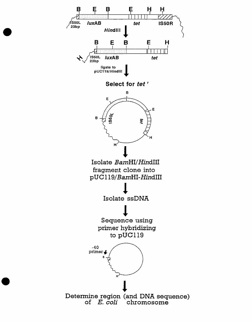
tigate to pUC119/Hindlll 1
Select for tet
Isolate BamHIIHindIII fragment clone into
pUC 1 19IBamHI-HindIII
Isolate ssDNA
Sequence using primer hybridizing
to pUC119
Determine region (and DNA sequence) of E. coli chromosome
Figure 4. Maps illustrating Tn5-IuxAB insertion sites in relation to the published
restriction rnap of E. coli (Rudd, 1992) for clones (A) CHLl (LF20206), (B) CHL2
(LF20207) and (C) DlNl (LF20204). Numbers below maps indicate minutes on
the E. coli chromosome. Unshaded Arrows indicate proximal identified genes in
their study and their orientations. Shaded arrows represent genes characterized
in this study and their orientation. Restriction enzyme sites are abbreviated as
foliows: BamHl (B), EcoRl (E), Sall (S), Hindlll (H), Bgn (G), EcoRV 0, Psd
(P), Kpnl (K), and Pvull (Pv). Taken from Costanzo, 1995.

B E B S E H
PvH K P H V I l !

Figure 5. Nucleotide sequence of the region adjacent to the Tn54uxAB
insertion of clone CHLI (LF20206). Amino acid sequence of the putative protein
encoded within this region is illustrated below the nucleotide sequence. i n 5
indicates the exact site of insertion Translation begins at an ATG codon and
teminates at a TAA codon. -35 and -10 promoter consensus sequences are
underlined and typed in bold face. Taken from Costanzo, 1995.

1 TCA CCT ATt T î C T M AGA TGA M A T'TA TTA GAG TCG CAT T M A M l'GA GCT M T
55 Ti? G A l ACT GGT tAT CTT GTG AtT ATT ?TC T M TGA CCC CGT GM C'TG A M CCC -35 - 1 O
109 TCC ATC CTt MT ATA AGG TGG AK; GAA AU; TGA rrC CllU ACT CAC TCA GTG GCC
163 AGA TCT t T A K A M T ATG M C AGG TTG M A M ACA GTA ACT TTG CK; TTT TTT 1 Met Asn Arg Leu Lys Lys Thr V a l Thr Leu k u Phe Phe
217 ATA CTT M C CAC TAT GCA TTA A% CTG CGT TAT PK: ATG A î G CCt M G MA M C 14 Ile Leu l u n Hfs Tyr Ala Leu H t t Leu k g Tyr Phe Met Met Pro L y s Lys AJn
271 CAC CGT TAC GCA M T GGT CM CGC TGG TTT TA7 CCG GTA CGT K;C M T TAT TT7 32 Gln A r g Tyr Ala h n Gly Gln k g Trp Phe Tyr Pro Val h g Cys A m Tyr Phe
325 TTA GCA GAA CCC GCT ICT M T AîT G M AîG ATT GAG M C MC GTA MG CAT ACC SU Lcu M a G l u Pro A l a Set Asn 116 Glu Thr I l e Glu h n b n Val L y s Hi8 Thr
379 GCT CCC U A & ACC AG& ACT CGC GM GGA AT* M *AT CCC ATG K G GTC TGC 68 M a Pro Lys Pso 7hr Arg Thr A r g G l u Gly I l e Lys Tyr Pro Met Set Val CyS
433 GCC ATT TCC GCA AGA M C GAT GCC AGG A M AGG M C AGG CAG GTG N ACG 86 N a I l c Ser Ala b g A m Asp Ala Arp Lys Arg Gln k n k g Gln Val Lys Thr
487 GGA ATC AK: GGT ATT Cl% T'TG GCT AAC G U CAG GTA CGT CTC CAT ACC AGT GCC 104 Gly I l e flc Gly I l e Leu Leu A l a Asn Glu G l n V a l k g Leu Wis Rtr Ser Ala
541 AGT AGC CA(; ACT TTT GAG M T ATG CTC T M CAG ATC ATT CCG AGG C M ATA AGA 122 Ser Ser Gln Thr Phe Glu Asn Met L e u * * *
595 ATA ATG CCG GGA GCC ACT CGG GCG CTT GCA TCC G U CTG ACT AAG ACG TAT ATC
649 CCC TGC AG?' ACC GTG ATT GAG CCG AGA lCAA ATA. AC(: C M TAA CAC CAC AGC CAG
703 TGT TCT T t A CTT C;AA MC GTA TTA CGT
Figure 6. Light emissions of 1-cm2 patches of strains LF20204 (DINI), LF20205
(DIN2), LF20206 (CHLI), LF20207 (CHU), and LF20208 (BRO), grown in t h e
presence of O, 1, and 10 PPM (mgfml) of the herbicides, bromacil and dinoseb,
and the pesticide, chlordane. See Materials and Methods for experimental
details.
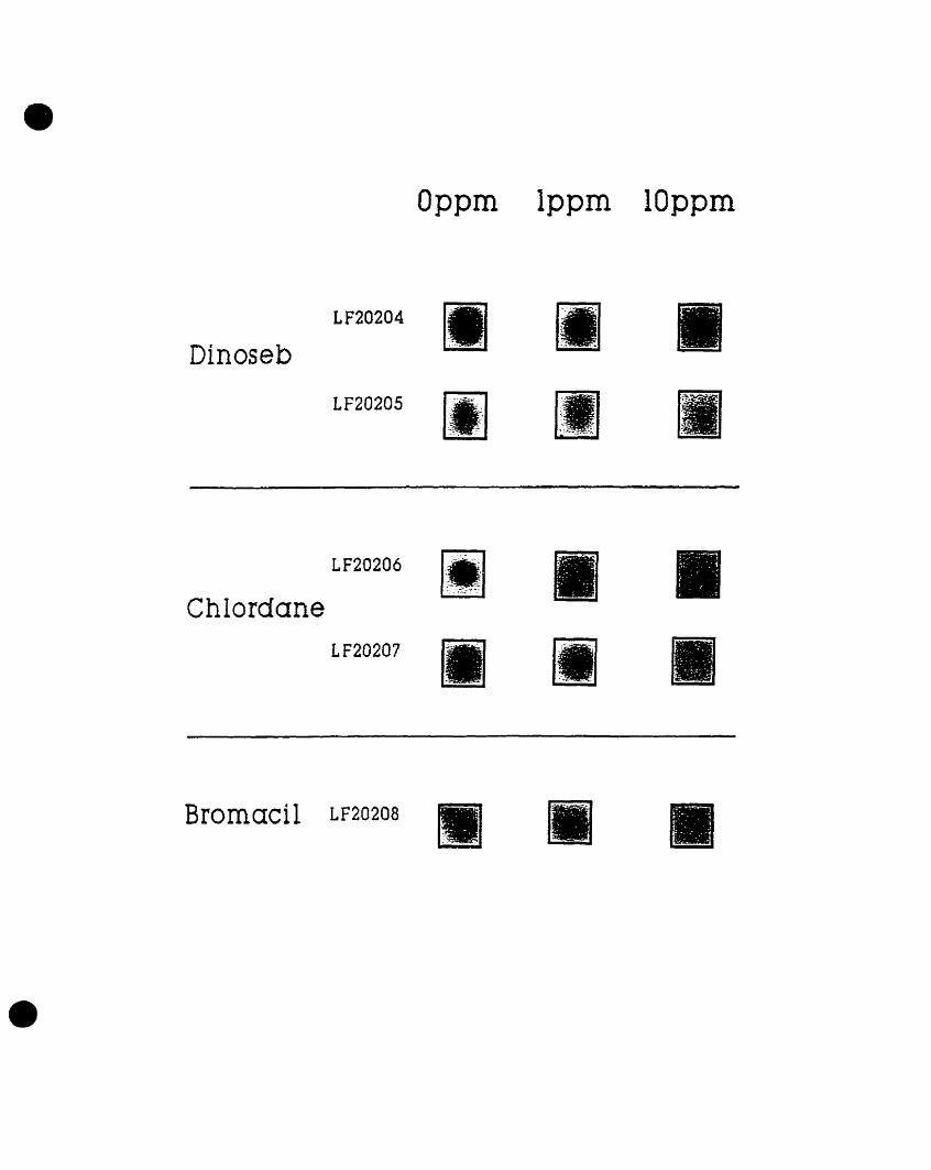
Dinoseb
Bromacil
Figure 7. Densitometric analysis of luminescence ernitted by strains (A) Dl NA and (B) DIN2 grown in the presence of O, 1. and 10 PPM (mglml) dinoseb.
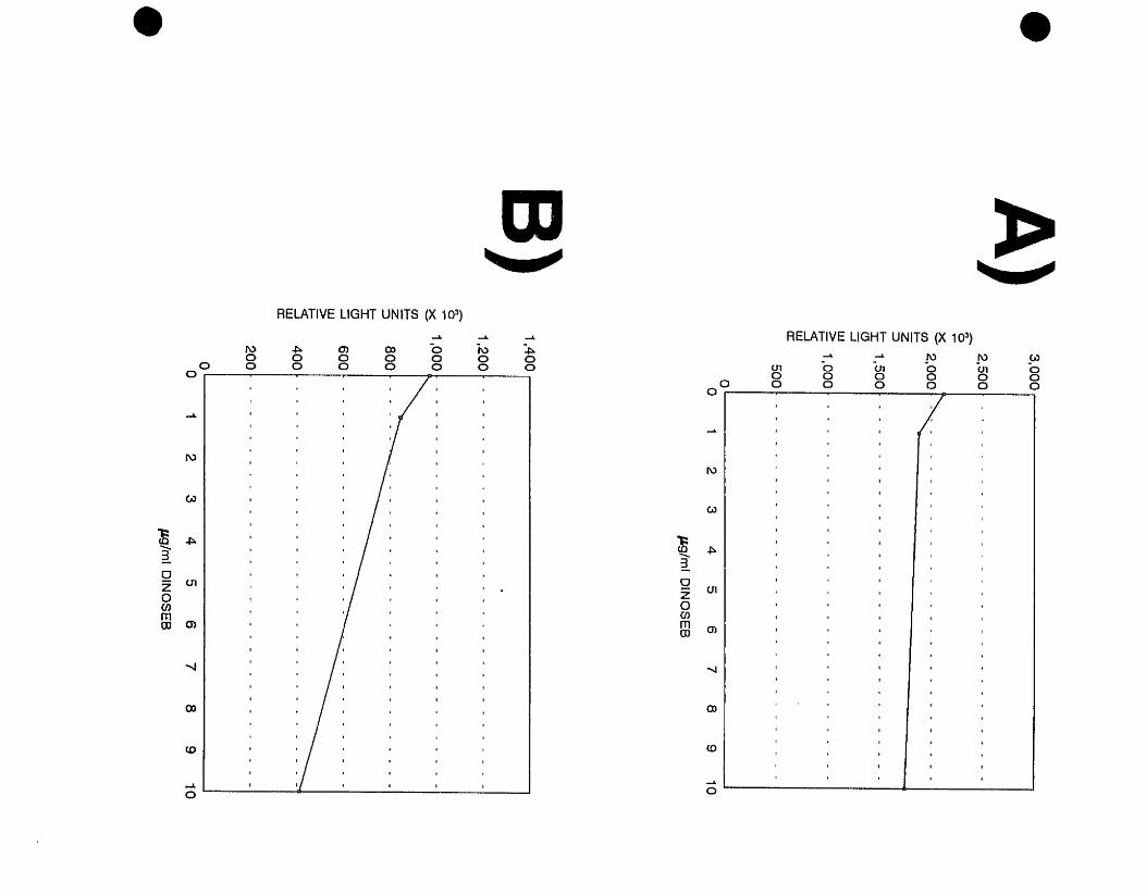
Figure 8. Densitometric analysis of luminescence ernitted by strains (A) CHLl and (B) CH12 grown in the presence of O, 1, and 10 PPM (mg/ml) chlordane.

O 1 2 3 4 5 6 7 8 9 1 0
pglmi CHLORDANE
l O 1 2 3 4 5 6 7 8 9 1 0
m / m i CHLORDANE
Figure 9. Densitometric analysis of luminescence emitted by strain BR0 grown in the presence of 0: 1, and 10 PPM (mglml) bromacil.
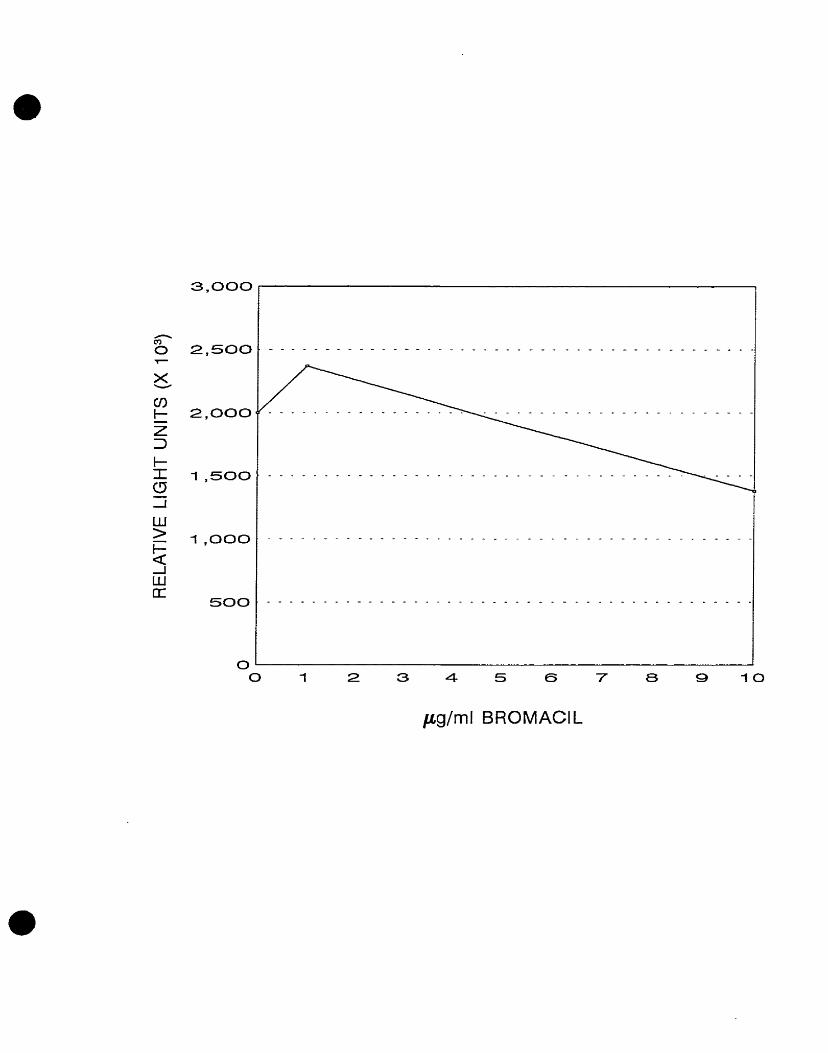
pg/ml BROMACIL
Figure 10. (A) Southern blot analysis of EcoRI- and Sali-cleaved cellular DNA from clones CHLl (LF20206). CHL2 (LF20207), and DlNl (LF20204). Sizes in Kb of marker DNAs are indicated at the left. Restriction enzymes used are abbreviated as foltows: (E) EcoRI, and (S) San. See Materials and Methods for experimental details. (B) Schematic diagram of the Tn5-IuxAB element inserted within the host chromosome. EcoRl and San sites are indicated
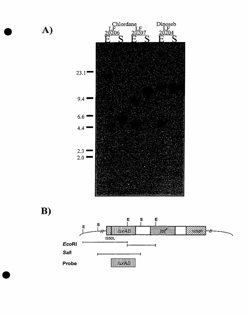
Dinoseb T C
Probe

Figure 11. Light emissions of 1 -cmZ patches of MG1 655 transductant strains LF20209 (DINI), LF20210 (CHLI), and LF20211 (CHL2), grown in the presence of O, 1, and 10 PPM (mglml) of the herbicide, dinoseb, or the pesticide, chlordane. See Materials and Methods for experimental details.
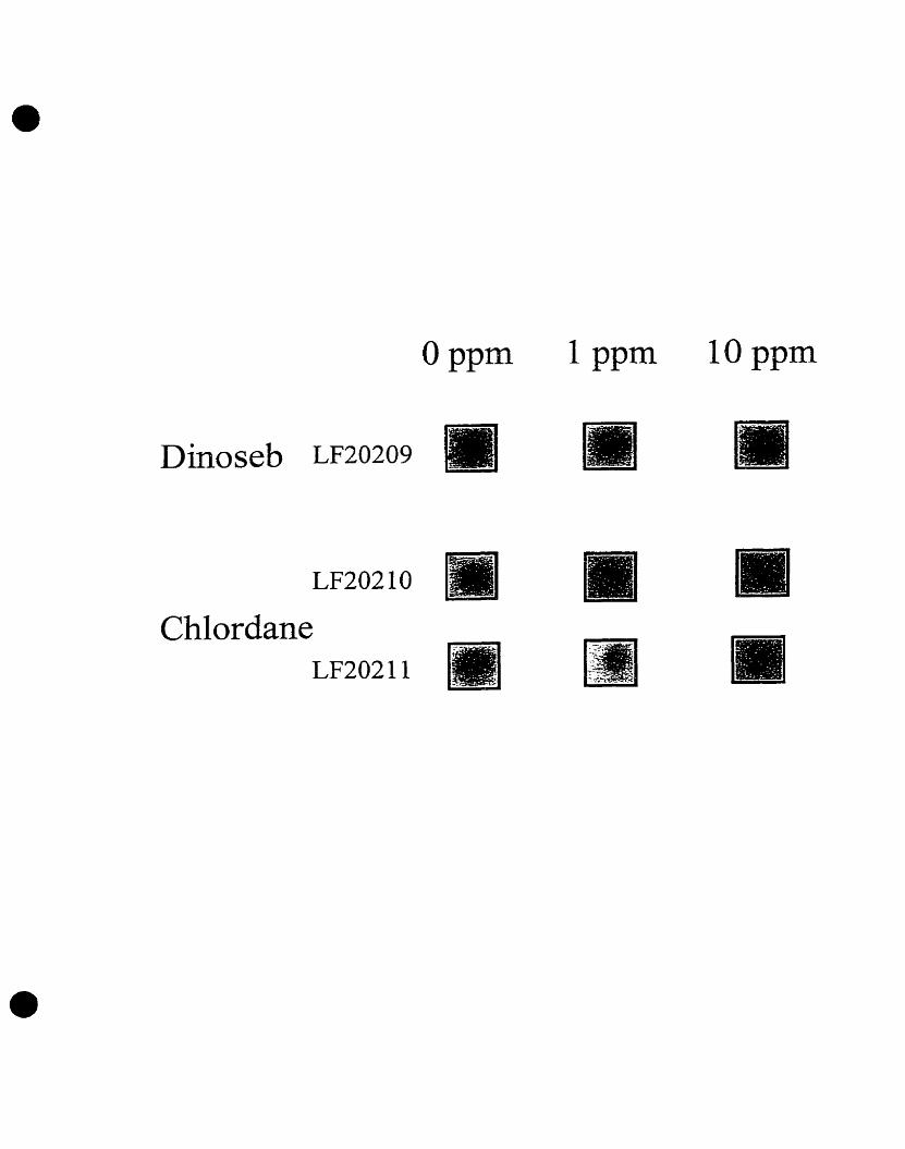
Dinoseb LF20209
Chlordane LF20211

Figure 12. Densitometric analysis of luminescence ernitted by the MG1655 transductant strain, DlNl (LF20209) grown in the presence of O, 1. and 10 PPM (rng/ml) dinoseb.
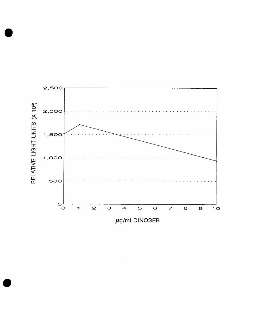
Figure 13. Densitometric analysis of luminescence ernitted by MG1 655 transductant strains (A) CH11 (LF20210) and (B) CHL2 (LF20212) grown in the presence of O, 1, and 10 PPM (mgfml) chlordane.

irglrnl CHLORDANE
O 1 2 3 4 5 6 7 8 9 1 0
pglrnl CHLORDANE

CHAPTER III
Use of an Arsenic Oxyanion-responsive Escherichia coli Luciferase Clone for the Detection of Arsenic Compounds in
an Environmental Aquatic Sample.
INTRODUCTION
Arsenic is a ubiquitous element with metalloid properties. In water,
arsenic is usually found in its trivalent (arsenite) or pentavalent (arsenate)
f o m (Andrae, 1977). The toxic properties of these arsenic oxyanions have
been well-documented. These include acute, subacute, and chronic effects
involving the respiratory, gastrointestinal, cardiovascular, nervous, and
haematopoietic systems (Done and Peart, 1971 ; Woods and Fowler, 1977;
Hine et al-, 1977). Adverse health effects of arsenic may therefore be local or
çystemic. Acute and subacute effects caused by the ingestion of inorganic
arsenic compounds, mainly arsenic (III) oxide, are also welf-documented in
the literature. The major lesion is profound gastrointestinal damage (Hine ef
al., 1977). Other acute symptoms and signs include muscular cramps, facial
oederna, and cardiac abnormalities (Done and Peart, 1971). Chronic effects,
although not as well studied, include local damage to the respiratory system,
skin, liver, cardiovascular and nervous systems upon inhalation andlor
ingestion of arsenic compounds (Harneda and Horiguchi, 1976). There is also
sufficient evidence to associate exposure to inorganic arsenic with carcinomas
of the lung and skin (Blot and Fraumeni, 1972; Friberg and Cederlof; 1978;
Jackson and Grarnge, 1975; Lee and Frauneni, 1969; Mabuchi et al., 1979;
Sommers and McManus, 1953; Reymann et al., 1978). Given the devastating
effects of these inorganic arsenic oxyanions, the sensitive and rapid detection
of arsenic in the environment is essential.
In 1973, Hedges and Braumberg (1973) found that gram positive and
gram negative bacteria both have plasmid-mediated resistance to arsenic and
antimony. It was later discovered that this resistance was due to a cluster of
genes, termed the ars operon. The plasmid-encoded ars operon is made up of
five open reading frarnes, desig nated arsRDABC. This operon is transcribed
as a single transcriptional unit initiated at a site just upstream of arsR. ArsR is

a trans-acting repressor which controls basal level of expression of the operon
(SanFrancisco et al., 1990; Wu and Rosen, 1991; Wu and Rosen, 1993a).
ArsD is a negative regulator of the operon (Wu and Rosen, 1993b). ArsA
encodes an arsenite- and antimonite-inducible ATPase (Rosen et al., 1988;
Mei-Hsu et al., 1991). ArsB encodes a membrane pump for efflux of arsenite
and antimonite ions (Wu and Rosen, 1992; Dey and Rosen, 1995). Finally,
ArsC encodes an arsenate reductase (Rosen et al., 1991; Gladysheva et al.,
1994). Several groups have recently found a chromosomally-located ars
operon homologue in Eschenchia coli and other gram negative bacteria!
species (Sophia et al., 1994; Carlin et al., 1995; Diorio et al., 1995). This
finding established a potential evolutionary relatedness between the plasmid-
encoded and the chromosomal ars operons in bacteria, the only difference
being that the E. coli chrornosomal ars operon comprises three open reading
frames which share high homology with the arsR, arsB, and arsC genes.
Finaily, expression of this operon in E. coli has been shown to be induced by
arsenic and antimonite oxyanions and the inducibility of arsenical cornpounds
appears to refiect their relative toxicity (Diorio et al., 1995; Cai and DuBow,
1996).
In this study, we therefore examined whether the arsB::luxAB clone,
described by Cai and DuBow (1996), could be used in the monitoring of
aquatic samples for arsenical compounds. These results were also compared
with results frorn R-galactosidase assays in which an arsB::lacZ fusion clone
was used to monitor the same contaminated samples. Finally, results
obtained from these biosensor experiments were compared to data from
chernical analyses performed by Environment Canada.
MATERIALS AND METHQDS
1 .O. Bacterial Strains and Gaowth Media
Table 1 describes bacterial strains and plasmids used. Media used in
the luminescence assays include Luria Bertani Broth (LB) [1% (w/v) tryptone,
1% (w/v) NaCI, 0.5% (wfv) Bacto yeast extract, 2 N NaOH] (unless othennrise
specified). Antibiotics were used at the following concentrations: ampicillin
(0.04 mg/ml) and tetracycline (0.01 mg/ml).
2.0. Liquid Luminescence Assay
Escherichia coli strains, LF20012, MG1 655, and 40 were grown at 37OC
overnight in 10 ml of Luria Bertani (LB) broth A solution of 2X LB broth [2%
(w/v) tryptone, 2% (wh) NaCI, 1% (w/v) Bacto-yeast extract] was prepared.
The overnight E. coli cultures were diluted 20-fold into the autoclaved solution
of 2X LB broth containing tetracyline. The bacterial cultures were then divided
into 5 flasks and grown for 1 h at 37OC to ensure logarithmic growth of the
culture (A,,=0.2-0.3). Cells were then exposed to equal volumes of either a
0.1 pgfml (PPM) arsenite solution (American Chernical Ltd., Montréal,
Canada), a 1 .O pg/ml (PPM) arsenate solution (Fisher Scientific, Ltd.,
Montréal, Canada), an equal volume of filtered and unfiltered arsenic-
contaminated water (Goodfellow Effluent, Environment Canada), or an equal
volume of sterile delH,O. Samples of contaminated water effluent were
collected by Environment Canada from the St. Lawrence River, Québec
(Goodfellow Effluent). Samples of contaminated water were filtered uçing
0.45pm nitrocellulose membranes. Samples contained tetracycline at a final
concentration of 0.01 mg/rnl. AI1 samples were then incubated for 1.5 h at
37OC. Following incubation, samples were diluted in LB broth to a final A, of
0.05 and then aliquoted into luminometer cuvets to be measured in a Tropix
Optocomp I luminometer (MGM Instruments. Hamden, Conn.). Total relative

light units (RLUs) were determined for 10-s intervals upon injection of 100 pl
of dodecyl aldehyde (diluted 1:100 LB broth) (Aldrich, Milwaukee, USA).
Luciferase activity is expressed as the number of photons emitted per second
per A,, unit.
3.0. p-galactosidase Assay
E. coli strains LF20001, MG1 655 and 40 were grown at 32OC overnight
in LB broth in the presence of ampicillin (0.04 mglml). Eighty pl of the
overnight cultures were then added to 5 ml fresh medium containing either 0.1
pgfml (PPM) arsenite (American Chemicals Ltd., Montréal, Canada), 1 .O pglrnl
(PPM) arsenate (Fisher Scientific Ltd., Montréal, Canada), an equal volume of
filtered and unfiltered arsenic-contaminated water (Goodfellow Effiuent,
Environment Canada), or an equal volume of sterile delH,O. Cells were then
grown at 32OC until they reached early exponential phase (2-5 X ?O8 cellslrnl).
The cultures were then cooled on ice for 20 min. The cell density was
recorded (A,,). An aliquot of 0.2 ml of each culture was immediately added to
1.8 ml Z buffer [60 mM Na,HP04.7 H,O, 40 mM NaH2P04*H20, 10 mM KCI, 1
mM MgSOp7 H20, 50 mM P-mercaptoethanol]. Eighty pl of chloroform and 40
pl of z 0.1% sodium dodecyl sulfate (SDS) solution were added to each assay
mixture. Tubes were vortexed for 10 seconds and placed in a 28OC water bath
for 5 min. The reaction was started by adding 0.4 ml ONPG [4 rnglml in 100
mM phosphate buffer]. After sufficient yellow colour had developed, the time
of the reaction was recorded. The reaction was stopped by adding 1 ml of a 1
M Na,CO, solution. A control solution containing 0.2 ml ONPG and 1.8 ml Z
buffer was also incubated along with the basal-level cultures. The optical
density was recorded (A4,,). Light scattering was corrected for by obtaining the
absorbance at another wavelength (A,,) where there is no contribution from
O-nitrophenol. P-galactosidase enzyme units were calculated as follows:
1 000X(OD420 - 1.75XOD5,,) / t X v X OD,,, (Miller, 1 992).

RESULTS
Luminescence Assays With Filtered and Unfiltered Arsenic-
Contarninated Water Samples
The biosensor potential of a previously constructed arsB::luxAB E. coli
clone (Diorio et al., 1995; Cai and DuBow, 1996) was tested for its ability to
detect the presence of arsenic compounds in a contarninated water sample
(Goodfellow Effluent, Environment Canada). Luminescence assays were
performed using three E. coli strains, E. coli 40 (Alac), E. coli MG1655 (wt),
and the arsB::luxAB E. co/ifusion clone, LF20012. Light emitted from the three
strains was measured following their exposure to sterile delH,O, a 0.1 PPM
arsenite solution, a 1.0 PPM arsenate solution, an unfiltered sarnple of the
arsenic-contarninated water effluent, and a filtered sample derived from the
same arsenic-contaminated effluent.
E. coli LF20012, containing the arsf3::iuxAB chromosomal fusion,
exhibited a 17-fold increase in light emission, following a 1.5 hr exposure to
the 0.1 PPM arsenite solution and to the 1.0 PPM arsenate solution (Figure
1). Increases in light emission were also observed following exposure of strain
LF20012 to the filtered and unfiltered arsenic-contaminated water effluents.
Exposure to the unfiltered water sample resulted in a 3-fold increase in
luminescence, while exposure to the filtered water sample resulted in a 4-fold
increase in light ernitted from the E. coli LF20012 culture (Figure 1). Only
basal levels of light emission were observed when the E. coli IuxAB fusion
clone was grown in the presence of sterile delH,O (Figure 1). Unlike E xoli
strain LF20012, E. coli 40 and MG1655 did not exhibit any significant
increases in luminescence (data not shown).

B-galactosidase Assays With Filtered and Unfiltered Arsenic-
Contaminated Water Samples
P-galactosidase assays were performed using three E. coli strains, E.
coli 40, E. coli MG1655, and the arsB::/acZ E. coli fusion clone, LF20001. P- galactosidase activity wss measured foilowing exposure to sterile delH,O, a
0.1 f PM arsenite solution, a I .O PPM arsenate solution, an unfiltered sample
of the arsenic-contaminated water effluent, and a filtered sample derived from
the same arsenic-contaminated effluent. MG1655 exhibited basal level P- galactosidase activity. Furthemore, this level of activity was not affected by
the presence of the arsenite, arsenate, or the arsenic-contaminated water
effluent samples (Figure 2). E. coli 40, a dac strain, did not exhibit any P- galactosidase activity (Figure 2). Unlike E. coli MG1 655 and E. coli 40, E. coli
LF20001 exhibited a 34-fold increase in P-galactosidase activity following
exposure to the 0.1 PPM arsenite solution and a 31-fold increase in the
presence of the 1 .O PPM arsenate solution (Figure 2). Exposure to the filtered
arsenic-contaminated water sample resulted in a 5-fold increase in B- galactosidase activity of the E. coli 1acZ::arsB fusion clone (Figure 2). The
unfiltered water sample also stimulated a 5-fold increase in P-galactosidase
(Figure 2). Finally, sterile delH,O did not stimulate any P-galactosidase activity
in the arsB::lacZ fusion clone (Figure 2).
DISCUSSION
In this study, we examined the potential of the Eschenchia coli
arsB::/uxAB clone (LF20012) described by Cai and DuBow (1996), and the
Eschenchia coli arsB::lacZ clone (LF2000 1) described by Diorio et al. (1 995),
to detect the presence of arsenical compounds in aquatic samples. The
results obtained from these experiments were then compared with chernical
data obtained from Environment Canada.
In the absence of any arsenic-containing solutions, the arsB::luxAB
clone exhibited basal levels (less than 1 X I O 6 RLUs) of light emission (Figure
1). In previous experirnents, it was shown that concentrations of 0.1 PPM
arsenite and 1.0 PPM arsenate rnaximally induced the expression of the
arsB::/uxAB fusion and thus, these concentrations were used as positive
controls (Cai and DuBow, 1996). As expected, drarnatic increases in light
emission (approxirnately 1.7 X I O 7 RLUs) were observed following exposure
of the arsf3::IuxAB clone to the arsenite and arsenate solutions (Figure 1).
Following exposure of the a&::/uxAB clone to filtered and unfiltered samples
of the arsenic-containing water effluent, approximately 3.6 X 1 O6 and 4 X I O6
RLUs were emitted, respectively (Figure 1). Chernical analyses performed by
Environment Canada revealed that the unfiltered water effiuent contained a
total arsenic concentration of 0.28 PPM (data not shown). The species and
nature of arsenic present in the water sample has not yet been determined.
However, it was previously shown that in oxygenated water, arsenic is
prevalent in iis pentavalent, arsenate form (Cullen and Reirner, 1989). If this is
the case, the results obtained from the luminescence assays described here
correlate well with the available chemical data. (Figure 1). However, further
chemical analyses, determining the exact nature and species of the arsenic
present in the effluent, is necessary to make any firm conclusions.
To confimi the results from the luminescence assays and to eliminate
the possibiliiy that luminescence was due to a luxAB-specific phenomenon, P- galactosidase assays were also perforrned. In these experiments, the E. coli
arsB::lacZ clone (Diorio e l a/., 1995) was exposed to the same arsenic-
containing water effluents described above. In the absence of any arsenic-
containing solutions, the ars5::lacZ clone exhibited only basal levels (0.5
Miller Units) of f3-galactosidase expression (Figure 2). However, in the
presence of 0.1 PPM arsenite and 1 .O PPM arsenate, drarnatic increases of
approximately 34 and 31 Units of P-galactosidase were observed, respectively
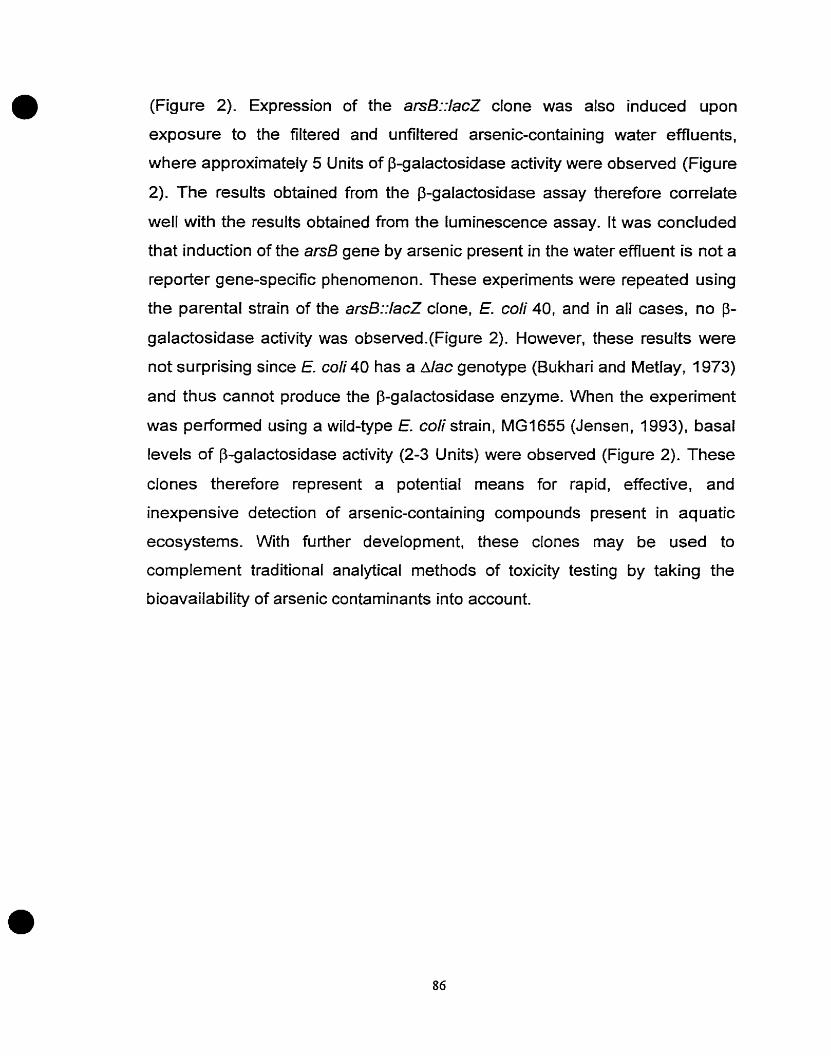
(Figure 2). Expression of the arsB::lacZ clone was also induced upon
exposure to the filtered and unfiltered arsenic-containing water effluents,
where approximately 5 Units of P-galactosidase activity were observed (Figure
2). The results obtained from the f3-galactosidase assay therefore correlate
well with the results obtained from the luminescence assay. It was concluded
that induction of the arsB gens by arsenic present in the water effiuent is not a
reporter gene-specific phenornenon. These experiments were repeated using
the parental strain of the arsS::lacZ clone. E. coli 40, and in al1 cases, no P- galactosidase activity was observed.(Figure 2). However. these results were
not surprising since E. coli 40 has a dac genotype (Bukhari and Metlay, 1973)
and thus cannot produce the P-galactosidase enzyme. When the experiment
was perfonned using a wild-type E. coli strain, MG1655 (Jensen, 1993). basal
levels of B-galactosidase activity (2-3 U nits) were observed (Figure 2). These
clones therefore represent a potential means for rapid, effective, and
inexpensive detection of arsenic-containing cornpounds present in aquatic
ecosystems. With further development, these clones may be used to
complement traditional analytical methods of toxicity testing by taking the
bioavailability of arsenic contarninants into account.
Table 1 : Bacterial Strains
Strain C haracteristics Reference
E. cd i MG1655 F-, h- Jensen, 1993
E. coli 40 F-, Apro-lac, rpsL, trp Bukhari and Metlay, 1973
E. coli LF20012 arsB::luxAB in E. coli 40 Cai and DuBow, 1996
E. coli LF20001 arsB::lacZ in E. coli 40 Diorio et al., 1995
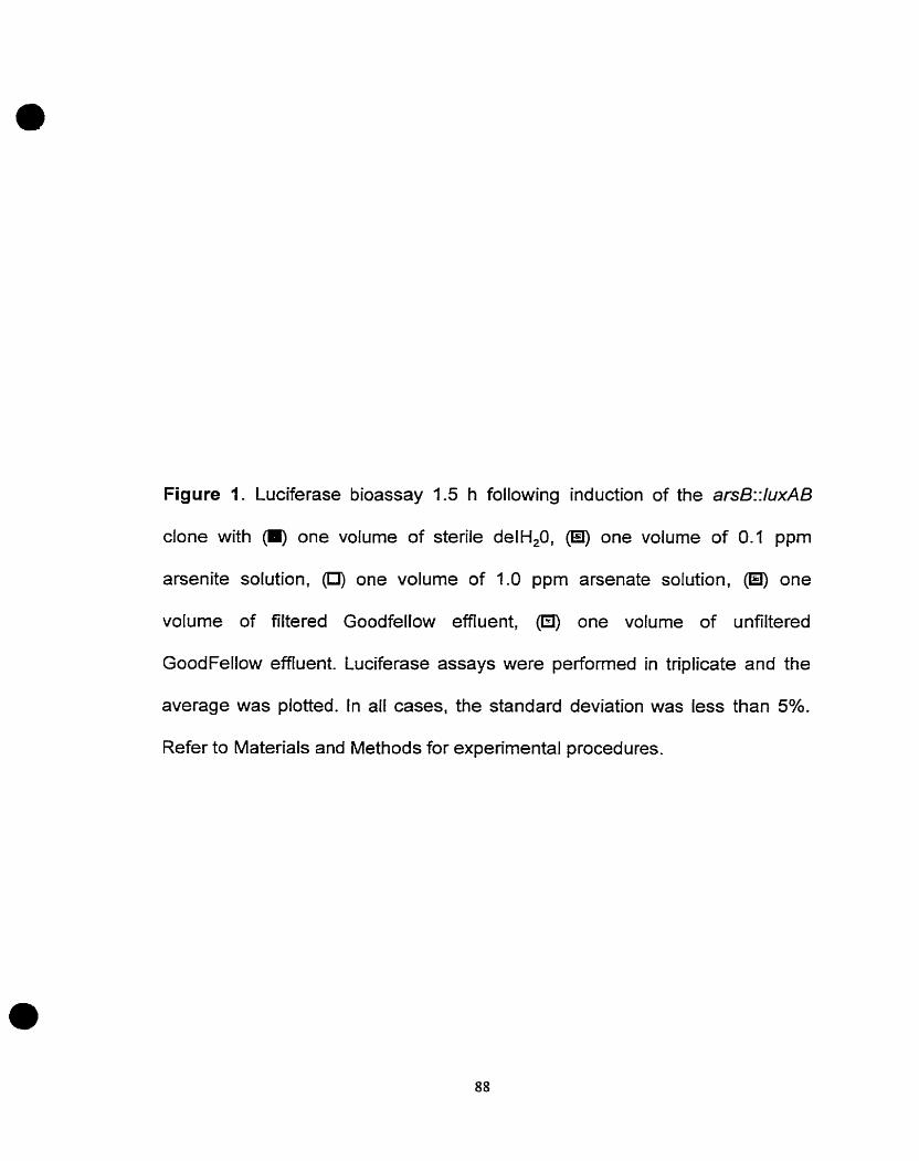
Figure 1. Luciferase bioassay 1.5 h following induction of the ars8::luxAB
clone with (O) one volume of sterile delH,O, 0 one volume of 0.1 ppm
arsenite solution, (U) one volume of 1.0 ppm arsenate solution, (Ei) one
volume of filtered Goodfellow effiuent, one volume of unfiltered
GoodFellow effluent. Luciferase assays were performed in triplicate and the
average was plotted. In al1 cases, the standard deviation was less than 5%.
Refer to Materials and Methods for experirnental procedures.

+ve(ite) +ve(ate) filtered u nfiltered

Figure 2: P-galactosidase bioassay 1.5 h following induction of the arsB::lacZ
clone with one volume of sterîle delH,O, O one volume of 0.1 ppm
aisenite solution, @) one volume of 1.0 pprn arsenate solution, 0 one
volume of filtered Goodfellow effluent, one volume of unfiltered
GoodFellow effluent. P-galactosidase assays were performed in triplicate and
the average was plotted. Error bars depict the calculated standard deviation.
In al1 cases, the standard deviation was less than 5%. Refer to MATERIALS
and METHODS for experimental procedures.


CHAPTER IV
Conclusions and Future Directions

Our environment is constantly being exposed to a myriad of chemicals,
such as pesticides and herbicides. However, Iittle is known about their
biological effects on cells. These effects may involve the induction of gene
expression producing proteins which alter the cell's physiology, allowing it to
cope with the pesticide- or herbicide-induced stress. The first part of the thesis
therefore focused on the characterization of genes in Escherichia coli whose
expression was altered upon cellular exposure to the herbicides, bromacil and
dinoseb, and to the pesticide, chlordane. To identify these genetically-
programmed responses, a modified Tn5-IuxAB E. coli gene fusion library
(Guuo and DuBow, 1991) was screened in the presence of increasing
concentrations of bromacil, chlordane, and dinoseb (Costanzo, 1995). Using
this 3000 clone library, Our laboratory has identified several genetically-
programmed responses to various inorganic, toxic, metal compounds. (Guzzo
et al., i991; Guzzo and DuBow, 1994a). However, the library had never been
screened for the presence of E. coli clones which respond to chemically
complex, organic compounds such as pesticides and herbicides.
Five distinct clones which exhibited increases in light emission following
exposure to either bromacil, dinoseb, or chlordane were isolated from this
Iibrary (Costanzo, 1995). Characterization of these pesticide- and herbicide-
responsive genes involved cloning, rnapping, and sequencing. Following partial
characterization of these genes, luminescence experirnents were repeated.
However, unlike the dose-dependent increases in Iight ernission originally
obsewed, luminescence assays performed with new lots of compounds were
inconsistent yielding erratic patterns of light emissions. Thus, experirnents were
performed to determine the cause of these inconsistencies. Southern blotting
analyses and P l vir transduction experiments were performed to determine if a
genetic phenomenon was responsible for fluctuations in luciferase expression
and luminescence patterns. Frorn these experiments, it was concluded that
inconsistent light emissions were not caused by secondary transposition of the
Tn5-luxAB element or genomic rearrangements or by the accumulation of point
mutations within the chromosomes of the responsive clones. Furthemore,
luminescence assays were performed under varying conditions to determine
the effects of media and temperature on luciferase expression. Based on
results presented in chapter two, it was hypothesized that the complex nature
of the cornpounds tested coupled with the extreme sensitivity of the
bioluminescence pathway may have been responsible for the variability
observed in luciferase expression and luminescence.
Future experiments to further characterize the pesticide- and herbicide-
responsive genes should involve performing luminescence assays following
exposure of the clones to the pure, individual components comprising the
complex pesticides and herbicides. Moreover, additional experiments should
incorporate the use of other well characterized, more defined reporter genes,
such as IacZ, to minimize luxAB artifacts. Following characterization of the
luminescence reactions, future stud ies rnay involve over-expression of the
proteins encoded by the two chlordane-responsive, putative open read ing
frames. Additional experiments should also be designed to study the regulatory
cascade involved in cspB induction. These experiments may ultimately
elucidate the functions and relationships of these genes to the pesticide,
chlordane, and the herbicide, dinoseb. Following characterization of these
genes and their corresponding gene products, assays, such as those described
in chapter three, should also be developed to test the potential of these
luminescent clones as biosensors to detect the presence of chlordane and
dinoseb in the environment-
With increased world-wide industrialization and a concomitant higher
demand for chemicals, both developed and developing countries are facing
increasing ecological and toxicological problems from the release of toxic
contaminants into the environment (Bitton and Koopman, 1986). In response to
these expanding stresses, a multitude of biological assays have been

developed to assess the impact of toxic chemicals on our ecosystems. The
long-term effects of chemicals discharged into aquatic environments are now
being realized and research efforts are now being directed to short-terni toxicity
tests which can alert monitoring agencies of the potential dangers associated
with toxic chemical release (Bitton and Koopman, 1986). The second part of the
thesis therefore focused on determining whether a well characterized
arsenicfantimony-responsive E. coli ars8::luxAB fusion clone, LF20012 (Cai
and DuBow, 1996), could be used as a biosensor to detect arsenic compounds
present in an aquatic sample obtained from the St Lawrence River, Québec
(Goodfellow Effluent, Environment Canada).
Liquid luminescence assays were performed in which the arsenic-
responsive €. coli clone was exposed to an arsenic-contaminated water
sample, as well as several control sarnples, containing known concentrations of
arsenite or arsenate. A three- to four-fold increase in light emission was
detected following exposure of the clone to either filtered or unfiltered water
effluents. If the arsenic present in the water effiuent is predominantly in its
pentavalent form (Cullen and Reimer, 1989), then luminescence results
obtained from Our assay correspond well with chemical data obtained from
Environment Canada. However, further investigations and chemical analyses
are necessary to deterrnine the exact nature and species of arsenic present in
the water effiuent. Similar experiments were perforrned in which P- galactosidase activity was measured in an E. coli arsB::lacZ fusion clone,
LF20001 (Diorio et al., 1995), following exposure to the same arsenic-
contarninated water effluent and control sarnples. Results obtained from the P- galactosidase assay confirmed that the light emission observed in the presence
of the arsenic-contaminating water effiuent was not due to a reporter gene-
specific p henomenon.
The results obtained from these experiments suggest that rnetal-
responsive E. coli clones, identified and isolated in our laboratory, have the
potential to be exploited as biosensors. Furthemore, the application of these
biosensors in the environment may complement existing bioassays by
detecting the presence of intermediate, biologically-relevant concentrations of
environmental contaminants, while at the same time taking the bioavailability of
these contarninants into account.
REFERENCES
Agency for Toxic Substances and Disease Registry. 1989. Toxicologicai Profile for Chlordane (ATSDRiTP-89/06). US Dept. of Commerce, National Technical Information Service, Springfield, VA
Alleman, J. E. 1988. Respiration-based evaluation of nitrification inhibition using enriched Nitrosornonas cultures. In: Scholze, R. J., Smith, E. D., Bandy, J., T., Yu, Y. C., and Basilico. J., V. (eds) Biotechnology for Degradation of toxic Chernicals in Hazardous Waste Sites. Noyes Dat. Corp., Park Ridge, NJ, pp. 642-650.
AISOP, G. M., Waggy, G., T m y and Conway, R. A. 1980. Bacterial growth inhibition test. J. Water Pollut. Control Fed. 52: 2452-2456.
Altman, F. P. 1976. Tetrazoliurn salts and formazans. Prog. Histochern. Cytochern. 9: 1
Andrae, M. 0. 1977. Determination of arsenic species in natural waters. Anal. Chem. 49: 820-825.
Ang, D. and Georgopopoulos, C. 1989. The heat-shock regulated grpE gene of Escherichia coli is required for bacterial growth at al1 temperatures but is dispensible in certain mutant backgrounds. J. Bacteriol. 171 : 2748-2755.
Baker, T. A., Howe, M. M., and Gross, C. A. (1983) Mu dX, a derivative of Mu d l (lac ~ p ~ ) which makes stable lacZ fusions at high temperature. J. Bacfenol. 156, 970-974.
Barrineau, P., Gilbert, P., Jackson, J., Jones, S., Surnmers, A. O., and Wisdom, S. 1984. The DNA sequence of the mercury resistance operon of the IncFll plasmid NRI. J. Mol. Appl. Genet. 2: 601-619.
Benjamin, M. M., Woods, S. L., and Ferguson, J. F. 1984. Anaerobic toxicity and biodegradability of pulp mil1 waste constituents. Water Res. 8: 601-607.
Bewley, R. J. F. and, Stotzky, G. 1983. Effects of cadmium and zinc on microbial activity in soil: Influence of clay minerals. Part II: Metals added sirnuitaneously. Sci. Total Environ. 31 : 57-69.
Bitton, G. and Dutka, B. J. 1986. Introduction and review of microbial and biochemical toxicity screening procedures. In: Toxicity Testing Using Microorganisrns. Vol 1 Bitton, G. and Dutka, B., J. (eds.), CRC Press, Boca Raton, Fia. pp. 1-9.
Bitton, G., Dutton, R. J., and Kooprnan, 5. 1988. Cell pemeability to toxicants: An important parameter in toxicity testing using bateria. CRC Crit. Rev. Environ. Control. 18: 177-1 88.
Bitton, G., and Koopman, B. 1992. Bacterial and Enzymatic Bioassays for Toxicity Testing in the Environment. Rev. of Environ. Contam. Toxicol. 125: 1- 22,
Bitton, G., Dutka, B., J., and Hendricks, C. W. 1989. Microbial toxicity tests. In: Warren-Hicks, W., Parkhurst, B., R., and Baker, S. S. (eds.). Ecological assessrnent of hazardous waste sites, US. E.P.A., Corvallis, OR, Report EPA 60013-891013. Pp. 6.44-6.66.
Bitton, G., and Koopman, B. 1986. Bacterial Tests for Toxicity Screening. In: Toxicity Testing Using Microorganisms. Vol 1 Bitton G., and Dutka, B. J. (eds.), CRC Press, Boca Raton, Fla. pp. 27-55.
Blake, C., Legault, R., Bemingham, N., van Coillie, Ra, and Vasseur, P. 1986. A simple microplate algal assay technique for aquatic testing assessment. Tox. Assess. 1 : 261 -281.
Blot, W. J. and Fruarneni, J. F. 1975. Arsenical air pollution and lung cancer. Lancet. 26: 142-144.
Blum, L. J., Gautier, S. M., and Coulet, P. R. 1993. Design of biolumiescence-based fiber optic sensors for flow-injection analysis. J. Biotechnol. 31 : 357-368.
Boehringer Mannheim, 1994. The DIG System Technique and Product Guide.
Brezonik, P. L., and Patterson, J. W. 1972. Activated sludge ATP: Effects of environmental stresses. J. San. Eng. Div. Proc. Am. Soc. Civil Eng. 97: 81 3- 821.
Briscoe, S. Fm, Diorio, C., and DuBow, M.S. 1996. Luminescent biosençors for the detection of tributyltin and dimethyl sulfoxide and the elucidation of their mechanisms of toxicity. In: Environmental Biotechnology: Principles and Applications. Moo-Young, M., Anderson, W. A., and Chakrabarty, A. M. (eds.), Kluwer Academic publishers, Dordrecht, pp. 645-655.
Bukhari, A. 1. and Metlay, M. 1973. Genetic mapping of prophage Mu. Virology 5 4 ~ 1 09-1 16.
Bulich, A. A. 1986. Bioluminescent assays. In: Bitton, G., and Dutka, B. J. (eds.) Toxicity Testing Using Microorganisms, Vol. 1. CRC Press, Boca Raton, Fla. pp. 57-74.
Bulich, A. A. 1979. Use of luminescent bacteria for determining toxicity in aquatic environments. In: Markings, L. L., Kirnerie, R. A., (eds.) Aquatic Toxicology. Am. Soc. Test Mat., Philadelphia, PA
Burlage, R. S., Sayler, G. S., and Larimer, F. 1990. Monitoring of naphthalene catabolism by bioluminescence with nah-lux transcriptional fusions. J. Bacteriol. 172: 4749-4757.
Burns, R. G., (ed). 1978. Soil Enzymes. Acadernic Press, London.
Cai, J. and DuBow, M.§. 1996. Expression of the Eschenchia coli chromosornal ars operon. Can. J. Micr~biol. 42: 662-671.
Carlin, A., Shi, W., Dey, S., and Rosen, B. P. 1995. The ars operon of Eschenchia coli confers arsenical and antimonial resistance. J. Bacteriol. 177: 981 -986.
Carlisle, S. M., and Trevors, J. T. 1986. Effects of the herbicide glyphosate on nitrification, denitrification, and acetyl reduction in soil. Water Air Soil Pollut. 29: 189-203.
Cenci, G., Moroui, G., and Caldini, G. 1985. lnjury by heavy metals in EschenChia coli. Bull. Environ. Contam. Toxicol. 34: 188-1 95.
Codina, J. C., Perez-Garcia, A., Rornero, P., and de Vicente, 1993. A Cornparison of Microbial Bioassays for the Detection of Meta1 Toxicity. Arch. Environ. Contam. Toxicol. 25: 250-254.
Cohen, S. 2. 1984. List of potential groundwater contaminants. Office of Pesticides and Toxic Substances. Washington, DC.
Coleman, W. E., and Qureshi, A. Am, 1985. Microtox and Spnlium volutans tests for assessing toxicity of environmental samples. Bull. Environ. Contam. Toxicol- 35: 443-451.
Collard, J., M., Corbisier, P., Diels, L., Dong, Q., Jeanthon, C., Mergeay, M., Taghavi, S., van der Lelie, D., Wilmotte, A., and Wuerh, S. 1994. Plasmids for heavy metal resistance in Alcaligenes eutrophus Ch34 mechanisms and applications. FEMS Microbiol. Rev. 14: 405414.
Corbisier, P., Ji, G., Nuyts, G., Mergeay, M., and Silver, S. 1993. IuxAB gene fusions with the arsenic and cadmium resistance operon of Sfaphlococcus aureus plasmid p1258. FEMS Microbiol. Let;. 110: 231-238.
Costanzo, M. A. 1995. Characterization of luciferase biosensors for the detection of pesticides and herbicides. Honors Research Project. Department of Microbiology and Imrnunoiogy. McGiII University.
Coulet, P. R. and Blum, L. J. 1992. Bioluminescence/chemilurninescence based sensors, Trends in Anal. Chern. 11 : 57-61.
Christensen, 6. M., Olson, D., and Reidel, B. 1982. Chemical effects on the activity of eight enzymes: A review and a discussion relevant to environmental monitoring. Environ. Res. 29: 247-255.
Craig, E. A. and G~OSS, C. A. 1991. 1s hsp70 the cellular thermometer? TIBS. 16: 135-140,
Derrner, O. C., Curtis, V. S., and Leach, F. R. 1980. Biochemical indicators of subsurface pollution. Ann Arbor Sci. Pub. Ann Arbor, MI.
Dey, S., and Rosen, B. P. 1995. Dual mode of energy coupling by the oxyanion-translocating ArsB protein. J. Bacteriol. 177: 385-389.
Diorio, C., Cai, J., Marmor, J m , Shinder, R., and DuBow, M. 1995. An Escherichia coli chromosornai ars operon homologue is functional in arsenite detoxification and conserved in gram negative bacteria. J. Bacteriol. 177: 2050- 2056.
Done, A. K. and Peart, A. J. 1971. Acute toxicities of arsenical herbicides. Clin. Toxicol. 4: 343-355.
DuBow, M. S. 1993. Antituberculosis drug screening. The Lancet. 342: 448- 449.
Dutka, B. J. and Gorrie, J. F. 1989. Assessrnent of toxicant activity in sediments by the ECHA Bioside Monitor. Environ. Pollut. 57: 1-7.
Dutton, R. J., Bitton, G., Koopman, 8., and Agami, 0. 1990. Effect of environmental toxicants on enzyme biosynthesis: A cornparison of P- galactosidase, a-glucosidase and tryptophanase. Arch. Environ. Contarn. TOXÏCOI. 5: 253-264.
Dutton, R. J., Bitton, Ga, and Koopman, B. 1988. Enzyme biosynthesis versus enzyme activity as a basis for rnicrobial toxicity testing. Tox. Assess. 3: 245- 253.
Elnabarawy, M. T., Robideau, R. R., and Beach, S. A. 1988. Cornparison of three rapid toxicity test procedures: Microtox, Polytox, and activated sludge respiration inhibition. Tox. Assess. 3: 361-370.
Elvin, C.M., NE. Dixon, and Rosenberg, f i . 1986. Molecular cloning of the phosphate (inorganic) transport (pif) gene of Eschenchia coli K12. Identification of the pit gene product and physical rnapping of the pit-gor region of the chromosome. Mol. Gen. Genet. 204: 477-484.
Feinberg, A.P., and Vogelstein, B. 1983. A technique for radiolabeling DNA restriction endonuclease fragments to hig h specific activity. Anal. Biochem. 132: 6-1 3.
Fernandea, A., Tejedor, C., Cabrera, F., and Chordi, A. 1995. Assessrnents of toxicity of river water and effluents by the bioluminescence assay using Photobacterium Phosphoreum. Water Res. 29: 1281 -1 286.
Ferrano, A.V., and Wittell, F. P. 1976. Effect of the RelA gene on the synthesis of individual proteins in vivo. Cell. 8: 12 5-122.
Fitzwater Tm, Tamm, J. and Polisky, B. 1984. RNA1 is sufficient to mediate plasmid ColEl incornpatibility in vivo. J. Mol. Biol. 175: 409-417.
Fonberg, A. J., Pavitt, G. D., and Higgins, C. F. 1994. Use of transcriptional fusions to rnonitor gene expression: A cautionary tale. J. Bacteriol. 176: 2128- 21 32.
Freidberg, E. C., Walker, G. C., and Siede, W. 1995. DNA Repair and Mutagenesis. ASM Press, Washington, D.C.
Friberg, L. and Cederlof, R. 1978. Late effects of air pollution with special reference to lung cancer. Environ. Health Perspect. 22: 45-66.
Gasiewicz, T. A. 1991. Nitro compounds and reiated phenolic pesticides, in: handbook of pesticide toxicology, vol. 3, classes of pesticides. Wayland J. Hayes and Edward R. Laws (eds.). Academic Press, NY.
Gladysheva, T. B., Oden, K. L. and Rosen, B. P. 1994. Properties of the arsenate reductase of plasmid R773. Biochemistry 33:7288-7293.
Gosselin, R. E. 1984. Clinical toxicology of commercial products. Fifth edition. Baltimore, MD. Williams and Wilkins.
Gough, J., and Murray, N. 1983. Sequence diversity among related genes for recognition of specific targets in DNA molecules. J. Mol. Biol. 166: 1-1 9.
GU, M. B., Dhurjati, P. S., VanDyk, T. K., and LaRossa, R. A. 1996. A miniature bioreactor. Biotechnol. Prog. 12: 393-397
Guilbault, G. G. and Kramer, D. N. 1964. Fluorirnetn'c detemination of lipase, acylase, a- and y-chymotrypsin and inhibitors of these enzymes. Anal. Chem. 36: 409-412,
Guuo, A. and DuBow, M. S. 1994. A IuxAB transcriptional fusion to the crypti ce/F gene of Escherichia coli displays increased luminescence in the presence of nickel. Mol. Gen. Genet. 242: 455460.
Guuo, A. and DuBow, M. S. 1991. Construction of stable. single-copy luciferase gene fusions in Eschenchia coli Arch. Microbiol. 156: 444-448.
Guuo, A., Diorio, C., and DuBow, M. S. 1991. Transcription of the Escherichia coli niC gene regulated by metal ions. Appl. Environ. Microbiol. 57: 2255-2259.
Guszo, J., Gurzo, A. and DuBow, M. S. 1992. Characterization of the effects of aluminum on luciferase biosensors for the detection of ecotoxicity. Toxicolog y Letters. 64165: 687-693.
Hall, L. 1978. Toxicol. Appl. Pham. 45235-236.
Hameda, T. and Horiguchi, S. 1976. Occupational chronic arsenical poisoning. On the cutaneous manifestations. Jpn. J. Ind. Health 18: 103-1 15.
Hanahan, D. 1983. Studies on transformation of Eschenchia coli with plasmids. J. Mol. Biol. 166: 557-580.
Hardell, L., Liljegren, Ge, Lindstrorn, G., Vanbavel, B., Broman, Km, Fredrikson, M., Hagberg, H., Nordstrom, M., and Johansson, B. 1996. lncreased concentrations of chlordane in adipose tissue from non-hodgkins lymphoma patients compared with controls without a malignant disease. International Journal of Oncology. 9: 1 139-1 142.
Hartley, D. and Kidd, H. (eds.). 1983. The agrochernicals handbook. Nottingham, England: Royal Society of Chernistry.
Hassoun, E. M., Bagchi, D., Bagchi, S., and Stohs, S.J. 1993. Comparitive studies on lipid peroxidation and DNA-single strand breaks induced by lindane, DDT, chlordane and endrin in rats. Comparative Biochemistry & Physiology - C: Comparative Phannacol. Toxicol. 104: 427431.
Hastings, J., W., Potrikas, C., J., Grupta, S. C., Kurfurst, M., and Makernson, J., C. 1985. Biochemistry and physiology of bioluminescent bacteria. Adv. Microbiol. Physiol. 26: 235-291.
Hayes, W. J. and Laws, E. R. (eds.). 1990. Handbook of Pesticide Toxicology, vol. 3, Classes of Pesticides. Acadernic Press, Inc., NY
Hedges, R. W., and Baurnberg, S. 1973. Resistance to arsenic compounds conferred by a plasrnid transmissible between Eschenchia coli J. Bacteriol. 11 5: 459-460.
Heitzer, A., Malachowsky, K., Thonnard, J. E., Bienkowski, P. R., White, D. C., and Sayler, G. S. 1994. Optical biosensor for environmental on-line monitoring of naphthalene and salicylate bioavailability with an immobilized bioluminescent catabolic reporter bacterium. Appl. Env. Microbiol. 60(5): 1487-1 494.
Hine, C. H., Pinto, S. S. and Nelson, K., W. 1977. Medical problems associated with arsenic exposure. J. Occup. Med. 19: 391 -396.
Hodgson, E. 1987. lntroductibn to Toxicology in: Textbook of Modem Toxicoiogy. Hodgson, E. and Levi, P. E. (eds.). Elsevier, NY, pp. 1-21.
Hoiland, H. T., Coppage, D. L., and Butler, P. A. (1967). Use of fish brain acetylcholiesterase to monitor pollution by organophosphorous pesticides. Bull. Environ. Contam. Toxicol. 2: 156-1 62.
Jackson, R., and Grarnge, J. W. 1975. Arsenic and Cancer. Can. Med. ASSOC. J. 113: 396-401.
Jacobs, W. R., Jr., Barletta, R. Go, Udani, R., Chan, J., Kalkut, G., Sosne, G., Kieser, Tor Sarkiç, G. J., Hatfull, G. F., and Bloom, 8. R. 1993. Rapid assessment of drug susceptibilities of Mycobactenum tuberculosis by means of luciferase reporter phages. Science 260: 819-822.
Jacob, F. and Monod, J. 1961. Genetic regulatory mechanisms in the synthesis of proteins. J. Mol. Biol. 3: 318-356.
Jacobs, W. R., Barletta, R. G., Udani, R., Chan, J., Kalkut, G. S., Kieser, T., Sarkis, G. Jm, Halfull, G. F., and Bloom, B. R. 1993. Rapid assessment of drug susceptibilities of Mycobactenum tuberculosis by means of luciferase reporter phages. Science. 260: 81 9-821.
Jensen, K. F. 1993. The Eschenchia coli K-12 "Wild Types" W3110 and MG1655 have an rph frameshift mutation that leads to pyrimidine starvation due to low p y E expression levels J. Bacteriol. 175: 3401-3407
Jones, P.G., and Inouye, M. 1994. The cold-shock response: A hot topic. Mol. Microbiol. II: 81 1-818.
Kahru, A., Kurvet, M., and Kulm, 1. 1996. Toxicity of phenolic wastewater to luminescent bacteria Photobacterium phosphoreum and activated sludges. Water Sci. Technot. 33: 139-146.
Kahru, A., Tomson, K., Pall, Tg, and Kulm, 1. 1996. Study of toxicity of pesticides using luminescent bacteria Phofobacterium phosphoreum. Water Sci. Tech. 33: 147-154.
Kahru, A. 1993. In vitro toxicity testing using marine luminescent bacteria (Photobacterium phosphoreum): the BiotoxTM test. ATLA 21 : 21 0-21 5.
Kenyon, C.J., and Walker, G. C. 1980. DNA-damaging agents stimulate gene expression at specific loci in Escherichia coli Proc. Natl. Acad. Sci. USA 77: 28 A 9-2823.
King, J. M. H., DiGraria, P. M., Applegate, B., Burlage, Roy Sanseverino, J., Dunbar, P., Larimer, F., and Sayler, G. S. 1990. Rapid, sensitive bioluminescence reporter technology for naphthalene exposure and biodegradation. Science. 249: 778-78 1.

King, E. F., and Dutka, B. J. 1986. Respirometnc techniques. In Bitton, G., and Dutka, B.. J. (eds.). Toxicity testing using Microorganisms. Vol 1. CRC Press, Boca Raton, Fla. pp. 74-1 33.
Kobatake, E., Niimi, T., Haruyama, T., Ikariyama, Y., and Aizawa, M. 1995. Biosensing of benzene derivatives in the environment by luminescent Eschenchia coli. Biosensors and Bioelectronics I O : 601 -605.
Kogama, T., Farr, S. B., Joyce, K. M., and Natvig, D. 0. 1988. Isolation of gene fusions (soi::lacZ) inducible by oxidative stress in Eschenchia coli. Proc. Natl. Acad. Sci. USA 85: 4799-4803.
Kohara, Y., Akiyama, K., and Isono, K. 1987. The physical map of the whole E. coli chromosome: application of a new strategy for rapid analysis and sorting of a large genomic library. Cell 50: 495-508.
Langer, Tm, Lu, C., Echols, Hm, Flanagan, J., Hayer, M. K., and Harti, F. U. 1992. Successive action of DnaK, DnaJ, and GroEL along the pathway of chaperone-rnediated protein folding. Nature 356: 683-689.
LaRossa, R. A., and Van Dyk, T. K. 1991. Physiological roles of the DnaK and GroE stress proteins: catalysts of protein folding or rnacromolecular sponges. Mol. Microbiol. 5: 529-534.
Lee, A. M., and Fraumeni, J. F. 1969. Arsenic and respiratory cancer in man: an occupational study. J. Natl. Cancer Inst. 42: 1045-1052.
Lee, S.J., Xie, A., Jiang, W., Etchegaray, J. P., Jones, P. G. and Inouye, M. 1994. Family of the major cold-shock protein, CspA (CS 7.4). of Eschenchia coli, whose rnernbers show a high sequence similarity with the eukaryotic Y-box binding proteins. Mol. Micobiol. 11 : 833-839.
Lehnard, G,. 1968. A standardized procedure for the determination of dehydrogenase activity in samples from anaerobic treatment systems. Water Res. 2: 161.
Levi, Y., Henriet, C., Coutant, J., P., Lucas, M., and Leger, G. 1989. Monitoring acute toxicity in rivers with the help of the Microtox test. Water Supply. 7: 25-31.
Li, F. 1982. Technical data subrnitted in support of the San Luis drain report of waste discharge. File Report. Branch of Scientific Resources, USBR Departrnent of Intenor. Sacramento, CA
Liberek, K., Marszalek, J., Ang, D., and Geogopouplous, C. 1991. Eschenchia coli DnaJ and GrpE heat shock proteins jointly stimulate ATPase activity of DnaK. Proc. Natl. Acad. Sci. USA. 88: 2874-2878.
Link, M. 1988. Detreminacion de la Toxidad mediante el sistema de las fotobacterias. Manual del Sisterna Dr. Lange "LUMIStox".
Loessner, M. J., Rees, C. E. D., Stewart, G. S. A. B., and Scherer, S. 1996. Construction of luciferase reporter bacteriophage A51 1 ::luxAB for rapid and sensitive detection of viable Listena cells. Appl. Environ. Microbiol. 62, 11 33- 1140.
Maillard, K. I., Benedik, M. J., and Willson, R. C. 1996. Rapid detection of mutagens by induction of luciferase-bearing prophage in Escherichia coli. Environ. Sci. Technol. 30, 2479-2483.
Martin, M. H., Duncan, E. M., and Coughtrey, P. J. 1982. The distribution of heavy metals in a contaminated woodland ecosystem. Environ. Pollut. Sci. 3: 147-1 57.
McConnachie, P.R. and Zahalsky, A.C. 1992. Immune alterations in humans exposed to the termicide technical chlordane. Archives of Environmental Health 47: 295-30 1.
McEwen, EL., and Stephenson, G. R. 1979. The Pesticide Dilemma, p. 8-19. In: The Use and Significance of Pesticides in the Environment. John Wiley and Sons Publications. New York.
Meighen, E.A. 2 991. Molecular biology of bacterial bioluminescence. Microbiol. Rev. 55: 123-142.
Mei-Hsu, C., Kaur, P., Karkaria, C. E., Steiner, R. F m y and Rosen, B. P. 1991. Substrate-induced dimerization of the ArsA protein, the catalytic cornponent of an anion-translocating ATPase. J. Biol. Chem. 266: 2327-2332.
Meister, R. T. (ed.). 1992 Farm Chernicals Handbook '92. Meister Publishing Company, Willoughby, Ohio.
Menzie, C. M. 1974. Metabolism of pesticides. US. Department of the Interior. Fish and Wildlife Service Special Scientific Report: Wldlife. Washington, DC: U.S. Govemment Printing Office.

Mifler, J.H. '1992. A short course in bacterial genetics. Cold Spring Habor Laboratory Press. Cold Spring Habour, USA.
Mobuchi, K., Lilienfeld, A., M. and Snell, L., M. 1979. Lung cancer among pesticide workers exposed to inorganic arsenicals. Arch. Environ., Health 34: 312-31 9,
Moreau, P., Bailone, A., Devoret, R. 1976. Prophage lambda induction of Eschenchia coli KI2 envA uvrB: a highly sensitive test for potential carcinogens. Proc. Natl. Acad. Sci. USA. 73: 3700-3704.
Morimoto, R. I., Tissieres, A., and Georgopoulos, C. 1990. The stress response, function of the proteins, and perspectives, p. 1-36. In: R.I. Morimoto, A. Tissieres, and C. Georgopoulos (eds.). Stress proteins in biology and medicine. Cold Spring Harbor Laboratory Press. Cold Spring Harbor, NY.
Munkittrick, K. R., Power, E. A., and Sergy, G. A. 1991. The relative sensitivity of Microtox. daphnid, rainbow trout and fathead minnow acute lethality tests. Environ. Toxicol. Water Qual. 6: 35-62.
Neidhardt, F. C., Ingraham, J. L., and Schaechter, M. 1990. Physiology of the bacterial cell. Sinauer Associates, Inc. Massachusetts.
Neidhardt, F.C., and VanBogelen K A . 1987. Heat shock response, p.1334- 1345. In: Eschenchia coli and Salmonella typhimukrn. Cellular and Molecular Biology. F.C. Neidhardt, J.L. Ingraham, K.B. Low, B. Magasanik, M. Schaechter and H. E. Umbarger (eds.), American Society for Microbiology, Washington, D.C.
Nikaido, H. and Vaara, M. 1985. Molecular basis of bacterial outer membrane permeability. Microbiol. Rev. 49: 1-32.
Obst, U., Holzapfel-Pschorn, A., Wiegand-Rosinus, M. 1988. Application of enzyme assays for toxicological water testing. Tox. Assess. 3:81-91.
Oh, M. G., Holder, B. B., and Gordon, H. L. 1974. Respiratory cancer and occupational exposure to arsencials. Arch. Environ. Health 29: 250-255.
Owen, W. F., Stuckey, D. C., Healy, .!= B., Youngj L. Y., and McCarty, P. L. 1979. Bioassay for monitoring biochemical methane potential and anaerobic toxicity. Water Res. 13: 485-492.

Parker, C. E. and Pribyl, E. J. 1984. Assessment of bacterial ATP response as a measurernent of aquatic toxicity. In: Liu, D. and Dutka, B., J. (eds.). Toxicity Screening Procedures Using Bactefial Systems, Marcel Dekker, NY. pp. 283- 293.
Paulson, G. D. 1975. Metabolic fates of herbicides in anirnals. NY: Springer- Verlag .
Peltier W. H., Weber, C. 1. 1985. Methods for rneasuring the acute toxicity of effluents to freshwater and marine organisms, 3* ed., US. E.P.A., Cincinnati, OH. Report EPA-60014-85/013.
Peterson, H. G., Nyholm, N., Nelson, M., Powell, R., Huang, P. M., and Scroggins, R. (1 996) Development of aquatic plant bioassays for rapid screening and interpretive risk assessments of metal mining liquid waste water. Water Sei, Technol. 33: 1 55-1 61.
Pichon, J., J. Marvaldi, C. Coeroli, A. Cozzone, and G. Marchis-Mouren, 1977. On the control of ribosomal protein biosynthesis in E. coli. 1. Studies on ribosomal protein biosynthesis in arnino acid-starved cells. Biochem. Biophys. Acta. 475: 21 7-227.
Quillardet, P., Huisman, O., D'Ari, R. and Hofnung, M. 1982. SOS Chromotest, a direct assay of induction of an SOS kinction in Eschenchia coli K-12 to measure genotoxicity, Proc. Natl. Acad. Sci. USA 79: 5971-5975.
Rao, N.N., 2nd A. Torrianni, 1990. Molecular aspects of phosphate transport in Eschenchia coli. Mol. Microbiol. 4: 1083-1 090.
Reidel, B., and Chistensen, G. 1979. Effects of selected water toxicants and other chernicals upon adenosinetfiphosphatases activity in vifro. Bull. Environ. Contam. Toxicol. 23: 365-368
Reinhartz, A., Lampert, l., Herzberg, M., and Fish, F. 1987. A new short-term, sensitive bacterial assay kit for the detection of toxicants. Tox. Assess. 2: 193- 206.
Reymann, F., Moller, R., and Nielson, A. 1978. Relationship between arsenic intake and interna1 malignant neoplasms. Arch. Dematol. 114: 378- 381.
Reznikoff, W. S. 1993. The Tn5 Transposon. Ann. Rev. Microbiol. 47: 945- 963.

Rosen, B. P., Weigel, U., Karkaria, C., and Garnola, P. 1988. Molecular characterization of an anion pump. The arsA gene product is an arsenite (antirnonate)-stimulated ATPase. J. Biol. Chem. 263: 3067-3070.
Rosen, B., P. Weigiel, U., Monticello, R. A., and Edwards, B. P. F. 1991. Molecular analysis of an anion pump: purification of the ArsC protein. Arch. Biochem. Biophys. 284: 381 -385.
Rouch, D. A., Parkhill, J., and Brown, N. L. 1995. Induction of mercury- and copper-responsive promoters: Functional differences between inducible systems and implications for their use in gene-fusions for in vivo metal biosensors. J. of Industrial Microbiol. 14: 349-353.
Rudd, K. E. 1992. Alignment of E. coli DNA sequences to a revised, integrated genomic restriction map, section 2.3-2.43. In: A short course in Bacterial Genetics. A Handbook for Escherichia coli and related bacteria, J. H. Miller (ed.). Cold Sprhg Harbor Laboratory Press, Cold Spring Harbor, USA.
Sambrook, J., Fritsch, E.F., and Maniatis, T. 1989. Molecular Cloning. Second Edition. Cold Spring Harbor Laboratory Press, Cold Spring Harbor, NY
SanFrancisco, M. J. D m , Hope, C. L., Owolabi, J. B., Tisa, 1. S., and Rosen, B. P. 1990. Identification of rnetalloregulatory element of the plasmid-encoded arsenic resistance operon. Nucleic. Acids Res. 18: 61 9-624.
Sarkar, D., Sharma, A., and Talukder, D. 1993. Differential protection of chlorophyllin against clastogenic effects of chromium and chlordane in mouse bone marrow in vivo. Mutations Research 301 : 33-38.
Seiifonova,O., Burlage, R., and Barkay, T. 1993. Bioluminescent Sensors for Detection of bioavailable Hg(ll) in the environment. Appl. Environ. Microbiol. 59: 3083-3090.
Silhavy, T. J. and Beckwith, J. R. 1985. Uses of lac fusions for the study of biological problems. Micro biol. Rev. 49: 39841 8.
Slauch, J. M. and Silhavy, T. J. 1991. Genetic fusions as Experimental Tools. Methods in Enymology 204: 21 3-248.
SIonczewski, J. L., Gonzolez, T. Nm, Bartholomew, F. M. and Holt, N. J. 1987. Mu d-directed IacZ fusions regulated by low pH in Eschenchia coli J. Bacteriol. 169: 300 1-3006.

Smith, D-K., Kassem, T., Singh, B., and Ellis, J. F. 1992. Eschenchia coli has two homologous glutamate decarboxylase genes that rnap to distinct loci. J. Bacteriol. 174: 5'820-5826.
Sofia, H.J., Burland, V., DanieIs, D. L., Plunkett III, G., and Blattrier, F. R. 1994. Analysis of the Eschenchia coli genome. V. DNA sequence of the region from 76.0 to 81 -5 minutes. Nucleic Acids Res. 22: 2576-2586.
Sommers, S. C. and McManus, R. G. 1953. Multiple arsenical cancers of skin and internai organs. Cancer 6: 347-359.
Southern, E.M, 1975. Detection of specific sequences among DNA fragments separated by gel electrophoresis. J. Mol. Biol. 98: 503.
Stewart, G. S. A. B. and Williams, P. 1992. Lux genes and the applications of bacterial bioluminescence. J. Gen. Microbiol. 138: 1289-1 300,
Theus, S. A., Tabor, O. R., Soderberg, L. S., and Barnett, J. B. 1992. Macrophage tumoricidal mechanisms are selectively altered by prenatal chlordane exposure. Agents and Actions 1-2: 140-146,
TOXNET. 1985. National library of medicine's toxicology data network. Hazardous Substances Databank. Public Health Service. Nationa lnstitute of Health. US Dept. of Health and Human Services. Dethesda, MD: NLM.
Trevon, J. T. 1986. Bacterial growth and activity as indicators of toxicity. In: Bitton, G., and Dutka, B., J. (eds.) Toxicity Testing Using Microorganisms. Vol 1. CRC Press, Boca Raton, Fla. pp. 9-25.
Tromberg, B. J., Sepaniak, M. Jm, Alarie, J. P., Vo-Dinh, T., and Santella, R. M. 1988. Development of antibody-based fiber-optic sensors for detection of a benzo(a)pyrene metabolite. Anal. Chem. 60: 1901 -1 908.
Tyler, G. 1976. Heavy metal pollution, phosphatase activity, and mineralization of organic phosphorus in forest soils. Soi1 Biol. Biochem. 8: 327-332.
Ulitzur, S. and Kuhn, J. 1987. Introduction of lux genes into bacteria, a new approach for specific determination of bacteria and their antibiotic susceptibility, in Bioluminescence and Cherniluminescence: New Perspectives (Schlomerich,J., Andreesen, R., Kapp, A., Ernest, M.. and Woods, W.G., eds.), Bristol-Wiley, pp. 463472.
US Environmental Protection Agency. 1990. National Pesticide Survey: Chlordane. Offices of Water and of Pesticides and Toxic Substances, US EPA, Washington, DC.
Van Dyk, T. K., Majarian, W. R., Konstintinov, K. B, Young, R. M., Dhurjati, P. S. and LaRossa. R. A. 1994 Rapid and sensitive pollutant detection by induction of heat shock gene-bioluminesce~ce gene fusions. Appl. Environ. Microbiol. 60: 14144420.
Venkat, J. A., Shami, S., Davis, K., Nayak, M., Plimmer, J. R., Pfeil, R., and Nair, P. P. 1995. Relative genotoxic activities of pesticides evaluated by a modified SOS microplate assay. Environmental and Molecular Mutagenesis. 25: 67-76.
Verschueren, K., 1983. Environmental data, p. 350-352. In: Handbook of Environmental Data on Organic Chemicals. Van Nostrand Reinhold Comapany, New York.
Vieira, J. and J. Messing, 1982. The pUC plasmids, an M13mp-derived system for insertion mutagenisis and sequencing with synthetic universal primers. Gene 19: 259-268.
Vieira, J. and J. Messing, 1987. Production of single-stranded plasmid DNA. Methods in Enzymology. 153: 3-11.
Vo-Dinh, T., Tromberg, B. J., Griffm, G. D., Ambrose, K. R., Sepaniak, M. J., and Gardenhire, E. M. 1987. Antibody-based fiberoptics biosensor for the carcinogen benzo(a)pyrene. Appl. Spectroscopy 41 : 735-738.
Wainwright, M. 1978. A review of the effects of pesticides on microbial activity in soiis. J. Soil Sci. 29: 287-298.
Walker, G. 1995. SOS-regulated proteins in translesion DNA sysntesis and mutagenesis. TlBS 20: LC? 6420.
Walker, G. 1987. The SOS response of Eschenchia coli, p.1346-1357. In: Escherichia coli and Salmonella typhimunum. Cellular and Molecular Biology. F.C. Neidhardt, J.L. Ingraham, K.B. Low, B. Magasanik, M. Schaechter and H.E. Umbarger (eds.), American Society for Microbiology, Washington, D.C.
Walker, M. M. and Keith, L. H. 1991. EPAYs Pesticide Fact Sheet Database. Lewis Publishers. Chelsea, MI.

Welch, W. 1990. The marnmalian stress response: cell physiology and biochemistry of stress proteins. P. 223-278. In: R. I . Morimoto, TÏssieres, A., and Georgopoulos, C. (eds.). Stress Proteins in Biology and Medicine. Cold Spring Harbor Laboratory Press. Cold Spring Harbor, NY.
Williamson, K. J. and Johnson, D. G. 1981. A bacterial bioassay for assessrnent of waste-water toxicity. Water Res. 15: 383-390.
Wolffe, A. P., Tafuri, S., Ranjan, M., and Familari, M. 1992. The Y-box factors: A family of nucleic acid binding proteins conserved from Eschenchia coli to man. New Biol. 4: 290-298.
Woods, J. S. and Fowler, B. A. 1977. Effects of chronic arsenic exposure on hematopoetic system in adult mammalian liver. Environ. Health Perspect. 19: 209-21 3.
Wu, J., and Rosen, B., P. 1991. The ArsR protein is a trans-acting regulatory protein. Mol Microbiol. 5: 1331 -1 336.
Wu, J., and Rosen, B. P. 1993a. Metalloregulated expression of the ars operon. J. Biol. Chem. J. 5iol. Chem. 268: 52-58.
Wu, J., and Rosen, B. P. 1993b. The arsD gene encodes a second trans- acting regulatory protein of the plasmid encoded arsenic resistance operon. Mol. Microbiol. 8: 615-623.
Xu, H., and Dutka, B. J. 1987. ATP-TOX system: A new rapid sensitive bacterial toxicity screening system based on the detemination of ATP. Tox. Assess. 2: 149-1 66.
IMAGE EVALUATION TEST TARGET (QA-3)
APPLlED - I M G E . lnc - - 1653 East Main Street - -- - - Rochester. NY 14609 USA -- -- - - Phone: 71 6/482-0300 -- -- - - Fx- 71 61288-5989
8 1993. Applied Image. lm. All Rights Reserved