North Central Region WOCN 2018 Conference
39
9/11/2018 1 North Central Region WOCN 2018 Conference Children’s Hospitals and Clinics of Minnesota Deanna Johnson MA, APRN, NNP-BC, CWON
Transcript of North Central Region WOCN 2018 Conference

9/11/2018
1
North Central RegionWOCN 2018 Conference
Children’s Hospitals and Clinics of Minnesota
Deanna Johnson MA, APRN, NNP-BC, CWON

9/11/2018
2
3 | © 2015
Basic concept in neonatal skin and wound management
Identification and treatment of hospital acquired wounds and skin injuries
Trauma birth wounds
Congenital wounds
Neonatal ostomy basics
Complex pouching
Topics for Discussion
4 | © 2015
Skin of the Preterm Infant

9/11/2018
3
5 | © 2015
Stratum Corneum
The adult stratum corneum in adults is 10 – 20 layers thick providing protection from infection and dehydration
In neonates <28 weeks gestation, the stratum corneum is 2 – 3 layers; at 23 – 24 weeks gestation this layer is negligible
The dermis is thin and underdeveloped, only 60% the thickness of an adult
6 | © 2015
Poor cohesion between the dermis and epidermis
Preterm Skin
Fewer rete ridges&
dermal papilla
More vulnerable to edema, blistering, and epidermal stripping

9/11/2018
4
7 | © 2015
Communication/Body Image
Skin Immune System
Thermoregulation
Sensation
Metabolism
Barrier Protection
Functions of Skin
8 | © 2015
Barrier Function
Transepidermalwater loss
ToxinsMicrobes

9/11/2018
5
9 | © 2015
Preterm Infant Skin Transition
The visible progression of preterm infant skin transition (Cornification)
Takes 2-8 weeks, longer with decreased gestation
10 | © 2015
Term Infants and Newborns
At term the epidermis is almost the thickness of an adult, but does not yet have full barrier function

9/11/2018
6
11 | © 2015
Historical Lessons
1945, Dye Poisoning in Infancy
On February 28th 35 infant became acutely cyanotic with no signs of respiratory distress. On March 3rd, nurse alerted medical staff that diapers were freshly stamped with ink containing Aniline dye. Infants diagnosed with Analine poisoning leading to methemoglobinemia.Resulted in the death of one premature infant.
On September 15th, three more infants become acutely cyanotic. Traced back to freshly stamped washcloths which had been used to cleanse the diaper area. They were the smallest babies on the ward and had significant skin breakdown related to diarrhea.
2 of the 3 died.
Scott, E.P., et al Dye Poisoning in Infancy. J Pedatr. 1945;713-718
12 | © 2015
Errors in Neonatology
1886/1945: Diaper Dye-Methemoglobinemia
1953: Sulfisoxazole-kernicterus
1956: Chloramphenicol-“gray baby”syndrome
1957: Novobiocin-Jaundiced
1952: Hexaclorophene-brain lesions
1969: Diaper Laundering-“Sweating” Syndrome
1972: Equipment cleaning-Jaundiced
12
“The errors of the past are the success and wisdom of our future” –Tyron Edwards

9/11/2018
7
13 | © 2015
Risks of today- Silver Sulfadiazine
Sulfonamides compete with bilirubin for binding to plasma albumin. Unbound bilirubin can cross the blood-brain barrier, leading to hyperbilirubinemea and kernicterus.
Premature infants are at especially high risk due poor barrier function and liver immaturity. Bilirubin stained brain
https://www.accessdata.fda.gov/drugsatfda_docs/label/2013/017381s050lbl.pdf
Christensen, R.D. Neonatal Death Suspected to be from Sepsis was Found to be Kernicterus with G6PD Deficiency. Pediatrics. 2013;132:e1694-e1698
Symptoms of acute bilirubin encephalopathy: lethargy, poor feeding, hypo/hypertonia, apnea, seizures, coma, death. Developmental delay and deafness.
14 | © 2015
Agent Toxicity Comments
Alcohols (topicalantiseptics)
Hemorrhagic Necrosis Primary risk to occluded skin
Aniline (diaper dye) Methemoglobinemia Dye stamps from freshly labeled diapers
Boric Acid (diaper powder)
Vomiting, diarrhea, severe dermatitis, death
Neomycin Ototoxicity, deafness Premature infants
Povidone-iodine (topical antiseptic)
Hypothyroxinemia, goiter Especially in preterm infants
Silver sulfadiazine Hyperbilirubinemia,Kernicterus
Not recommended for infants <2 month
Lidocaine-prilocaine local anesthetic cream
SeizuresMethemoglobinemia,
Mancini, A. Skin. Pediatrics. 2004; 113(3):1114

9/11/2018
8
15 | © 2015
What can be used?
Produced during the third trimester, as the sebaceous glands become more active.
Made up of hydrated fetal skin cells that hold a large volume of water, embedded in lipid matrix.
Thought to have a critical role in utero the formation of the stratum corneum
Decreases in amount at term, as the pulmonary surfactants facilitate detachment from fetal skin.
Vernix Caseosa
Protective substance that facilitates barrier function following delivery.
16 | © 2015
Skin Care for Babies
Keep it very simple
Bland: Scent-Free, very few ingredients, and free of potential allergens
Natural: Free from (or minimal) preservatives and potential toxins
Neutral: pH balanced to the skin ~ 5.5
Don’t put anything on baby’s skin that you would not put on wounded skin (or in your eye).

9/11/2018
9
17 | © 2015
Skin Care for BabiesCleansers: Neutral pH
Emollients: Dimethicone or petrolatum based. Avoid products with lanolin.
Diaper wipes: Warm water and soft disposable cloth. Prewarmed saline wipes for preterm infants.
18 | © 2015
Wound Products for Infants
Safe antimicrobial dressings
Honey
Honey alginates and hydrogels
Silver: safe in low amounts
Soft silicone dressings with Ag
Hydrofiber or alginate dressings with Ag
Antimicrobial impregnated guaze (AMD). Polyhexamethylene biguanide
(PHMB)
Use products containing iodine with caution

9/11/2018
10
Identification and treatment of hospital acquired wounds and skin injuries
Pressure Injuries

9/11/2018
11
21 | © 2015
Medical Device Related Pressure Injuries
85% of HAPIs at Children’s MN2018 YTD
22 | © 2015
• Intact skin with a localized area of non-blanchable erythema, which may appear differently in darkly pigmented skin. Presence of blanchableerythema or changes in sensation, temperature, or firmness may precede visual changes. Color changes do not include purple or maroon discoloration; these may indicate deep tissue pressure injury.
Pressure Injuries- Stage 1
9/11/2018
12
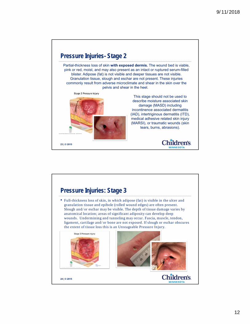
23 | © 2015
Pressure Injuries- Stage 2 Partial-thickness loss of skin with exposed dermis. The wound bed is viable, pink or red, moist, and may also present as an intact or ruptured serum-filled
blister. Adipose (fat) is not visible and deeper tissues are not visible. Granulation tissue, slough and eschar are not present. These injuries
commonly result from adverse microclimate and shear in the skin over the pelvis and shear in the heel.
This stage should not be used to describe moisture associated skin
damage (MASD) including incontinence associated dermatitis
(IAD), intertriginous dermatitis (ITD), medical adhesive related skin injury (MARSI), or traumatic wounds (skin
tears, burns, abrasions).
24 | © 2015
• Full-thickness loss of skin, in which adipose (fat) is visible in the ulcer and granulation tissue and epibole (rolled wound edges) are often present. Slough and/or eschar may be visible. The depth of tissue damage varies by anatomical location; areas of significant adiposity can develop deep wounds. Undermining and tunneling may occur. Fascia, muscle, tendon, ligament, cartilage and/or bone are not exposed. If slough or eschar obscures the extent of tissue loss this is an Unstageable Pressure Injury.
Pressure Injuries: Stage 3

9/11/2018
13
25 | © 2015
Staging pressure injuries in premature infants
Can this infant have a stage 2 pressure injury? If so, what would it look like?
What would a stage 3 pressure injury look like with very little subcutaneous tissue?
26 | © 2015
• Full-thickness skin and tissue loss with exposed or directly palpable fascia, muscle, tendon, ligament, cartilage or bone in the ulcer. Slough and/or eschar may be visible. Epibole (rolled edges), undermining and/or tunneling often occur. Depth varies by anatomical location. If slough or eschar obscures
the extent of tissue loss this is an Unstageable Pressure Injury.
Pressure Injuries: Stage 4

9/11/2018
14
27 | © 2015
• Full-thickness skin and tissue loss in which the extent of tissue damage within the ulcer cannot be confirmed because it is obscured by slough or eschar. If slough or eschar is removed, a Stage 3 or Stage 4 pressure injury will be revealed. Stable eschar (i.e. dry, adherent, intact without erythema or fluctuance) on the heel or ischemic limb should not be softened or removed.
Pressure Injuries- Unstagable
28 | © 2015
• Intact or non-intact skin with localized area of persistent non-blanchabledeep red, maroon, purple discoloration or epidermal separation revealing a dark wound bed or blood filled blister. Pain and temperature change often precede skin color changes. The wound may evolve rapidly to reveal the actual extent of tissue injury, or may resolve without tissue loss.
Pressure Injuries- Deep Tissue Pressure Injury
Do not use DTPI to describe vascular,
traumatic, neuropathic, or dermatologic
conditions.

9/11/2018
15
29 | © 2015
“Affected by microclimate, nutrition, perfusion, co-morbidities and condition of the soft tissue.”
30 | © 2015
• CPAP
• IV catheter hubs/StatLocks
• Fresh trachs and GTs
• vEEG
Medical Device-Related Pressure Injuries

9/11/2018
16
31 | © 2015
Deference to Expertise
WOCN- Expert on possible ways to off-load and cushion
Bedside nurse and medical team- expert on the patients
Respiratory therapist/vEEG techs/vascular access- expert on the
device
CNS- Expert on nursing process and policies
Nursing educator- Expert on knowledge communication to the bedside.
Informatics- Expert in clinical documentation
Medical device-related PI prevention
32 | © 2015
Joining Together
Share selfishly and steal shamelessly

9/11/2018
17
33 | © 2015
Nasal CPAP/Prongs
Return to the concept of microclimate… macerated skin is weak skin.
Can we remove the pressure?
Can we offload the pressure?
34 | © 2015
Bubble CPAP (bCPAP)

9/11/2018
18
35 | © 2015
Tracheostomy
Stage 3 HAPI
DTPI
36 | © 2015
Tracheostomy

9/11/2018
19
37 | © 2015
Tracheostomy
Which wound is pressure injury?
Should the wound care plan be different for the pressure injuryor can we use the same principles?
38 | © 2015
Video EEG Leads

9/11/2018
20
39 | © 2015
Bed Garbage
40 | © 2015
Congenital Pressure Injuries
“The injury occurs as a result of intense and/or prolonged pressure or pressure in combination with shear.”

9/11/2018
21
Moisture, Incontinence, Medical Adhesives, Friction, Chemical and thermal.
Everything Else…
42 | © 201542 | © 2015
“should not be used to describe moisture associated skin damage (MASD) including incontinence associated dermatitis (IAD), intertriginous dermatitis (ITD), medical adhesive related skin injury (MARSI), or traumatic wounds (skin tears, burns, abrasions).”
Remember….Pressure Injury

9/11/2018
22
43 | © 2015
Medical adhesive related skin injuries
Epidermal (skin) stripping
Tension blisters
Irritant contact dermatitis• Unusual in the neonatal period
Keep treatment simple. Treat epidermal stripping with frequent applications of petrolatum to soften crusting and facilitate healing.
Blisters usual flatten and heal without dressings.
Monitor for honey-colored crusting/drainage. The drainage may be yellow/green if infant has an elevated bilirubin level.
44 | © 2015
MARSI- Prevention
Do not use tackifiers
Protective barrier film under adhesives
Adhesive remover wipes
Use silicone tapes for non-lifelines
Consider backing aggressive tapes (or creating a landing-pad) with a hydrocolloid
Hydrogel EKG patches

9/11/2018
23
45 | © 2015
Intertriginois Dermatitis/Intertrigo
Moisture Associated Skin Damage (MASD)
May use antimicrobial moisture-wicking.
Consider topical antibiotic (mupirocin) if weeping/crusting is present.
46 | © 2015
MASD- Incontinence Associated Dermatitis
Irritant contact diaper dermatitis
Maceration
Denudement
Apply a thick layer of zinc oxide barrier paste

9/11/2018
24
47 | © 2015
Diaper Rash- YeastCandida
In skin folds
Satellite lesions and bright red patches
Check mouth and mother’s nipples for thrush
Treat with topical antifungal under barrier paste or a antifungal barrier ointment
Treat thrush
48 | © 2015

9/11/2018
25
49 | © 2015
Candida in the preterm infant
Hint: If this were a mucous membranesurface, what would you guess?
50 | © 2015
IV Infiltration and Extravasation• Infiltration: inadvertent administration of nonvesicant
solution or medication into surrounding tissue
• Non-vesicant solution, depending on volume, can cause deep skin damage
• Extravasation: inadvertent administration of vesicant solution or medication into surrounding tissue.
• In pediatric patients, occurs frequently due to immature skin structures

9/11/2018
26
51 | © 2015
Venous and Arterial Line Complications
52 | © 2015
Antiseptic Skin Preparations
Sardesai, S., Kornacka, M., Walas, W., & Ramanathan, R.; Iatrogenic skin injury in the neonatal intensive care unit, The Journal of Maternal-Fetal and Neonatal Medicine, 2010
Parsada Lashkari, H. Chow, P. Godame, S. Aqueous 2% chlorhexidine-induced chemical burns in an extremely premature infant. ADC Fetal& Neonatal. http://fn.bmj.com/content/97/1/F64. Downloaded on 9/15/2017
Betadine & Alcohol
2% Aqueous CHG
CHG & Alcohol
CHG & Alcohol
9/11/2018
27
53 | © 2015
Antiseptic Skin Preparations
Alcohol preparations can cause skin necrosis.
Betadine carriers a high risk for iodine overload. Preterm infants at greatest risks for hypothyroidism and possible neurodevelopmental delays.
Chlorhexidine gluconate can cause skin burns. Potential for CHG toxicity??
_____________________________________________________________
Gentle application, with minimal scrubbing. Allow to dry completely. Do not allow to pool in skin folds. Consider removing the antiseptic solution after the procedure with sterile saline.
At Children’s Minnesota we use Betadine and sterile saline for babies for less than 28 weeks gestations. We then transition to CHG.
Burns are treated with Medical Grade Honey and silicone foam dressings
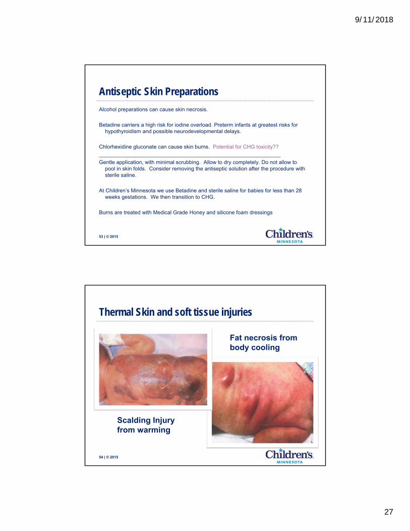
54 | © 2015
Thermal Skin and soft tissue injuries
Scalding Injury from warming
Fat necrosis from body cooling

9/11/2018
28
55 | © 2015
Skin scarring condition specific to extremely preterm infants, caused by traumatizing the dermis
Anetoderma of Prematurity
56 | © 2015
Anetoderma of Prematurity

9/11/2018
29
57 | © 2015
Anetoderma of Prematurity
58 | © 2015
DTI resulting in columellar scarring
Anetoderma of Prematurity
How are we staging PIs in premature infants?

9/11/2018
30
Birth TraumaForceps, vacuum, and other
60 | © 2015
Neonatal Birth Trauma
Birth related skin injuries very rarely require wound care and most often heal without scarring or alopecia.
Leave crusting as a biologic dressing and allow to heal spontaneously. Dressings offer poor wound coverage due to being suspended over the scalp by the hair. Also become tacky and gummy in the hair.

9/11/2018
31
61 | © 2015
Neonatal Birth Trauma
Why are these NOT pressure injuries?
62 | © 2015
Neonatal Birth Trauma
For full thickness wounds, use Bactroban to prevent infection. Otherwise allow to heal spontaneously. Can result in scarring and may require Plastics

9/11/2018
32
Congenital Wounds
64 | © 2015
Aplasia Cutis-
Congenital absence of skin

9/11/2018
33
65 | © 2015
Giant Omphalocele
Neonatal Ostomies

9/11/2018
34
67 | © 2015
Congenital anamalies
Imperforate Anus
Hirschprungs disease
Cloacal defect
______________________
Complications of prematurity and infectionNecrotizing Enterocolitis (NEC)
Spontaneous intestinal perforation
Common reasons for GI ostomies
68 | © 2015
Goals- Same as adults
Protect Surrounding Skin
Control Drainage
Patient Comfort
Attitudes and perspectives
Ostomy Care

9/11/2018
35
69 | © 2015
Types of Ostomies
End Stoma
Loop Stoma Double-Barrel Stoma
70 | © 2015
Barrier Ring
- Start soft and flexible
Elastic Barrier strips
- Babies are abdominal
breathers and require flexible barriers
Integrated gas filter
-Babies are gassy!
Helpful Accessories

9/11/2018
36
71 | © 2015
Product Considerations
One stoma or two (or three or more!)
Type of ostomy: some barriers limit cutting surface
Size of the abdomen: parents will want to start small, but this may not always be in their best interest.
Type of Effluent: for high stomas, consider a urostomy pouch with a high endurance barrier.
Ostomy Care
72 | © 2015
Application Pearls
Dry Skin
Warm barrier before and after placement
Trouble Shooting
Fill creases
Creases change as baby grows
Read the back of the barrier
Ostomy Care- Neonatal Pitfalls

9/11/2018
37
73 | © 2015
Before pouching…
74 | © 2015
Pouching example with two barriers

9/11/2018
38
75 | © 2015
Pouching example
76 | © 2015
Thank You

9/11/2018
39
77 | © 2015
References•Lund C, Kuller J, Lane A, Lott JW, Raines D. Neonatal skin care: the scientific basis for practice. JOGNN. 1999;28: 241-254.•Kalia Y, Nonato L, Lund C, Guy R. Development of skin barrier function in preterm infants. J Invest Dermatol. 1998: 111; 320-326. •Fluhr JW, Darlenski R, Taieb A, Hachem J-P, Baudouin C, Msika P, De Belilovsky C, Berardesca E. Functional skin adaptation in infancy- almost complete but not fully complete. Experimental Dermatology. 2010; 19: 483-492.•Fluhr JW, Darlenski R, Lachmann N, Baudouin C, Msika P, De Belilovsky C, Hachem J-P. Infant epidermal skin physiology: adaptation after birth. Br J Dermatol. 2012; 166: 483-490.•Lund C. Medical adhesives in the NICU. Newborn & Infant Nursing Reviews. 2014; 14:160-165.•Goujon E, Beer F, Gay S, Sandre D, Gouyon J-B, Vabres P. Anetoderma of prematurity: An iatrogenic consequences of neonatal intensive care. Arch Dermatol. 2010; 5: 565-567.•Maffeis L, Pugni L, Pietrasanta C, Ronchi A, Fumagalli M, Gelmetti C, Mosca F. Iatrogenic anetoderma of prematurity: a case report and review of the literature. Case Reports in Dermatological Medicine. 2014; Article ID 781493, 4 pages.•Boralevi F. Hubiche T. Leaute-Labreze C, Saubusse E, Fayon M, Roul S, Maurice-Tison S, Taieb A. Epicutaneous aeroallergen sensitization in atopic dermatitis infants- determining the role of epidermal barrier impairment. Allergy. 2008; 63: 205-210. •Spergel J, Paller A, Atopic dermatitis and the atopic march. J Allery Clin Immunol. 2003; 112:S118-27.•Chang M, Nakrani R, Six children with allergic contact dermatitis to methylisothiazolinone in wet wipes (baby wipes). Pediatrics. 2014; 133(2): e434-e438.•Ponnusamy V, Venkatesh V, Clarke P. Skin antisepsis in the neonate: what should we use? Curr Opin Infectious Disease. 2014; 27: 244-250.•Chapman A, Aucott S, Gilmore M, Advani S, Clarke W, Milstone A. Absorption and tolerability of aqueous chlorhexidine gluconate used for skin antisepsis prior to catheter insertion in preterm neonates. J Perinatol. 2013; 33(10):768-771.•O’Grady NP, Alexander M, Burns LA, et al. Guidelines for the prevention of intravascular catheter-related infections. Healthcare infection control practices advisory committee. http://www.cdc.gov/hicpac/pdf/guidelines/bsi-guidelines-2011.pdf. Published 2011. Accessed July 7, 2016.•Chapman A, Aucutt S, Milstone A. Safety of chlorhexidine gluconate used for skin antisepsis in the preterm infant. J Pernitol. 2012; 32: 4-9.•Aitken J, Williams F, A systematic review of thyroid dysfunction in preterm neonates exposed to topical iodine. Arch Dis Child Fetal Neontal Ed. 2014; 99: F21-F28.•Kiechl-Kohlendorfer U, Berger C, Inzinger R. The effect of daily treatment with an olive oil/lanolin emollient on skin integrity in preterm infants: a randomized controlled trial. Pediatric Dermatology. 2008; 25 (2): 174-178.• Connor JM, Soll R, Edwards WH. Topical ointment for preventing infection in preterm infants. The Cochrane Collaboration. 2009, Issue 3. •Cleminson J, McGuire W. Topical emollient for preventing infection in the preterm infant. The Cochrane Collbaoration. 2016, Issue 1, Art Nu: CD001150•Robertson, AF. Reflections on Errors in Neonatology: I. The “Hands-off “ years, 1920 to 1950. J of Perinatology. 2003; 23:48-55•Robertson, AF. Reflections on Errors in Neonatology: II. The “Heroic” Years, 1950 to 1970. J of Perinatology. 2003 23:154-161