
NORMAL LABOR AND DELIVERY Mrs.Mahdia Samaha Kony.
90
NORMAL LABOR AND DELIVERY Mrs.Mahdia Samaha Kony
-
Upload
felix-carpenter -
Category
Documents
-
view
225 -
download
3
Transcript of NORMAL LABOR AND DELIVERY Mrs.Mahdia Samaha Kony.

NORMAL LABOR AND DELIVERY
Mrs.Mahdia Samaha Kony

Definition
Labor is the process by which contractions of the gravid uterus expel the fetus.
A term pregnancy delivers between 37 and 42 weeks from the last menstrual period (LMP).
Preterm labor is that occurring before 37 weeks of gestational age.
Postdate pregnancy occurs after 42 weeks gestation and requires careful monitoring.
Termination of pregnancy before 20 weeks of gestation is defined as either spontaneous or elective abortion.

Definition
Primigravida - pregnant for first time Multigravida - pregnant more than once Viability - able to survive outside the
womb (24+ weeks gestation) Nulliparous - never carried a pregnancy
to viability Multiparous - has had two or more
deliveries that were carried to viability

The initiation of labor
Labor is influenced by combination of factors include:
- Uterine stretching- Progesterone withdrawal- increased oxytocin sensitivity- increased level of prostaglandins

Theories that explain initiation of labor:
Change in estrogen-to- progesterone ratio. Which facilitate coordination of uterine contraction and myometrium stretching.
Prostaglandin level increase in late pregnancy secondary to elevated level of estrogen. It stimulates smooth muscle contraction of the uterus: Leads to myometrium contraction Reduce cervical resistance The cervix becomes soft, thin out and dilate
during labor.

Theories that explain initiation of labor:
Increased number of oxytocin receptors late in pregnancy, this increased the sensitivity to oxytocin as its also increased in response to estrogen rising.
Oxytocin also aid in stimulation of prostaglandin synthesis in the decidua.

Oxytocin effect
The hormone oxytocin stimulates and enhances labor contractions. As the baby moves toward the vagina (birth canal), pressure receptors within the cervix (muscular outlet of uterus) send messages to the brain to produce oxytocin.
Oxytocin travels to the uterus through the bloodstream, stimulating the muscles in the uterine wall to contract stronger (increase of ideal normal value).
The contractions intensify increase until the baby is outside the birth canal.
When the stimulus to the pressure receptors ends, oxytocin production stops and labor contractions cease.


Premonitory signs of labor Cervical changes
softening and dilation with descent of the presenting part into the pelvic. This stage occurs one month to one hour before actual labor.
The cervix becomes shortened and thinned segment


Premonitory signs of labor Lightening: occurs when the fetal presenting
part begins to descend into the maternal pelvic. The uterus lowers and moves into a more anterior position. this change will cause:
Breathing becomes easier Increased pelvic pressure Cramping and low backache Lower extremities edema Increased vaginal secretion More frequent urination
In PG it occurs 2 weeks ore more before labor. In MP it occurs during labor

Premonitory signs of labor Increased energy level : many women will
focus this energy in preparation by cleaning, cooking, preparing the nursery…it is usually occur 24-48 hours before labor.
Bloody show: the mucus plug of the cervical canal during pregnancy is expelled as a result of cervical softening and increased pressure of the presenting part. The exposed cervical capillaries release a small amount of blood that mix with the mucus, resulting in bloody show.

Premonitory signs of labor Braxton Hicks Contraction: these
contractions aid in moving the cervix from the posterior position to the anterior position, they also help in ripining and softening of the cervix.
The contractions are irregular and diminished by walking, voiding, eating, increasing fluid intake, or changing position.

Premonitory signs of labor Spontaneous rupture of membrane: one in four
women experience SROM before onset of labor. This reduces the capacity of the uterus, thickens the uterine wall, and increases uterine irritability. Labor usually follows.
At term, 90% will be in labor within 24 h after membrane rupture.
If labor does not begin in 24 h, the case must be considered complicated by prolonged rupture of the membranes because of the increased risk of ascending infection.
Risk of cord prolapses is increased if engagement of the presenting part not occur.

True versus false labor:
Differentiating True Labor and False Labor
Factors True labor False labor
Contractions timing
Regular intervals, becoming close together, usually 4-6 minutes apart, lasting 30-60 seconds.
Irregular intervals, not occurring close together
Contraction strength
Becomes stronger with time, vaginal pressure is usually felt Frequently weak, not getting strong with time
Contraction discomfort
Start in the back and radiates around toward the front of the abdomen Usually felt in the front of the abdomen
Position changes Contractions continue no matter what positional changes is made Contraction may stop or slow down with walking or changing position
Effect of analgesia Not terminated by sedation Frequently abolished by sedation
Cervical change Progressive effacement and dilation No change

Factors affecting the labor process:
Labor entails the interaction of the so-called 5Ps:
Passageway( birth canal) Passenger( fetus and placenta) Power( contractions) Position( maternal) Psychological rersponse
Passageway:
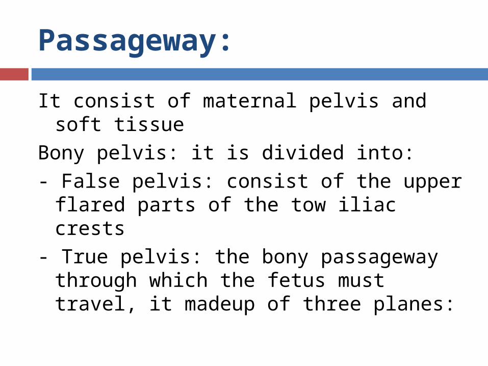
It consist of maternal pelvis and soft tissueBony pelvis: it is divided into:- False pelvis: consist of the upper flared
parts of the tow iliac crests - True pelvis: the bony passageway
through which the fetus must travel, it madeup of three planes:

Passageway:
1- Pelvic inlet: allow entrance to the true pelvis. Is measured clinically by attempting to touch the sacral promontory with the vaginal examining finger while simultaneously noting where the inferior border of the symphysis touches the examining finger. A measurement >12 cm suggests adequacy.

Passageway:
2- Mid pelvis: the mid pelvis (cavity) occupies the space between the inlet and outlet.
The interspinous diameter is estimated by palpating the ischial spines. An estimated distance >9 cm suggests midpelvis contraction. Experience is required to estimate this diameter with accuracy.

Passageway:
3- Outlet The outlet is limited anteriorly by the arch
of the symphysis pubis, posteriorly by the tip of the coccyx, and laterally by the ischial tuberosities.
This transverse diameter of the outlet can be estimated by placing a clenched fist between the two ischial tuberosities. A measurement of 8 cm or more suggests an adequate diameter.


Pelvic shape:
Anthropoid pelvis: is common in men and occurs in 20-30% of women. This pelvis is usually favorable for a vaginal delivery.
Android pelvis: common in men and occurs approximately in 20% of women, it has a heart shape inlet with narrow side walls. It is called a funnel pelvis and produces a difficult vaginal delivery.
Gynecoid pelvis: is less common in men and is considered the true female pelvis, although only about half of all women have this type, vaginal birth is most favorable with this type.
Platypelloid or flat pelvis is the least common type of pelvic structure among men and women with incidence of 5%. Women with this pelvis require C/S.


The soft tissue
The cervix (effacement and dilation): Effacement: How thin is the cervix (in cm or %)
The pelvic floor muscles: help the fetus to rotate anteriorly as it passes
through the birth canal.The vagina: the soft tissue of the vagina
expands to accommodate the fetus during birthing.

Passenger
a. Fetal skull: is the largest presenting part and least compressible fetal structure, making it an important factor in relation to labor and birth.
Bones – 6 bones: S – sphenoid, F – frontal – sinciput, E – ethmoid, O –occuputal – occiput, T – temporal, P – parietal 2 x
Measu rement fetal head:1. transverse diameter – 9.25cm- biparietal – largest transverse- bitemporal 8 cm2. bimastoid 7cm smallest transverse

Passenger
Sutures – intermembranous spaces that allow molding.
1.) Sagittal suture – connects 2 parietal bones 2.) Coronal suture – connect parietal & frontal
bone 3.) Lambdoidal suture – connects occipital &
parietal boneMoldings: the overlapping of the sutures of
the skull to permit passage of the head to the pelvis

Passenger
Fontanels:1.) Anterior fontanel – bregma, diamond
shape, 3 x 4 cm, (> 5 cm – hydrocephalus), 12 – 18 months after birth- close
2.) Posterior fontanel or lambda – triangular shape, 1 x 1 cm.
closes – 2 – 3 months after birth

Passenger
Anteroposterior diameter 1. suboccipitobregmatic 9.5 cm, complete
flexion, smallest AP2. occipitofrontal 12cm partial flexion3. occipitomental – 13.5 cm hyper
extension submentobragmatic-face presentation

Fetal attitude
Is another important consideration related to passenger.
It refers to the posturing (flexion or extension) of the joints and the relationship of fetal parts to one another.

Fetal Lie
The relationship of the long axis of the fetus to the long axis of the mother. The lie is longitudinal with a vertex or breech presentation or otherwise transverse or oblique, as with a shoulder presentation

Fetal Presentation
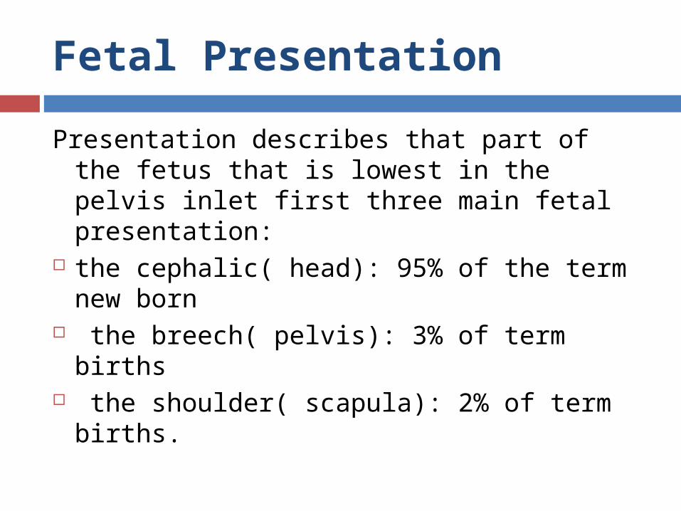
Presentation describes that part of the fetus that is lowest in the pelvis inlet first three main fetal presentation:
the cephalic( head): 95% of the term new born
the breech( pelvis): 3% of term births the shoulder( scapula): 2% of term
births.

Fetal Presentation
In cephalic presentation: the presenting part is usually the occiput portion of the fetal head. Vertex presentation is most common: the portion of the head that is covered by wearing beani cap.
Brow or face presentation is a variation on vertex, but with deflexion of the fetal head, allowing the brow or face to enter the pelvis first.

Fetal Presentation
In a breech presentation, the fetal buttocks (the breech) are the presenting part. The breech presentation has several variations:
Frank breech: the fetal legs are extended above the fetal pelvis with the breech as the presenting part
Complete breech: the feet and buttocks present together
Single-footling breech: one leg/foot is extended and presenting
Double-footling breech: both legs/feet are extended and presenting.


Although all abnormal presentations have an increased incidence of cord prolapse, footling breeches are especially at risk.
A shoulder presentation implies a transverse lie.
Compound presentations (e.g., vertex and an extremity together) rarely are seen with term pregnancies.
The position of the presenting part is best determined by vaginal

Fetal position:
Position of the presenting part is described as the relationship between a certain landmark on the fetal presenting part and the maternal pelvis, as follows:
Anterior, closest to the symphysis Posterior, closest to the coccyx Transverse, closest to the left or right vaginal
sidewall.

The index landmarks are: In a vertex presentation is the occiput,
which is identified by palpating the lambdoid sutures forming a Y with the sagittal sutureThe sacrum in a breech presentationthe mentum (or chin) in a face presentation

The index landmarks are:
The designations of anterior, posterior, left, and right refer to the maternal pelvis. Therefore, right occiput transverse implies the occiput is directed toward the right side of the maternal pelvis.
Breech and face presentations are described in a similar fashion (e.g., right sacrum transverse, right mentum transverse).


Fetal engagement:
Engagement is determined by pelvic examination. It occurs tow weeks before term in primigravida and several weeks before the onset of labor or not until before labor beginStation refers to the relationship between the fetal presenting part and pelvic landmarks.

Fetal engagement:
When the presenting part is at zero station, it is at the level of the ischial spines, which are the landmarks for the midpelvis. This is important in the vertex presentation because it implies that the largest dimension of the fetal head, the biparietal diameter, has passed through the smallest dimension of the pelvis, the pelvic inlet.

Fetal engagement:
In 1988, the American College of Obstetricians and Gynecologist introduced a classification dividing the pelvis into 5-cm segments above and below the spines: If the presenting part is 1 cm above the spines, it is
described as -1 station. If it is 2 cm below the spines, the station is +2. At -5 station, the presenting part is described as floating. At +5 station, the presenting part is on the perineum,
and it may distend the vulva with a contraction and be visible to an observer.


Cardinal movement of labor:
The process of labor and delivery is marked by characteristic changes in fetal position or cardinal movements in relation to the maternal pelvis. These spontaneous adjustments are made to effect efficient passage through the pelvis as the fetus descends.

Cardinal movement of labor:
Engagement is the descent of the largest transverse diameter, the biparietal diameter, to a level below the pelvic inlet. An occiput below the ischial spines is engaged.
Descent of the head is a discontinuous process occurring throughout labor. Because the transverse diameter of the pelvic inlet is wider than the AP diameter, and because the greatest diameter of the unflexed fetal head is the AP diameter, in most instances the fetus enters the pelvis in an occiput transverse alignment.


Cardinal movement of labor:
Flexion decreases the AP diameter of the fetal head. It occurs as the head encounters the levator muscle sling, thereby decreasing the diameter by approximately 1.5 to 2.5 cm (occipitomental, 12.0 cm, to occipitofrontal, 10.5 cm). Later, further flexion occurs, reducing the diameter to 9.5 cm (suboccipitobregmatic).

Cardinal movement of labor:
Internal rotation occurs in the midpelvis. The architecture of the midpelvic passageway changes so that the AP diameter of the maternal pelvis at this level is greater than the transverse diameter. The fetus accommodates to this change by rotation of the head from a transverse orientation (occiput transverse) to an AP alignment (usually occiput anterior), thus accomplishing internal rotation. Further descent to the level of the perineum occurs with the head aligned in the AP plane.

Cardinal movement of labor:
Extension of the head allows delivery of the head from the usual occiput anterior position through the introitus. The face appears over the perineal body Occurs once fetus has descended to the level
of the introitus Base of occiput in contact with inferior margin
of symphysis pubis

Cardinal movement of labor: External rotation occurs after delivery
of the head, when the fetal head rotates back, or restitutes, toward the original transverse orientation (external rotation or restitution) when the bisacromial diameter (fetal shoulders) is aligned in an AP orientation with the greatest diameter of the pelvic outlet.

Cardinal movement of labor: Expulsion: The remainder of the
delivery proceeds with presentation of the anterior shoulder beneath the symphysis pubis and the posterior shoulder across the posterior fourchette


The Powers
Forces generated by uterine musculature Frequency, amplitude, and duration of ctx’s Observation, manual palpation,
tocodynamometry, intrauterine pressure catheter (IUPC)
Contractions cause complete dilation and effacement of the cervix.

Uterine contraction:
Uterine contraction is involuntary and there fore cannot be controlled by the experiencing women.ut. Cont. is intermittent and rhythmic with a period of relaxation. Uterine cont.has three phases:
Increment: building up of the contraction Acme: peak or highest intensity Decrement: descent or relaxation of the
uterine muscle fibers


Parameters of uterine contraction: Interval
10 to 20 minutes between contractions: early labor
3 to 5 minutes between contractions: late labor Duration
20 second long contraction: early labor 40 to 80 second long contraction: late labor
Quality Uterus can be dented (poor quality): early labor Uterus is hard (good quality): late labor

Intra-abdominal pressure: Increased intra-abdominal pressure
(voluntary muscle contraction) compresses the uterus and adds to the power of the expulsion forces of the uterine contraction.

Psychological responses:
The birth experience influence the woman's self confidence, self esteem, and her view of life, her relationships, and her children.
Factors influencing a positive birth experience include: clear information on procedure positive support, not being alone sense of mastery, self- confidence trust in staff caring for her positive reaction to the pregnancy personal control over breathing Preparation for childbirth experience.

Maternal position:
Changing positions and moving around during birth offer several benefits, it facilitate fetal descend and rotation
Squatting position enlarges the pelvic outlet by approximately 25% .
The use of upright or lateral position compared with supine or lithotomy positions may:
reduce the duration of the second stage of labor reduce the number of assisted
deliveries( vacuum and forceps)

Maternal position:
reduce episiotomies and perineal tear contribute to fewer abnormal fetal heart increase comfort/ reduce request for pain
medication enhance a sense of control reported by
mothers alert the shape and size of the pelvis,
which assist descent assist gravity to move the fetus downward reduce the length of labor

Physiologic responses to labor:

Maternal responses:
Increased heart rate by 10 to 18 bpm Increased cardiac output by l 0 to 15% during the
first stage of labor and increased by 30 to 50% during the second stage of labor
Increased blood pressure by 10 to 30 mm Hg during uterine contractions in all labor stages
Increase in white blood cell count to 25,000 to 30,000 cells/mm3 perhaps as a result of tissue trauma
Increased respiratory rate along with greater oxygen consumption related to the increase in metabolism

Maternal responses:
Decreased gastric motility and food absorption, which may increase the risk of nausea and vomiting during the transition stage of labor
Decreased gastric emptying and gastric pH which, which increase the risk of vomiting and aspiration
Slight elevation in temperature possibly as a result of an increase in muscle activity.
Muscular aches/cramps as a result of a stressed musculoskeletal system involved in the labor process.
Increased BMR and decreased blood glucose level because of the stress of labor.

Fetal responses
1. Periodic fetal heart rate accelerations and slight decelerations related to fetal movement, fundal pressure, and uterine contractions.
2. Decrease in circulation and perfusion to the fetus secondary to uterine contractions.
3. Increased in arterial carbon dioxide pressure(PCO2)
4. Decrease in fetal breathing movements throughout labor.
5. Decrease in fetal oxygen pressure with a decrease in the partial pressure of oxygen (PO2).

Stages of labor:
The first stage of labor begins with the onset of labor and ends with complete (10 cm) dilatation of the cervix.
Duration of the first stage: The first stage is the longest, averaging 8–12 h for
primigravidas or 6–8 h for multiparas. However, the first stage of labor may be markedly
shorter or longer depending on the 4Ps. Labor is a very dynamic process, and contractions
should increase steadily in regularity, intensity, and duration. This is not always the case, and one must set limits concerning the progress of labor.

Phases of the first stage labor:
Latent phase of labor begins with the onset of regular uterine contractions and extends to the start of the active phase of cervical dilatation (_0-3 cm).
Contractions may or may not be painful( mild) Cervical effacement from 0-40% Dilate very slowly Can talk or laugh through contractions May last days or longer May be treated with sedation, hydration, ambulation,
rest, or pitocin Nullipara lasting up to 9 hours, multipara lasting up to
5 to 6 hours. prolonged latent phase: defined as greater than 20
hours in a nullipara and greater than 14 hours in a parous woman

Phases of the first stage labor: Active phase of labor: lasts from 4 to 7 cm
dilation, moderate contractions. Regular, frequent, usually painful contractions cervical dilation rate of 1.2 cm/hr for nulliparas
and 1.5 cm/hr for parous women cervical effacement 40 to 80% nullipara lasting lasting up to 6 hours, multipara
lasting up to 4 hours. Contraction frequency every 2 to 5 min. Contraction duration 45 to 60 seconds. Are not comfortable with talking or laughing
during their contractions

Phases of the first stage labor:
Transition phase: is from 8 to 10cm dilation, strong uterine contraction. Cervical effacement from 80 to 100% Nullipara lasting up to 1 hour, multi Para
lasting up to 30 minutes. Contraction duration 60 to 90 seconds Contraction intensity hard by palpation.

Stages of labor
(2) The second stage of labor begins when the cervix becomes fully dilated and ends with the complete birth of the infant. The second stage normally lasts up to 1 hour. While one should be concerned when the second stage extends longer than 1 h (based on fetal morbidity and mortality). Safety for the fetus may be assured by thoughtful monitoring.

The second stage of labor
Pelvic phase: period of fetal descend Perineal phase: period of active pushing Nullipara lasts up to 1 hour, multipara
lasts up to 30 minutes. Contraction frequency every 2 to 3
minutes or less Contraction duration 60 to 90 seconds. Strong urge to push in perineal phase.

Stages of labor
(3) The third, or placental, stage of labor is the period from birth of the infant to 1 h after delivery of the placenta. The rapidity of separation and means of recovery of the placenta determine the duration of the third stage
(4) Fourth stage of labor: is 1 to 4 hours after birth, time of maternal physiologic adjustment.

ADMISSION PROCEDURE
-One of the most critical diagnoses in obstetrics is the accurate diagnosis of labor:
History Physical examination: Fundal height measurement Uterine contraction (duration, frequency, intensity) fetus (presentation, heart rate, size) fetal membrane, vaginal bleeding & leakage The fetal heart rate should be checked, especially at the
end of a contraction and immediately, thereafter, to identify pathological slowing of the heart rate
Pain level

Laboratory studies:
CBC Blood type and RH UA (pretein, glucose) Syphilis, hepatitis B, HIV

Management the fist stage of labor: Ambulation OK with intact membranes If in bed, lie on one side or the other…
not flat on her back Check vital signs every 4 hours (if
membrane rupture or high temperature: hourly)

Oral intake
- food should be withheld during active labor and delivery
- in labor & analgesics are administered :gastric emptying time is prolonged :not absorbed ,vomited, and aspiration -sips of clear liquids, occasional ice chips, and lip
moisturizers are permitted
Intravenous fluids -there is seldom any real need for such in the
normally pregnant at least until analgesia is administered

During early labor, for low risk patients, note the fetal heart rate every 1-2 hours.
During active labor, evaluate the fetal heart every 30 minutes

Urinary bladder function
-bladder distention should be avoided : obstructed labor subsequent bladder hypotonia and
infection -ambulation: self voiding, if not, intermittent catheterization


Evaluation of labor progress: Vaginal examination:
Dilatation and effacement of the cervix: Rupture of membrane Fetal descent and presenting part
Uterine contraction Leopold's Maneuvers

Leopold’s Maneuver
Purpose: is done to determine the attitude, fetal presentation lie, presenting part, degree of descent, an estimate of the size, and number of fetuses, position, fetal back & fetal heart tone
- use palm! Warm palm.Prep mother:1. Empty bladder2. Position of mother in supine with knee
flex (dorsal recumbent – to relax abdominal muscles)
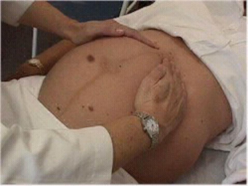
1st maneuver:
place patient in supine position with knees slightly flexed; put towel under head and right hip; with both hands palpate upper abdomen and fundus. Assess size, shape, movement and firmness of the part to determine presentation

2nd Maneuver:
with both hands moving down, identify the back of the fetus ( to hear fetal heart sound) where the ball of the stethoscope is placed to determine FHT. Get V/S(before 2nd maneuver) PR to diff: Uterine soufflé – maternal H rate


3rd Maneuver:
using the right hand, grasp the symphis pubis part using thumb and fingers. To determine degree of engagement.
Assess whether the presenting part is engaged in the pelvis )Alert : if the head is engaged it will not be movable).


4th Maneuver:
the Examiner changes the position by facing the patient’s feet. With two hands, assess the descent of the presenting part by locating the cephalic prominence or brow. To determine attitude – relationship of fetus to one another.


