Nonspecific interstitial pneumonia ... - PneumologieNonspecific interstitial pneumonia. Diagnosis,...
19
Nonspecific interstitial pneumonia. Diagnosis, differential diagnosis and treatment. Martina Vašáková, Martina Šterclová Department of Respiratory Medicine 1st Medical School Charles University Thomayer Hospital The presentation is supported by grant IGA MZČR 1207 and 1302
Transcript of Nonspecific interstitial pneumonia ... - PneumologieNonspecific interstitial pneumonia. Diagnosis,...
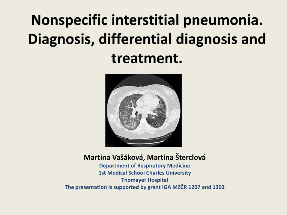
Nonspecific interstitial pneumonia. Diagnosis, differential diagnosis and
treatment.
Martina Vašáková, Martina Šterclová Department of Respiratory Medicine 1st Medical School Charles University
Thomayer Hospital The presentation is supported by grant IGA MZČR 1207 and 1302
Idiopathic pulmonary fibrosis and nonspecific interstitial pneumonia
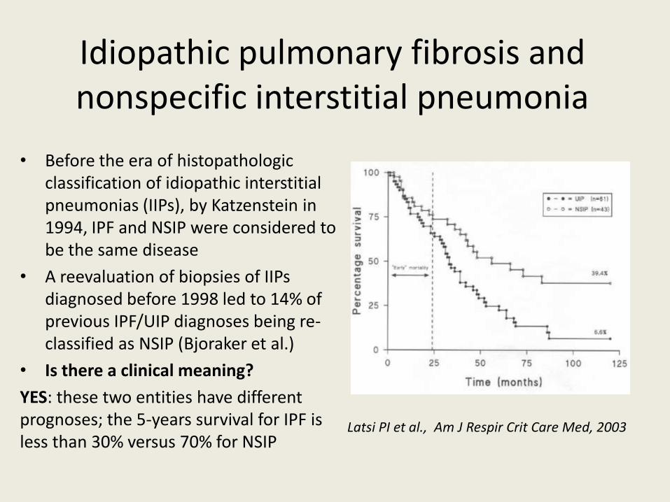
• Before the era of histopathologic classification of idiopathic interstitial pneumonias (IIPs), by Katzenstein in 1994, IPF and NSIP were considered to be the same disease
• A reevaluation of biopsies of IIPs diagnosed before 1998 led to 14% of previous IPF/UIP diagnoses being re-classified as NSIP (Bjoraker et al.)
• Is there a clinical meaning?
YES: these two entities have different prognoses; the 5-years survival for IPF is less than 30% versus 70% for NSIP
Latsi PI et al., Am J Respir Crit Care Med, 2003
Are there any differences in etiopathogenesis in IPF and NSIP?
• Nonspecific reactions of lung tissue to both external and internal injury are present in disposed individual. Effects depends on underlying conditions/disease
• Neovascularization is substantially greater in IPF than in NSIP – accompanied by greater expression of VEGF-A mRNA and MMP-2 mRNA Takahashi et al., Pathol Int 2013
• Proteomic analysis - UIP vs. NSIP – involves different subtypes of vimentin Ohara et al., Histol Histopathol 2013
History of NSIP • NSIP was first described as a unique subunit of idiopathic
interstitial pneumonias in 1994 Katzenstein, Fiorelli- ATS/ERS International Multidisciplinary
Consensus Classification of Idiopathic Interstitial Pneumonias, Am J Respir Crit Care Med 2002
• NSIP was previously considered as provisory, i.e. autoimmune undifferentiated connective tissue disease (UCTD)
duBois R, King TE Challenges in pulmonary fibrosis. 5: The NSIP/UIP debate, Thorax 2005
• 2008- “There is now sufficient data to justify several clinicoradiological syndromes for NSIP; these include NSIP with a IPF-like profile or overlap (NSIP/IPF), NSIP with an organized pneumonia profile (NSIP/OP) and NSIP with a hypersensitivity profile (NSIP/HP)“ Wells AU, Hirani N on behalf of BTS ILDs guideline group, Thorax 2008
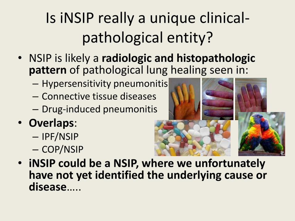
Is iNSIP really a unique clinical- pathological entity?
• NSIP is likely a radiologic and histopathologic pattern of pathological lung healing seen in: – Hypersensitivity pneumonitis – Connective tissue diseases – Drug-induced pneumonitis
• Overlaps: – IPF/NSIP – COP/NSIP
• iNSIP could be a NSIP, where we unfortunately have not yet identified the underlying cause or disease…..
If iNSIP exists, how is it diagnosed? • Multidisciplinary approach
Flaherty K et al Am J Respir Crit Care Med 2004
• Optimally: a HRCT NSIP pattern, a histopathologic NSIP pattern and exclusion of possible causes of NSIP
• Histopathological findings might reveal a UIP pattern in HRCT NSIP pattern or a NSIP pattern in HRCT UIP pattern
• Unrepresentative biopsy (unrevealed UIP) are also possible
• !!! We must exclude other diseases with a NSIP healing pattern, which seem to be fairly common
iNSIP vs. NSIP as a manifestation of other diseases
• Connective tissue diseases (CTDs) – the NSIP pattern more common than the UIP, i.e. systemic sclerosis, rheumatoid arthritis, Sjogren syndrome, UCTD Nakamura Y et al. Sarcoidosis Vasc Diffuse Lung Dis 2003
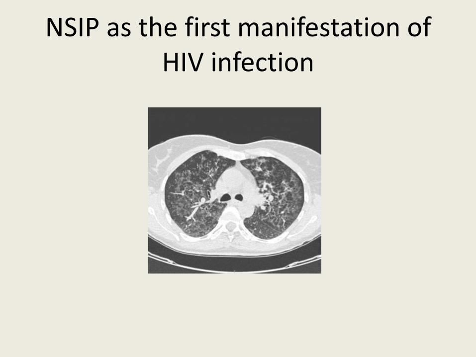
• Hypersensitivity pneumonitis (HP) • Drug-induced ILDs • Systemic infections - HIV- LIP and NSIP- mainly before antiretroviral
treatment Doffman SR et al Clin Chest Med 2013
• Immunopathologic states – IgG4 autoimmune disease - high IgG4 concentrations, sclerosing
inflammation with numerous IgG4+ plasma cells with frequent autoimmune pancreatitis, sclerosing sialoadenitis retroperitoneal fibrosis, sclerosing cholangitis
Takat H et al. Intern Med 2008
– hypo and dysgammaglobulinemia - CVID
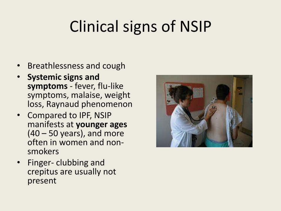
Clinical signs of NSIP
• Breathlessness and cough • Systemic signs and
symptoms - fever, flu-like symptoms, malaise, weight loss, Raynaud phenomenon
• Compared to IPF, NSIP manifests at younger ages (40 – 50 years), and more often in women and non-smokers
• Finger- clubbing and crepitus are usually not present
Laboratory and functional workup
• Laboratory findings- autoantibodies (Aab) can be detected – AECA- anti-endothelial Aab – present in ≈ 50% of patients with
iNSIP and NSIP in CTDs (Matsui et al., Respir Med 2008) – ANA – present in 34.5% patients with NSIP - a titre higher than
1:320 predicts, with high probability, further evolution of CTDs (RR 6.4), but has no influence on prognosis
– RF is present in 13.2% patients with NSIP – Other AAbs are present in 0.7- 6.8% patients with NSIP (Kang BH
et al J Korean Med Sci 2013) – Anti-HSP 47 Aab – has a higher titre in NSIP compared to IPF and
is more frequent in fibrotic NSIP versus cellular NSIP ( Kakugawa T et al BMC Pulm Med 2008)
• Functional parameters – has a similar pattern as that seen in IPF, i.e. a restrictive ventilatory pattern with impairment of diffusion capacity
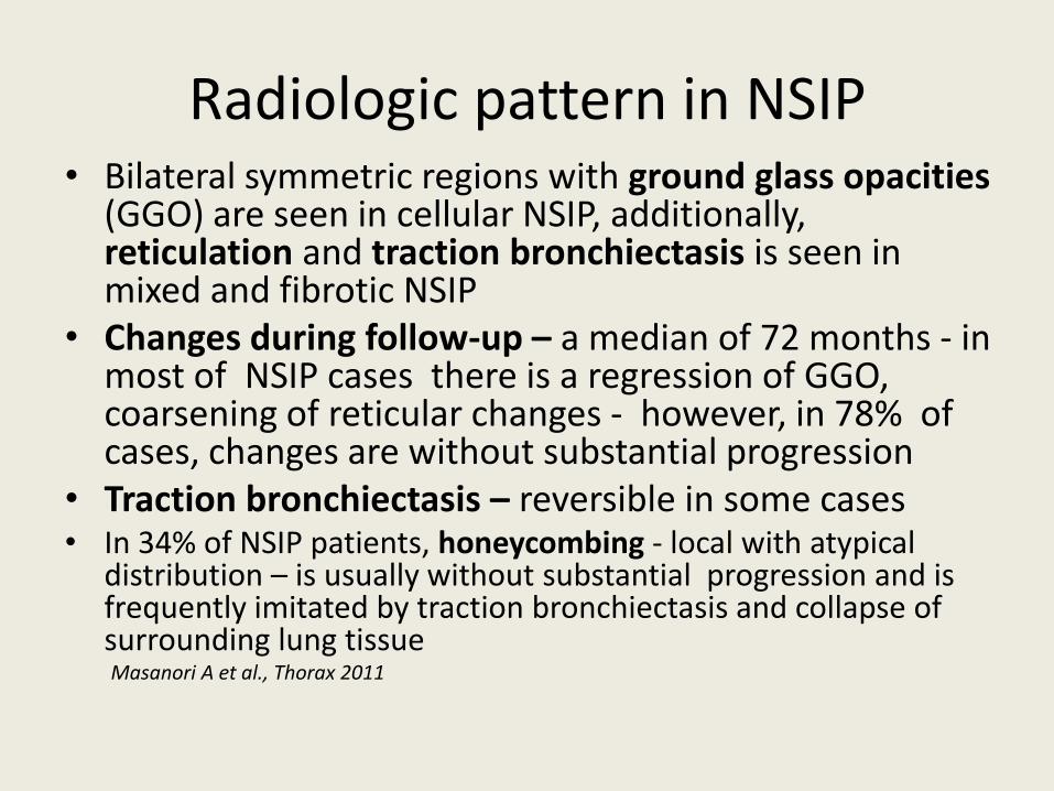
Radiologic pattern in NSIP • Bilateral symmetric regions with ground glass opacities
(GGO) are seen in cellular NSIP, additionally, reticulation and traction bronchiectasis is seen in mixed and fibrotic NSIP
• Changes during follow-up – a median of 72 months - in most of NSIP cases there is a regression of GGO, coarsening of reticular changes - however, in 78% of cases, changes are without substantial progression
• Traction bronchiectasis – reversible in some cases • In 34% of NSIP patients, honeycombing - local with atypical
distribution – is usually without substantial progression and is frequently imitated by traction bronchiectasis and collapse of surrounding lung tissue Masanori A et al., Thorax 2011
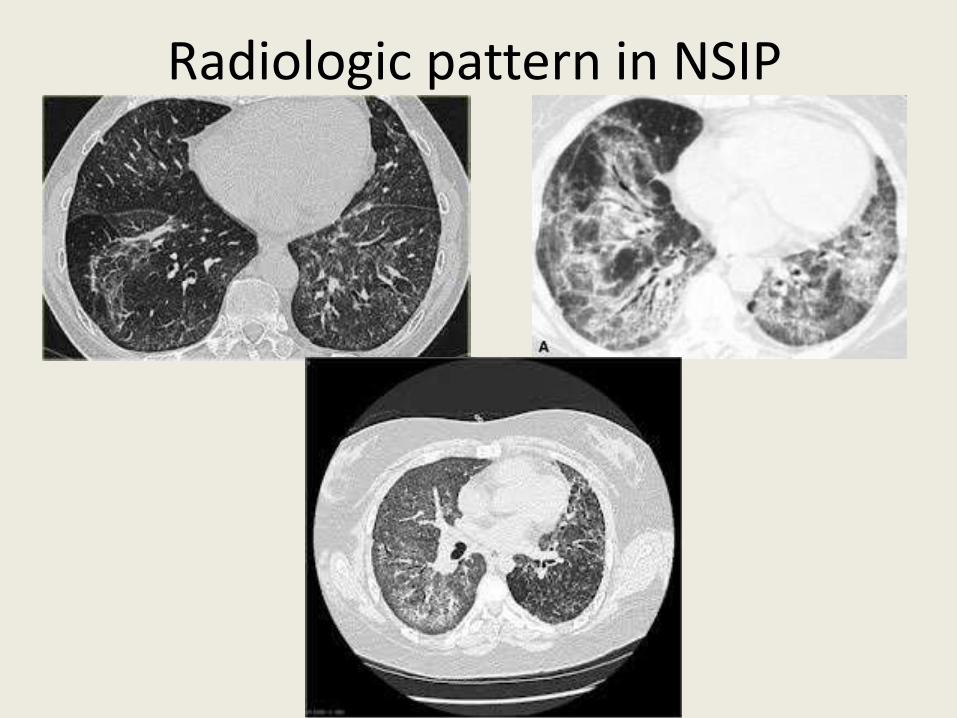
Radiologic pattern in NSIP
NSIP as the first manifestation of HIV infection
Findings in bronchoalveolar lavage fluid (BALF)
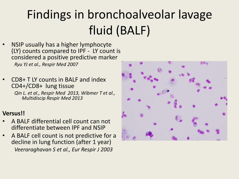
• NSIP usually has a higher lymphocyte (LY) counts compared to IPF - LY count is considered a positive predictive marker
Ryu YJ et al., Respir Med 2007
• CD8+ T LY counts in BALF and index
CD4+/CD8+ lung tissue Qin L. et al., Respir Med 2013, Wibmer T et al.,
Multidiscip Respir Med 2013
Versus!! • A BALF differential cell count can not
differentiate between IPF and NSIP • A BALF cell count is not predictive for a
decline in lung function (after 1 year) Veeraraghavan S et al., Eur Respir J 2003
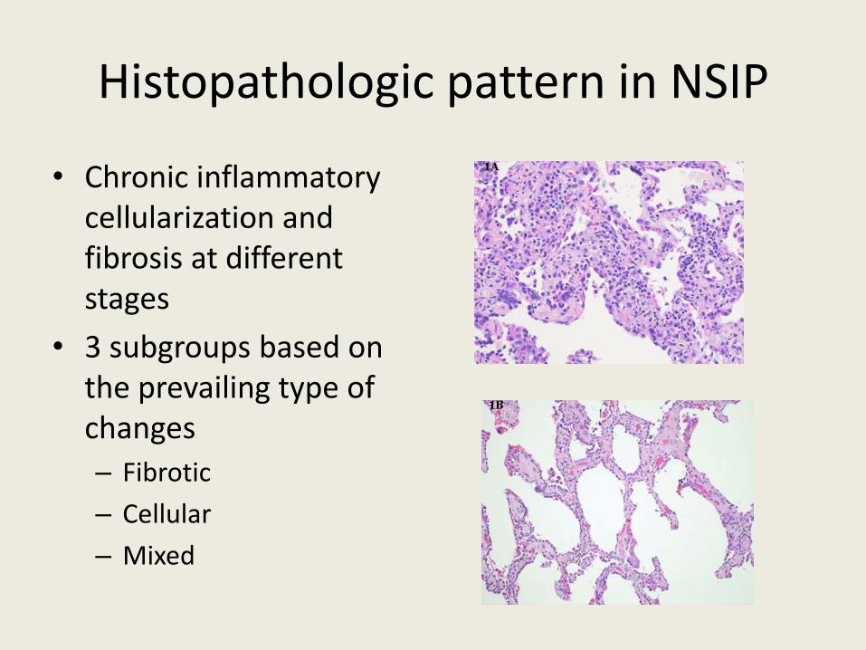
Histopathologic pattern in NSIP
• Chronic inflammatory cellularization and fibrosis at different stages
• 3 subgroups based on the prevailing type of changes
– Fibrotic
– Cellular
– Mixed
Diagnosis of iNSIP
• Diagnosis of iNSIP is always a diagnosis per exclusion
• Corresponding clinical, HRCT, and histopathological phenotype
• Exclusion of: – Connective tissue diseases
– Lung disorders in immunocompromised host (HIV, IgG4, CVID)
– Drug-induced pneumonitis
– Hypersensitivity pneumonitis
• Sometimes patients with iNSIP can also present with CTD during their disease course
Treatment
• iNSIP usually responds well to antiinflammatory treatment, i.e. corticosteroids (prednisone 0,5 mg/kg with subsequent tapering) or eventually in combination with cytotoxic drugs and immunosuppressants (azathioprine 150 mg p.o./day)
• Cellular NSIP responds better to treatment than fibrotic NSIP
• In cases of NSIP with CTDs, combined immunosuppressive treatment with cyclophosphamide (2,5 mg/kg/day or pulses 600 mg i.v. /month) should be considered
• ???Antibifibrotic treatment » Wells AU, Hirani N. Interstitial lung disease guideline: the British Thoracic Society
in collaboration with the Thoracicc Society of Australia and Hew Zealand and the Irish Thoracic Society, Thorax 2008
Prognosis
• Even though iNSIP and NSIP in CTDs look similar, NSIP in CTDs usually responds better to corticosteroid treatment and has a better prognosis than iNSIP
• Patients with cellular iNSIP have a substantially better prognosis than those with mixed or fibrotic NSIP
• In patients with fibrotic NSIP, the extent of fibrosis is one of the most important negative predictors for survival
• 5-years survival in NSIP is 80% and 10-years survival is 73% • IPF/UIP vs. fibrotic NSIP – 5 year survival is 43% vs. 90%,
and 10 year survival is 15% vs. 35% • Cellular iNSIP - 5 and 10 year survival is 100%
Travis WD et al., Am J Surg Pathol 2000
??
• Is iNSIP really a clinical and pathological entity, or only a pattern of healing common in different pulmonary and systemic disease?
• Can fibrotic NSIP be treated by antifibrotic treatment?

Thank you for your kind attention