Noninvasive Ventilation Weaning in Acute Hypercapnic...
11
Research Article Noninvasive Ventilation Weaning in Acute Hypercapnic Respiratory Failure due to COPD Exacerbation: A Real-Life Observational Study Paola Faverio , 1 Anna Stainer, 1 Federica De Giacomi, 1 Grazia Messinesi, 1 Valentina Paolini, 1 Anna Monzani, 1 Paolo Sioli, 1 Irdi Memaj, 1 Oriol Sibila, 2 Paolo Mazzola, 3 and Alberto Pesci 1 1 School of Medicine and Surgery, University of Milan Bicocca, Respiratory Unit, San Gerardo Hospital, ASST Monza, Monza, Italy 2 Respiratory Department, Hospital de La Santa Creu I Sant Pau, Autonomous University of Barcelona and Biomedical Research Institute Sant Pau (IIB Sant Pau), Barcelona, Spain 3 School of Medicine and Surgery, University of Milan Bicocca, Acute Geriatrics Unit, San Gerardo Hospital, ASST Monza, Monza, Italy Correspondence should be addressed to Paola Faverio; [email protected] Received 27 September 2018; Revised 1 February 2019; Accepted 26 February 2019; Published 25 March 2019 Academic Editor: Pierachille Santus Copyright © 2019 Paola Faverio et al. is is an open access article distributed under the Creative Commons Attribution License, which permits unrestricted use, distribution, and reproduction in any medium, provided the original work is properly cited. e most recent British oracic Society/Intensive Care Society (BTS/ICS) guidelines on the use of noninvasive ventilation (NIV) in acute hypercapnic respiratory failure (AHRF) suggest to maximize NIV use in the first 24 hours and to perform a slow tapering. However, a limited number of studies evaluated the phase of NIV weaning. e aim of this study is to describe the NIV weaning protocol used in AHRF due to acute exacerbation of chronic obstructive pulmonary disease (AE-COPD), patients’ characteristics, clinical course, and outcomes in a real-life intermediate respiratory care unit (IRCU) setting. We performed a retrospective study on adult patients hospitalized at the IRCU of San Gerardo Hospital, Monza, Italy, from January 2015 to April 2017 with a diagnosis of AHRF due to COPD exacerbation. e NIV weaning protocol used in our institution consists of the interruption of one of the three daily NIV sessions at the time, starting from the morning session and finishing with the night session. e 51 patients who started weaning were divided into three groups: 20 (39%) patients (median age 80 yrs, 65% males) who completed the protocol and were discharged home without NIV (Completed Group), 20 (39%) did not complete it because they were adapted to domiciliary ventilation (Chronic NIV Group), and 11 (22%) interrupted weaning ex abrupto mainly due to NIV intolerance (Failed Group). Completed Group patients were older, had a higher burden of comorbidities, but a lower severity of COPD compared to Chronic NIV Group. Failed Group patients experienced higher frequency of delirium after NIV discontinuation. None of the patients who completed weaning had AHRF relapse during hospitalization. While other NIV weaning methods have been previously described, our study is the first to describe a protocol that implies the interruption of a ventilation session at the time. e application of a weaning protocol may prevent AHRF relapse in the early stages of NIV interruption and in elderly frail patients. 1. Introduction Bilevel noninvasive ventilation (NIV) is a milestone in the treatment of acute hypercapnic respiratory failure (AHRF) in patients with acute exacerbation of chronic obstructive pulmonary disease (AE-COPD) [1, 2]. Unfortunately, only a limited number of studies evaluated the weaning from NIV once acidosis and respiratory distress are solved, proposing different weaning methods but not clearly establishing the superiority of one over the others [3–6]. A recently pub- lished randomized controlled trial (RCT) questioned the utility of the weaning phase [5]. In this RCT, Sellares et al. compared the prolongation of NIV for three nights after recovery from an AHRF episode to direct NIV Hindawi Canadian Respiratory Journal Volume 2019, Article ID 3478968, 10 pages https://doi.org/10.1155/2019/3478968
Transcript of Noninvasive Ventilation Weaning in Acute Hypercapnic...
Research ArticleNoninvasive Ventilation Weaning in Acute HypercapnicRespiratory Failure due to COPD Exacerbation: A Real-LifeObservational Study
Paola Faverio ,1 Anna Stainer,1 Federica De Giacomi,1 Grazia Messinesi,1
Valentina Paolini,1 Anna Monzani,1 Paolo Sioli,1 Irdi Memaj,1 Oriol Sibila,2
Paolo Mazzola,3 and Alberto Pesci1
1School of Medicine and Surgery, University of Milan Bicocca, Respiratory Unit, San Gerardo Hospital, ASST Monza,Monza, Italy2Respiratory Department, Hospital de La Santa Creu I Sant Pau,Autonomous University of Barcelona and Biomedical Research Institute Sant Pau (IIB Sant Pau), Barcelona, Spain3School of Medicine and Surgery, University of Milan Bicocca, Acute Geriatrics Unit, San Gerardo Hospital, ASST Monza,Monza, Italy
Correspondence should be addressed to Paola Faverio; [email protected]
Received 27 September 2018; Revised 1 February 2019; Accepted 26 February 2019; Published 25 March 2019
Academic Editor: Pierachille Santus
Copyright © 2019 Paola Faverio et al. (is is an open access article distributed under the Creative Commons Attribution License,which permits unrestricted use, distribution, and reproduction in any medium, provided the original work is properly cited.
(e most recent British(oracic Society/Intensive Care Society (BTS/ICS) guidelines on the use of noninvasive ventilation (NIV)in acute hypercapnic respiratory failure (AHRF) suggest to maximize NIV use in the first 24 hours and to perform a slow tapering.However, a limited number of studies evaluated the phase of NIV weaning. (e aim of this study is to describe the NIV weaningprotocol used in AHRF due to acute exacerbation of chronic obstructive pulmonary disease (AE-COPD), patients’ characteristics,clinical course, and outcomes in a real-life intermediate respiratory care unit (IRCU) setting. We performed a retrospective studyon adult patients hospitalized at the IRCU of San GerardoHospital, Monza, Italy, from January 2015 to April 2017 with a diagnosisof AHRF due to COPD exacerbation. (e NIV weaning protocol used in our institution consists of the interruption of one of thethree daily NIV sessions at the time, starting from the morning session and finishing with the night session. (e 51 patients whostarted weaning were divided into three groups: 20 (39%) patients (median age 80 yrs, 65%males) who completed the protocol andwere discharged home without NIV (Completed Group), 20 (39%) did not complete it because they were adapted to domiciliaryventilation (Chronic NIV Group), and 11 (22%) interrupted weaning ex abrupto mainly due to NIV intolerance (Failed Group).Completed Group patients were older, had a higher burden of comorbidities, but a lower severity of COPD compared to ChronicNIV Group. Failed Group patients experienced higher frequency of delirium after NIV discontinuation. None of the patients whocompleted weaning had AHRF relapse during hospitalization.While other NIV weaning methods have been previously described,our study is the first to describe a protocol that implies the interruption of a ventilation session at the time. (e application of aweaning protocol may prevent AHRF relapse in the early stages of NIV interruption and in elderly frail patients.
1. Introduction
Bilevel noninvasive ventilation (NIV) is a milestone in thetreatment of acute hypercapnic respiratory failure (AHRF)in patients with acute exacerbation of chronic obstructivepulmonary disease (AE-COPD) [1, 2]. Unfortunately, only alimited number of studies evaluated the weaning from NIV
once acidosis and respiratory distress are solved, proposingdifferent weaning methods but not clearly establishing thesuperiority of one over the others [3–6]. A recently pub-lished randomized controlled trial (RCT) questioned theutility of the weaning phase [5]. In this RCT, Sellares et al.compared the prolongation of NIV for three nights afterrecovery from an AHRF episode to direct NIV
HindawiCanadian Respiratory JournalVolume 2019, Article ID 3478968, 10 pageshttps://doi.org/10.1155/2019/3478968
discontinuation in COPD patients without previous do-miciliary ventilation [5]. No differences were observed inregard to prevention of AHRF relapse, hospital readmission,or mortality [5]. Furthermore, the application of a weaningphase resulted in a prolonged intermediate respiratory careunit (IRCU) stay. (ese results seem to suggest that NIV canbe directly discontinued once the acute episode is solved.However, as in all RCTs, very elderly patients, not fullycooperative and with several comorbidities, have rarely beenincluded, although they often represent a consistent part ofthe COPD population with AHRF admitted to IRCUs [5]. Insuch a frail group, many factors may play a role in AHRFrelapse after NIV discontinuation, including other re-spiratory comorbidities, delirium, and sarcopenia [7].(erefore, the actual need of weaning from NIV in a real-lifecohort of patients with AHRF and AE-COPD and the bestway to perform it still need to be determined.
Our aim is to present and test the feasibility of the NIVweaning strategy used in patients with AHRF due to AE-COPD admitted in our IRCU. Furthermore, we describe thecharacteristics, clinical course, and outcomes of the cohort ofpatients who underwent the NIV weaning protocol.
2. Materials and Methods
2.1. Study Design. (is was a retrospective cohort study onadult patients hospitalized in the IRCU of San GerardoHospital, Monza, Italy, from January 2015 to April 2017 witha diagnosis of AE-COPD. (e study was approved by thelocal Institutional Review Board (#684-May 2017) and didnot require patients’ informed consent due to the retro-spective design.
(e following data were retrospectively collected frompatients’ charts: demographics, comorbidities and Charlsoncomorbidity index [8], COPD characteristics (includingpulmonary function tests performed at the previous avail-able outpatient visit during clinical stability, number ofexacerbations and hospitalizations during the prior year, andCOPD phenotype—emphysema, chronic bronchitis,asthma-COPD overlap, and bronchiectasis-COPD overlap[9, 10]), severity scores at admission (acute physiology andchronic health disease classification system II (APACHE II)[11] and sequential organ failure assessment (SOFA) [12]),radiological imaging during hospitalization (either chestX-ray, computerized tomography scan, or both), arterialblood gases (ABG), days of NIV application and hospitallength of stay, presence of delirium during hospitalization,and NIV parameters. Follow-up data, including three-month mortality and hospitalization rate, were also retro-spectively collected from outpatient clinic charts. All pa-tients discharged from our IRCU with a diagnosis of AE-COPD were followed-up after 3months from discharge atour COPD outpatient clinic; those who did not present at thevisit received a phone call to reprogram it.
2.2. AE-COPD Definition. AE-COPD was defined as acuteworsening of respiratory symptoms that resulted in addi-tional therapy, including antibiotics, systemic
corticosteroids, and increased bronchodilator therapy [13].In our study population, AE-COPD was the primary ad-mission diagnosis according to the physician in charge.Patients who also had pneumonia or congestive heart failureas secondary diagnoses were not excluded from the study,and these variables were reported in the Results section.
2.3. NIV Initiation Criteria and Indication to Chronic Do-miciliary NIV. In accordance with the protocol adopted byour institution, bilevel NIV was initiated in patients withAHRF (arterial carbon dioxide partial pressure (PaCO2)> 45mmHg) due to AE-COPD in case of respiratory acidosis(pH≤ 7.35) and/or respiratory distress. Respiratory distresswas defined as the respiratory rate (RR)> 25 associated withuse of respiratory accessory muscles. In case of pH< 7.25,decision regarding NIV initiation was shared with the in-tensive care unit (ICU) team.
All patients were ventilated with pressure supportventilation modality. (e ventilators available in our IRCUwere Respironics Esprit and Resmed Elisee 150. Full face andoronasal interfaces were used.
In our institution, indications for domiciliary NIV inCOPD patients were persistent daytime hypercapnia after atleast two episodes of AHRF requiring NIV in the prior year;and comorbidities such as obstructive sleep apnea orobesity-hypoventilation syndrome, in accordance withguidelines for each disease [1, 14, 15].
2.4. NIV Disconnection and NIVWeaning Initiation Criteria.NIV disconnection was attempted when all the followingwere concurrently met:
(a) Adequate oxygenation defined as arterial oxygenpartial pressure to fraction of inspired oxygen ratio(PaO2/FiO2)> 200mmHg during NIV with fractionof inspired oxygen (FiO2)< 0.5
(b) pH> 7.35, RR< 25 without use of respiratory ac-cessory muscles
(c) Hemodynamic stability (assessed through heart rateand blood pressure)
(d) Kelly score [16]≤ 2
(e weaning phase began when all the following criteriawere reached after at least 1 hour of disconnection fromNIVand by administering oxygen through a venturi mask with aFiO2 titrated in order to maintain peripheral oxygen satu-ration (SpO2) 88–92%:
(a) pH> 7.35, RR< 25 without use of respiratory ac-cessory muscles
(b) Hemodynamic stability(c) Kelly score [16]≤ 2
2.5. NIV Weaning Protocol. (e weaning protocol used inour institution is summarized in Figure 1. (e term “NIVsession” was used to indicate the continuous application ofNIV for at least 3 hours to the maximum duration tolerated
2 Canadian Respiratory Journal
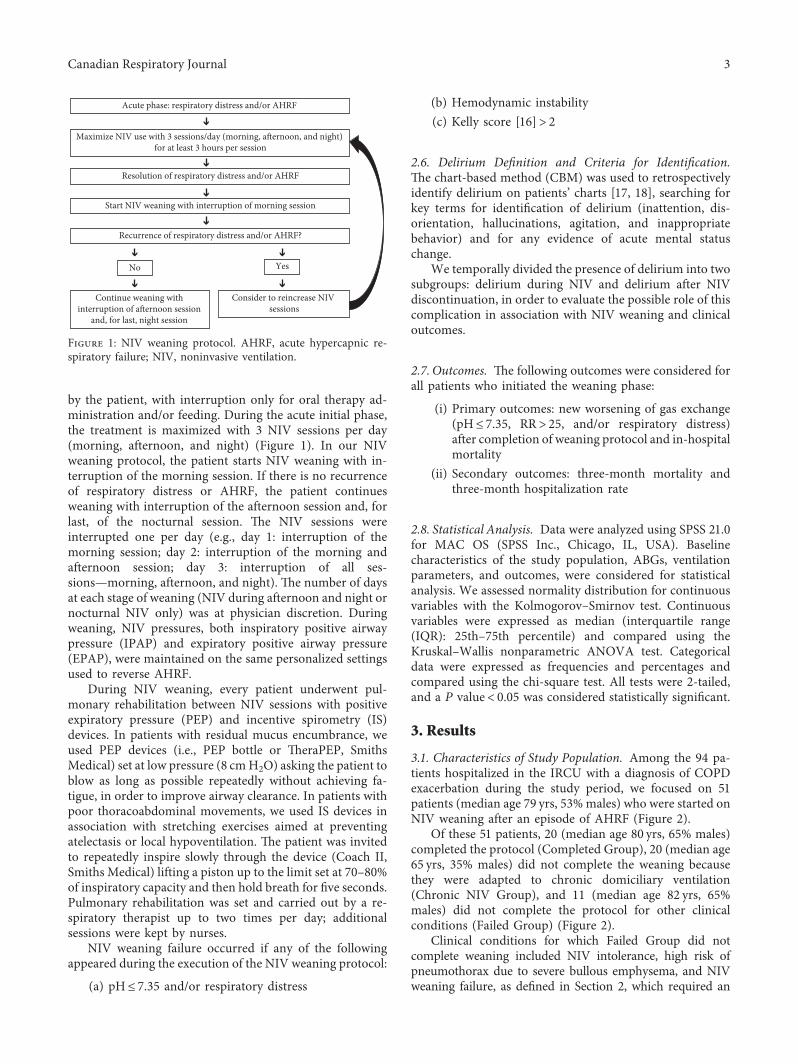
by the patient, with interruption only for oral therapy ad-ministration and/or feeding. During the acute initial phase,the treatment is maximized with 3 NIV sessions per day(morning, afternoon, and night) (Figure 1). In our NIVweaning protocol, the patient starts NIV weaning with in-terruption of the morning session. If there is no recurrenceof respiratory distress or AHRF, the patient continuesweaning with interruption of the afternoon session and, forlast, of the nocturnal session. (e NIV sessions wereinterrupted one per day (e.g., day 1: interruption of themorning session; day 2: interruption of the morning andafternoon session; day 3: interruption of all ses-sions—morning, afternoon, and night). (e number of daysat each stage of weaning (NIV during afternoon and night ornocturnal NIV only) was at physician discretion. Duringweaning, NIV pressures, both inspiratory positive airwaypressure (IPAP) and expiratory positive airway pressure(EPAP), were maintained on the same personalized settingsused to reverse AHRF.
During NIV weaning, every patient underwent pul-monary rehabilitation between NIV sessions with positiveexpiratory pressure (PEP) and incentive spirometry (IS)devices. In patients with residual mucus encumbrance, weused PEP devices (i.e., PEP bottle or (eraPEP, SmithsMedical) set at low pressure (8 cmH2O) asking the patient toblow as long as possible repeatedly without achieving fa-tigue, in order to improve airway clearance. In patients withpoor thoracoabdominal movements, we used IS devices inassociation with stretching exercises aimed at preventingatelectasis or local hypoventilation. (e patient was invitedto repeatedly inspire slowly through the device (Coach II,Smiths Medical) lifting a piston up to the limit set at 70–80%of inspiratory capacity and then hold breath for five seconds.Pulmonary rehabilitation was set and carried out by a re-spiratory therapist up to two times per day; additionalsessions were kept by nurses.
NIV weaning failure occurred if any of the followingappeared during the execution of the NIV weaning protocol:
(a) pH≤ 7.35 and/or respiratory distress
(b) Hemodynamic instability(c) Kelly score [16]> 2
2.6. Delirium Definition and Criteria for Identification.(e chart-based method (CBM) was used to retrospectivelyidentify delirium on patients’ charts [17, 18], searching forkey terms for identification of delirium (inattention, dis-orientation, hallucinations, agitation, and inappropriatebehavior) and for any evidence of acute mental statuschange.
We temporally divided the presence of delirium into twosubgroups: delirium during NIV and delirium after NIVdiscontinuation, in order to evaluate the possible role of thiscomplication in association with NIV weaning and clinicaloutcomes.
2.7. Outcomes. (e following outcomes were considered forall patients who initiated the weaning phase:
(i) Primary outcomes: new worsening of gas exchange(pH≤ 7.35, RR> 25, and/or respiratory distress)after completion of weaning protocol and in-hospitalmortality
(ii) Secondary outcomes: three-month mortality andthree-month hospitalization rate
2.8. Statistical Analysis. Data were analyzed using SPSS 21.0for MAC OS (SPSS Inc., Chicago, IL, USA). Baselinecharacteristics of the study population, ABGs, ventilationparameters, and outcomes, were considered for statisticalanalysis. We assessed normality distribution for continuousvariables with the Kolmogorov–Smirnov test. Continuousvariables were expressed as median (interquartile range(IQR): 25th–75th percentile) and compared using theKruskal–Wallis nonparametric ANOVA test. Categoricaldata were expressed as frequencies and percentages andcompared using the chi-square test. All tests were 2-tailed,and a P value< 0.05 was considered statistically significant.
3. Results
3.1. Characteristics of Study Population. Among the 94 pa-tients hospitalized in the IRCU with a diagnosis of COPDexacerbation during the study period, we focused on 51patients (median age 79 yrs, 53%males) who were started onNIV weaning after an episode of AHRF (Figure 2).
Of these 51 patients, 20 (median age 80 yrs, 65% males)completed the protocol (Completed Group), 20 (median age65 yrs, 35% males) did not complete the weaning becausethey were adapted to chronic domiciliary ventilation(Chronic NIV Group), and 11 (median age 82 yrs, 65%males) did not complete the protocol for other clinicalconditions (Failed Group) (Figure 2).
Clinical conditions for which Failed Group did notcomplete weaning included NIV intolerance, high risk ofpneumothorax due to severe bullous emphysema, and NIVweaning failure, as defined in Section 2, which required an
Acute phase: respiratory distress and/or AHRF
Maximize NIV use with 3 sessions/day (morning, afternoon, and night) for at least 3 hours per session
Resolution of respiratory distress and/or AHRF
Start NIV weaning with interruption of morning session
Recurrence of respiratory distress and/or AHRF?
No Yes
Continue weaning with interruption of afternoon session
and, for last, night session
Consider to reincrease NIV sessions
Figure 1: NIV weaning protocol. AHRF, acute hypercapnic re-spiratory failure; NIV, noninvasive ventilation.
Canadian Respiratory Journal 3
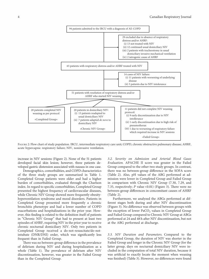
increase in NIV sessions (Figure 2). None of the 51 patientsdeveloped facial skin lesion; however, three patients de-veloped gastric distension associated with nausea in one case.
Demographics, comorbidities, and COPD characteristicsof the three study groups are summarized in Table 1.Completed Group patients were older and had a higherburden of comorbidities, evaluated through the Charlsonindex. In regard to specific comorbidities, Completed Grouppresented the highest frequency of cardiovascular diseases,while Chronic NIV Group showed more frequently obesity-hypoventilation syndrome and mood disorders. Patients inCompleted Group presented more frequently a chronicbronchitis phenotype and had a lower number of COPDexacerbations and hospitalizations in the prior year. How-ever, this finding is related to the definition itself of patientsin “Chronic NIV Group” that had to present at least twoepisodes of AHRF, requiring NIV in the prior year to receivechronic nocturnal domiciliary NIV. Only two patients inCompleted Group received a do-not-resuscitate/do-not-intubate (DNR/DNI) order, which was significantly lessfrequent than in Failed Group.
(ere was no between-group difference in the prevalenceof delirium during NIV and during hospitalization as awhole (Table 1). (e prevalence of delirium after NIVdiscontinuation, however, was greater in the Failed Groupthan in the Completed Group.
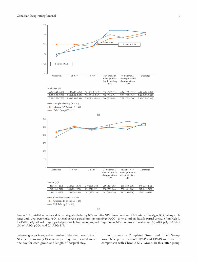
3.2. Severity on Admission and Arterial Blood GasesEvaluation. APACHE II score was greater in the FailedGroup compared to the other two study groups. In contrast,there was no between-group difference in the SOFA score(Table 2). Also, pH values of the ABG performed at ad-mission were lower in Completed Group and Failed Groupin comparison with Chronic NIV Group (7.30, 7.29, and7.35, respectively; P value<0.01) (Figure 3). (ere were nobetween-group differences in concomitant causes of AHRF(Table 2).
Furthermore, we analyzed the ABGs performed at dif-ferent stages both during and after NIV discontinuation(Figure 3). No difference was observed between groups withthe exception of lower PaCO2 values in Completed Groupand Failed Group compared to Chronic NIVGroup at ABGsperformed at 24 and 48 h after NIV discontinuation, but notat the ABG performed at discharge.
3.3. NIV Duration and Parameters. Compared to theCompleted Group, the duration of NIV was shorter in theFailed Group and longer in the Chronic NIV Group (for thelatter group, days on nocturnal domiciliary NIV were in-cluded in the computation of total NIV duration, because itwas artificial to exactly locate the moment when weaningwas finished) (Table 3). However, no differences were found
94 patients admitted to the IRCU with a diagnosis of AE-COPD
65 patients with respiratory distress and/or AHRF treated with NIV
51 patients with resolution of respiratory distress and/or AHRF who started NIV weaning
«Failed Group»
20 patients completed NIV weaning as per protocol
«Completed Group»
«Chronic NIV Group»
29 excluded due to absence of respiratorydistress and/or AHRF:
13 not treated with NIV12 continued usual domiciliary NIV2 patients with tracheostomy in usualdomiciliary invasive mechanical ventilation2 iatrogenic cause of AHRF
(i)(ii)
(iii)
14 cases of NIV failure: 11 patients with worsening of underlying disease3 patients due to NIV intolerance
(i)
11 patients did not complete NIV weaningprotocol:
9 early discontinuation due to NIV intollerance1 early discontinuation due to high risk ofpneumothorax1 due to worsening of respiratory failurewhich required increase in NIV sessions
(i)
(ii)
(iii)
20 patients in domiciliary NIV: 13 patients readapted to usual domiciliary NIV7 patients adapted de novo to domiciliary NIV
(i)
(ii)
(ii)
(iv)
Figure 2: Flow chart of study population. IRCU, intermediate respiratory care unit; COPD, chronic obstructive pulmonary disease; AHRF,acute hypercapnic respiratory failure; NIV, noninvasive ventilation.
4 Canadian Respiratory Journal
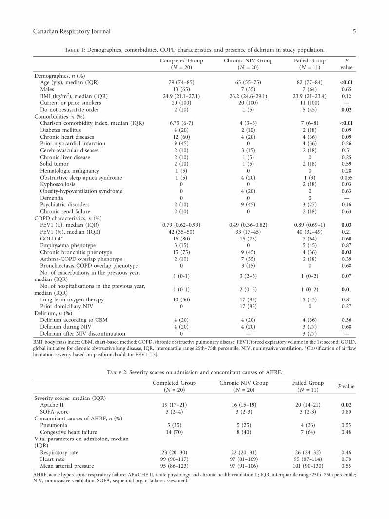
Table 1: Demographics, comorbidities, COPD characteristics, and presence of delirium in study population.
Completed Group(N � 20)
Chronic NIV Group(N � 20)
Failed Group(N � 11)
P
valueDemographics, n (%)Age (yrs), median (IQR) 79 (74–85) 65 (55–75) 82 (77–84) <0.01Males 13 (65) 7 (35) 7 (64) 0.65BMI (kg/m2), median (IQR) 24.9 (21.1–27.1) 26.2 (24.6–29.1) 23.9 (21–23.4) 0.12Current or prior smokers 20 (100) 20 (100) 11 (100) —Do-not-resuscitate order 2 (10) 1 (5) 5 (45) 0.02
Comorbidities, n (%)Charlson comorbidity index, median (IQR) 6.75 (6-7) 4 (3–5) 7 (6–8) <0.01Diabetes mellitus 4 (20) 2 (10) 2 (18) 0.09Chronic heart diseases 12 (60) 4 (20) 4 (36) 0.09Prior myocardial infarction 9 (45) 0 4 (36) 0.26Cerebrovascular diseases 2 (10) 3 (15) 2 (18) 0.51Chronic liver disease 2 (10) 1 (5) 0 0.25Solid tumor 2 (10) 1 (5) 2 (18) 0.59Hematologic malignancy 1 (5) 0 0 0.28Obstructive sleep apnea syndrome 1 (5) 4 (20) 1 (9) 0.055Kyphoscoliosis 0 0 2 (18) 0.03Obesity-hypoventilation syndrome 0 4 (20) 0 0.63Dementia 0 0 0 —Psychiatric disorders 2 (10) 9 (45) 3 (27) 0.16Chronic renal failure 2 (10) 0 2 (18) 0.63
COPD characteristics, n (%)FEV1 (L), median (IQR) 0.79 (0.62–0.99) 0.49 (0.36–0.82) 0.89 (0.69–1) 0.03FEV1 (%), median (IQR) 42 (35–50) 33 (17–45) 40 (32–49) 0.21GOLD 4∗ 16 (80) 15 (75) 7 (64) 0.60Emphysema phenotype 3 (15) 0 5 (45) 0.87Chronic bronchitis phenotype 15 (75) 9 (45) 4 (36) 0.03Asthma-COPD overlap phenotype 2 (10) 7 (35) 2 (18) 0.39Bronchiectasis-COPD overlap phenotype 0 3 (15) 0 0.68No. of exacerbations in the previous year,
median (IQR) 1 (0-1) 3 (2–5) 1 (0–2) 0.07
No. of hospitalizations in the previous year,median (IQR) 1 (0-1) 2 (0–5) 1 (0–2) 0.01
Long-term oxygen therapy 10 (50) 17 (85) 5 (45) 0.81Prior domiciliary NIV 0 17 (85) 0 0.27
Delirium, n (%)Delirium according to CBM 4 (20) 4 (20) 4 (36) 0.36Delirium during NIV 4 (20) 4 (20) 3 (27) 0.68Delirium after NIV discontinuation 0 — 3 (27) —
BMI, body mass index; CBM, chart-based method; COPD, chronic obstructive pulmonary disease; FEV1, forced expiratory volume in the 1st second; GOLD,global initiative for chronic obstructive lung disease; IQR, interquartile range 25th–75th percentile; NIV, noninvasive ventilation. ∗Classification of airflowlimitation severity based on postbronchodilator FEV1 [13].
Table 2: Severity scores on admission and concomitant causes of AHRF.
Completed Group(N � 20)
Chronic NIV Group(N � 20)
Failed Group(N � 11) P value
Severity scores, median (IQR)Apache II 19 (17–21) 16 (15–19) 20 (14–21) 0.02SOFA score 3 (2–4) 3 (2-3) 3 (2-3) 0.80
Concomitant causes of AHRF, n (%)Pneumonia 5 (25) 5 (25) 4 (36) 0.55Congestive heart failure 14 (70) 8 (40) 7 (64) 0.48
Vital parameters on admission, median(IQR)Respiratory rate 23 (20–30) 22 (20–34) 26 (24–32) 0.46Heart rate 99 (90–117) 97 (81–109) 95 (87–114) 0.78Mean arterial pressure 95 (86–123) 97 (91–106) 101 (90–130) 0.55
AHRF, acute hypercapnic respiratory failure; APACHE II, acute physiology and chronic health evaluation II; IQR, interquartile range 25th–75th percentile;NIV, noninvasive ventilation; SOFA, sequential organ failure assessment.
Canadian Respiratory Journal 5
Median (IQR)
100
90
80
70
60
50
40Admission 1h NIV 3h NIV 24h a�er NIV
interruption/1stday domiciliary
NIV
Discharge48h a�er NIVinterruption/2ndday domiciliary
NIV
52 (43–66) 66 (62–72) 65 (57–72) 64 (56–69) 61 (55–66) 63 (60–65)51 (46–60) 67 (61–70) 59 (52–73) 61 (56–63) 63 (55–68) 62 (59–67)53 (41–61) 65 (61–69) 63 (61–78) 62 (55–66) 67 (60–75) 60 (58–61)
Completed Group (N = 20)Chronic NIV Group (N = 20)Failed Group (N = 11)
(a)
Median (IQR)
Admission 1h NIV 3h NIV 24h a�er NIVinterruption/1stday domiciliary
NIV
Discharge48h a�er NIVinterruption/2ndday domiciliary
NIV
65 (58–73) 60 (51–69) 56 (47–66) 50 (48–53) 51 (47–54) 50 (45–54)63 (57–73) 62 (51–70) 63 (56–68) 62 (49–66) 57 (51–63) 53 (49–61)65 (54–78) 56 (46–68) 59 (43–65) 47 (43–51) 45 (44–56) 52 (48–55)
Completed Group (N = 20)Chronic NIV Group (N = 20)Failed Group (N = 11)
P value = 0.01
P value = 0.04
70
65
60
55
50
45
40
35
30
(b)
Figure 3: Continued.
6 Canadian Respiratory Journal
between groups in regard to number of days with maximizedNIV before weaning (3 sessions per day) with a median ofone day for each group and length of hospital stay.
For patients in Completed Group and Failed Group,lower NIV pressures (both IPAP and EPAP) were used incomparison with Chronic NIV Group. In this latter group,
Median (IQR)
Admission 1h NIV 3h NIV 24h a�er NIVinterruption/1stday domiciliary
NIV
Discharge48h a�er NIVinterruption/2ndday domiciliary
NIV
7.30 (7.26–7.34) 7.32 (7.29–7.36) 7.34 (7.32–7.38) 7.42 (7.39–7.46) 7.42 (7.40–7.44) 7.41 (7.39–7.43)7.35 (7.30–7.38) 7.35 (7.31–7.37) 7.34 (7.32–7.37) 7.38 (7.36–7.41) 7.39 (7.37–7.41) 7.41 (7.38–7.42)7.29 (7.27–7.35) 7.34 (7.31–7.38) 7.36 (7.31–7.42) 7.40 (7.36–7.42) 7.38 (7.33–7.40) 7.40 (7.36–7.44)
Completed Group (N = 20)Chronic NIV Group (N = 20)Failed Group (N = 11)
7.45
7.4
7.35
7.3
7.25
7.2
P value < 0.01
P value < 0.01P value = 0.02
(c)
Median (IQR)
Admission 1h NIV 3h NIV 24h a�er NIVinterruption/1stday domiciliary
NIV
Discharge48h a�er NIVinterruption/2ndday domiciliary
NIV
225 (185–287) 244 (214–269) 240 (208–282) 250 (227–292) 236 (230–270) 273 (228–296)217 (184–247) 255 (214–278) 233 (216–271) 250 (238–266) 252 (231–266) 263 (243–293)189 (132–223) 248 (224–290) 241 (225–339) 263 (214–280) 285 (200–226) 271 (218–321)
Completed Group (N = 20)Chronic NIV Group (N = 20)Failed Group (N = 11)
300
250
200
150
100
0
50
(d)
Figure 3: Arterial blood gases at different stages both during NIV and after NIV discontinuation. ABG, arterial blood gas; IQR, interquartilerange 25th–75th percentile; PaO2, arterial oxygen partial pressure (mmHg); PaCO2, arterial carbon dioxide partial pressure (mmHg); P/F�PaO2/FiO2, arterial oxygen partial pressure to fraction of inspired oxygen ratio; NIV, noninvasive ventilation. (a) ABG: pO2, (b) ABG:pH, (c) ABG: pCO2, and (d) ABG: P/F.
Canadian Respiratory Journal 7

NIV pressures used during the acute phase were similar tothose titrated for the chronic domiciliary NIV use, as shownin Table 3.
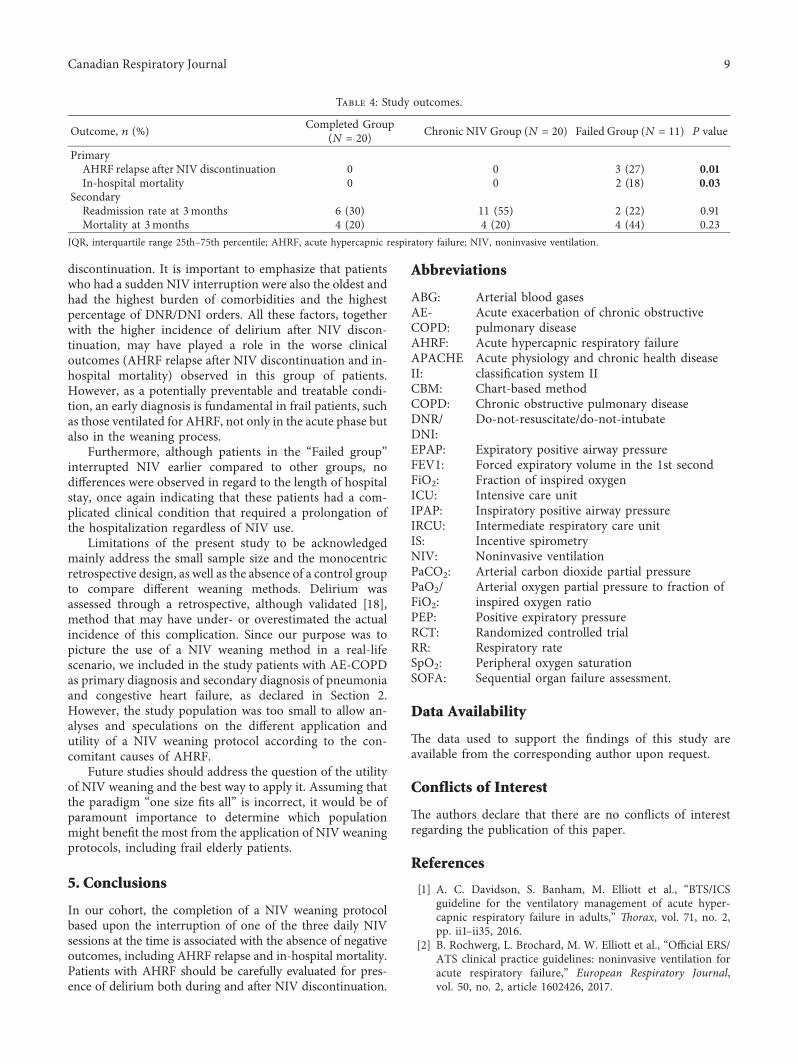
3.4. Outcomes. (ree patients, all in Failed Group, experi-enced relapse of AHRF during the first 48 hours from NIVdiscontinuation (P< 0.01), and two of them died during thehospitalization (both of them had received a DNR/DNIorder due to the severity of comorbidities) (Table 4). Nodifference was observed between groups in regard to sec-ondary outcomes.
4. Discussion
While other NIV weaning methods have been previouslydescribed [3–6], our study is the first to describe a protocolthat implies the interruption of a ventilation session at thetime. None of the patients who completed weaning expe-rienced AHRF relapse or died in the course of hospitali-zation. A considerable proportion of patients, particularlythose younger, with a lower burden of comorbidities, but ahigher severity of COPD, was initiated on chronic domi-ciliary NIV. Delirium should be taken into account amongthe factors that can complicate the clinical course of ven-tilated patients with AHRF due to AE-COPD even in theweaning phase.
Despite the wide range of studies available on weaningfrom invasive mechanical ventilation [19], few studies haveinvestigated the strategies to wean patients from NIV [3–6].Similarly to Damas et al., who described a weaning protocolthat maintains the same number of sessions per day butprovides a progressive reduction of hours for each NIVsession, we did not observe recurrence of AHRF duringhospitalization after weaning [4]. Two recently publishedRCTs by Sellares et al. and Lun et al. comparing immediatevs. stepwise NIV withdrawal did not show any benefit fromthe use of a weaning strategy in preventing relapse of AHRFafter NIV discontinuation, suggesting that NIV can be di-rectly discontinued in COPD patients once the episode ofAHRF is solved [5, 6]. However, when looking at theweaning phase itself, in our population, only one patient
failed the first weaning attempt because of worsening re-spiratory failure. In the studies by Sellares et al. and Lunet al., 19% and 44% of patients, respectively, in the directdiscontinuation groups failed the first withdrawal attempt[5, 6]. (ese percentages were higher than in the stepwisewithdrawal (5% and 26%, respectively) [5, 6]. In spite of thisvariable not being included among study outcomes, it isimportant to consider that the application of a weaningprotocol may prevent respiratory function deteriorationeven in the early stages of NIV interruption.
Furthermore, both the aforementioned studies recruitedonly fully cooperative patients with a limited burden ofcomorbidities. In a real-life setting, it is often necessary toapply NIV to elderly patients with a high burden ofcomorbidities (median Charlson comorbidity index in ourpopulation ranged between 4 and 7), not fully cooperative(as demonstrated by the prevalence of delirium in our co-hort) and who received a DNR/DNI order (16% of cases inour cohort). (ese frail patients often excluded from clinicaltrials could be the ones most benefiting from weaningprotocols.
Only a minority of patients in our study completed theweaning protocol, while a considerable proportion (39%)was initiated on chronic domiciliary NIV. (ese patientswere younger, with a lower burden of comorbidities but ahigher severity of COPD compared to patients who com-pleted weaning. (ese data suggest that patients who aremost likely to receive chronic domiciliary NIV are those witha longer life expectancy and more severe COPD.
According to the CBM, 23% of patients in our cohort hada diagnosis of delirium. A meta-analysis published in 2012reported a prevalence of delirium of 37% in patients on NIV,which was higher than the one generally presented inhospitalized nonventilated patients (10–31%) [20]. Fur-thermore, the risk of NIV failure in patients with deliriumwas 2.12 times higher than in those without delirium. Ourstudy groups showed similar prevalence of delirium duringNIV, whereas, after NIV discontinuation, patients whodiscontinued NIV ex abrupto showed a higher prevalencecompared to those who completed the weaning protocol.Wepostulate that delirium, on a case-by-case basis, might beboth a cause and a consequence of NIV intolerance and
Table 3: Hospital length of stay and NIV duration, NIV parameters, and presence of delirium.
Completed Group(N � 20)
Chronic NIV Group(N � 20)
Failed Group(N � 11) P value
Hospital length of stay and NIV duration,median (IQR)Hospital length of stay, days 10 (8–14) 11 (10–14) 11 (7–15) 0.76NIV duration, days 4 (3–6) 11 (10–14) 2 (1–3) <0.01Number of days with 3 NIV sessions 1 (1-2) 1 (0–3) 1 (0–3) 0.96Number of days with 2 NIV sessions 1 (1-2) 2 (1–9) 0 (0-1) <0.01Number of days with 1 NIV session 1 (1-2) 4 (0–5) 0 (0-1) < 0.01
NIV parameters, median (IQR)Maximum IPAP (cm H2O) 16 (14–19) 21 (18–23) 17 (15–19) <0.01Maximum EPAP (cm H2O) 6 (5–7) 8 (7-8) 6 (5–7) <0.01IPAP (cm H2O) with domiciliary NIV — 19 (17–23) — —EPAP (cm H2O) with domiciliary NIV — 8 (5–8) — —
EPAP, expiratory positive airway pressure; IQR, interquartile range 25th–75th percentile; IPAP, inspiratory positive airway pressure; NIV, noninvasiveventilation.
8 Canadian Respiratory Journal
discontinuation. It is important to emphasize that patientswho had a sudden NIV interruption were also the oldest andhad the highest burden of comorbidities and the highestpercentage of DNR/DNI orders. All these factors, togetherwith the higher incidence of delirium after NIV discon-tinuation, may have played a role in the worse clinicaloutcomes (AHRF relapse after NIV discontinuation and in-hospital mortality) observed in this group of patients.However, as a potentially preventable and treatable condi-tion, an early diagnosis is fundamental in frail patients, suchas those ventilated for AHRF, not only in the acute phase butalso in the weaning process.
Furthermore, although patients in the “Failed group”interrupted NIV earlier compared to other groups, nodifferences were observed in regard to the length of hospitalstay, once again indicating that these patients had a com-plicated clinical condition that required a prolongation ofthe hospitalization regardless of NIV use.
Limitations of the present study to be acknowledgedmainly address the small sample size and the monocentricretrospective design, as well as the absence of a control groupto compare different weaning methods. Delirium wasassessed through a retrospective, although validated [18],method that may have under- or overestimated the actualincidence of this complication. Since our purpose was topicture the use of a NIV weaning method in a real-lifescenario, we included in the study patients with AE-COPDas primary diagnosis and secondary diagnosis of pneumoniaand congestive heart failure, as declared in Section 2.However, the study population was too small to allow an-alyses and speculations on the different application andutility of a NIV weaning protocol according to the con-comitant causes of AHRF.
Future studies should address the question of the utilityof NIV weaning and the best way to apply it. Assuming thatthe paradigm “one size fits all” is incorrect, it would be ofparamount importance to determine which populationmight benefit the most from the application of NIV weaningprotocols, including frail elderly patients.
5. Conclusions
In our cohort, the completion of a NIV weaning protocolbased upon the interruption of one of the three daily NIVsessions at the time is associated with the absence of negativeoutcomes, including AHRF relapse and in-hospital mortality.Patients with AHRF should be carefully evaluated for pres-ence of delirium both during and after NIV discontinuation.
Abbreviations
ABG: Arterial blood gasesAE-COPD:
Acute exacerbation of chronic obstructivepulmonary disease
AHRF: Acute hypercapnic respiratory failureAPACHEII:
Acute physiology and chronic health diseaseclassification system II
CBM: Chart-based methodCOPD: Chronic obstructive pulmonary diseaseDNR/DNI:
Do-not-resuscitate/do-not-intubate
EPAP: Expiratory positive airway pressureFEV1: Forced expiratory volume in the 1st secondFiO2: Fraction of inspired oxygenICU: Intensive care unitIPAP: Inspiratory positive airway pressureIRCU: Intermediate respiratory care unitIS: Incentive spirometryNIV: Noninvasive ventilationPaCO2: Arterial carbon dioxide partial pressurePaO2/FiO2:
Arterial oxygen partial pressure to fraction ofinspired oxygen ratio
PEP: Positive expiratory pressureRCT: Randomized controlled trialRR: Respiratory rateSpO2: Peripheral oxygen saturationSOFA: Sequential organ failure assessment.
Data Availability
(e data used to support the findings of this study areavailable from the corresponding author upon request.
Conflicts of Interest
(e authors declare that there are no conflicts of interestregarding the publication of this paper.
References
[1] A. C. Davidson, S. Banham, M. Elliott et al., “BTS/ICSguideline for the ventilatory management of acute hyper-capnic respiratory failure in adults,” 8orax, vol. 71, no. 2,pp. ii1–ii35, 2016.
[2] B. Rochwerg, L. Brochard, M. W. Elliott et al., “Official ERS/ATS clinical practice guidelines: noninvasive ventilation foracute respiratory failure,” European Respiratory Journal,vol. 50, no. 2, article 1602426, 2017.
Table 4: Study outcomes.
Outcome, n (%) Completed Group(N � 20) Chronic NIV Group (N � 20) Failed Group (N � 11) P value
PrimaryAHRF relapse after NIV discontinuation 0 0 3 (27) 0.01In-hospital mortality 0 0 2 (18) 0.03
SecondaryReadmission rate at 3months 6 (30) 11 (55) 2 (22) 0.91Mortality at 3months 4 (20) 4 (20) 4 (44) 0.23
IQR, interquartile range 25th–75th percentile; AHRF, acute hypercapnic respiratory failure; NIV, noninvasive ventilation.
Canadian Respiratory Journal 9
[3] J. Duan, X. Tang, S. Huang, J. Jia, and S. Guo, “Protocol-directed versus physician-directed weaning from noninvasiveventilation,” Journal of Trauma and Acute Care Surgery,vol. 72, no. 5, pp. 1271–1275, 2012.
[4] C. Damas, C. Andrade, J. P. Araujo, J. Almeida, andP. Bettencourt, “Desmame de ventilação não invasiva:experiencia com perıodos de descontinuação,” Revista Por-tuguesa de Pneumologia, vol. 14, no. 1, pp. 49–53, 2008.
[5] J. Sellares, M. Ferrer, A. Anton et al., “Discontinuing non-invasive ventilation in severe chronic obstructive pulmonarydisease exacerbations: a randomised controlled trial,” Euro-pean Respiratory Journal, vol. 50, no. 1, article 1601448, 2017.
[6] C.-T. Lun, V. L. Chan,W.-S. Leung et al., “A pilot randomizedstudy comparing two methods of non-invasive ventilationwithdrawal after acute respiratory failure in chronic ob-structive pulmonary disease,” Respirology, vol. 18, no. 5,pp. 814–819, 2013.
[7] J. James, “Comprehensive geriatric assessment duringemergency admission,” Nursing Older People, vol. 28, no. 2,pp. 16–22, 2016.
[8] M. E. Charlson, P. Pompei, K. L. Ales, and C. R. MacKenzie,“A new method of classifying prognostic comorbidity inlongitudinal studies: development and validation,” Journal ofChronic Diseases, vol. 40, no. 5, pp. 373–383, 1987.
[9] J. R. Hurst, J. S. Elborn, and A. D. Soyza, “COPD-bronchiectasis overlap syndrome,” European RespiratoryJournal, vol. 45, no. 2, pp. 310–313, 2015.
[10] A. Segreti, E. Stirpe, P. Rogliani, and M. Cazzola, “Definingphenotypes in COPD: an aid to personalized healthcare,”Molecular Diagnosis & 8erapy, vol. 18, no. 4, pp. 381–388,2014.
[11] W. A. Knaus, E. A. Draper, D. P. Wagner, andJ. E. Zimmerman, “Apache II: a severity of disease classifi-cation system,” Critical Care Medicine, vol. 13, no. 10,pp. 818–829, 1985.
[12] J.-L. Vincent, R. Moreno, J. Takala et al., “(e SOFA (Sepsis-related Organ Failure Assessment) score to describe organdysfunction/failure,” Intensive Care Medicine, vol. 22, no. 7,pp. 707–710, 1996.
[13] Global Initiative for Chronic Obstructive Lung Disease,GOLD 2017 Global Strategy for the Diagnosis, Managementand Prevention of COPD, Global Initiative for ChronicObstructiveLung Disease-OLD, 2018, https://goldcopd.org/gold-2017-global-strategy-diagnosis-management-prevention-copd/.
[14] L. J. Epstein, D. Kristo, P. J. Strollo et al., “Clinical guidelinefor the evaluation, management and long-term care of ob-structive sleep apnea in adults,” Journal of Clinical SleepMedicine, vol. 5, no. 3, pp. 263–276, 2009.
[15] A. Chanda, J. S. Kwon, A. J. Wolff, and C. A. Manthous,“Positive pressure for obesity hypoventilation syndrome,”Pulmonary Medicine, vol. 2012, Article ID 568690, 9 pages,2012.
[16] B. J. Kelly and M. A. Matthay, “Prevalence and severity ofneurologic dysfunction in critically III patients,” Chest,vol. 104, no. 6, pp. 1818–1824, 1993.
[17] A. Morandi, L. M. Solberg, R. Habermann et al., “Docu-mentation and management of words associated with de-lirium among elderly patients in postacute care: a pilotinvestigation,” Journal of the American Medical DirectorsAssociation, vol. 10, no. 5, pp. 330–334, 2009.
[18] S. K. Inouye, L. Leo-Summers, Y. Zhang, S. T. Bogardus,D. L. Leslie, and J. V. Agostini, “A chart-based methodfor identification of delirium: validation compared with
interviewer ratings using the confusion assessment method,”Journal of the American Geriatrics Society, vol. 53, no. 2,pp. 312–318, 2005.
[19] J.-M. Boles, J. Bion, A. Connors et al., “Weaning from me-chanical ventilation,” European Respiratory Journal, vol. 29,no. 5, pp. 1033–1056, 2007.
[20] M. Charlesworth, M. W. Elliott, and J. D. Holmes, “Non-invasive positive pressure ventilation for acute respiratoryfailure in delirious patients: understudied, underreported, orunderappreciated? A systematic review and meta-analysis,”Lung, vol. 190, no. 6, pp. 597–603, 2012.
10 Canadian Respiratory Journal

Stem Cells International
Hindawiwww.hindawi.com Volume 2018
Hindawiwww.hindawi.com Volume 2018
MEDIATORSINFLAMMATION
of
EndocrinologyInternational Journal of
Hindawiwww.hindawi.com Volume 2018
Hindawiwww.hindawi.com Volume 2018
Disease Markers
Hindawiwww.hindawi.com Volume 2018
BioMed Research International
OncologyJournal of
Hindawiwww.hindawi.com Volume 2013
Hindawiwww.hindawi.com Volume 2018
Oxidative Medicine and Cellular Longevity
Hindawiwww.hindawi.com Volume 2018
PPAR Research
Hindawi Publishing Corporation http://www.hindawi.com Volume 2013Hindawiwww.hindawi.com
The Scientific World Journal
Volume 2018
Immunology ResearchHindawiwww.hindawi.com Volume 2018
Journal of
ObesityJournal of
Hindawiwww.hindawi.com Volume 2018
Hindawiwww.hindawi.com Volume 2018
Computational and Mathematical Methods in Medicine
Hindawiwww.hindawi.com Volume 2018
Behavioural Neurology
OphthalmologyJournal of
Hindawiwww.hindawi.com Volume 2018
Diabetes ResearchJournal of
Hindawiwww.hindawi.com Volume 2018
Hindawiwww.hindawi.com Volume 2018
Research and TreatmentAIDS
Hindawiwww.hindawi.com Volume 2018
Gastroenterology Research and Practice
Hindawiwww.hindawi.com Volume 2018
Parkinson’s Disease
Evidence-Based Complementary andAlternative Medicine
Volume 2018Hindawiwww.hindawi.com
Submit your manuscripts atwww.hindawi.com