Nonalcoholic fatty liver disease: an emerging threat to obese and diabetic individuals
17
Ann. N.Y. Acad. Sci. ISSN 0077-8923 ANNALS OF THE NEW YORK ACADEMY OF SCIENCES Issue: The Year in Diabetes and Obesity Nonalcoholic fatty liver disease: an emerging threat to obese and diabetic individuals Howard C. Masuoka and Naga Chalasani Division of Gastroenterology and Hepatology, Department of Medicine, Indiana University School of Medicine, Indianapolis, Indiana Address for correspondence:Naga Chalasani, M.D., Professor of Medicine, and Cellular & Integrative Physiology, Director, Division of Gastroenterology and Hepatology, Indiana University School of Medicine, 1050 Wishard Blvd, RG 4100, Indianapolis, IN, 46202. [email protected] Nonalcoholic fatty liver disease (NAFLD) is the most common liver disease in the Western world and its incidence is increasing rapidly. NAFLD is a spectrum ranging from simple steatosis, which is relatively benign hepatically, to nonalcoholic steatohepatitis (NASH), which can progress to cirrhosis. Obesity, insulin resistance, type 2 diabetes mellitus, and dyslipidemia are the most important risk factors for NAFLD. Due to heavy enrichment with metabolic risk factors, individuals with NAFLD are at significantly higher risk for cardiovascular disease. Individuals with NAFLD have higher incidence of type 2 diabetes. The diagnosis of NAFLD requires imaging evidence of hepatic steatosis in the absence of competing etiologies including significant alcohol consumption. Liver biopsy remains the gold standard for diagnosing NASH and for determining prognosis. Weight loss remains a cornerstone of treatment. Weight loss of ∼5% is believed to improve steatosis, whereas ∼10% weight loss is necessary to improve steatohepatitis. A number of pharmacologic therapies have been investigated to treat NASH, and agents such as vitamin E and thiazolidinediones have shown promise in select patient subgroups. Keywords: steatosis; steatohepatitis; fatty liver; thiazolidinediones Introduction Nonalcoholic fatty liver disease (NAFLD) is the name given to a spectrum of liver disorders asso- ciated with hepatic steatosis that is not due to sig- nificant alcohol consumption or other secondary causes, such as steatogenic medication, or inborn errors of metabolism (Table 1). This disorder en- compasses a wide range of diseases, from simple steatosis, which is relatively benign, to hepatic in- flammation, hepatocyte injury, and fibrosis, a syn- drome referred to as nonalcoholic steatohepatitis (NASH), which can progress to cirrhosis. 1–3 Over the last three decades, NAFLD has emerged as one of the leading causes of cirrhosis in the United States, and a large proportion of individuals who previously had been classified as having cryptogenic cirrhosis are now believed to have cirrhosis due to NASH. 4 NAFLD is becoming an increasingly impor- tant health issue. In Western nations, including the United States, NAFLD has become the most common cause of chronic liver disease. The rate of NAFLD is increasing likely due to the rising prevalence of associated conditions such as obe- sity and type 2 diabetes mellitus (T2DM). It has been projected that, within the next two decades, NASH will become the predominant cause of cirrhosis requiring orthotopic liver transplanta- tion. 5,6 This review focuses on clinical aspects of NAFLD, such as epidemiology, natural history, need for liver biopsy, and treatment options. A detailed discussion of pathogenesis, genetic investigations, and histological classification is beyond the scope of this review, and interested readers are referred to several recent excellent re- views. 7–14 A recently published multi-society prac- tice guideline offers guidance on the diagnosis and management of NAFLD for practicing healthcare providers. 15 doi: 10.1111/nyas.12016 106 Ann. N.Y. Acad. Sci. 1281 (2013) 106–122 c 2013 New York Academy of Sciences.
Transcript of Nonalcoholic fatty liver disease: an emerging threat to obese and diabetic individuals

Ann. N.Y. Acad. Sci. ISSN 0077-8923
ANNALS OF THE NEW YORK ACADEMY OF SCIENCESIssue: The Year in Diabetes and Obesity
Nonalcoholic fatty liver disease: an emerging threatto obese and diabetic individuals
Howard C. Masuoka and Naga ChalasaniDivision of Gastroenterology and Hepatology, Department of Medicine, Indiana University School of Medicine, Indianapolis,Indiana
Address for correspondence: Naga Chalasani, M.D., Professor of Medicine, and Cellular & Integrative Physiology, Director,Division of Gastroenterology and Hepatology, Indiana University School of Medicine, 1050 Wishard Blvd, RG 4100,Indianapolis, IN, 46202. [email protected]
Nonalcoholic fatty liver disease (NAFLD) is the most common liver disease in the Western world and its incidenceis increasing rapidly. NAFLD is a spectrum ranging from simple steatosis, which is relatively benign hepatically, tononalcoholic steatohepatitis (NASH), which can progress to cirrhosis. Obesity, insulin resistance, type 2 diabetesmellitus, and dyslipidemia are the most important risk factors for NAFLD. Due to heavy enrichment with metabolicrisk factors, individuals with NAFLD are at significantly higher risk for cardiovascular disease. Individuals withNAFLD have higher incidence of type 2 diabetes. The diagnosis of NAFLD requires imaging evidence of hepaticsteatosis in the absence of competing etiologies including significant alcohol consumption. Liver biopsy remainsthe gold standard for diagnosing NASH and for determining prognosis. Weight loss remains a cornerstone oftreatment. Weight loss of ∼5% is believed to improve steatosis, whereas ∼10% weight loss is necessary to improvesteatohepatitis. A number of pharmacologic therapies have been investigated to treat NASH, and agents such asvitamin E and thiazolidinediones have shown promise in select patient subgroups.
Keywords: steatosis; steatohepatitis; fatty liver; thiazolidinediones
Introduction
Nonalcoholic fatty liver disease (NAFLD) is thename given to a spectrum of liver disorders asso-ciated with hepatic steatosis that is not due to sig-nificant alcohol consumption or other secondarycauses, such as steatogenic medication, or inbornerrors of metabolism (Table 1). This disorder en-compasses a wide range of diseases, from simplesteatosis, which is relatively benign, to hepatic in-flammation, hepatocyte injury, and fibrosis, a syn-drome referred to as nonalcoholic steatohepatitis(NASH), which can progress to cirrhosis.1–3 Overthe last three decades, NAFLD has emerged as oneof the leading causes of cirrhosis in the United States,and a large proportion of individuals who previouslyhad been classified as having cryptogenic cirrhosisare now believed to have cirrhosis due to NASH.4
NAFLD is becoming an increasingly impor-tant health issue. In Western nations, including
the United States, NAFLD has become the mostcommon cause of chronic liver disease. The rateof NAFLD is increasing likely due to the risingprevalence of associated conditions such as obe-sity and type 2 diabetes mellitus (T2DM). It hasbeen projected that, within the next two decades,NASH will become the predominant cause ofcirrhosis requiring orthotopic liver transplanta-tion.5,6
This review focuses on clinical aspects ofNAFLD, such as epidemiology, natural history,need for liver biopsy, and treatment options.A detailed discussion of pathogenesis, geneticinvestigations, and histological classification isbeyond the scope of this review, and interestedreaders are referred to several recent excellent re-views.7–14 A recently published multi-society prac-tice guideline offers guidance on the diagnosis andmanagement of NAFLD for practicing healthcareproviders.15
doi: 10.1111/nyas.12016106 Ann. N.Y. Acad. Sci. 1281 (2013) 106–122 c© 2013 New York Academy of Sciences.

Masuoka & Chalasani Nonalcoholic fatty liver disease
Table 1. Common causes of hepatic macrovesicularsteatosis
- Obesity, type 2 diabetes, and dyslipidemia (NAFLD)
- Excessive alcohol consumption
- Hepatitis C (genotype 3)
- Wilson’s disease
- Lipodystrophy
- Starvation
- Parenteral nutrition
- Abetalipoproteinemia and hypobetalipoproteinemia
- Medications (e.g., amiodarone, methotrexate,
tamoxifen, corticosteroids)
Epidemiology of NAFLD
The epidemiology and natural history of NAFLDremain incompletely understood, although studieshave begun to more clearly elucidate them. The inci-dence of fatty liver disease has been examined in onlya few studies. In an early study from Japan, the in-cidence of NAFLD was estimated to be 31 per 1,000person-years based on the incidence of elevatedaminotransferases as a surrogate for NAFLD.16 In amore recent Japanese study, which employed bien-nially abdominal ultrasound examination of 1,635Nagasaki atomic bomb survivors without NAFLDat baseline, the incidence of NAFLD was 19.9 per1000 person-years (22.3 for men, 18.6 for women)and peaked in the sixth decade of life.17 The annualincidence of NAFLD in England was estimated to be29 per 100,000 individuals in one study, but this fre-quency is likely an underestimation because it wasbased on outpatient hepatology referrals.18 In thisstudy, the incidence of NAFLD was much higherthan other types of chronic liver disease.
The estimated prevalence of NAFLD varies widelylikely secondary to difference in the populationstudied and the method used to detect NAFLD.The prevalence of suspected NAFLD based on el-evated aminotransferases without imaging is be-tween 7% and 11%, but this likely is an un-derestimation because aminotransferases can benormal in individuals with NAFLD.19 The preva-lence of significant hepatic steatosis in potentialliving donors for liver transplantation was 20%by liver biopsy.20 The prevalence of NAFLD inparticipants of the Dallas Heart Study, a multi-ethnic population based study in Dallas County,
Texas, was 34% using magnetic resonance spec-troscopy for hepatic triglyceride quantitation.21
The estimated prevalence of NASH is lower, rang-ing from 3 to 5% of the general populationwhereas the prevalence of NASH-related cirrhosis isunknown.19
Ethnicity has a significant impact on the preva-lence of NAFLD. In the Dallas Heart Study, theprevalence of hepatic steatosis was 45% in Hispan-ics, 33% in non-Hispanic Caucasians, and 24% inAfrican Americans.21 This difference in prevalencewas only partially explained by differences in obesityand insulin resistance especially in African Ameri-cans where the prevalence of NAFLD was lower thanin Caucasians with similar risk factors. This condi-tion is highly prevalent in Asian population as well.For example, in a Korean study of potential liverdonors who underwent liver biopsy, the presence ofNAFLD was 51%, with 10% revealing >30% steato-sis and 2.2% with NASH.22
Gender has a significant impact on the prevalenceof NAFLD, with most epidemiologic studies demon-strating almost twice the prevalence of NAFLD inmales compared with females. In a study of 26,527subjects undergoing medical checkups in China, theprevalence of NAFLD by abdominal ultrasound was31% in men and 16% in women.23 Similarly, a pop-ulation based study in India demonstrated a 25%prevalence of NAFLD in men compared with 14%in women.24 The prevalence of NAFLD in the Dal-las Heart Study was 42% in white men comparedwith only 24% in white women and this differencewas not attributable to differences in body weightor insulin sensitivity.21 However, this study foundno gender difference in the prevalence of NAFLDin Hispanic and Black Americans. Studies have sug-gested that estrogen may reduce the risk of develop-ing NAFLD.25
Jejunoileal bypass surgery has long been recog-nized as a cause of NAFLD likely due to the rapid-ity of weight loss and bacterial overgrowth leadingto increased levels of endotoxin in the portal cir-culation.26,27 This procedure has now been aban-doned, and the current foregut bariatric surgicalprocedures are not believed to cause or significantlyworsen NAFLD. The efficacy and role of foregutbariatric surgery in individuals with NAFLD willbe discussed later in detail. Pancreaticoduodenec-tomy has also been associated with an increasedrisk of subsequent development of NAFLD. In one
Ann. N.Y. Acad. Sci. 1281 (2013) 106–122 c© 2013 New York Academy of Sciences. 107

Nonalcoholic fatty liver disease Masuoka & Chalasani
Table 2. Risk factors associated with NAFLD
Major risk factors
Conditions with emerging
association
Truncal obesity and Hypothyroidism
insulin resistance Obstructive sleep apnea
Type 2 diabetes Hypopituitarism
mellitus Hypogonadism
Hypertriglyceridemia Pancreaticoduodenal
Metabolic syndrome resection
Polycystic ovary syndrome
series from Japan, the incidence of NAFLD afterpancreatico-duodenectomy NAFLD was 37% with10% exhibiting NASH.28
A number of studies have examined the preva-lence of NAFLD in patients attending obesity clin-ics or undergoing bariatric surgery. The reportedprevalence of NAFLD in this group ranges from57% to 91% whereas the prevalence of NASH rangedfrom 26% to 37%.22,29–31 Unsuspected cirrhosis was1.6–1.7%.29,30
Although obesity, insulin resistance, T2DM,and dyslipidemia are the most important riskfactors, other endocrine conditions, such ashypothyroidism, hypopituitarism, hypogonadism,and polycystic ovary syndrome, are also associatedwith NAFLD (Table 2).32–35
Relationship of NAFLD to obesityand diabetes
The majority of patients with NAFLD havemetabolic risk factors, such as obesity, T2DM, anddyslipidemia. Conversely, the presence of NAFLDis a risk factor for the subsequent development ofsome metabolic disorders such as T2DM. T2DM isnot only a risk factor for the development of NAFLDbut also a risk factor for the development of cirrhosisand hepatocellular carcinoma.36,37
Obesity and dyslipidemia are well-establishedrisk factors for NAFLD. A Japanese study found thatobesity, low high-density lipoprotein-cholesterol,hypertriglyceridemia, glucose intolerance, and hy-pertension were risk factors for the development ofNAFLD, though in the multivariate analysis, onlyobesity, hypertriglyceridemia, and hypertension re-mained predictive.17 Similarly, a Korean study ofliving donors found obesity, older age, and hyper-
triglyceridemia were independent risk factors forNAFLD.22 The prevalence of NAFLD in patients re-ferred to a lipid clinic was found to be 50% in oneseries.38 Visceral fat accumulation appears to be asignificant risk for the development of NAFLD. Astudy from Japan found that the severity of hep-atic steatosis by ultrasound was positively correlatedwith visceral fat accumulation and insulin resistancein both obese and nonobese subjects, suggesting thathepatic steatosis may be influenced by visceral fataccumulation regardless of body mass index.39
Metabolic syndrome, as defined by the AdultTreatment Panel (ATP) III criteria, is defined bythe presence of three or more of the following:(1) waist circumference greater than 102 cm inmen or greater than 88 cm in women; (2) triglyc-eride level greater than 150 mg/dL (1.7 mmol/L) ordrug treatment for elevated triglycerides; (3) high-density lipoprotein (HDL) cholesterol level less than40 mg/dL (1.03 mmol/L) in men and less than50 mg/dL (1.29 mmol/L) in women or on drugtreatment for low HDL; (4) systolic blood pressure≥130 mm Hg or diastolic pressure ≥85 mm Hg ortreatment for hypertension; and (5) fasting plasmaglucose level ≥110 mg/dL or drug treatment for ele-vated blood glucose.40 Patients with metabolic syn-drome have an increased prevalence of NAFLD with86% of patients with metabolic syndrome havingNAFLD, 24% exhibiting steatohepatitis, and unex-pected cirrhosis in 2% by liver biopsy.41 An observa-tional study from Japan demonstrated that men andwomen with metabolic syndrome at baseline weremore likely to develop NAFLD during a 14 monthfollow-up with an adjusted odds ratio of 4.0 and11.2, respectively.42 Conversely, NAFLD increasesthe risk for subsequent development of metabolicsyndrome.43
Insulin resistance and diabetes are both very im-portant risk factors for the development of NAFLD.Several studies have demonstrated that an elevatedinsulin resistance index, either HOMA-IR greaterthan 5.8 or FAIR score of 2 or greater, is a risk factorfor the development of NAFLD in overweight non-diabetic individuals.29,30 In one series of patientsundergoing gastric bypass, the odds of NASH were128 times greater and the odds of severe fibrosis 75times greater in patients with T2DM than in thosewithout T2DM.31 Several recent studies have ob-served that adipose tissue insulin resistance (AdipoIR) may be an important predictor of liver histology
108 Ann. N.Y. Acad. Sci. 1281 (2013) 106–122 c© 2013 New York Academy of Sciences.
Masuoka & Chalasani Nonalcoholic fatty liver disease
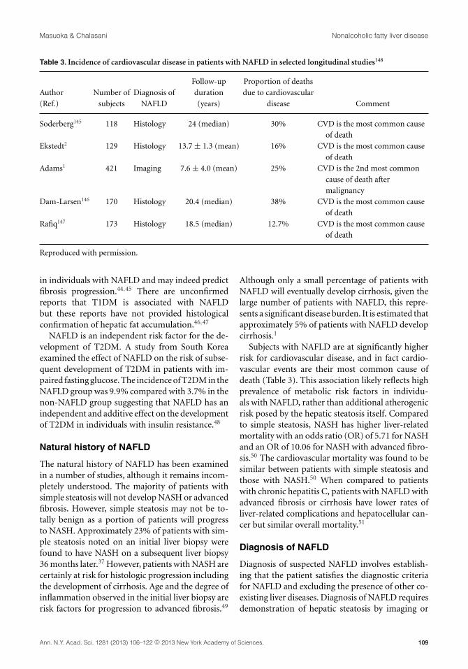
Table 3. Incidence of cardiovascular disease in patients with NAFLD in selected longitudinal studies148
Follow-up Proportion of deaths
Author Number of Diagnosis of duration due to cardiovascular
(Ref.) subjects NAFLD (years) disease Comment
Soderberg145 118 Histology 24 (median) 30% CVD is the most common cause
of death
Ekstedt2 129 Histology 13.7 ± 1.3 (mean) 16% CVD is the most common cause
of death
Adams1 421 Imaging 7.6 ± 4.0 (mean) 25% CVD is the 2nd most common
cause of death after
malignancy
Dam-Larsen146 170 Histology 20.4 (median) 38% CVD is the most common cause
of death
Rafiq147 173 Histology 18.5 (median) 12.7% CVD is the most common cause
of death
Reproduced with permission.
in individuals with NAFLD and may indeed predictfibrosis progression.44,45 There are unconfirmedreports that T1DM is associated with NAFLDbut these reports have not provided histologicalconfirmation of hepatic fat accumulation.46,47
NAFLD is an independent risk factor for the de-velopment of T2DM. A study from South Koreaexamined the effect of NAFLD on the risk of subse-quent development of T2DM in patients with im-paired fasting glucose. The incidence of T2DM in theNAFLD group was 9.9% compared with 3.7% in thenon-NAFLD group suggesting that NAFLD has anindependent and additive effect on the developmentof T2DM in individuals with insulin resistance.48
Natural history of NAFLD
The natural history of NAFLD has been examinedin a number of studies, although it remains incom-pletely understood. The majority of patients withsimple steatosis will not develop NASH or advancedfibrosis. However, simple steatosis may not be to-tally benign as a portion of patients will progressto NASH. Approximately 23% of patients with sim-ple steatosis noted on an initial liver biopsy werefound to have NASH on a subsequent liver biopsy36 months later.37 However, patients with NASH arecertainly at risk for histologic progression includingthe development of cirrhosis. Age and the degree ofinflammation observed in the initial liver biopsy arerisk factors for progression to advanced fibrosis.49
Although only a small percentage of patients withNAFLD will eventually develop cirrhosis, given thelarge number of patients with NAFLD, this repre-sents a significant disease burden. It is estimated thatapproximately 5% of patients with NAFLD developcirrhosis.1
Subjects with NAFLD are at significantly higherrisk for cardiovascular disease, and in fact cardio-vascular events are their most common cause ofdeath (Table 3). This association likely reflects highprevalence of metabolic risk factors in individu-als with NAFLD, rather than additional atherogenicrisk posed by the hepatic steatosis itself. Comparedto simple steatosis, NASH has higher liver-relatedmortality with an odds ratio (OR) of 5.71 for NASHand an OR of 10.06 for NASH with advanced fibro-sis.50 The cardiovascular mortality was found to besimilar between patients with simple steatosis andthose with NASH.50 When compared to patientswith chronic hepatitis C, patients with NAFLD withadvanced fibrosis or cirrhosis have lower rates ofliver-related complications and hepatocellular can-cer but similar overall mortality.51
Diagnosis of NAFLD
Diagnosis of suspected NAFLD involves establish-ing that the patient satisfies the diagnostic criteriafor NAFLD and excluding the presence of other co-existing liver diseases. Diagnosis of NAFLD requiresdemonstration of hepatic steatosis by imaging or
Ann. N.Y. Acad. Sci. 1281 (2013) 106–122 c© 2013 New York Academy of Sciences. 109

Nonalcoholic fatty liver disease Masuoka & Chalasani
histology and exclusion of significant alcohol use orother secondary causes of steatosis. In addition toalcohol consumption, secondary causes of hepaticsteatosis include medications, chronic hepatitis Cinfection, parenteral nutrition, and Wilson’s disease(Table 1).
Liver biochemistries and other laboratorytestingIn patients with suspected NAFLD, initial labora-tory evaluation typically involves obtaining liverbiochemistries and exclusion of chronic viral hep-atitis, hemochromatosis, Wilson’s disease, and au-toimmune hepatitis via appropriate diagnostic tests.
In patients with NAFLD, serum aminotrans-ferases can be normal or mildly elevated. Serumaminotransferases generally wax and wane and theyrarely exceed 200 U/L. Typically, ALT exceeds ASTalthough the serum AST frequently exceeds ALTwhen there is advanced fibrosis.52 Alkaline phos-phatase can also be elevated as well, and patientscan have only an isolated elevation of serum alkalinephosphatase with normal aminotransferase levels.53
NAFLD is the most common cause of incidentallyfound abnormal liver biochemistries in the primarycare setting.54 The sensitivity of abnormal amino-transferases in detecting NAFLD is poor since 55%to 79% of individuals with NAFLD may have nor-mal transaminase levels.21 In patients with NAFLD,neither the degree of elevation nor the pattern ofabnormal liver biochemistries are reliable in deter-mining that the disease activity and risk of diseaseprogression. In addition, changes of aminotrans-ferase levels do not parallel changes in fibrosis stagepreventing them from being a reliable surrogate forfibrosis progression.55
Mildly elevated serum ferritin is common inpatients with NAFLD though it is typically notassociated with increased hepatic iron stores.56
However, elevated serum ferritin and transferrinsaturation in patients with suspected NAFLD shouldprompt genetic testing for hereditary hemochro-matosis. A liver biopsy should be considered in apatient with suspected NAFLD who is homozygousor compound heterozygous for the C282Y mutationin the HFE gene to assess hepatic iron concentra-tion and to evaluate for significant liver injury andfibrosis.
Mild elevations of autoantibodies are relativelycommon in patients with NAFLD. A similar phe-
nomenon has been observed in liver disorders suchas viral hepatitis and drug-induced liver injury andis generally considered as an epiphenomenon. A re-cent study from the NASH Clinical Research Net-work (NASH CRN), found that positive serumautoantibodies, defined as antinuclear antibody(ANA) titer ≥1:160 or antismooth muscle antibody(ASMA) ≥1:40, were present in 21% of patientswith biopsy-proven NAFLD and they were not as-sociated with more advanced histologic features.57
However, if there are additional features suggestiveof autoimmune hepatitis, such as markedly elevatedaminotransferase, high �-globulin, or high serumimmunoglobulin G, then a liver biopsy may be con-sidered necessary to firmly establish the diagnosis.
Several models have been developed that com-bine laboratory testing, demographic variables, andclinical data to predict NASH with advanced fi-brosis, but their detailed discussion is beyond thescope of this review article. The NAFLD fibrosisscore (http://nafldscore.com/) is a promising bed-side tool for identifying NAFLD patients who are athigh risk for advanced fibrosis, and it employs sixeasily available variables (age, hyperglycemia, bodymass index, platelet count, albumin, and AST/ALTratio).58 In a recent meta-analysis of 13 publishedstudies, the NAFLD fibrosis score had a pooled areaunder the curve of a receiver operating characteristic(AUROC) of 0.85 for predicting advanced fibro-sis (stage 3 or 4). The NAFLD fibrosis score lessthan 1.455 had 90% sensitivity and 60% specificityto exclude advanced fibrosis, while a score greaterthan 0.676 had 67% sensitivity and 97% speci-ficity to identify the presence of advanced fibro-sis.50 Enhanced liver fibrosis (ELF) panel employsautomated immunoassay of three serum markers ofmatrix constituents and mediators of matrix remod-eling (hyaluronic acid, amino-terminal propeptideof type III collagen, and tissue inhibitor of matrixmetalloproteinase 1).59 The ELF panel has an AU-ROC of 0.90 for detection of advanced fibrosis witha threshold of 0.3576 associated with a sensitivity of80%, a specificity of 90%, a positive predictive valueof 71%, and a negative predictive value of 94%.60
Studies suggest that hepatocyte apoptosis playsan important role in the pathogenesis of NASH.Hepatocyte apoptosis results in caspase 3 generatedcleavage fragment of cytokeratin-18 (CK-18) beingreleased. The serum CK-18 fragments can be mea-sured by ELISA, and they significantly increased in
110 Ann. N.Y. Acad. Sci. 1281 (2013) 106–122 c© 2013 New York Academy of Sciences.

Masuoka & Chalasani Nonalcoholic fatty liver disease
patients with NASH compared with simple steato-sis and normal controls.61 The circulating levels ofCK-18 fragments have been shown to reflect diseaseactivity, and change in their level may correlate withthe change in NAFLD activity score.37 In a recentmeta-analysis, the pooled AUROC of serum CK-18for detection of NASH was 0.82 with a sensitivityand specificity of 78% and 87%, respectively.50
ImagingA number of imaging modalities have been em-ployed in the diagnosis of NAFLD. Although sev-eral different imaging techniques are valuable indemonstrating steatosis, the ability of current imag-ing technologies to evaluate fibrosis and especiallyinflammatory activity is limited.
Abdominal ultrasound is a relatively inexpensive,noninvasive diagnostic test to demonstrate steatosiswith excellent sensitivity in individuals with mod-erate to severe hepatic steatosis. Steatosis is visual-ized as increased echogenicity with bright liver echopattern on ultrasound B-mode examination andincreased attenuation.62 In a study of ultrasoundpaired with liver biopsy the reported sensitivity ul-trasound for the detection steatosis of 64% to 91%,and a specificity of 93% to 97%.63,64 Morbid obesitywas associated with a lower sensitivity, and higherdegrees of steatosis with a greater sensitivity.
Noncontrast abdominal CT is also useful indemonstrating hepatic steatosis. When contrast en-hanced CT is performed, the portal phase imagesshould be employed for the determination of steato-sis though contrast may result in decreased sen-sitivity and specificity compared with noncontrastscans.65 On a CT scan, hepatic steatosis is visualizedas decreased attenuation of the liver, resulting in theliver appearing darker than the spleen and is associ-ated with decreased liver attenuation index, which isthe difference between the mean hepatic and splenicattenuation in Hounsfield units. The sensitivity of aCT scan for detecting steatosis greater than 30% wasas high as 82% in a study involving potential livingliver donors.65
Abdominal magnetic resonance imaging (MRI)is a sensitive technique for demonstrating steatosis.T1-weighted gradient-echo magnetic resonance im-ages are acquired with an echo time such that waterand lipid spins are in phase or opposed phase allow-ing lipid quantitation by relative loss of signal inten-sity on opposed-phase images compared with that
on in-phase images.66 In a study of potential livingdonors MRI demonstrated a sensitivity, specificity,and accuracy of 100%, and 92.3%, and 93%, re-spectively, for detection of steatosis in patients withgreater than 20% steatosis by liver biopsy.67 Mag-netic resonance spectroscopy has been employed toquantitate hepatic steatosis by measuring hepatictriglyceride content although its use remains pri-marily investigational.68
Heterogeneity of hepatic steatosis including focalsparing is a relatively common finding with eachof the imaging modalities.69–74 The most commonlocations are the gallbladder fossa, and the areas ad-jacent to the porta hepatis and falciform ligament.Conversely, focal fatty infiltration can also be ob-served where steatosis is increased in only one regionof the liver.
Transient elastography is an ultrasound-basednoninvasive method of assessing of fibrosis throughmeasurement of liver stiffness. Although insensitivefor detection of early fibrosis, transient elastogra-phy can be useful in screening for advanced fibrosisin many patients. In a study involving patients withNAFLD, transient elastography demonstrated a sen-sitivity of 91% and a specificity of 75% for detectingstage 3 or higher fibrosis with positive and negativepredictive values of 52% and 97%, respectively.75
Unfortunately, body habitus can limit the applica-tion of this study since there is an increased failurerate in obtaining a successful transient elastogra-phy measurement with increasing degree of obesity.Also, transient elastography is not currently com-mercially available in the United States. Magneticresonance elastography is a promising techniquethat measures liver stiffness over a larger region ofthe liver than transient elastography; this researchtool is currently available at a limited number ofcenters.
Liver biopsyLiver biopsy remains the gold standard for char-acterizing the histology of NAFLD. It can play animportant role in the diagnosis of NAFLD, butit is expensive and carries risk of morbidity andvery rarely mortality. Histological examination ofliver tissue allows for exclusion of competing eti-ologies as well as for the assessment of coexistingliver diseases. Currently, liver biopsy is the only toolavailable for assessing the degree of inflammationand cell injury and to stage for the degree of fibrosis.
Ann. N.Y. Acad. Sci. 1281 (2013) 106–122 c© 2013 New York Academy of Sciences. 111

Nonalcoholic fatty liver disease Masuoka & Chalasani
It is invaluable to differentiate simple steatosis fromNASH.
Macrovesicular steatosis is a predominant featureof NAFLD, and the presence of steatosis in greaterthan 5% of hepatocytes is generally accepted asfatty liver (Fig. 1A).76 In addition to steatosis,common histologic findings in NASH includehepatocyte ballooning, lobular inflammation that iseither mixed type or neutrophil predominant, andvarying degrees of fibrosis (Figs. 1B and C).77 Thereare no histologic features that reliably differentiateNASH for alcoholic hepatitis, and the term NASHwas originally employed in a report from the MayoClinic regarding 20 patients with a liver diseasethat histologically mimicked alcoholic hepatitisin patients without significant alcohol intake.78 Ahistopathological classification system for NASHwas originally developed by Brunt et al., withhistologic features distinctive to NASH employedin determining the necroinflammatory activity(grade) and architectural alterations (stage).79 TheNAFLD activity score (NAS), an unweighted sum ofsteatosis, inflammation, and ballooning scores, wasdeveloped by the NASH CRN as a tool to quantifychanges in liver histology in NAFLD therapeutictrials.76 There is not a threshold value of NAS thatreliably identifies the presence of NASH.80
There can be significant sampling error due tothe small portion of the liver sampled by a biopsyand the inhomogeneous distribution of the histo-logic lesions of NASH. This sample variability ismoderate for hepatocyte ballooning and perisinu-soidal fibrosis and is somewhat higher for lobularinflammation.81,82
Pathogenesis of NAFLD
Several pathophysiologic mechanisms have beenproposed to explain the basis for fat accumulation,liver injury, and fibrosis in NAFLD, but their detaileddiscussion is beyond the scope of this review article.Interested readers are alerted to several recent com-prehensive reviews on this subject.7,8,10,83,84 Despiteconsiderable research in this area, the pathogenesisof NAFLD remains incompletely understood. It hasbeen very challenging to differentiate causative fac-tors from associated phenomena and downstreameffects. Although there is an increased understand-ing of the pathogenesis of hepatic fat accumulation,there are critical knowledge gaps in our understand-ing of mediators and mechanisms of hepatocyte
Figure 1. (A) Liver histology demonstrating moderatemacrovesicular steatosis around the central vein. Hematoxylinand eosin staining, with magnification of 200×. (B) Liver his-tology demonstrating active steatohepatitis with steatosis, bal-looned hepatocytes, inflammatory infiltrate, and Mallory’s Hya-line. Hematoxylin and eosin staining, with magnification of400×. (C) Liver histology demonstrating steatohepatitis withextensive pericellular fibrosis. Trichrome staining, with magni-fication 400×. Figure courtesy of David Kleiner, MD, NationalCancer Institute.
112 Ann. N.Y. Acad. Sci. 1281 (2013) 106–122 c© 2013 New York Academy of Sciences.

Masuoka & Chalasani Nonalcoholic fatty liver disease
injury, mediators of stellate cell activation, andfibrosis. It remains a puzzle why some individualswith NAFLD have advanced histological featuresand develop cirrhosis whereas others with compara-ble risk factor profile have simple steatosis with min-imal or no disease progression. A genetic basis forinter-individual phenotypic variability is stronglysuspected, but genetic studies are very limited inindividuals with histologically characterized NASH.
Insulin resistance is nearly universal in NAFLDand it is believed to play a crucial role in the patho-genesis of NAFLD. In adipocytes, insulin resistanceresults in increased activity of hormone-sensitive li-pase, which results in lipolysis of triglycerides andrelease of free fatty acids into circulation. Fatty acidstaken up by hepatocytes from circulation and pro-duced by de novo lipogenesis undergo esterifica-tion resulting in hepatocytes steatosis. Initially itwas proposed that NASH resulted from a “two-hit”mechanism with hepatocyte steatosis being the ini-tial metabolic insult that then allows a second injuryleading to NASH.85 However, subsequent researchhas cast significant doubt on this paradigm, and itis now widely believed that free fatty acids and theirmetabolic products (e.g., diacylglycerol) and seque-lae (e.g., free radicals) are the likely mediators ofhepatocyte injury.8,86–89
At a cellular level, several different mechanismshave been proposed for causing hepatocyte injury,including apoptosis, perturbations in autophagy,mitochondrial dysfunction, alterations in naturalkiller T cell and Kupffer cell function, and an in-crease in inflammatory cytokines.8,90–96 Apopto-sis appears to play an important role in hepato-cyte death in NAFLD, and free fatty acids couldbe the primary mediators of hepatocyte apopto-sis (lipoapoptosis).83,90 In addition, phagocytosis ofhepatocyte apoptotic bodies by stellate cells leads totheir activation and likely plays an important role infibrosis.
Treatment of NALFD
Lifestyle modificationLifestyle modification is the cornerstone of treat-ment of NAFLD. These interventions are not onlyeffective in improving NAFLD but also associatedconditions such as metabolic syndrome, T2DM, andthe related risk of cardiovascular disease.
Weight reduction plays an important role in thetreatment of NASH. Weight loss has been shown
to decrease hepatic steatosis and improve abnor-mal aminotransferase levels.97–99 Weight loss can bean effective treatment to improve the histology ofNASH if patients can attain sufficient weight reduc-tion. In a study by Harrison et al., subjects withbiopsy-proven NASH who lost 5% of body weighthad improvement in insulin sensitivity and hep-atic steatosis compared with those who lost lessthan 5% of their body weight.100 However, it wasonly in subjects who achieved at least 9% weightreduction that there was significant improvementin inflammation, ballooning, and NAS. A random-ized controlled trial (RCT) involving patients withbiopsy-proven NASH by Promrat et al. examinedthe efficacy of lifestyle intervention using a combi-nation of diet, exercise, and behavior modificationcompared to the control group that received struc-tured education.101 The primary end-point in thisstudy was improvement in liver histology. Partici-pants who achieved the study weight loss goal of atleast 7% had significant improvements in steatosis,lobular inflammation, and ballooning injury. Per-cent weight reduction correlated significantly withimprovement in NAS. Weight loss has been shownto prevent progression of fibrosis in NASH.37
However, very rapid weight loss may lead to in-creased portal inflammation and fibrosis. In a smallstudy of severely obese patients with NAFLD whowere placed on a very low calorie formula diet re-sulting in a median weight loss of 34 kg over an8-week period, 24% of patients developed mild por-tal inflammation or portal fibrosis.102 Therefore,one should be cautious in recommending very lowcalorie diets for individuals with NAFLD.
Weight reduction surgeryBecause NAFLD is present in the majority of pa-tients who undergo bariatric surgery, there has beenan interest in foregut bariatric surgery as a potentialtreatment option for NASH. Currently, there are noRCTs that have examined foregut bariatric surgeryas a treatment option for NAFLD or NASH. How-ever, several retrospective and prospective cohortstudies have compared liver histology in the severelyobese individuals before and after bariatric surgery.Unfortunately, a majority of them do not haveuniform histologic evaluation by post-bypass liverbiopsies and instead performed biopsies at varyingintervals and only in selected patients undergoingother surgical procedures such as abdominal hernia
Ann. N.Y. Acad. Sci. 1281 (2013) 106–122 c© 2013 New York Academy of Sciences. 113

Nonalcoholic fatty liver disease Masuoka & Chalasani
repair. However, one exception is the seminal studyby Mathurin et al., that prospectively correlated clin-ical and metabolic data with liver histology beforeand 1 and 5 years after bariatric surgery in 381 adultpatients with severe obesity.103 There was a signif-icant improvement in steatosis and ballooning at 1and 5 years following bariatric surgery compared tobaseline. In patients with probable or definite NASHat baseline, there was significant improvement insteatosis, ballooning, and NAS and resolution ofprobable or definite NASH at 1 and 5 years follow-ing bariatric surgery. The majority of histologicalbenefits were present at 1 year with no differencesin liver histology between 1 and 5 years followingbariatric surgery. Because no patient in this studyhad cirrhosis at baseline, the effect of bariatricsurgery in patients with cirrhosis could not beevaluated.
There are two meta-analyses that evaluated theinfluence of bariatric surgery on liver histologyin adults with NAFLD. Mummadi et al. foundthat steatosis, steatohepatitis, and fibrosis improveor completely resolve after bariatric surgery in asignificant proportion of patients.104 However, aCochrane review concluded that lack of RCTs orother high-quality clinical studies prevents defini-tive determination of benefits and risks of bariatricsurgery as a treatment option for patients withNASH.105
A recently published multi-society practiceguideline concluded that it is premature to considerforegut bariatric surgery as an established optionto specifically treat NASH.15 However, it concludedthat foregut bariatric surgery is not contraindicatedin otherwise eligible obese individuals with NAFLDor NASH.
Vitamin EOxidative stress has been proposed as an impor-tant mediator of hepatic injury in NASH.88,106,107
Vitamin E comprises a series of closely related com-pounds with antioxidant activity that have beenemployed in several therapeutic trials of NASH,although small sample sizes, differences in vita-min E preparations, and differences in endpointshave made them difficult to compare. The PIVENStrial, a recent randomized double blind placebocontrolled trial, is the largest study to investigatethe effectiveness of vitamin E supplementation onnondiabetic adults with histologically confirmed
NASH.108 This study employed unmodified RRR-alpha-tocopherol administered as a once daily doseof 800 IU given for 96 weeks. Vitamin E supplemen-tation resulted in significant improvement in patho-logic features of NASH with improvement in NASseen in 42% of patients receiving vitamin E com-pared with 19% of patients receiving placebo witha number needed to treat of 4.4. Compared withplacebo, vitamin E significantly improved amino-transferases as well. Vitamin E was well toleratedin this trial. The effectiveness of vitamin E supple-mentation has not been evaluated in diabetic pa-tients with NASH or in patients with NASH-relatedcirrhosis.
Some concerns have been raised regarding thelong-term safety of vitamin E, although current datasuggest that serious toxicity from vitamin E is likelyvery small if present. A recent RCT of vitamin Eadministered at a dose of 400 IU/day found a sta-tistically non-significant increase in prostate cancerrisk in the vitamin E group with an absolute in-creased risk of 1.6 per 1,000 person years of vita-min E use.109 Whether vitamin E supplementationmay increase all-cause mortality remains contro-versial. While some early meta-analyses suggesteda possible increase in all-cause mortality associ-ated with vitamin E supplementation, subsequentstudies have failed to demonstrate any increasedmortality.110–115
Insulin-sensitizing agentsInsulin-sensitizing agents have been investigated ex-tensively in therapeutic trials since insulin resistanceis believed to play an important role in the patho-genesis of NAFLD.
Metformin has been employed in a number oftherapeutic trials of NASH. While several smallopen-label studies suggested some improvementin aminotransferase levels with metformin ther-apy, a study in which only the metformin armunderwent biopsy suggested that it might lead tohistologic improvement.116 However, subsequentrandomized, placebo controlled clinical trials havefailed to show a significant difference in liver histol-ogy in nondiabetic patients with insulin resistanceand NASH.117,118 Because metformin does not havea significant effect on liver histology in patients withNASH compared with lifestyle modification alone,the use of metformin as therapy for NASH is notrecommended.15
114 Ann. N.Y. Acad. Sci. 1281 (2013) 106–122 c© 2013 New York Academy of Sciences.

Masuoka & Chalasani Nonalcoholic fatty liver disease
Thiazolidinediones (TZDs) are oral antidiabeticmedications that increase insulin sensitivity by ac-tivation of peroxisome proliferator-activated recep-tors present in a number of tissues, including liver,skeletal muscle, and adipose tissue. An early non-randomized trial, involving 22 patients with biopsy-proven NASH (including 50% with impairedglucose tolerance and diabetes) treated with rosigli-tazone 4 mg twice daily for 48 weeks, demon-strated improvement in inflammation, hepatocyteballooning, and fibrosis on an end-of-treatmentliver biopsy.119 However, weight gain occurred in67% of patients with a median body weight increaseof 7.3%.
The RCTs of TZDs have generally shown his-tologic improvement with TZDs in patients withNASH, although there has been variability in cer-tain parameters, such as improvement in inflamma-tion and fibrosis. In an early double-blind placebocontrolled study by Belfort et al., pioglitazone(45 mg/day) combined with a hypocaloric diet sig-nificantly improved steatosis, hepatocyte balloon-ing, and inflammation compared with a hypocaloricdiet alone in patients with biopsy-proven NASH andeither T2DM or insulin resistance.120 Improvementin the NAS was seen in 73% of patients treated withpioglitazone compared to 24% of placebo-treatedpatients (P < 0.001), and there was a trend towardimprovement in fibrosis in patients receiving pi-oglitazone (P < 0.08). A randomized trial of 63patients with biopsy-proven NASH by Ratziu et al.,found that rosiglitazone treatment (4 mg/day for thefirst month and 8 mg/day thereafter) for one yearimproved aminotransferases and hepatic steatosis,but not necroinflammation, fibrosis or NAS.121 Atwo-year open-label extension phase of this studydemonstrated similar results with no significant im-provement in hepatocyte ballooning, intralobularinflammation, fibrosis, or NAS seen in the rosiglita-zone treatment group compared with the controlgroup.122 Aithal et al., conducted a randomized,placebo-controlled trial of lifestyle interventionwith either pioglitazone (30 mg/day) or placebo for12 months in a total of 74 nondiabetic patients withNASH.123 Although steatosis did not improve sig-nificantly compared to placebo, cellular injury andfibrosis improved significantly. Weight gain was ob-served in the TZD group in each of these studiesand ranged from 1.5 to 2.77 kg, whereas the placebocontrol groups lost 0.55 to 1 kg.
The PIVENS study is a recent large RCT thatrandomized 247 nondiabetic patients with biopsy-proven NASH to pioglitazone (30 mg/day), vitaminE (800 IU/day), or placebo for 24 months.124 Theprimary endpoint was an improvement in NAS byat least 2 points, with at least a one-point improve-ment in hepatocellular ballooning and a one-pointimprovement in either the lobular inflammationor steatosis score, and no increase in the fibrosisscore.108 This endpoint was achieved in 34% of thepioglitazone group (P = 0.04 vs. placebo) and 43%of the vitamin E group (P = 0.001 vs. placebo) com-pared with 19% in the placebo group. The resolutionof NASH, a key secondary end point, was achieved insignificantly higher percentage of patients receivingpioglitazone compared with placebo (47% vs. 21%,P = 0.001). Similar to prior trials, pioglitazone wasassociated with a 4.7 kg weight gain compared toplacebo. A recent meta-analysis that included fourhigh quality randomized placebo controlled trialsshowed that TZDs significantly improved steato-sis (OR 3.39, 95% 2.19–5.25), inflammation (OR2.58, 95% CI: 1.68–3.97), and ballooning (OR 2.11,95% CI:1.33–3.36), but not fibrosis (combined OR1.40, 95% CI 0.87–2.24).125 When studies involvingpioglitazone alone were analyzed, there was statisti-cally significant improvement in fibrosis (combinedOR 1.68, 95% CI: 1.02–2.77).
The addition of metformin to rosiglitazone hasrecently been investigated but metformin did not of-fer additional histologic improvement over rosigli-tazone treatment alone in two open-labeled RCTsand more importantly metformin did not mitigateTZD associated weight gain.118,126
There is considerable debate about the long-term safety of TZDs with reference to increasedrisk of cardiovascular events, congestive heartfailure (CHF), bladder cancer, and bone loss.A meta-analysis of trials involving rosiglitazonedemonstrated a significant increase in the rate ofmyocardial infarction (OR 1.43, 95% CI 1.03 to 1.98,P = 0.03).127 This is distinct from the meta-analysisof 19 trials of pioglitazone enrolling a total of 16,390patients with T2DM, pioglitazone treatment was as-sociated with a significant reduction in the primaryoutcome of death, myocardial infarction, or stroke(P = 0.005).128 However, there was increasedincidence of CHF with pioglitazone (2.3% vs.1.8% in the control group, P = 0.002). Therefore,caution must be exercised when considering
Ann. N.Y. Acad. Sci. 1281 (2013) 106–122 c© 2013 New York Academy of Sciences. 115

Nonalcoholic fatty liver disease Masuoka & Chalasani
TZDs in patients with preexisting cardiac dis-ease. Owing to increased risk of cardiac events,rosiglitazone availability is highly restricted inthe United States and is no longer marketed inEurope.
Statins, omega-3 fatty acids, andursodeoxycholic acidDyslipidemia is almost universal in patients withNAFLD, and the effect of lipid-lowering therapywith statins on NALFD has been evaluated in severalstudies. The St. Francis Heart Study demonstratedthat atorvastatin 20 mg combined with vitaminsC and E is effective in reducing the odds of hav-ing hepatic steatosis as determined by CT althoughno histologic evaluation was performed.129 Thepost hoc analysis of the Greek Atorvastatin and Coro-nary Heart Disease Evaluation (GREACE) study of227 patients with possible NAFLD based on moder-ately abnormal liver tests at baseline treated with astatin demonstrated improvement inliver tests with-out an increase in liver-related adverse effects com-pared with controls.130 Several other studies havedemonstrated that statins are safe in patients withliver disease, and there is no evidence that patientswith NAFLD are at increased risk for serious liver in-jury from statins compared with those without liverdisease.131–134 There are no RCTs with histologicalend points that have examined the use of statinsin the treatment of NASH, and thus statins specif-ically to treat NASH cannot be advocated at thistime.15
Treatment of NAFLD with fish oil supplemen-tation or polyunsaturated fatty acids that are en-riched in fish oil omega 3 fatty acids has beeninvestigated in animal studies and a small num-ber of preliminary human studies. Epidemiologicstudies have suggested that there may be an in-verse relationship between the level of fish oilintake and risk of NAFLD.135,136 However, the as-sociation is relatively modest and was not statis-tically significant in some studies after adjustingfor confounding factors. Several small nonrandom-ized open-label study of omega-3 fatty acids aloneor with supplements such as olive oil have foundimprovement in liver tests, serum triglycerides,and steatosis by ultrasound.137–140 A large multi-center trial of the omega-3 fatty acid eicosapen-tanoic acid to treat NASH is ongoing in the UnitedStates. At this point, it is premature to recommend
omega-3 fatty acids for the treatment of NAFLD al-though they may be considered for the treatment ofhypertriglyceridemia.
There has been interest in the use of ursodes-oxycholic acid (UDCA) to treat NAFLD althoughstudies to date have yielded disappointing results.UDCA is a secondary bile acid that is approved forthe treatment of primary biliary cirrhosis and haseffects on cholesterol absorption and inflammation.Initial small uncontrolled clinical studies suggestedthat UDCA may offer benefit to individuals withNASH. However, a two-year prospective, double-blind trial of UDCA (13–15 mg/kg per day) of166 patients failed to demonstrate improvementin laboratory data or liver histology.141 Subsequentstudies have employed high dose UDCA with mixedresults. A RCT of high-dose UDCA (28–35 mg/kgper day) given for 12 months in patients with NASHby Ratziu et al., demonstrated that UDCA improvedtransaminase levels and markers of insulin resis-tance and fibrosis.142 Critically, no histologic eval-uation was performed. Leuschner et al. performeda double-blind, randomized, placebo-controlledtrial of high-dose UDCA (23–28 mg/kg/day) in185 patients with histologically proven NASH.143
The treatment was provided over 18 monthswith both pre- and posttreatment liver biopsies.Although lobular inflammation was improvedin patients in the treatment group, there wasno improvement in fibrosis and no significantdifference in NAS between the treatment andcontrol group. In summary, there is no evidencethat UDCA is effective to treat NASH.
Emerging therapiesTable 4 describes selected compounds that are beingtested in large phase 2/3 studies. A large randomizedplacebo controlled trial of two doses of eicosapen-tanoic acid is near completion in the United Statesand its results are eagerly awaited. Pentoxifylline hasshown encouraging histological benefits in severalsmall studies and it is a suitable candidate for fur-ther testing in large multicenter RCTs. The NASHCRN is conducting a multicenter RCT of obeticholicacid in adults with NASH and a multicenter RCT ofcysteamine bitartrate in children with NASH. Theirresults will not be available until 2014 and 2015, re-spectively. Obeticholic acid is a novel FXR agonist,whereas cysteamine bitartrate is a potent antioxi-dant.
116 Ann. N.Y. Acad. Sci. 1281 (2013) 106–122 c© 2013 New York Academy of Sciences.

Masuoka & Chalasani Nonalcoholic fatty liver disease
Table 4.Selected compounds with high therapeutic potential that are currently being investigated in phase 2/3 studies
Compound Nature of the trial
Potential
mechanism of
action Primary end point Comment
Eicosapentanoic
acid
Multicenter phase 2/3 study
in the United States;
sponsored by Mochida
Pharmaceuticals
Decreased
lipogenesis and
improved insulin
sensitivity
Liver histology To be completed
soon; results
awaited
Pentoxifylline Several small studies have
shown histological
benefits
Anti-TNF-� Liver histology Suitable agent
for large-scale
definitive
studies
Obeticholic acid Large placebo-controlled,
phase 2b is under way in
the United States;
conducted by the NASH
CRN under a CRADA
agreement with Intercept
Pharmaceuticals
Farsenoid X
receptor agonist
Liver histology Results will
become
available in
2014
Cysteamine
bitartrate
Large placebo-controlled,
phase 2b trial
in children with NASH is
under way; conducted by
the NASH CRN under a
CRADA agreement with
Raptor Pharmaceuticals
Potent antioxidant Liver histology Results to
become
available in
2015
GFT 505 Multicenter,
placebo-controlled RCT
to be initiated soon;
sponsored by Genfit
GFT 505 is a dual
PPAR �/� agonist
Liver histology To be initiated
soon
GS 6624 Two separate phase 2b
studies to be initiated
internationally by Gilead
Pharmaceuticals
GS 6624 is a
parenteral
compound, and
it is a
monoclonal
antibody against
a lysyl
oxidase–like
molecule
Reversal of cirrhosis by
histology is the
primary end point
for the cirrhosis
study; however,
progression of
fibrosis is the end
point for the
advanced fibrosis
study
To be initiated
soon
In adults with compensated
cirrhosis
In adults with advanced
fibrosis
Surveillance for the developmentof complications
In addition to their risk of developing cirrhosis,liver failure, and hepatocellular cancer, patients withNAFLD are at significantly higher risk for develop-ing diabetes and cardiovascular disease, and thus
there should be heightened attention to monitoringfor the development of these conditions.
In patients with NASH related cirrhosis, regularsurveillance for cirrhosis related complications suchas hepatocellular carcinoma (HCC) and esophagealvarices should be performed. As with other causes
Ann. N.Y. Acad. Sci. 1281 (2013) 106–122 c© 2013 New York Academy of Sciences. 117

Nonalcoholic fatty liver disease Masuoka & Chalasani
of cirrhosis, surveillance for HCC should be per-formed every 6 months with abdominal ultrasound,or intravenous contrast enhanced abdominal CT orMRI. Yearly cumulative incidence of HCC in pa-tients with NASH-related cirrhosis was found to be2.6% in one series. This was less than the yearlycumulative incidence in patients with hepatitis Ccirrhosis of 4.0% though the difference was not sta-tistically significant.144
Conclusions
Individuals with obesity and T2DM are at signif-icantly higher risk for NAFLD. The incidence ofNAFLD is rapidly increasing throughout the worlddue to the increasing frequency of obesity andT2DM. The NAFLD is a spectrum of chronic liverdiseases ranging from simple steatosis, which is rel-atively benign from a liver standpoint, to NASH,which can progress to cirrhosis and liver failure.The diagnosis of NAFLD requires imaging evi-dence of hepatic steatosis while excluding com-peting etiologies, such as significant alcohol con-sumption, viral hepatitis, and hemochromatosis.Liver biopsy remains the gold standard for diag-nosing NASH and for assessing fibrosis. Recentadvances in laboratory testing and noninvasiveimaging have shown promise for identifying steato-hepatitis and advanced fibrosis in individuals withNAFLD. Weight loss of at least 5% is required to im-prove steatosis, whereas weight loss in the range of7–10% may be needed to improve steatohepatitis. Anumber of pharmacologic therapies have been eval-uated in NASH, and agents such as vitamin E andTZDs have shown some promise. Ongoing studieshold promise for developing more effective diag-nostic tests and therapies.
Acknowledgment
This work is in part supported by NIH K24DK069290A Grant to NC.
Conflicts of interest
N. Chalasani received fees from Merck, GlaxoSmithKline, Biolex, J & J, Mochida, Salix, Aegerion,and Sanofi-Aventis for providing consulting relatedto NAFLD/NASH or drug hepatotoxicity in the last12 months. He has received research support fromLilly, Cumberland Pharmaceuticals, and Intercept.H. Masuoka has no financial conflicts to declare.
References
1. Adams, L.A., J.F. Lymp, J. St Sauver, et al. 2005. The naturalhistory of nonalcoholic fatty liver disease: a population-based cohort study. Gastroenterology 129: 113–121.
2. Ekstedt, M., L.E. Franzen, U.L. Mathiesen, et al. 2006. Long-term follow-up of patients with NAFLD and elevated liverenzymes. Hepatology 44: 865–873.
3. Ratziu, V. & T. Poynard. 2006. Assessing the outcome ofnonalcoholic steatohepatitis? It’s time to get serious. Hepa-tology 44: 802–805.
4. Caldwell, S.H., D.H. Oelsner, J.C. Iezzoni, et al. 1999. Cryp-togenic cirrhosis: clinical characterization and risk factorsfor underlying disease. Hepatology 29: 664–669.
5. Charlton, M. 2004. Nonalcoholic fatty liver disease: a reviewof current understanding and future impact. Clin. Gastroen-terol. Hepatol. 2: 1048–1058.
6. Charlton, M.R., J.M. Burns, R.A. Pedersen, et al. 2011. Fre-quency and outcomes of liver transplantation for nonalco-holic steatohepatitis in the United States. Gastroenterology.141: 1249–1253.
7. Nguyen, T.A. & A.J. Sanyal. 2012. Pathophysiology guidedtreatment of nonalcoholic steatohepatitis. J. Gastroenterol.Hepatol. 27(Suppl 2): 58–64.
8. Ibrahim, S.H., R. Kohli & G.J. Gores. 2011. Mechanisms oflipotoxicity in NAFLD and clinical implications. J. Pediatr.Gastroenterol. Nutr. 53: 131–140.
9. Brunt, E.M. 2011. Non-alcoholic fatty liver disease: what’snew under the microscope? Gut. 60: 1152–1158.
10. Cohen, J.C., J.D. Horton & H.H. Hobbs. 2011. Human fattyliver disease: old questions and new insights. Science 332:1519–1523.
11. Anstee, Q.M., A.K. Daly & C.P. Day. 2011. Genetics of alco-holic and nonalcoholic fatty liver disease. Semin. Liver Dis.31: 128–146.
12. Fujii, H. & N. Kawada. 2012. Inflammation and fibrogenesisin steatohepatitis. J. Gastroenterol. 47: 215–225.
13. Cusi, K. 2012. Role of obesity and lipotoxicity in the devel-opment of nonalcoholic steatohepatitis: pathophysiologyand clinical implications. Gastroenterology 142: 711–725e716.
14. Aly, F.Z. & D.E. Kleiner. 2011. Update on fatty liver diseaseand steatohepatitis. Adv. Anat. Pathol. 18: 294–300.
15. Chalasani, N., Z. Younossi, J.E. Lavine, et al. 2012. Thediagnosis and management of non-alcoholic fatty liver dis-ease: practice Guideline by the American Association forthe Study of Liver Diseases, American College of Gastroen-terology, and the American Gastroenterological Associa-tion. Hepatology 55: 2005–2023.
16. Suzuki, A., P. Angulo, J. Lymp, et al. 2005. Chronological de-velopment of elevated aminotransferases in a nonalcoholicpopulation. Hepatology 41: 64–71.
17. Tsuneto, A., A. Hida, N. Sera, et al. 2010. Fatty liver inci-dence and predictive variables. Hyper Res. 33: 638–643.
18. Whalley, S., P. Puvanachandra, A. Desai & H. Kennedy.2007. Hepatology outpatient service provision in secondarycare: a study of liver disease incidence and resource costs.Clin Med. 7: 119–124.
19. Vernon, G., A. Baranova & Z.M. Younossi. 2011. Sys-tematic review: the epidemiology and natural history of
118 Ann. N.Y. Acad. Sci. 1281 (2013) 106–122 c© 2013 New York Academy of Sciences.

Masuoka & Chalasani Nonalcoholic fatty liver disease
non-alcoholic fatty liver disease and non-alcoholic steato-hepatitis in adults. Aliment. Pharm. Ther. 34: 274–285.
20. Marcos, A., R.A. Fisher, J.M. Ham, et al. 2000. Selectionand outcome of living donors for adult to adult right lobetransplantation. Transplantation 69: 2410–2415.
21. Browning, J.D., L.S. Szczepaniak, R. Dobbins, et al. 2004.Prevalence of hepatic steatosis in an urban population inthe United States: impact of ethnicity. Hepatology 40: 1387–1395.
22. Lee, J.Y., K.M. Kim, S.G. Lee, et al. 2007. Prevalence andrisk factors of non-alcoholic fatty liver disease in poten-tial living liver donors in Korea: a review of 589 consec-utive liver biopsies in a single center. J. Hepatol. 47: 239–244.
23. Chen, Z.W., L.Y. Chen, H.L. Dai, et al. 2008. Relationshipbetween alanine aminotransferase levels and metabolic syn-drome in nonalcoholic fatty liver disease. J. Zhejiang Univ.Science B. 9: 616–622.
24. Amarapurkar, D., P. Kamani, N. Patel, et al. 2007. Prevalenceof non-alcoholic fatty liver disease: population based study.Ann. Hepatol. 6: 161–163.
25. Gutierrez-Grobe, Y., G. Ponciano-Rodriguez, M.H. Ramos,et al. 2010. Prevalence of non alcoholic fatty liver disease inpremenopausal, posmenopausal and polycystic ovary syn-drome women. The role of estrogens. Ann. Hepatol. 9: 402–409.
26. Piepkorn, M.W., N.K. Mottet & E.A. Smuckler. 1977. Fattymetamorphosis of the liver associated with jejunoileal by-pass. Report of five cases. Arch. Pathol. Lab. Med. 101: 411–415.
27. Peters, R.L., T. Gay & T.B. Reynolds. 1975. Post-jejunoileal-bypass hepatic disease. Its similarity to alcoholic hepaticdisease. Am. J. Clin. Pathol. 63: 318–331.
28. Kato, H., S. Isaji, Y. Azumi, et al. 2010. Development ofnonalcoholic fatty liver disease (NAFLD) and nonalco-holic steatohepatitis (NASH) after pancreaticoduodenec-tomy: proposal of a postoperative NAFLD scoring system.J. Hepato-biliary-pancreatic sciences. 17: 296–304.
29. Boza, C., A. Riquelme, L. Ibanez, et al. 2005. Predictorsof nonalcoholic steatohepatitis (NASH) in obese patientsundergoing gastric bypass. Obes. Surg. 15: 1148–1153.
30. Haentjens, P., D. Massaad, H. Reynaert, et al. 2009. Identi-fying non-alcoholic fatty liver disease among asymptomaticoverweight and obese individuals by clinical and biochem-ical characteristics. Acta clinica Belgica. 64: 483–493.
31. Beymer, C., K.V. Kowdley, A. Larson, et al. 2003. Prevalenceand predictors of asymptomatic liver disease in patientsundergoing gastric bypass surgery. Arch. Surg. 138: 1240–1244.
32. Loria, P., L. Carulli, M. Bertolotti & A. Lonardo. 2009. En-docrine and liver interaction: the role of endocrine pathwaysin NASH. Nat. Revs. 6: 236–247.
33. Barclay, J.L., C.N. Nelson, M. Ishikawa, et al. 2011. GH-dependent STAT5 signaling plays an important role in hep-atic lipid metabolism. Endocrinology. 152: 181–192.
34. Gabbi, C., F. Carubbi, L. Losi, et al. 2008. Nonalcoholicfatty liver disease induced by leuprorelin acetate. J. Clin.Gastroenterol. 42: 107–110.
35. Brzozowska, M.M., G. Ostapowicz & M.D. Weltman. 2009.An association between non-alcoholic fatty liver disease and
polycystic ovarian syndrome. J. Gastroenterol. Hepatol. 24:243–247.
36. Kawamura, Y., Y. Arase, K. Ikeda, et al. 2012. Large-scalelong-term follow-up study of Japanese patients with non-alcoholic fatty liver disease for the onset of hepatocellularcarcinoma. Am. J. Gastroenterol. 107: 253–261.
37. Wong, V.W., G.L. Wong, P.C. Choi, et al. 2010. Disease pro-gression of non-alcoholic fatty liver disease: a prospectivestudy with paired liver biopsies at 3 years. Gut. 59: 969–974.
38. Assy, N., K. Kaita, D. Mymin, et al. 2000. Fatty infiltration ofliver in hyperlipidemic patients. Digest. Dis. Sci. 45: 1929–1934.
39. Eguchi, Y., T. Eguchi, T. Mizuta, et al. 2006. Visceral fataccumulation and insulin resistance are important factorsin nonalcoholic fatty liver disease. J. Gastroenterol. 41: 462–469.
40. Grundy, S.M., J.I. Cleeman, S.R. Daniels, et al. 2005. Di-agnosis and management of the metabolic syndrome: anAmerican Heart Association/National Heart, Lung, andBlood Institute Scientific Statement. Circulation 112: 2735–2752.
41. Marceau, P., S. Biron, F.S. Hould, et al. 1999. Liver pathologyand the metabolic syndrome X in severe obesity. J. Clin.Endocrinol. Metabol. 84: 1513–1517.
42. Hamaguchi, M., T. Kojima, N. Takeda, et al. 2005. Themetabolic syndrome as a predictor of nonalcoholic fattyliver disease. Ann. Int. Med. 143: 722–728.
43. Hanley, A.J., K. Williams, A. Festa, et al. 2005. Liver markersand development of the metabolic syndrome: the insulinresistance atherosclerosis study. Diabetes. 54: 3140–3147.
44. Lomonaco, R., C. Ortiz-Lopez, B. Orsak, et al. 2012. Effectof adipose tissue insulin resistance on metabolic parametersand liver histology in obese patients with nonalcoholic fattyliver disease. Hepatology 55: 1389–1397.
45. Musso, G., M. Cassader, F. De Michieli, et al. 2012. Non-alcoholic steatohepatitis versus steatosis: Adipose tissue in-sulin resistance and dysfunctional response to fat inges-tion predict liver injury and altered glucose and lipoproteinmetabolism. Hepatology 56: 933–942.
46. Targher, G., L. Bertolini, R. Padovani, et al. 2010. Prevalenceof non-alcoholic fatty liver disease and its association withcardiovascular disease in patients with type 1 diabetes. J.Hepatol. 53: 713–718.
47. Regnell, S.E. & A. Lernmark. 2011. Hepatic steatosis in type1 diabetes. RDS. 8: 454–467.
48. Bae, J.C., E.J. Rhee, W.Y. Lee, et al. 2011. Combined effect ofnonalcoholic fatty liver disease and impaired fasting glucoseon the development of type 2 diabetes: a 4-year retrospectivelongitudinal study. Diabetes Care. 34: 727–729.
49. Argo, C.K., P.G. Northup, A.M. Al-Osaimi & S.H. Caldwell.2009. Systematic review of risk factors for fibrosis progres-sion in non-alcoholic steatohepatitis. J. Hepatol. 51: 371–379.
50. Musso, G., R. Gambino, M. Cassader & G. Pagano. 2011.Meta-analysis: natural history of non-alcoholic fatty liverdisease (NAFLD) and diagnostic accuracy of non-invasivetests for liver disease severity. Ann. Med. 43: 617–649.
51. Bhala, N., P. Angulo, D. van der Poorten, et al. 2011.The natural history of nonalcoholic fatty liver disease with
Ann. N.Y. Acad. Sci. 1281 (2013) 106–122 c© 2013 New York Academy of Sciences. 119

Nonalcoholic fatty liver disease Masuoka & Chalasani
advanced fibrosis or cirrhosis: an international collaborativestudy. Hepatology 54: 1208–1216.
52. Angulo, P., J.C. Keach, K.P. Batts & K.D. Lindor. 1999. In-dependent predictors of liver fibrosis in patients with non-alcoholic steatohepatitis. Hepatology 30: 1356–1362.
53. Pantsari, M.W. & S.A. Harrison. 2006. Nonalcoholic fattyliver disease presenting with an isolated elevated alkalinephosphatase. J. Clin Gastroenterol. 40: 633–635.
54. Armstrong, M.J., D.D. Houlihan, L. Bentham, et al. 2012.Presence and severity of non-alcoholic fatty liver diseasein a large prospective primary care cohort. J. Hepatol. 56:234–240.
55. Adams, L.A., S. Sanderson, K.D. Lindor & P. Angulo. 2005.The histological course of nonalcoholic fatty liver disease:a longitudinal study of 103 patients with sequential liverbiopsies. J. Hepatol. 42: 132–138.
56. Vuppalanchi, R. & N. Chalasani. 2009. Nonalcoholicfatty liver disease and nonalcoholic steatohepatitis: Se-lected practical issues in their evaluation and management.Hepatology 49: 306–317.
57. Vuppalanchi, R., R.J. Gould, L.A. Wilson, et al. 2011. Clin-ical significance of serum autoantibodies in patients withNAFLD: results from the nonalcoholic steatohepatitis clin-ical research network. Hepatol. Int. 6: 379–385.
58. Angulo, P., J.M. Hui, G. Marchesini, et al. 2007. The NAFLDfibrosis score: a noninvasive system that identifies liver fi-brosis in patients with NAFLD. Hepatology 45: 846–854.
59. Rosenberg, W.M., M. Voelker, R. Thiel, et al. 2004. Serummarkers detect the presence of liver fibrosis: a cohort study.Gastroenterology 127: 1704–1713.
60. Guha, I.N., J. Parkes, P. Roderick, et al. 2008. Noninvasivemarkers of fibrosis in nonalcoholic fatty liver disease: Val-idating the European Liver Fibrosis Panel and exploringsimple markers. Hepatology 47: 455–460.
61. Wieckowska, A., N.N. Zein, L.M. Yerian, et al. 2006. Invivo assessment of liver cell apoptosis as a novel biomarkerof disease severity in nonalcoholic fatty liver disease.Hepatology 44: 27–33.
62. Joseph, A.E., K.C. Dewbury & P.G. McGuire. 1979. Ultra-sound in the detection of chronic liver disease (the “brightliver”). British J. Radiol. 52: 184–188.
63. Joseph, A.E., S.H. Saverymuttu, S. al-Sam, et al. 1991.Comparison of liver histology with ultrasonography in as-sessing diffuse parenchymal liver disease. Clin. Radiol. 43:26–31.
64. Palmentieri, B., I. de Sio, V. La Mura, et al. 2006. The roleof bright liver echo pattern on ultrasound B-mode exam-ination in the diagnosis of liver steatosis. Digest. Liver Dis.38: 485–489.
65. Jacobs, J.E., B.A. Birnbaum, M.A. Shapiro, et al. 1998. Diag-nostic criteria for fatty infiltration of the liver on contrast-enhanced helical CT. AJR. 171: 659–664.
66. Outwater, E.K., R. Blasbalg, E.S. Siegelman & M. Vala. 1998.Detection of lipid in abdominal tissues with opposed-phasegradient-echo images at 1.5 T: techniques and diagnosticimportance. Radiographics 18: 1465–1480.
67. Kim, S.H., J.M. Lee, J.K. Han, et al. 2006. Hepaticmacrosteatosis: predicting appropriateness of liver dona-tion by using MR imaging–correlation with histopathologicfindings. Radiology 240: 116–129.
68. Szczepaniak, L.S., P. Nurenberg, D. Leonard, et al. 2005.Magnetic resonance spectroscopy to measure hepatictriglyceride content: prevalence of hepatic steatosis in thegeneral population. Am. J. Physiol. Endocrinol. Metab. 288:E462–E468.
69. Qayyum, A., M. Nystrom, S.M. Noworolski, et al. 2012.MRI steatosis grading: development and initial validationof a color mapping system. AJR. 198: 582–588.
70. Choi, J.S. & M.J. Kim. 2011. Education and imaging: hep-atobiliary and pancreatic: focal steatohepatitis mimicking ametastasis. J. Gastroenterol. Hepatol. 26: 415.
71. Tharayil, V. & L.R. Roberts. 2010. Evaluation of a focal lesionin the liver. answer to the clinical challenges and images in GIquestion: image 3: Focal fatty infiltration. Gastroenterology139: e10–e11.
72. Fasih, N., A.K. Shanbhogue, S. Thipphavong, et al. 2010.Gamut of focal fatty lesions in the liver: imaging manifesta-tions with emphasis on magnetic resonance imaging. Curr.Prob. Diag. Radiol. 39: 137–151.
73. Karcaaltincaba, M. & O. Akhan. 2007. Imaging of hepaticsteatosis and fatty sparing. Eur. J. Radiol. 61: 33–43.
74. Hamer, O.W., D.A. Aguirre, G. Casola, et al. 2006. Fatty liver:imaging patterns and pitfalls. Radiographics 26: 1637–1653.
75. Wong, V.W., J. Vergniol, G.L. Wong, et al. 2010. Diagnosisof fibrosis and cirrhosis using liver stiffness measurementin nonalcoholic fatty liver disease. Hepatology 51: 454–462.
76. Kleiner, D.E., E.M. Brunt, M. Van Natta, et al. 2005. Designand validation of a histological scoring system for nonalco-holic fatty liver disease. Hepatology 41: 1313–1321.
77. Brunt, E.M. 2004. Nonalcoholic steatohepatitis. Semin. liverDis. 24: 3–20.
78. Ludwig, J., T.R. Viggiano, D.B. McGill & B.J. Oh. 1980.Nonalcoholic steatohepatitis: Mayo Clinic experiences witha hitherto unnamed disease. Mayo Clin. Proc. 55: 434–438.
79. Brunt, E.M., C.G. Janney, A.M. Di Bisceglie, et al. 1999.Nonalcoholic steatohepatitis: a proposal for grading andstaging the histological lesions. Am. J. Gastroenterol. 94:2467–2474.
80. Brunt, E.M., D.E. Kleiner, L.A. Wilson, et al. 2011. Nonal-coholic fatty liver disease (NAFLD) activity score and thehistopathologic diagnosis in NAFLD: distinct clinicopatho-logic meanings. Hepatology 53: 810–820.
81. Ratziu, V., F. Charlotte, A. Heurtier, et al. 2005. Samplingvariability of liver biopsy in nonalcoholic fatty liver disease.Gastroenterology 128: 1898–1906.
82. Merriman, R.B., L.D. Ferrell, M.G. Patti, et al. 2006. Cor-relation of paired liver biopsies in morbidly obese patientswith suspected nonalcoholic fatty liver disease. Hepatology44: 874–880.
83. Alkhouri, N., C. Carter-Kent & A.E. Feldstein. 2011. Apop-tosis in nonalcoholic fatty liver disease: diagnostic and ther-apeutic implications. Expert Rev. Gastroenterol. Hepatol. 5:201–212.
84. Feldstein, A.E. 2010. Novel insights into the pathophysiol-ogy of nonalcoholic fatty liver disease. Semin. liver Dis. 30:391–401.
85. Day, C.P. & O.F. James. 1998. Steatohepatitis: a tale of two“hits”? Gastroenterology 114: 842–845.
86. Gorden, D.L., P.T. Ivanova, D.S. Myers, et al. 2011. In-creased diacylglycerols characterize hepatic lipid changes
120 Ann. N.Y. Acad. Sci. 1281 (2013) 106–122 c© 2013 New York Academy of Sciences.

Masuoka & Chalasani Nonalcoholic fatty liver disease
in progression of human nonalcoholic fatty liver disease;comparison to a murine model. PloS one. 6:e2 2775.
87. Kotronen, A., T. Seppanen-Laakso, J. Westerbacka, et al.2009. Hepatic stearoyl-CoA desaturase (SCD)-1 activityand diacylglycerol but not ceramide concentrations are in-creased in the nonalcoholic human fatty liver. Diabetes 58:203–208.
88. Rolo, A.P., J.S. Teodoro & C.M. Palmeira. 2012. Role ofoxidative stress in the pathogenesis of nonalcoholic steato-hepatitis. Free Rad. Biol. Med. 52: 59–69.
89. Soardo, G., D. Donnini, L. Domenis, et al. 2011. Oxidativestress is activated by free fatty acids in cultured humanhepatocytes. Metab. Synd. related Disord. 9: 397–401.
90. Malhi, H. & G.J. Gores. 2008. Molecular mechanisms oflipotoxicity in nonalcoholic fatty liver disease. Semin. liverDis. 28: 360–369.
91. Amir, M. & M.J. Czaja. 2011. Autophagy in nonalcoholicsteatohepatitis. Exp. Rev. Gastroenterol. Hepatol. 5: 159–166.
92. Grattagliano, I., O. de Bari, T.C. Bernardo, et al. 2012. Roleof mitochondria in nonalcoholic fatty liver disease–fromorigin to propagation. Clin. Biochem. 45: 610–618.
93. Kremer, M. & I.N. Hines. 2008. Natural killer T cells andnon-alcoholic fatty liver disease: fat chews on the immunesystem. WJG. 14: 487–488.
94. Zhan, Y.T. & W. An. 2010. Roles of liver innate immune cellsin nonalcoholic fatty liver disease. WJG. 16: 4652–4660.
95. Braunersreuther, V., G.L. Viviani, F. Mach & F. Montecucco.2012. Role of cytokines and chemokines in non-alcoholicfatty liver disease. WJG. 18: 727–735.
96. Ono, M. & T. Saibara. 2012. Is impaired Kupffer cell func-tion really important to the pathogenesis of nonalcoholicsteatohepatitis? J. Gastroenterol. Hepatol. 27: 622–624.
97. Palmer, M. & F. Schaffner. 1990. Effect of weight reductionon hepatic abnormalities in overweight patients. Gastroen-terology 99: 1408–1413.
98. Park, H.S., M.W. Kim & E.S. Shin. 1995. Effect of weightcontrol on hepatic abnormalities in obese patients with fattyliver. J. Kor. Med. Sci. 10: 414–421.
99. Ueno, T., H. Sugawara, K. Sujaku, et al. 1997. Therapeuticeffects of restricted diet and exercise in obese patients withfatty liver. J. Hepatol. 27: 103–107.
100. Harrison, S.A., W. Fecht, E.M. Brunt & B.A. Neuschwander-Tetri. 2009. Orlistat for overweight subjects with nonalco-holic steatohepatitis: a randomized, prospective trial. Hep-atology 49: 80–86.
101. Promrat, K., D.E. Kleiner, H.M. Niemeier, et al. 2010. Ran-domized controlled trial testing the effects of weight loss onnonalcoholic steatohepatitis. Hepatology 51: 121–129.
102. Andersen, T., C. Gluud, M.B. Franzmann & P. Christof-fersen. 1991. Hepatic effects of dietary weight loss in mor-bidly obese subjects. J. Hepatol. 12: 224–229.
103. Mathurin, P., A. Hollebecque, L. Arnalsteen, et al. 2009.Prospective study of the long-term effects of bariatricsurgery on liver injury in patients without advanced dis-ease. Gastroenterology 137: 532–540.
104. Mummadi, R.R., K.S. Kasturi, S. Chennareddygari & G.K.Sood. 2008. Effect of bariatric surgery on nonalcoholic fattyliver disease: systematic review and meta-analysis. Clin. Gas-troenterol. Hepatol. 6: 1396–1402.
105. Chavez-Tapia, N.C., F.I. Tellez-Avila, T. Barrientos-Gutierrez, et al. 2010. Bariatric surgery for non-alcoholicsteatohepatitis in obese patients. Cochrane Database SystRev.: CD007340.
106. Rezazadeh, A., R. Yazdanparast & M. Molaei. 2012. Amelio-ration of diet-induced nonalcoholic steatohepatitis in ratsby Mn-salen complexes via reduction of oxidative stress. J.Biomed. Sci. 19: 26.
107. Narasimhan, S., K. Gokulakrishnan, R. Sampathkumar,et al. 2010. Oxidative stress is independently associated withnon-alcoholic fatty liver disease (NAFLD) in subjects withand without type 2 diabetes. Clin. Biochem. 43: 815–821.
108. Sanyal, A.J., N. Chalasani, K.V. Kowdley, et al. 2010. Pioglita-zone, vitamin E, or placebo for nonalcoholic steatohepatitis.N. Engl. J. Med. 362: 1675–1685.
109. Klein, E.A., I.M. Thompson Jr., C.M. Tangen, et al. 2011.Vitamin E and the risk of prostate cancer: the Selenium andVitamin E Cancer Prevention Trial (SELECT). JAMA 306:1549–1556.
110. Miller, E.R. 3rd, R. Pastor-Barriuso, D. Dalal, et al. 2005.Meta-analysis: high-dosage vitamin E supplementation mayincrease all-cause mortality. Ann. Int. Med. 142: 37–46.
111. Bjelakovic, G., D. Nikolova, L.L. Gluud, et al. 2007. Mor-tality in randomized trials of antioxidant supplements forprimary and secondary prevention: systematic review andmeta-analysis. JAMA. 297: 842–857.
112. Berry, D., J.K. Wathen & M. Newell. 2009. Bayesian modelaveraging in meta-analysis: vitamin E supplementation andmortality. Clin. Trials. 6: 28–41.
113. Gerss, J. & W. Kopcke. 2009. The questionable associationof vitamin E supplementation and mortality–inconsistentresults of different meta-analytic approaches. Cell. Mol. Biol.(Noisy-le-grand). 55(Suppl): OL1111–1120.
114. Dietrich, M., P.F. Jacques, M.J. Pencina, et al. 2009. Vi-tamin E supplement use and the incidence of cardiovas-cular disease and all-cause mortality in the FraminghamHeart Study: does the underlying health status play a role?Atherosclerosis 205: 549–553.
115. Abner, E.L., F.A. Schmitt, M.S. Mendiondo, et al. 2011.Vitamin E and all-cause mortality: a meta-analysis. Curr.Ag. Sci. 4: 158–170.
116. Bugianesi, E., E. Gentilcore, R. Manini, et al. 2005. A ran-domized controlled trial of metformin versus vitamin E orprescriptive diet in nonalcoholic fatty liver disease. Am. J.Gastroenterol. 100: 1082–1090.
117. Shields, W.W., K.E. Thompson, G.A. Grice, et al. 2009. Theeffect of metformin and standard therapy versus standardtherapy alone in nondiabetic patients with insulin resistanceand nonalcoholic steatohepatitis (NASH): a pilot trial. Ther.Adv. Gastroenterol. 2: 157–163.
118. Omer, Z., S. Cetinkalp, M. Akyildiz, et al. 2010. Efficacy ofinsulin-sensitizing agents in nonalcoholic fatty liver disease.Eur. J. Gastroenterol. Hepatol. 22: 18–23.
119. Neuschwander-Tetri, B.A., E.M. Brunt, K.R. Wehmeier,et al. 2003. Improved nonalcoholic steatohepatitis after 48weeks of treatment with the PPAR-gamma ligand rosiglita-zone. Hepatology 38: 1008–1017.
120. Belfort, R., S.A. Harrison, K. Brown, et al. 2006. A placebo-controlled trial of pioglitazone in subjects with nonalcoholicsteatohepatitis. N. Engl. J. Med. 355: 2297–2307.
Ann. N.Y. Acad. Sci. 1281 (2013) 106–122 c© 2013 New York Academy of Sciences. 121

Nonalcoholic fatty liver disease Masuoka & Chalasani
121. Ratziu, V., P. Giral, S. Jacqueminet, et al. 2008. Rosiglitazonefor nonalcoholic steatohepatitis: one-year results of the ran-domized placebo-controlled Fatty Liver Improvement withRosiglitazone Therapy (FLIRT) Trial. Gastroenterology 135:100–110.
122. Ratziu, V., F. Charlotte, C. Bernhardt, et al. 2010. Long-termefficacy of rosiglitazone in nonalcoholic steatohepatitis: re-sults of the fatty liver improvement by rosiglitazone therapy(FLIRT 2) extension trial. Hepatology 51: 445–453.
123. Aithal, G.P., J.A. Thomas, P.V. Kaye, et al. 2008. Random-ized, placebo-controlled trial of pioglitazone in nondiabeticsubjects with nonalcoholic steatohepatitis. Gastroenterology135: 1176–1184.
124. Sanyal, A.J., N.Chalasani, K.V. Kowdley, et al. 2010. Pioglita-zone, vitamin E, or placebo for nonalcoholic steatohepatitis.N. Engl. J. Med. 362: 1675–1685.
125. Boettcher, E., G. Csako, F. Pucino, et al. 2012. Meta-analysis:pioglitazone improves liver histology and fibrosis in pa-tients with non-alcoholic steatohepatitis. Aliment. Pharma-col. Ther. 35: 66–75.
126. Torres, D.M., F.J. Jones, J.C. Shaw, et al. 2011. Rosiglitazoneversus rosiglitazone and metformin versus rosiglitazone andlosartan in the treatment of nonalcoholic steatohepatitis inhumans: a 12-month randomized, prospective, open- labeltrial. Hepatology 54: 1631–1639.
127. Nissen, S.E. & K. Wolski. 2007. Effect of rosiglitazone on therisk of myocardial infarction and death from cardiovascularcauses. N. Engl. J. Med. 356: 2457–2471.
128. Lincoff, A.M., K. Wolski, S.J. Nicholls & S.E. Nissen. 2007.Pioglitazone and risk of cardiovascular events in patientswith type 2 diabetes mellitus: a meta-analysis of randomizedtrials. JAMA 298: 1180–1188.
129. Foster, T., M.J. Budoff, S. Saab, et al. 2011. Atorvastatinand antioxidants for the treatment of nonalcoholic fattyliver disease: the St Francis Heart Study randomized clinicaltrial. Am. J. Gastroenterol. 106: 71–77.
130. Athyros, V.G., K. Tziomalos, T.D. Gossios, et al. 2010. Safetyand efficacy of long-term statin treatment for cardiovascularevents in patients with coronary heart disease and abnormalliver tests in the Greek Atorvastatin and Coronary HeartDisease Evaluation (GREACE) Study: a post-hoc analysis.Lancet 376: 1916–1922.
131. Chalasani, N., H. Aljadhey, J. Kesterson, et al. 2004. Patientswith elevated liver enzymes are not at higher risk for statinhepatotoxicity. Gastroenterology 126: 1287–1292.
132. Vuppalanchi, R., E. Teal, N. Chalasani. 2005. Patients withelevated baseline liver enzymes do not have higher frequencyof hepatotoxicity from lovastatin than those with normalbaseline liver enzymes. Am. J. Med. Sci. 329: 62–65.
133. Chalasani, N. 2005. Statins and hepatotoxicity: focus onpatients with fatty liver. Hepatology 41: 690–695.
134. Browning, J.D. 2006. Statins and hepatic steatosis: perspec-tives from the Dallas Heart Study. Hepatology 44: 466–471.
135. Zelber-Sagi, S., D. Nitzan-Kaluski, R. Goldsmith, et al. 2007.Long term nutritional intake and the risk for non-alcoholicfatty liver disease (NAFLD): a population based study. J.Hepatol. 47: 711–717.
136. Oya, J., T. Nakagami, S. Sasaki, et al. 2010. Intake of n-3 polyunsaturated fatty acids and non-alcoholic fatty liverdisease: a cross-sectional study in Japanese men and women.Eur. J. Clin. Nutr. 64: 1179–1185.
137. Capanni, M., F. Calella, M.R. Biagini, et al. 2006. Prolongedn-3 polyunsaturated fatty acid supplementation ameliorateshepatic steatosis in patients with non-alcoholic fatty liverdisease: a pilot study. Aliment. Pharmacol. Ther. 23: 1143–1151.
138. Spadaro, L., O. Magliocco, D. Spampinato, et al. 2008. Ef-fects of n-3 polyunsaturated fatty acids in subjects withnonalcoholic fatty liver disease. Digest. liver Dis. 40: 194–199.
139. Tanaka, N., K. Sano, A. Horiuchi, et al. 2008. Highly puri-fied eicosapentaenoic acid treatment improves nonalcoholicsteatohepatitis. J. Clin. Gastroenterol. 42: 413–418.
140. Sofi, F., I. Giangrandi, F. Cesari, et al. 2010. Effects of a1-year dietary intervention with n-3 polyunsaturated fattyacid-enriched olive oil on non-alcoholic fatty liver diseasepatients: a preliminary study. Inter. J. Food Sci. Nutr. 61:792–802.
141. Lindor, K.D., K.V. Kowdley, E.J. Heathcote, et al. 2004. Ur-sodeoxycholic acid for treatment of nonalcoholic steato-hepatitis: results of a randomized trial. Hepatology 39: 770–778.
142. Ratziu, V., V. de Ledinghen, F. Oberti, et al. 2011. A ran-domized controlled trial of high-dose ursodesoxycholicacid for nonalcoholic steatohepatitis. J. Hepatol. 54: 1011–1019.
143. Leuschner, U.F., B. Lindenthal, G. Herrmann, et al. 2010.High-dose ursodeoxycholic acid therapy for nonalco-holic steatohepatitis: a double-blind, randomized, placebo-controlled trial. Hepatology 52: 472–479.
144. Ascha, M.S., I.A. Hanouneh, R. Lopez, et al. 2010. Theincidence and risk factors of hepatocellular carcinoma inpatients with nonalcoholic steatohepatitis. Hepatology 51:1972–1978.
145. Soderberg, C., P. Stal, J. Askling, et al. 2010. Decreased sur-vival of subjects with elevated liver function tests during a28-year follow-up. Hepatology 51: 595–602.
146. Dam-Larsen, S., M. Franzmann, I.B. Andersen, et al. 2004.Long term prognosis of fatty liver: risk of chronic liver dis-ease and death. Gut. 53: 750–755.
147. Rafiq, N, C.H. Bai, Y. Fang, et al. 2009. Long-term follow-up of patients with non-alcoholic fatty liver. Clin. Gastro.Hepatol. 7: 234–238.
148. Chatrath, H., R. Vuppalanchi, N. Chalasani. 2012. Dys-lipidemia in patients with nonalcoholic fatty liver disease.Semin. Liver Dis. 32: 22–29.
122 Ann. N.Y. Acad. Sci. 1281 (2013) 106–122 c© 2013 New York Academy of Sciences.