Non Cardiogenic Pulmonary Oedema
53
Acute Respiratory Distress Syndrome Dr Mohammed Jamsheed
description
ARDS, Non-Cardiogenic pulmonary edema,
Transcript of Non Cardiogenic Pulmonary Oedema

Acute Respiratory Distress Syndrome
Dr Mohammed Jamsheed
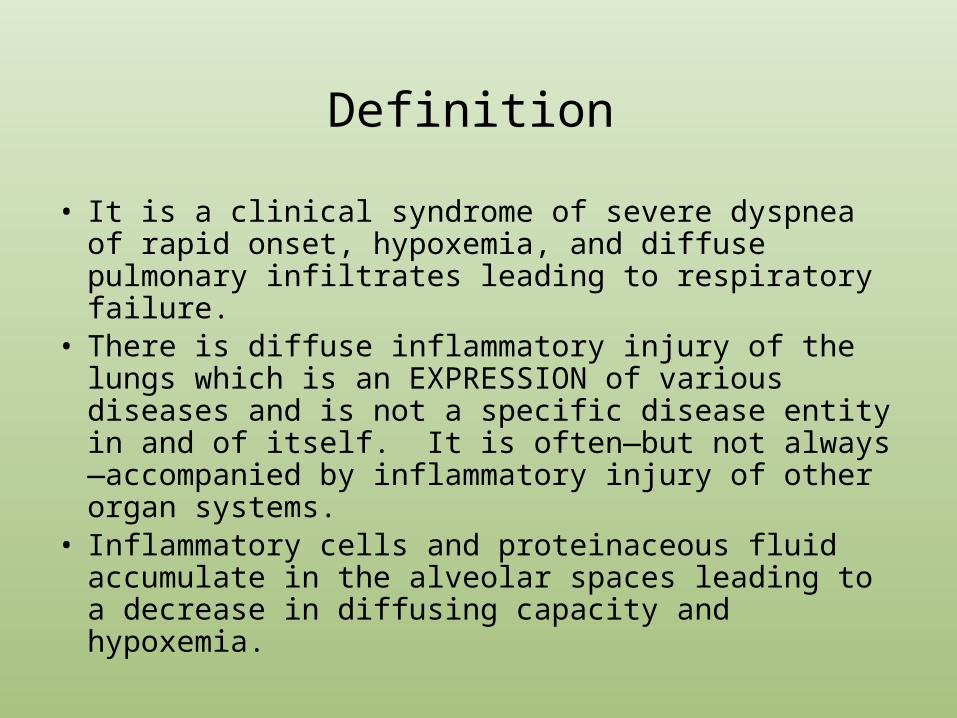
Definition
• It is a clinical syndrome of severe dyspnea of rapid onset, hypoxemia, and diffuse pulmonary infiltrates leading to respiratory failure.
• There is diffuse inflammatory injury of the lungs which is an EXPRESSION of various diseases and is not a specific disease entity in and of itself. It is often—but not always—accompanied by inflammatory injury of other organ systems.
• Inflammatory cells and proteinaceous fluid accumulate in the alveolar spaces leading to a decrease in diffusing capacity and hypoxemia.

Acute Lung Injury (ALI) vs. ARDS
• ALI is the term used for patients with significant hypoxemia (PaO2/FiO2 ratio of <300)
• ARDS is the term used for a subset of ALI patients with severe hypoxemia (PaO2/FiO2 ratio of <200)
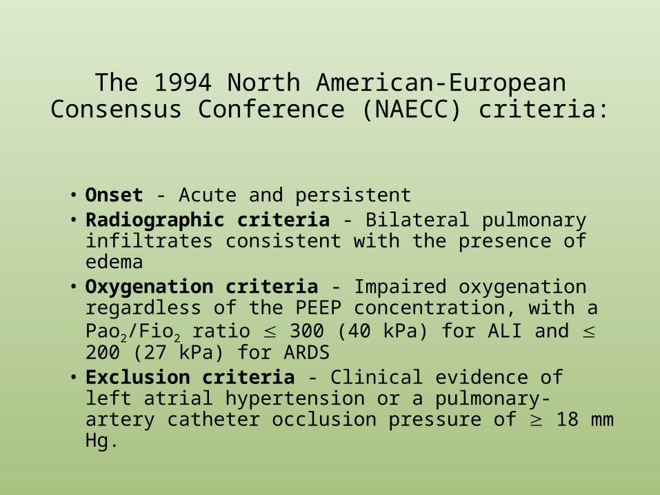
The 1994 North American-European Consensus Conference (NAECC) criteria:
• Onset - Acute and persistent• Radiographic criteria - Bilateral pulmonary infiltrates
consistent with the presence of edema• Oxygenation criteria - Impaired oxygenation regardless
of the PEEP concentration, with a Pao2/Fio2 ratio 300 (40 kPa) for ALI and 200 (27 kPa) for ARDS
• Exclusion criteria - Clinical evidence of left atrial hypertension or a pulmonary-artery catheter occlusion pressure of 18 mm Hg.

The ‘Berlin definition’
Adopted in 2011; initiated by European Society Of Intensive Care Medicine
3 mutually exclusive categories.. based on PaO2/FIO2 (at PEEP> 5)
• Mild- Between 300 and 200 mmHg.• Moderate- Between 100 and 200 mmHg.• Severe- Less than 100 mmHg.
JAMA. 2012;307(23):2526-2533/ doi:10.1001/jama.2012.5669

‘Berlin definition’ G Differences
• Acute Lung Injury.. No longer exists. It is replaced by mild ARDS.
• Onset must be within 7 days of some defined event, like sepsis, pneumonia or worsening of respiratory symptoms.
• Bilateral opacities consistent with Pulmonary edema must be present, but may be detected on CT scan.
• There is no need to exclude heart failure in this new definition. Patients with CHF or high PCOP can still have ARDS.
JAMA. 2012;307(23):2526-2533/ doi:10.1001/jama.2012.5669
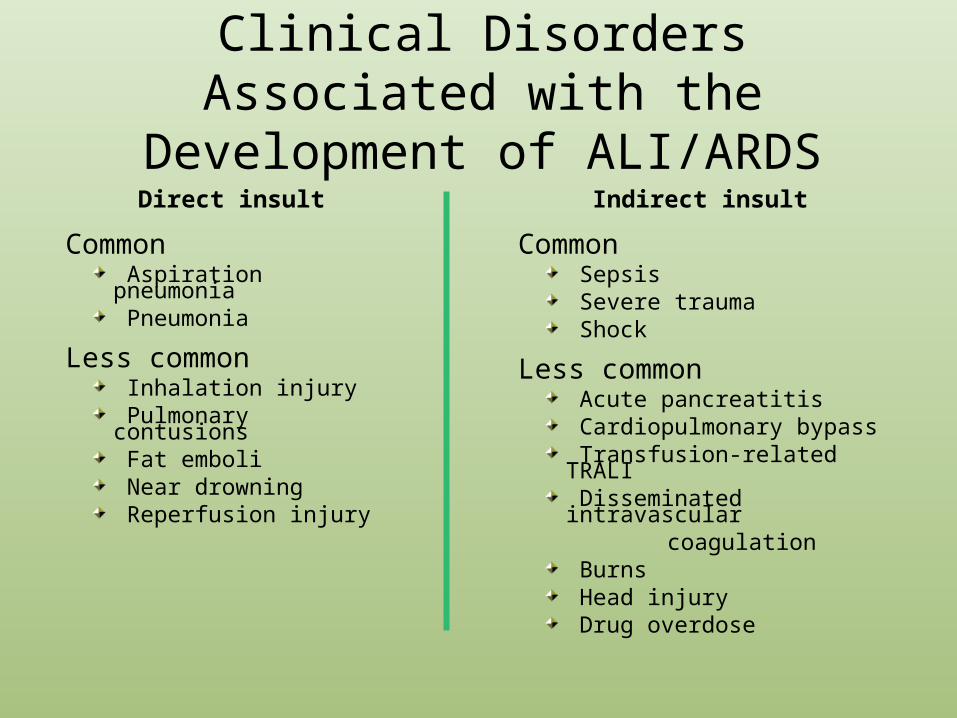
Clinical Disorders Associated with the Development of ALI/ARDS
Direct insult
Common Aspiration pneumonia Pneumonia
Less common Inhalation injury Pulmonary contusions Fat emboli Near drowning Reperfusion injury
Indirect insult
Common Sepsis Severe trauma Shock
Less common Acute pancreatitis Cardiopulmonary bypass Transfusion-related TRALI Disseminated intravascular
coagulation Burns Head injury Drug overdose
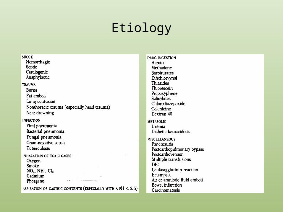
Etiology

ARDS - PATHOGENESIS
Insult (direct or indirect)
Activation of inflammatory cells & mediators
Damage to alveolar capillary membrane
Increased permeability of alveolar capillary membrane
Influx of protein rich edema fluid and inflammatory cells into air spaces
Dysfunction of surfactant


THE STAGES OF ARDS
1. Exudative: occurs over the first 2 to 4 days after onset of lung injury. There is accumulation in the alveoli of excessive fluid, protein and inflammatory cells that have entered the air spaces from the alveolar capillaries
2. Fibroproliferative (or proliferative): Connective tissue and other structural elements in the lungs proliferate in response to the initial injury. Under a microscope, lung tissue appears densely cellular. Also, at this stage, there is a danger of pneumonia sepsis and rupture of the lungs causing leakage of air into surrounding areas.

Stages cont’d…
3. Resolution and Recovery: The lung reorganizes and recovers. Lung function may continue to improve for as long as 6-12 months and sometimes longer, depending on the precipitating condition and severity of the injury
• Some experts recognize a fourth phase of ARDS. This is the period longer than 6-12 months after onset, when some patients experience continued health problems caused by the acute illness. These problems may include cough, limited exercise tolerance and fatigue. Others experience anxiety, depression and flashback memories of their critical illness, which are very similar to post-traumatic stress disorder.

How does the patient present?
• Unexplained tachypnoea (often the 1st sign)• Increasing hypoxemia with central cyanosis• Bilateral fine Inspiratory crackles throughout
the lung field• CXR: bilateral diffuse shadowing diffuse --
interstitial at first but subsequently alveolar pattern with air bronchogram. May progress to a picture of complete “white-out”.

Differential Diagnosis
• CARDIOGENIC PULMONARY EDEMA• Bronchopneumonia• Hypersensitivity pneumonitis• Pulmonary hemorrhage• Acute interstitial pneumonia (Hamman-Rich
Syndrome)

Cardiogenic vs. Non-Cardiogenic Edema
Cardiogenic• Patchy infiltrates appearing in the lung bases first• Effusions may be present• Clinical signs and symptoms lag behind radiographic evidence
(i.e. CXR is more impressive than the degree of hypoxemia)
Non-Cardiogenic• Infiltrates are more homogeneous• No pleural effusions• No Kerley B’s• Radiographic evidence lags behind clinical signs and
symptoms (i.e. the CXR is unimpressive given the degree of hypoxemia)

Clinical Features: CXR

Cardiogenic vs. Non-Cardiogenic Edema via CXR
Cardiogenic Non-Cardiogenic
Bilateral infiltrates predominately in lung bases. Kerley B’s. Cardiomegaly. Diffuse Bilateral patchy infiltrates
homogenously distributed throughout the lungs. Positive tube sign. No Kerley B’s.

Cardiogenic vs. Non-Cardiogenic Edema via CT
CardiogenicNon-Cardiogenic
http://rad.usuhs.edu/m
edpix/medpix_im
age.html?
mode=quiz&
imid=16078&
quiz=no&com
ebackto=mode=caption_li
st
No septal thickening. Diffuse alveolar infiltrates. Atelectasis of dependent lobes usually seen.Septal thickening. More severe in lung
bases.
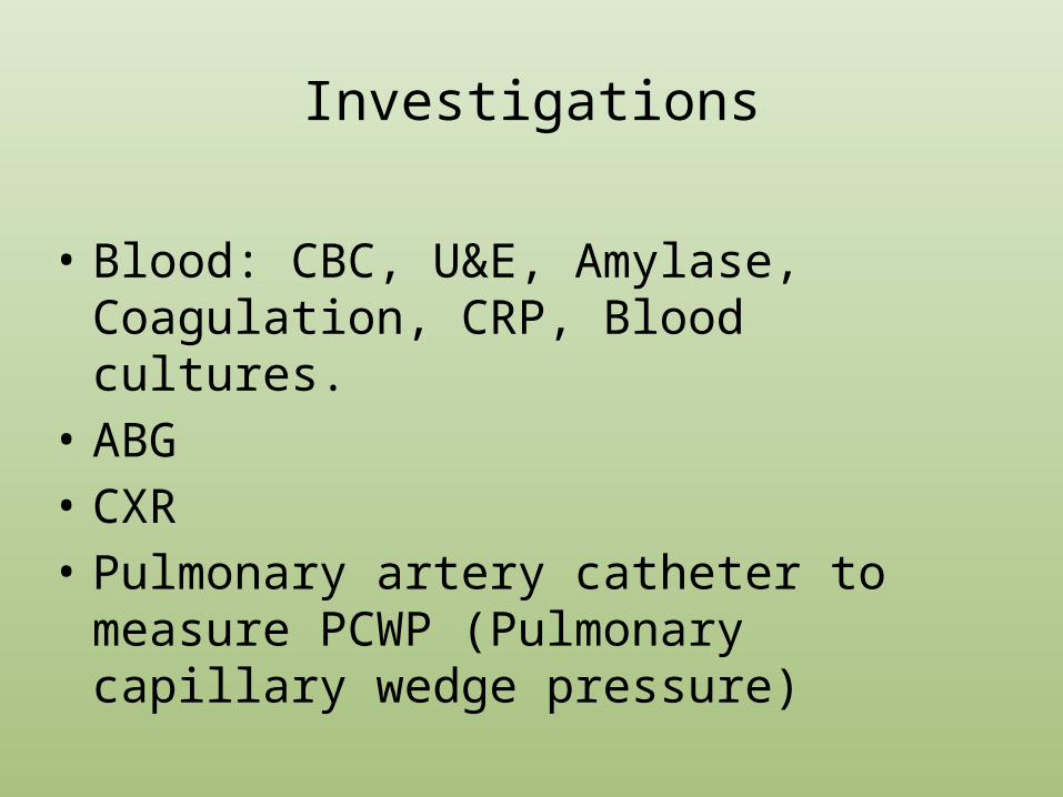
Investigations
• Blood: CBC, U&E, Amylase, Coagulation, CRP, Blood cultures.
• ABG• CXR• Pulmonary artery catheter to measure PCWP
(Pulmonary capillary wedge pressure)

Management of ARDS
• Admit to ICU• Treat underlying illness
– Sepsis, etc• Nutrition• Supportive care (respiratory support, circulatory support)• DVT prophylaxis• GI prophylaxis• Medications• Avoid complications (such as ventilator – associated lung
injury)

Respiratory support
• In early ARDS, CPAP with 40-60% O2 may be adequate to maintain oxygenation.
• However, most patients need invasive ventilation• Indications for ventilation: PaO2<8.3kPa despite 60%
O2• A low tidal-volume, pressure limited approach, with
either low or moderate high PEEP improves the outcomes

Management: Reducing Ventilator-Induced Lung Injury
• Low tidal volume mechanical ventilation– In ARDS there is a large amount of poorly compliant (i.e.
non-ventilating) lung and a small amount of healthy, compliant lung tissue. Large tidal volume ventilation can lead to over-inflation of the healthy lung tissue resulting in ventilator-induced lung injury of that healthy tissue.
• PEEP– Setting a PEEP prevents further lung injury due to shear
forces by keeping airways patent during expiration

Positive End-Expiratory Pressure (PEEP)
• Titrate PEEP to decrease FiO2– Goal sat 88% with FiO2 <60% (Minimize oxygen toxicity)– PEEP can improve lung recruitment and decrease end-
expiratory alveolar collapse (and therefore right-to-left shunt)
– Can also decrease venous return, cause hemodynamic compromise, worsen pulmonary edema
• ARDS-net PEEP trial of 549 patients show no difference in mortality or days on ventilator with high vs low PEEP
NEJM 2004:351(4):327-336

The Flip Side• Is there such thing as too low a TV?
– Tidal Volume must be sufficient for gas exchange to take place. Permissive hypercapnia is the term used to state that a certain degree of hypercapnia and its resulting acidemia can be allowed in order to maintain lung-protective TVs.
• Absolute limits is unclear, but a pH of 7.2-7.25 and a PCO2 of 60-70 mm Hg is a good cut off range.
• Is there such thing as too much PEEP? – PEEP serves to help open less compliant alveoli and keep alveoli open
during expiration, but it too can lead to overinflation of alveoli that are already maintaining aeration.
– Setting PEEP too high also increases intrathoracic pressure leading to decreased venous return.
• Start patients at a PEEP trial of 5 – 12 cm H2O and increase if needed.

Other Ideas in Ventilator Management
• Prone positioning– May be beneficial in certain subgroup, but complications including
pressure sores• RCT of 304 patients showed no mortality benefit
• High-frequency oscillatory ventilation– In RCT, improved oxygenation initially, but results not sustained after 24
hours, no mortality benefit• ECMO -- Extracorporeal membrane oxygenation, provides continuous
cardiopulmonary support on a long-term basis, typically days to weeks, as adjunctive management. The goal of therapy is to minimize ventilator-induced lung injury while allowing additional time to treat the underlying disease process and to permit recovery from acute injury. – RCT of 40 adults showed no benefit

Circulatory support
• Invasive haemodynamic monitoring • Conservative fluid management approach
improves outcomes• Inotropes to maintain Cardiac output and O2
delivery

Fluid management
• “Dry lungs are happy lungs” • ARDSnet RCT of 1000 patients (FACTT), Conservative vs
liberal fluid strategy using CVP or PAOP (Pulmonary artery occlusion pressure) monitoring to guide, primary outcome: death.
• Conservative fluids– Improved oxygenation– More ventilator-free days– More days outside ICU– No increase in shock or dialysis– No mortality effects

Diuretics—A Good or Bad Therapy in ARDS?
Yes• Diuretics have been shown to decrease any pulmonary edema
that is present, increase lung compliance, and improve gas exchange. However they have shown no survival benefit.
No• Diuretics are not anti-inflammatory agents: lung infiltrates in
ARDS are neutrophils and proteins, NOT edema• Hemodynamic compromise: tissue oxygenation is the #1
concern. Aggressive diuretics decrease venous pressures leading to decrease cardiac output and increased tissue ischemia

FACTT Study: The Fluid and Catheter Treatment Trial (FACTT)
• Large prospective trial addressed the use of conservative (higher, more frequent lasix doses) Vs liberal fluid management (more frequent fluid boluses).
• Outcomes: NO significant difference in 60-day mortality between the two groups, however the conservative fluid group had improved lung function, shorter durations of mechanical ventilation, and shorter ICU stays, SUPPORTING THE USE OF DIURETICS.
ARDS Clinical Trial Network. 2006. Comparison of Two Fluid-Management Strategies in Acute Lung Injury. N Engl J Med. 354 (24). pp 2564-75.

Management: Fluid Status
• Remember, the #1 goal in therapy is to decrease tissue ischemia. We must maintain ARDS patient’s cardiac output to insure tissue profusion.– In the FACTT study, conservative fluid
therapy was not followed if a patient was deemed to be in shock, in the presence of oliguria, or if a patient’s circulation was deemed inadequate.

Use of Pulmonary Arterial Catheters in ARDS
Swan-Ganz Catheter

Uses of the PAC in ARDS
• Used to aid in diagnosis– Traditionally placed to confirm non-cardiogenic
edema verses cardiogenic edema in cases of uncertainty
• If PCWP is elevated > 18 mm Hg then by diagnostic criteria—as set by the American-European Consensus—the edema is NOT noncardiogenic.
• Used to guide treatment

Should PCWP Be Used to Confirm the Diagnosis of ARDS?
• PCWP is an estimate of left atrium pressure– When the PAC balloon is inflated it occludes blood
flow through the lungs. The pressure measured in this closed circuit is equal to the pressure in the left-atrium
• In ARDS, PCWP is used to estimate pulmonary capillary pressure. PCWP CANNOT be equal to pulmonary capillary pressure and left atrial pressure. If this were true there would be no pressure gradient making forward blood flow through the pulmonary arteries possible. Therefore PCWP underestimates pulmonary capillary pressure.
• This suggests that wedge pressure should not be part of the diagnostic criterion for ARDS.

Should PCWP Be Used to Dictate the Treatment of ARDS?
• The Fluid and Catheter Treatment Trial (FACTT)– A randomized, multi-center trial comparing
outcomes of ARDS patients with use of PACs vs. CVCs (central venous catheters).

FACTT
• Result: No difference in mortality, number of days on the ventilator or in the ICU, lung or kidney function, rates of hypotension, ventilator settings or use of dialysis between two groups. The PAC group had ≈ twice as many catheter-related complications (mainly arrhythmias).
ARDS Clinical Trial Network. 2006. Pulmonary-Artery versus Central Venous Catheter to Guide Treatment of Acute Lung Injury. N Engl J Med. 354 (21). pp 2213-24.

FACTT Conclusions in Regards to PACs
• PAC-guided therapy for ARDS does not improve survival or organ-function, reduce ventilator time or decrease ICU-stays. Although associated with more complications, major harm did not occur from PAC use. The evidence does not favor the routine use of the PAC.

Drug therapy
• Agents studied:– Corticosteroids– Ketoconazole– Inhaled nitric oxide– Surfactant
• No benefit demonstrated

Steroids in ARDS
• Earlier studies showed no benefit to early use steroids, but small study in 1990s showed improved oxygenation and possible mortality benefit in late stage
• ARDSnet trial (Late Steroid Rescue Study “LaSRS” – “lazarus”) of steroids 7+ days out from onset of ARDS
• 180 patients enrolled, RCT methylprednisolone vs placebo• Overall, no mortality benefit
– Steroids increased mortality in those with sx >14 days
JAMA 1998;280:159-65, N Engl J Med 2006;354:1671-84

Other drugs in ARDS• Ketoconazole
– ARDSnet study of 234 patients, ketoconazole did NOT decrease mortality, duration of mechanical ventilation or improve lung function
• Surfactant– Multicenter trial, 725 patients with sepsis-induced ARDS,
surfactant had no effect on 30-day survival, ICU LOS, duration of mechanical ventilation or physiologic function
• Inhaled Nitric oxide– 177 patients RCT, improved oxygenation, but no effect on
mortality of duration of mechanical ventilation
N Engl J Med. 1996;334:1417-21. Crit Care Med. 1998;26:15-23.
ARDS - Prognosis
Improved survival in recent years – mortality was 50-60% for many years, now 25-40%
Improvements in supportive care, newer ventilatory strategies
Early deaths (3 days) usually from underlying cause of ARDS
Later deaths from nosocomial infections, sepsis, MOSF Severity of gas exchange at admission does not correlate
with mortality Respiratory failure only responsible for ~16% of fatalities Long-term survivors usually show mild abnormalities in
pulmonary function (DLCO), impaired neurocognitive function

Poor Prognostic Factors
• Failure to improve over 1st few days• Initially increased dead space• Advanced age• Sepsis• Multiple organ dysfunction (higher APACHE)• Steroids given prior to onset of ARDS• Blood transfusion• Not managed by Intensivist

1 year after ARDS survival
• Lung Function:– FEV1 and FVC were normal; DLCO minimally reduced– Only 20% had mild abnormalities on CXR
• Functionally:– Survivors’ perception of health was <70% of normals in:
• Physical Role: Extent to which health limits physical activity• Physical Functioning: Extent to which health limits work• Vitality: Degree of energy patients have
– 6 minutes walk remained low– Only 49% had returned to work
NEJM 2003: 348: 683-693
Summary
• ARDS is a clinical syndrome characterized by severe, acute lung injury, inflammation and scarring
• Significant cause of ICU admissions, mortality and morbidity
• Caused by either direct or indirect lung injury• Mechanical ventilation with low tidal volumes and
plateau pressures improves outcomes• So far, no pharmacologic therapies have demonstrated
mortality benefit• Ongoing large, multi-center randomized controlled trials
are helping us better understand optimal management

Thank you !

Complications in Managing ARDS patients
• Mechanical ventilation causes:– Overdistention of lungs (volutrauma)
• Further damaging epithelium• Increased fluid leak, indistinguishable from ARDS damage
– Barotrauma• Rupture alveolar membranes• Pneuomothorax, pneumomediastinum
– Sheer stress• Opening/closing alveoli• Inflammatory reaction, cytokine release
• Oxygen toxicity– Free radical formation

ARDS Network
• NIH-funded consortium of 10 centers, 24 hospitals, 75 intensive care units
• Goal to design large RCTs to determine effective treatments
• Key ARDSnet studies:– Ventilator volumes– Steroids– PEEP– Volume management/PA catheter

Pulmonary artery catheters
• Often used to help evaluate for cardiogenic pulmonary edema
• SUPPORT trial (retrospective study) first raised doubts about utility
• Two multicenter RCTs confirmed lack of mortality benefit of PA catheters in ARDS (ARDSnet FACTT)
• Monitoring CVP equally effective, so PAC not recommended in routine management
JAMA. 1996;276:889-97. N Engl J Med. 2006:354:2213-24

Ventilator management – ARDSnet protocol
• 861 patients randomized to Vt 10-12 mg/kg ideal body weight and plateau pressure ≤50cmH2O vs Vt 6-8 mg/kg IBW and plateau pressure ≤30cm H2O
• KEYS– Low tidal volumes – 6-8mL/kg ideal body weight– Maintain plateau (end-inspiratory) pressures <30cm H20– Permissive hypercapnia and acidosis
• Decreased mortality by 22%
NEJM 2000;342:1301-8.

ARDSnet Tidal Volume Study
NEJM 2000;342:1301-8.

Risk Factors in ARDS
Sepsis 3.8% Cardiopulmonary bypass 1.7% Transfusion 5.0% Severe pneumonia 12.0% Burn 2.3% Aspiration 35.6% Fracture 5.3% Intravascular coagulopathy 12.5% Two or more of the above 24.6%

Cardiogenic Edema:Weak ENDOthelium
• Vascular Endothelium breaks under stress easily, however it also repairs itself quickly
• Cardiogenic edema often develops quickly and can resolve quickly because vascular endothelium is able to repair itself quickly

Non-Cardiogenic Edema: strong EPIthelium
• Alveolar epithelium is quite resistant to damage. It withstands greater force before becoming damaged. However, once “broken” it takes much longer to heal than weenie man endothelium.
• Cellular damage in Non-Cardiogenic edema runs along a spectrum from predominately vascular endothelial damage to predominately alveolar epithelial damage