Noise drives sharpening of gene expression boundaries in...
12
Noise drives sharpening of gene expression boundaries in the zebrafish hindbrain Lei Zhang 1,2,3,4,7 , Kelly Radtke 5,7 , Likun Zheng 1,2,3 , Anna Q Cai 6 , Thomas F Schilling 2,5, * and Qing Nie 1,2,3, * 1 Department of Mathematics, University of California, Irvine, CA, USA, 2 Center for Complex Biological Systems, University of California, Irvine, CA, USA, 3 Center for Mathematical and Computational Biology, University of California, Irvine, CA, USA, 4 Department of Mathematics, City University of Hong Kong, Kowloon Tong, Hong Kong, 5 Department of Development and Cell Biology, University of California, Irvine, CA, USA and 6 Department of Applied Mathematics, School of Mathematics and Statistics, University of New South Wales, Sydney, New South Wales, Australia 7 These authors contributed equally to this work. * Corresponding authors. TF Schilling, Department of Development and Cell Biology, University of California, 4109 Natural Sciences II, Irvine, CA 92697-2300, USA. Tel.: þ 1 949 824 2479; Fax: þ 1 949 824 4709; E-mail: [email protected] or Q Nie, Department of Mathematics, University of California, 540F Rowland Hall, Irvine, CA 92697-3875, USA. Tel.: þ 1 949 824 5530; Fax: þ 1 949 824 7993; E-mail: [email protected] Received 16.3.12; accepted 16.8.12 Morphogens provide positional information for spatial patterns of gene expression during development. However, stochastic effects such as local fluctuations in morphogen concentration and noise in signal transduction make it difficult for cells to respond to their positions accurately enough to generate sharp boundaries between gene expression domains. During development of rhombomeres in the zebrafish hindbrain, the morphogen retinoic acid (RA) induces expression of hoxb1a in rhombomere 4 (r4) and krox20 in r3 and r5. Fluorescent in situ hybridization reveals rough edges around these gene expression domains, in which cells co-express hoxb1a and krox20 on either side of the boundary, and these sharpen within a few hours. Computational analysis of spatial stochastic models shows, surprisingly, that noise in hoxb1a/krox20 expression actually promotes sharpening of boundaries between adjacent segments. In particular, fluctuations in RA initially induce a rough boundary that requires noise in hoxb1a/krox20 expression to sharpen. This finding suggests a novel noise attenuation mechanism that relies on intracellular noise to induce switching and coordinate cellular decisions during developmental patterning. Molecular Systems Biology 8: 613; published online 25 September 2012; doi:10.1038/msb.2012.45 Subject Categories: simulation and data analysis; development Keywords: cellular decision; intracellular noise; morphogen; signal transduction network; stochastic fluctuation Introduction A fundamental feature of developing systems is that cells sense their positions along morphogen gradients and respond collectively to form precise domains of target gene expres- sion (Meinhardt, 2009; Wartlick et al, 2009). How do gene expression domains achieve such sharp boundaries? Cells near a future boundary experience fluctuations or ‘noise’ in: (1) morphogen concentration, due to varying synthesis and transport, (2) ability to respond, for example, due to differ- ences in numbers of receptors, (3) transcription and trans- lation rates of target genes, and (4) feedback (Kepler and Elston, 2001; Elowitz et al, 2002; Kaern et al, 2005). Various mechanisms have been proposed to attenuate these sources of noise to generate consistent gene expression domains in every individual. In spatial patterning systems, noise is generally considered as detrimental to the ultimate goal of the system. However, for systems without spatial constraints, noise can regulate biological switches between high and low gene expres- sion states, and noise can be attenuated by an ultrasensitive signal (Hasty et al, 2000; Thattai and van Oudenaarden, 2002; To and Maheshri, 2010). Could similar switches be operating in spatial patterning systems? Bistability (distinct steady states of a regulatory gene network within a cell) can have a critical role in spatial patterning and lead to sharp borders between gene expression domains in deterministic models (Meinhardt, 1978, 1982; Ferrell, 2002; Lopes et al, 2008). Spatially constrained stochastic models, such as for segmentation of the Drosophila embryo, suggest that noise predominantly depends on trans- cription and translation dynamics of target gene expression (Holloway et al, 2011), but external fluctuations in signals also have an important role in these downstream responses (He et al, 2012). However, very few studies have addressed mechanisms of noise attenuation in the formation of gene expression boundaries in any system. Here, we investigate interactions between noise in a morpho- gen (i.e., retinoic acid—RA) and noise in its downstream, bistable regulatory gene network in boundary sharpening. RA specifies rough boundaries between segments (called rhombomeres) of the zebrafish hindbrain in a concentration- dependent manner, which subsequently become razor sharp (Giudicelli et al, 2001; Cooke and Moens, 2002; White et al, 2007; White and Schilling, 2008). Two genes downstream of RA, hoxb1a (r4) and krox20 (r3 and r5), cross-inhibit one Molecular Systems Biology 8; Article number 613; doi:10.1038/msb.2012.45 Citation: Molecular Systems Biology 8:613 & 2012 EMBO and Macmillan Publishers Limited All rights reserved 1744-4292/12 www.molecularsystemsbiology.com & 2012 EMBO and Macmillan Publishers Limited Molecular Systems Biology 2012 1
-
Upload
phungnguyet -
Category
Documents
-
view
214 -
download
0
Transcript of Noise drives sharpening of gene expression boundaries in...
Noise drives sharpening of gene expressionboundaries in the zebrafish hindbrain
Lei Zhang1,2,3,4,7, Kelly Radtke5,7, Likun Zheng1,2,3, Anna Q Cai6, Thomas F Schilling2,5,* and Qing Nie1,2,3,*
1 Department of Mathematics, University of California, Irvine, CA, USA, 2 Center for Complex Biological Systems, University of California, Irvine, CA, USA, 3 Center forMathematical and Computational Biology, University of California, Irvine, CA, USA, 4 Department of Mathematics, City University of Hong Kong, Kowloon Tong,Hong Kong, 5 Department of Development and Cell Biology, University of California, Irvine, CA, USA and 6 Department of Applied Mathematics, School of Mathematicsand Statistics, University of New South Wales, Sydney, New South Wales, Australia7These authors contributed equally to this work.* Corresponding authors. TF Schilling, Department of Development and Cell Biology, University of California, 4109 Natural Sciences II, Irvine, CA 92697-2300, USA.Tel.: þ 1 949 824 2479; Fax: þ 1 949 824 4709; E-mail: [email protected] or Q Nie, Department of Mathematics, University of California, 540F Rowland Hall, Irvine,CA 92697-3875, USA. Tel.: þ 1 949 824 5530; Fax: þ 1 949 824 7993; E-mail: [email protected]
Received 16.3.12; accepted 16.8.12
Morphogens provide positional information for spatial patterns of gene expression duringdevelopment. However, stochastic effects such as local fluctuations in morphogen concentrationand noise in signal transduction make it difficult for cells to respond to their positions accuratelyenough to generate sharp boundaries between gene expression domains. During development ofrhombomeres in the zebrafish hindbrain, the morphogen retinoic acid (RA) induces expression ofhoxb1a in rhombomere 4 (r4) and krox20 in r3 and r5. Fluorescent in situ hybridization revealsrough edges around these gene expression domains, in which cells co-express hoxb1a and krox20 oneither side of the boundary, and these sharpen within a few hours. Computational analysis of spatialstochastic models shows, surprisingly, that noise in hoxb1a/krox20 expression actually promotessharpening of boundaries between adjacent segments. In particular, fluctuations in RA initiallyinduce a rough boundary that requires noise in hoxb1a/krox20 expression to sharpen. This findingsuggests a novel noise attenuation mechanism that relies on intracellular noise to induce switchingand coordinate cellular decisions during developmental patterning.Molecular Systems Biology 8: 613; published online 25 September 2012; doi:10.1038/msb.2012.45Subject Categories: simulation and data analysis; developmentKeywords: cellular decision; intracellular noise; morphogen; signal transduction network; stochasticfluctuation
Introduction
A fundamental feature of developing systems is that cellssense their positions along morphogen gradients and respondcollectively to form precise domains of target gene expres-sion (Meinhardt, 2009; Wartlick et al, 2009). How do geneexpression domains achieve such sharp boundaries? Cellsnear a future boundary experience fluctuations or ‘noise’ in:(1) morphogen concentration, due to varying synthesis andtransport, (2) ability to respond, for example, due to differ-ences in numbers of receptors, (3) transcription and trans-lation rates of target genes, and (4) feedback (Kepler andElston, 2001; Elowitz et al, 2002; Kaern et al, 2005). Variousmechanisms have been proposed to attenuate these sources ofnoise to generate consistent gene expression domains in everyindividual. In spatial patterning systems, noise is generallyconsidered as detrimental to the ultimate goal of the system.
However, for systems without spatial constraints, noise canregulate biological switches between high and low gene expres-sion states, and noise can be attenuated by an ultrasensitivesignal (Hasty et al, 2000; Thattai and van Oudenaarden, 2002;To and Maheshri, 2010). Could similar switches be operating in
spatial patterning systems? Bistability (distinct steady states ofa regulatory gene network within a cell) can have a critical rolein spatial patterning and lead to sharp borders between geneexpression domains in deterministic models (Meinhardt, 1978,1982; Ferrell, 2002; Lopes et al, 2008). Spatially constrainedstochastic models, such as for segmentation of the Drosophilaembryo, suggest that noise predominantly depends on trans-cription and translation dynamics of target gene expression(Holloway et al, 2011), but external fluctuations in signalsalso have an important role in these downstream responses(He et al, 2012). However, very few studies have addressedmechanisms of noise attenuation in the formation of geneexpression boundaries in any system.
Here, we investigate interactions between noise in a morpho-gen (i.e., retinoic acid—RA) and noise in its downstream,bistable regulatory gene network in boundary sharpening.RA specifies rough boundaries between segments (calledrhombomeres) of the zebrafish hindbrain in a concentration-dependent manner, which subsequently become razor sharp(Giudicelli et al, 2001; Cooke and Moens, 2002; White et al,2007; White and Schilling, 2008). Two genes downstream ofRA, hoxb1a (r4) and krox20 (r3 and r5), cross-inhibit one
Molecular Systems Biology 8; Article number 613; doi:10.1038/msb.2012.45Citation: Molecular Systems Biology 8:613& 2012 EMBO and Macmillan Publishers Limited All rights reserved 1744-4292/12www.molecularsystemsbiology.com
& 2012 EMBO and Macmillan Publishers Limited Molecular Systems Biology 2012 1
another and auto-activate their own expression to form abistable switch (Barrow et al, 2000; Giudicelli et al, 2001;Alexander et al, 2009). With a stochastic model that incorpo-rates these interactions we estimate the switching probabilitybetween hoxb1a and krox20 expression at different RA concen-trations based on an exponential function of Minimum ActionPaths (MAPs) between stable and unstable states (Freidlin andWentzell, 1998). Exploration of the stochastic models revealsthat noise in the RA morphogen gradient can lead to roughgene expression boundaries initially, and that sharpening isdriven by noise in the expression of hoxb1a and krox20, due toinduced switching between expression of one gene and theother. These results reveal an unexpected positive role fornoise in boundary sharpening that may be common for manypatterning systems.
Results
hoxb1a and krox20 co-expression duringrhombomere boundary sharpening
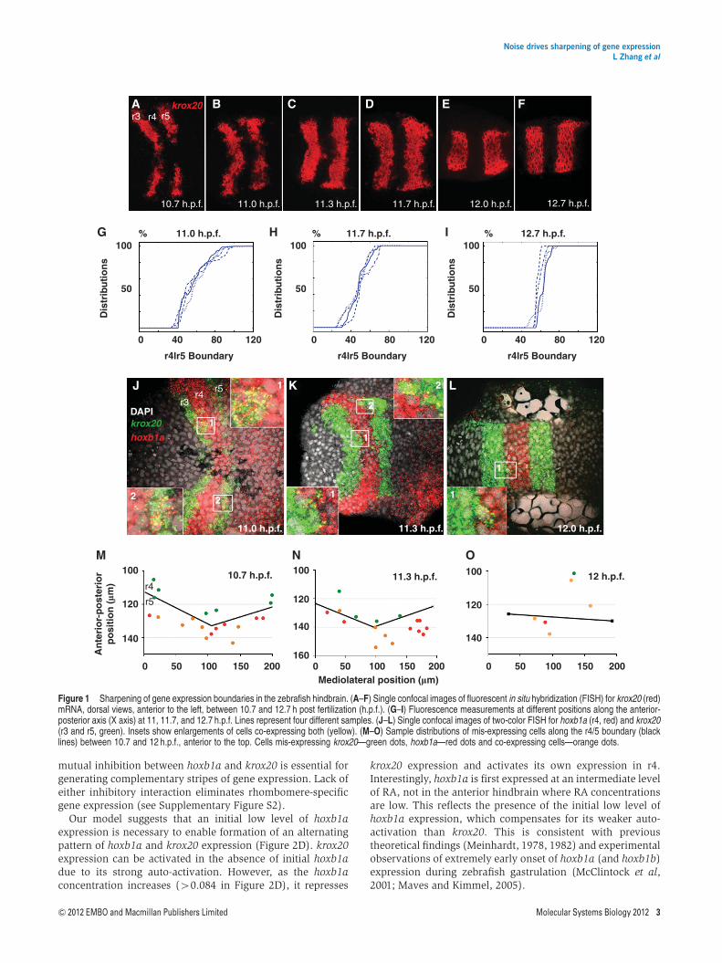
To determine the temporal dynamics of hoxb1a and krox20expression in the embryonic zebrafish hindbrain, we per-formed fluorescent in situ hybridization (FISH) analysis.Previous studies showed that initial boundaries of hoxb1a inr4 and krox20 expression in r3 and r5 are rough but becomerazor sharp between 10 and 14 h post fertilization (h.p.f.)(Figure 1A–F; Cooke and Moens, 2002; Cooke et al, 2005).Cells that find themselves on the wrong side of a boundary(i.e., surrounded by neighbors with a different pattern of geneexpression) may go through a transient phase in which theyexpress both genes and subsequently downregulate one or theother to enable sharpening (Schilling et al, 2001; Cooke andMoens, 2002). To quantify sharpness in krox20 expression,confocal stacks were collected for a minimum of 10 embryos at6 different stages (between 10.7 and 12.7 h.p.f.) (Figure 1A–F)and the fluorescence was measured at different positions alongthe anterior-posterior (A-P) axis focusing on the r4/5 boundary(Figure 1G–I). This analysis demonstrated quantitatively howkrox20 expression sharpens at rhombomere boundaries overtime.
To examine this more closely, we used confocal analysis andtwo-color FISH to colocalize hoxb1a and krox20 near the r3/4and r4/5 boundaries at 20-min intervals between 10.6 and12 h.p.f. (Figure 1J–L). hoxb1a expression is initiated broadlyin the early gastrula (6.5 h.p.f.; Maves and Kimmel, 2005), andis preceded by its close relative hoxb1b, which is the first geneinduced by RA in this system and activates hoxb1a transcrip-tion directly (McClintock et al, 2001). By 10.5 h.p.f. hoxb1aexpression resolves into a strong r4 stripe 4–6 cells wide alongthe A-P axis while krox20 is expressed in flanking r3 and r5stripes that overlap with hoxb1a at its edges (Figure 1J–L).Higher magnification images demonstrated that krox20and hoxb1a mRNAs colocalize in many of these cells nearfuture boundaries (insets) and occasional colocalizationwas observed as late as 12.0 h.p.f. This revealed an initial‘transition zone’ containing a mixture of hoxb1a, krox20 andco-expressing cells that was B40 mm in length along the A-Paxis and later reduced to 5–10mm (1 cell diameter) by 12 h.p.f.Similar numbers of co-expressing cells were identified at
10.7 h.p.f. (average 7 cells, n¼ 3) and 11.3 h.p.f. (average 7.3cells, n¼ 3), however, by 12 h.p.f. the number of co-expressingcells decreased (average 3, n¼ 3). Conversely, the percentageof mis-specified cells that were expressing both genesincreased from 36% and 34% at 10.7 and 11.3 h.p.f. to 56%at 12 h.p.f. The co-expressing cells were more prevalent in ther5 domain at both 10.7 and 11.3 h.p.f. (Figure 1M and N) whilethis bias was not observed at 12 h.p.f. (Figure 1O).
Induction of stripes of hoxb1a and krox20expression by RA requires bistability and initialexpression of Hoxb1
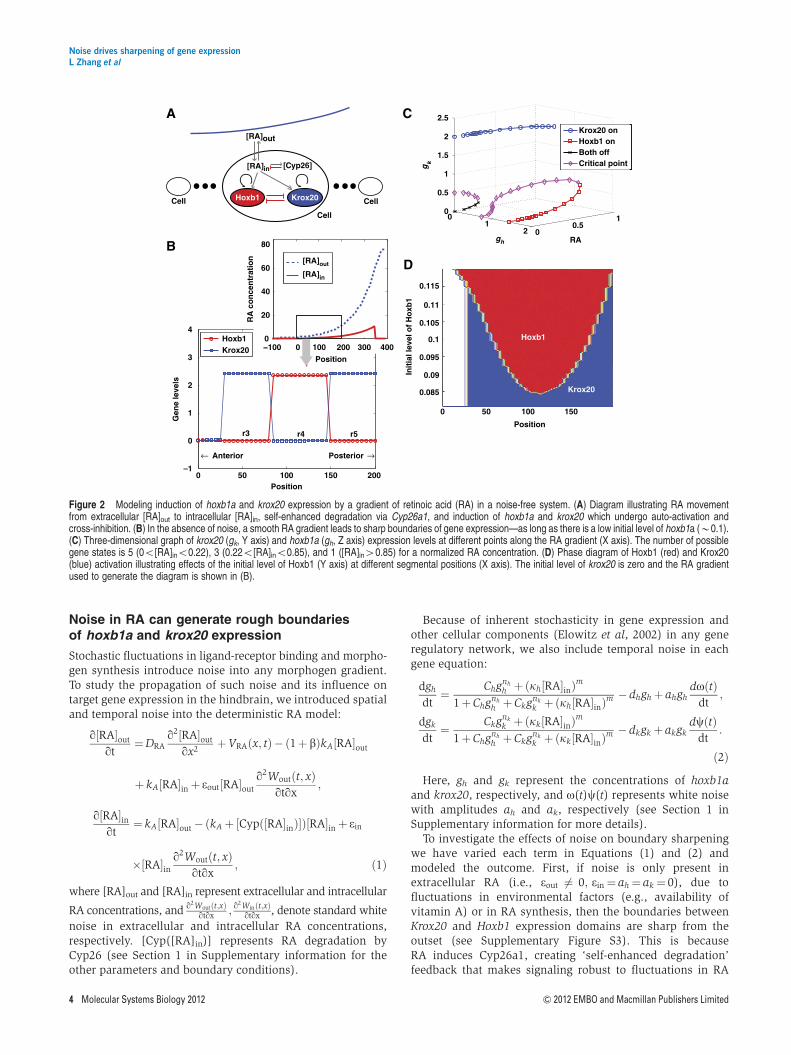
RA activates hoxb1a expression in r4 (directly) and krox20 inr3 and r5 (indirectly through Vhnf1 and MafB) in a concentra-tion-dependent manner (Niederreither et al, 2000; Begemannet al, 2001; Hernandez et al, 2004; Labalette et al, 2011). Ourdeterministic model is based on a previous continuum modelof the RA signaling network that consists of diffusiveextracellular and intracellular RA, and self-enhanced degrada-tion through the enzyme Cyp26a1 (White et al, 2007), withoutinclusion of downstream signal responses (see Equation S1.1in Supplementary information). In the new model, RAactivates hoxb1a and krox20 expression, which in turn bothpositively regulate their own expression and negativelyregulate each other (Barrow et al, 2000; Giudicelli et al,2001; Alexander et al, 2009; Figure 2A). Such positive auto-regulation and mutual inhibition have been modeled andshown to result in only one gene remaining active in aparticular cell (Meinhardt, 1978, 1982). Here, the dynamics ofboth genes are modeled using rate equations along with Hillfunctions for regulation, with RA as input (see Equation S1.2 inSupplementary information).
Exploration of the model reveals that the system robustlyresolves into a striped pattern of gene expression withhoxb1a in r4 and krox20 in r3 and r5 (Figure 2B). Thisdemonstrates that by simply including two bistable steadystates and an anteriorly declining RA gradient, one can specifyalternating gene expression patterns with sharp boundaries.Simulations in two dimensions show a similar striped pattern(Supplementary Figure S1). Auto-activation and mutual inhi-bition between Hoxb1 and Krox20 allow one to switch fromthe off to the on state, or vice versa, within a range of RA.In particular, at a low RA concentration (RAo0.22 mM), thereare three stable states (hoxb1a-on, krox20-on, or both off) andtwo unstable critical transition states (Figure 2C). As the RAconcentration increases above 0.22 mM, both the off stateand one unstable transition state merge and disappear, whileother states (hoxb1a-on, krox20-on, and another unstabletransition state) remain (Figure 2C). If the RA concentration ishigh (larger than 0.85 mM in Figure 2C), then the hoxb1a-onand unstable transition states disappear and only krox20 isactivated.
Because of the monotonic spatial distribution of RA (andadditional influences from Fgf signaling) (Hernandez et al,2004; White et al, 2007; Labalette et al, 2011), the activation(and auto-activation) of krox20 is likely stronger than hoxb1a,at least in r5. However, our models suggest that enhancedactivation alone cannot create this bistability. Rather, the
Noise drives sharpening of gene expressionL Zhang et al
2 Molecular Systems Biology 2012 & 2012 EMBO and Macmillan Publishers Limited
mutual inhibition between hoxb1a and krox20 is essential forgenerating complementary stripes of gene expression. Lack ofeither inhibitory interaction eliminates rhombomere-specificgene expression (see Supplementary Figure S2).
Our model suggests that an initial low level of hoxb1aexpression is necessary to enable formation of an alternatingpattern of hoxb1a and krox20 expression (Figure 2D). krox20expression can be activated in the absence of initial hoxb1adue to its strong auto-activation. However, as the hoxb1aconcentration increases (40.084 in Figure 2D), it represses
krox20 expression and activates its own expression in r4.Interestingly, hoxb1a is first expressed at an intermediate levelof RA, not in the anterior hindbrain where RA concentrationsare low. This reflects the presence of the initial low level ofhoxb1a expression, which compensates for its weaker auto-activation than krox20. This is consistent with previoustheoretical findings (Meinhardt, 1978, 1982) and experimentalobservations of extremely early onset of hoxb1a (and hoxb1b)expression during zebrafish gastrulation (McClintock et al,2001; Maves and Kimmel, 2005).
r3 r4 r5
10.7 h.p.f. 11.0 h.p.f. 11.3 h.p.f. 11.7 h.p.f. 12.0 h.p.f. 12.7 h.p.f.
12.7 h.p.f.11.7 h.p.f.11.0 h.p.f.% % %100
50
0
r4lr5 Boundary
Dis
trib
uti
on
s
100
50D
istr
ibu
tio
ns
100
A
G
M N O
H I
J K L
B C D E F
50
Dis
trib
uti
on
s
40 80 1200
r4lr5 Boundary
40 80 1200
r4lr5 Boundary
40 80 120
krox20
r5r4
r3
2 2
1
1
1
2
2
1 1
1
11.0 h.p.f. 11.3 h.p.f. 12.0 h.p.f.
12 h.p.f.
200150100500200150100500Mediolateral position (�m)
An
teri
or-
po
ster
ior
po
siti
on
(�m
)
200150100500
140140
160
120120
100100
140
120
10011.3 h.p.f.10.7 h.p.f.
r4
r5
DAPIkrox20hoxb1a
Figure 1 Sharpening of gene expression boundaries in the zebrafish hindbrain. (A–F) Single confocal images of fluorescent in situ hybridization (FISH) for krox20 (red)mRNA, dorsal views, anterior to the left, between 10.7 and 12.7 h post fertilization (h.p.f.). (G–I) Fluorescence measurements at different positions along the anterior-posterior axis (X axis) at 11, 11.7, and 12.7 h.p.f. Lines represent four different samples. (J–L) Single confocal images of two-color FISH for hoxb1a (r4, red) and krox20(r3 and r5, green). Insets show enlargements of cells co-expressing both (yellow). (M–O) Sample distributions of mis-expressing cells along the r4/5 boundary (blacklines) between 10.7 and 12 h.p.f., anterior to the top. Cells mis-expressing krox20—green dots, hoxb1a—red dots and co-expressing cells—orange dots.
Noise drives sharpening of gene expressionL Zhang et al
& 2012 EMBO and Macmillan Publishers Limited Molecular Systems Biology 2012 3
Noise in RA can generate rough boundariesof hoxb1a and krox20 expression
Stochastic fluctuations in ligand-receptor binding and morpho-gen synthesis introduce noise into any morphogen gradient.To study the propagation of such noise and its influence ontarget gene expression in the hindbrain, we introduced spatialand temporal noise into the deterministic RA model:
q½RA�out
qt¼DRA
q2½RA�out
qx2þVRAðx; tÞ� ð1þ bÞkA½RA�out
þ kA½RA�inþ eout½RA�out
q2Woutðt; xÞqtqx
;
q½RA�inqt
¼ kA½RA�out�ðkAþ ½Cypð½RA�inÞ�Þ½RA�inþ ein
�½RA�inq2Woutðt; xÞ
qtqx; ð1Þ
where [RA]out and [RA]in represent extracellular and intracellular
RA concentrations, and q2Woutðt;xÞqtqx ; q
2Winðt;xÞqtqx , denote standard white
noise in extracellular and intracellular RA concentrations,respectively. [Cyp([RA]in)] represents RA degradation byCyp26 (see Section 1 in Supplementary information for theother parameters and boundary conditions).
Because of inherent stochasticity in gene expression andother cellular components (Elowitz et al, 2002) in any generegulatory network, we also include temporal noise in eachgene equation:
dgh
dt¼ Chgnh
h þðkh½RA�inÞm
1þChgnh
h þCkgnk
k þðkh½RA�inÞm � dhghþ ahgh
doðtÞdt
;
dgk
dt¼ Ckgnk
k þðkk½RA�inÞm
1þChgnh
h þCkgnk
k þðkk½RA�inÞm � dkgkþ akgk
dcðtÞdt
:
ð2Þ
Here, gh and gk represent the concentrations of hoxb1aand krox20, respectively, and o(t)c(t) represents white noisewith amplitudes ah and ak, respectively (see Section 1 inSupplementary information for more details).
To investigate the effects of noise on boundary sharpeningwe have varied each term in Equations (1) and (2) andmodeled the outcome. First, if noise is only present inextracellular RA (i.e., eout 6¼ 0; ein¼ ah¼ ak¼ 0), due tofluctuations in environmental factors (e.g., availability ofvitamin A) or in RA synthesis, then the boundaries betweenKrox20 and Hoxb1 expression domains are sharp from theoutset (see Supplementary Figure S3). This is becauseRA induces Cyp26a1, creating ‘self-enhanced degradation’feedback that makes signaling robust to fluctuations in RA
0 50 100 150 200–1
0
1
2
3
4
Position
Gen
e le
vels
← Anterior Posterior →
Hoxb1Krox20 −100 0 100 200 300 400
0
20
40
60
80
Position
RA
co
nce
ntr
atio
n
[RA]out
[RA]in
r3 r4 r5
[Cyp26]
Cell
Krox20Hoxb1 CellCell
[RA]in
[RA]out
Position
Init
ial l
evel
of
Ho
xb1
0 50 100 150
0.085
0.09
0.095
0.1
0.105
0.11
0.115
Hoxb1
Krox20
01
2 00.5
10
0.5
1
1.5
2
2.5
RAgh
gk
A
B
C
D
Krox20 onHoxb1 onBoth offCritical point
Figure 2 Modeling induction of hoxb1a and krox20 expression by a gradient of retinoic acid (RA) in a noise-free system. (A) Diagram illustrating RA movementfrom extracellular [RA]out to intracellular [RA]in, self-enhanced degradation via Cyp26a1, and induction of hoxb1a and krox20 which undergo auto-activation andcross-inhibition. (B) In the absence of noise, a smooth RA gradient leads to sharp boundaries of gene expression—as long as there is a low initial level of hoxb1a (B0.1).(C) Three-dimensional graph of krox20 (gk, Y axis) and hoxb1a (gh, Z axis) expression levels at different points along the RA gradient (X axis). The number of possiblegene states is 5 (0o[RA]ino0.22), 3 (0.22o[RA]ino0.85), and 1 ([RA]in40.85) for a normalized RA concentration. (D) Phase diagram of Hoxb1 (red) and Krox20(blue) activation illustrating effects of the initial level of Hoxb1 (Y axis) at different segmental positions (X axis). The initial level of krox20 is zero and the RA gradientused to generate the diagram is shown in (B).
Noise drives sharpening of gene expressionL Zhang et al
4 Molecular Systems Biology 2012 & 2012 EMBO and Macmillan Publishers Limited

(Eldar et al, 2003; White et al, 2007) resulting in a smoothgradient of intracellular RA concentration along the A-P axis.Consistent with this idea, simulations in which we havevaried spatial noise demonstrate that self-enhanced degrada-tion provides excellent noise attenuation for fluctuations inextracellular RA (see Supplementary Figures S3 and S4).
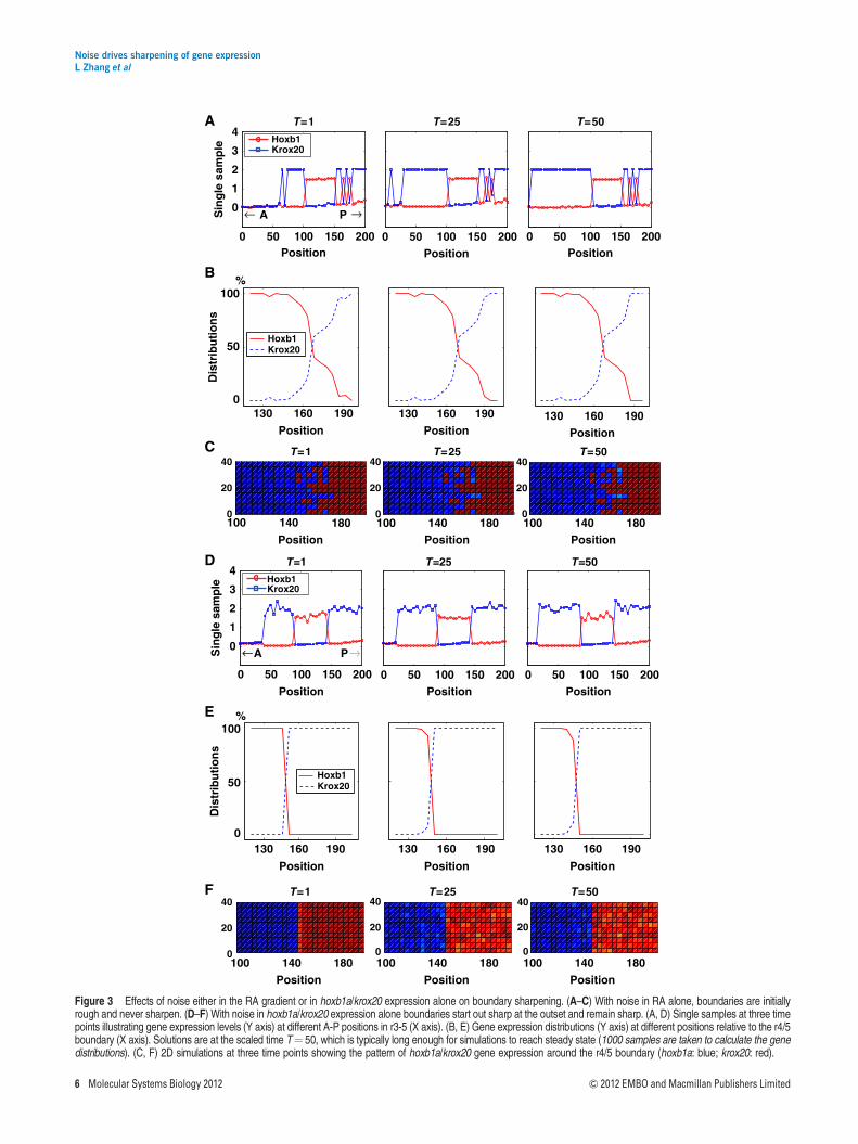
In contrast, if noise is introduced into the intracellular RAconcentration, [RA]in (i.e., eout 6¼ 0; ein 6¼ 0; ah¼ ak¼ 0), forexample, due to fluctuations in RA transport into cells, thenboundaries between hoxb1a and krox20 never sharpen(Figure 3A). In this case, the r3 domain of krox20 expressionexpands as fluctuations in the RA gradient reach the thresholdthat induces krox20. Monte Carlo simulations indicate thatthere is a large variation in the distribution of gene expres-sion around the r4/5 boundary over time when [RA]in isnoisy (Figure 3B). Two-dimensional simulations also showthat hoxb1a and krox20 expression domains initially form arough r4/5 boundary, which does not sharpen (Figure 3C).An initial noisy distribution of hoxb1a expression at thisboundary can also disrupt sharpening (see SupplementaryFigure S5). However, if noise is only restricted to later hoxb1aand krox20 expression, and not local RA concentration(i.e., eout¼ ein¼ 0; ah 6¼ 0; ak 6¼ 0), then boundaries tendto sharpen from the outset (Figure 3D and F) and MonteCarlo simulations confirm this prediction (Figure 3E). Theseresults suggest that rough boundaries of gene expressionbetween r4 and r5 arise due to noise in [RA]in or initial hoxb1aexpression.
Noise in Hoxb1/Krox20 expression enables noiseattenuation during boundary sharpening
Rhombomeres form lineally-restricted compartments (Fraseret al, 1990; Jimenez-Guri et al, 2010) and single hindbraincells can upregulate or downregulate their Hox expressionaccording to their host environment/rhombomere (Trainorand Krumlauf, 2000; Schilling et al, 2001). This suggests thatsimilar gene expression ‘switches’ occur in cells on either sideof a noisy rhombomere boundary. For example, cells expres-sing krox20 isolated among neighbors expressing hoxb1a(Figure 1J–L) may downregulate the former and upregulatethe latter, thereby attenuating the noise and sharpening theborder. To study such switching from one stable gene expres-sion state to another, we employed an MAP analysis basedon the Wentzell–Freidlin theory of large deviation (Freidlinand Wentzell, 1998). This theory allows one to estimate theprobability of a transition j between two stable states X1, X2 ina stochastic dynamic system (e.g., with a form of Equation (2)).The most probable path j* requires the least action and iscalled an MAP (see Section 2 in Supplementary informationfor more details). MAP analysis has been used primarily tomodel phase transitions between two states in stochasticchemical kinetics (E et al, 2004). Here, we adapt it to estimatethe switching probability between two stable gene expressionstates.
The likelihood that a system switches from X1 to X2 relieson its ability to pass through the unstable critical point Xc
that lies between X1 and X2 along the path j*. The distance|j*(X1)�j*(Xc)| is the minimum barrier to the stochastic
transition from one state to the other. For a smooth RA gradientand a simple bistable gene expression state (Figure 2C), wecalculate MAPs (E et al, 2004) at different levels of RA. At lowRA concentration (e.g., at RA¼ 0.1 mM), three MAPs connecteach pair of stable states, with each MAP passing through oneunstable critical point (Figure 4A). Based on MAP theory, theactivation of Krox20 (Krox20-on) from a ‘both-off’ staterequires less action (a lower barrier) than activation of Hoxb1,which helps explain why the r3 domain of Krox20 expressionexpands when noise increases in our models (Figure 3C). Incontrast, at intermediate RA concentrations a single MAPconnects the two steady states and the action to switching fromHoxb1-on to Krox20-on decreases from RA¼ 0.5 to 0.8 mM(Figure 4A), indicating that it becomes easier to switch in thisdirection as RA increases. When RA levels are high, Krox20-onis the only stable state.
To quantify such switching capability, we estimate theswitching probability from X1 to X2 within a time interval [0, T]through an exponential of the minimal barrier:
PX1!X2¼ exp ð� a� j j�ðX1Þ�j�ðXcÞ j nÞ: ð3Þ
The switching probability from X2 to X1 is defined in asimilar manner:
PX2!X1¼ exp ð� a� j j�ðX2Þ�j�ðXcÞ j nÞ: ð4Þ
We estimate the Hoxb1/Krox20 gene switching probabilitiesPH-K and PK-H using the MAP calculation of Equation 2 for anormalized RA concentration. Our models indicate that PH-K
increases exponentially when [RA] is high and PK-H is low,and cells have a high probability of switching from Hoxb1 toKrox20 expression. On the other hand, Krox20 expression ismore stable due to a cell’s low switching (to Hoxb1 expression)probability (Figure 4B). Together, this suggests that noise inHoxb1/Krox20 expression drives cells from occasionally co-expressing Hoxb1/Krox20 expression to a uniform Krox20expression when RA concentrations are high, leading to asharp boundary. In support of this analysis, direct Monte Carlosimulations of the gene system (2) of switching probability atthe same time intervals provide similar MAP estimates basedon Equations (3) and (4) (Figure 4B; see Section 2 inSupplementary information for more details).
Thus, surprisingly, our models suggest that the combinationof noise in both [RA]in and Krox20/Hoxb1 expression (i.e.,eout 6¼ 0; ein 6¼ 0; ah 6¼ 0; ak 6¼ 0), synergize to reducenoise during boundary sharpening, at least at the r4/5boundary (Figure 4C–E). Interestingly, the initial boundary(T¼ 1) is established at 160±10 mm along the A–P axis and,following sharpening, the boundary is located at 144 mm(T¼ 50) (Figure 4E). This suggests that sharpening preferen-tially drives cells near the initial, rough boundary to krox20expression due to the irreversibility of gene switching. Similardirectional boundary shifts in gene expression have also beenobserved in Drosophila (Jaeger et al, 2004). This fits well withour in vivo observation of a higher percentage of hoxb1a/krox20 co-expressing cells on the posterior side of the putativer4/5 boundary at 10.7 and 11.3 h.p.f. (Figure 1M and N), whichmight predict that the forming boundary shifts anteriorly.
To quantify boundary sharpening more systematically, wedefine a ‘Sharpness Index’ (S), which resembles the standarddeviation. To define S, we calculate the ‘mean’ location of the
Noise drives sharpening of gene expressionL Zhang et al
& 2012 EMBO and Macmillan Publishers Limited Molecular Systems Biology 2012 5
0 50 100 150 200
0
1
2
3
4
Sin
gle
sam
ple
← A P →
A T=1 T=25 T=50
0
50
100
Dis
trib
uti
on
s
Hoxb1Krox20
Hoxb1Krox20
0 50 100 150 200Position
0 50 100 150 200PositionPosition
%B
D
E
130 160 190
Position130 160 190
Position130 160 190
Position
0
50
100
Dis
trib
uti
on
s
%
130 160 190
Position
130 160 190
Position
130 160 190
Position
0 50 100 150 200
0
1
2
3
4
Sin
gle
sam
ple
0 50 100 150 200Position
0 50 100 150 200PositionPosition
←A P→
Hoxb1Krox20
Hoxb1Krox20
T=1 T=25 T=50
Position Position Position
C
100 140 1800
20
40
100 140 1800
20
40
100 140 1800
20
40F
T=1 T=25 T=50
T=1 T=25 T=50
100 140 1800
20
40
100 140 1800
20
40
100 140 1800
20
40
Position Position Position
Figure 3 Effects of noise either in the RA gradient or in hoxb1a/krox20 expression alone on boundary sharpening. (A–C) With noise in RA alone, boundaries are initiallyrough and never sharpen. (D–F) With noise in hoxb1a/krox20 expression alone boundaries start out sharp at the outset and remain sharp. (A, D) Single samples at three timepoints illustrating gene expression levels (Y axis) at different A-P positions in r3-5 (X axis). (B, E) Gene expression distributions (Y axis) at different positions relative to the r4/5boundary (X axis). Solutions are at the scaled time T¼ 50, which is typically long enough for simulations to reach steady state (1000 samples are taken to calculate the genedistributions). (C, F) 2D simulations at three time points showing the pattern of hoxb1a/krox20 gene expression around the r4/5 boundary (hoxb1a: blue; krox20: red).
Noise drives sharpening of gene expressionL Zhang et al
6 Molecular Systems Biology 2012 & 2012 EMBO and Macmillan Publishers Limited
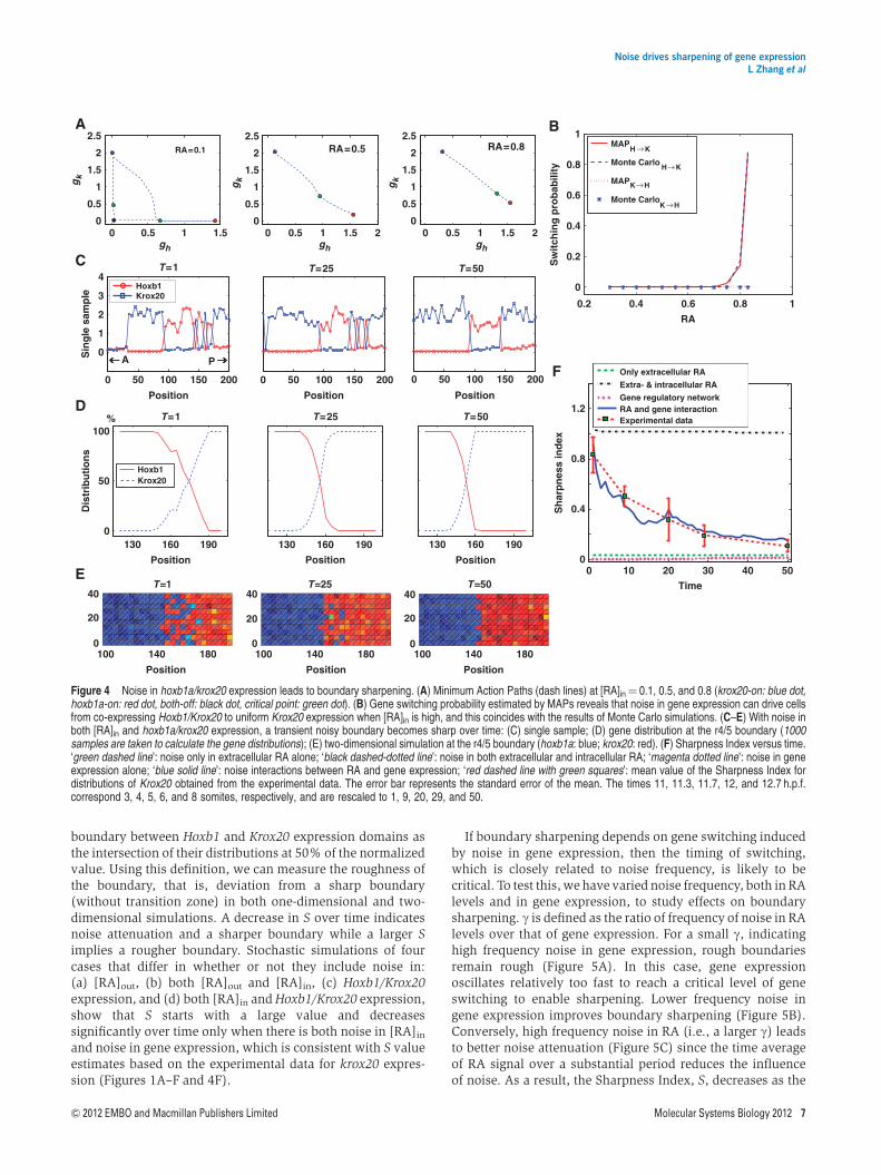
boundary between Hoxb1 and Krox20 expression domains asthe intersection of their distributions at 50% of the normalizedvalue. Using this definition, we can measure the roughness ofthe boundary, that is, deviation from a sharp boundary(without transition zone) in both one-dimensional and two-dimensional simulations. A decrease in S over time indicatesnoise attenuation and a sharper boundary while a larger Simplies a rougher boundary. Stochastic simulations of fourcases that differ in whether or not they include noise in:(a) [RA]out, (b) both [RA]out and [RA]in, (c) Hoxb1/Krox20expression, and (d) both [RA]in and Hoxb1/Krox20 expression,show that S starts with a large value and decreasessignificantly over time only when there is both noise in [RA]in
and noise in gene expression, which is consistent with S valueestimates based on the experimental data for krox20 expres-sion (Figures 1A–F and 4F).
If boundary sharpening depends on gene switching inducedby noise in gene expression, then the timing of switching,which is closely related to noise frequency, is likely to becritical. To test this, we have varied noise frequency, both in RAlevels and in gene expression, to study effects on boundarysharpening. g is defined as the ratio of frequency of noise in RAlevels over that of gene expression. For a small g, indicatinghigh frequency noise in gene expression, rough boundariesremain rough (Figure 5A). In this case, gene expressionoscillates relatively too fast to reach a critical level of geneswitching to enable sharpening. Lower frequency noise ingene expression improves boundary sharpening (Figure 5B).Conversely, high frequency noise in RA (i.e., a larger g) leadsto better noise attenuation (Figure 5C) since the time averageof RA signal over a substantial period reduces the influenceof noise. As a result, the Sharpness Index, S, decreases as the
100 140 1800
20
40
100 140 1800
20
40
100 140 1800
20
40
0 10 20 30 40 500
0.4
0.8
1.2
Time
Sh
arp
nes
s in
dex
Only extracellular RA
Extra- & intracellular RA
Gene regulatory networkRA and gene interactionExperimental data
0.2 0.4 0.6 0.8 1
0
0.2
0.4
0.6
0.8
1
RA
Sw
itch
ing
pro
bab
ility
MAP
H →K
Monte CarloH→K
MAPK→H
Monte CarloK→H
Hoxb1Krox20
0 50 100 150 200
Position
0 50 100 150 200
Position
0 50 100 150 200
Position
0
1
2
3
4
Sin
gle
sam
ple
Hoxb1Krox20
0
50
100
Dis
trib
uti
on
s
%
130 160 190
Position
130 160 190
Position
130 160 190
Position
A B
C T=1 T=25 T=50
0 0.5 1 1.50
0.5
1
1.5
2
2.5
gk
RA=0.1 RA=0.5
0 0.5 1 1.5 20
0.5
1
1.5
2
2.5
0 0.5 1 1.5 2
RA=0.8
0
0.5
1
1.5
2
2.5
D
FA P
E
Position Position Position
T=1 T=25 T=50
T=1 T=25 T=50
gk
gk
gh gh gh
Figure 4 Noise in hoxb1a/krox20 expression leads to boundary sharpening. (A) Minimum Action Paths (dash lines) at [RA]in¼ 0.1, 0.5, and 0.8 (krox20-on: blue dot,hoxb1a-on: red dot, both-off: black dot, critical point: green dot). (B) Gene switching probability estimated by MAPs reveals that noise in gene expression can drive cellsfrom co-expressing Hoxb1/Krox20 to uniform Krox20 expression when [RA]in is high, and this coincides with the results of Monte Carlo simulations. (C–E) With noise inboth [RA]in and hoxb1a/krox20 expression, a transient noisy boundary becomes sharp over time: (C) single sample; (D) gene distribution at the r4/5 boundary (1000samples are taken to calculate the gene distributions); (E) two-dimensional simulation at the r4/5 boundary (hoxb1a: blue; krox20: red). (F) Sharpness Index versus time.‘green dashed line’: noise only in extracellular RA alone; ‘black dashed-dotted line’: noise in both extracellular and intracellular RA; ‘magenta dotted line’: noise in geneexpression alone; ‘blue solid line’: noise interactions between RA and gene expression; ‘red dashed line with green squares’: mean value of the Sharpness Index fordistributions of Krox20 obtained from the experimental data. The error bar represents the standard error of the mean. The times 11, 11.3, 11.7, 12, and 12.7 h.p.f.correspond 3, 4, 5, 6, and 8 somites, respectively, and are rescaled to 1, 9, 20, 29, and 50.
Noise drives sharpening of gene expressionL Zhang et al
& 2012 EMBO and Macmillan Publishers Limited Molecular Systems Biology 2012 7
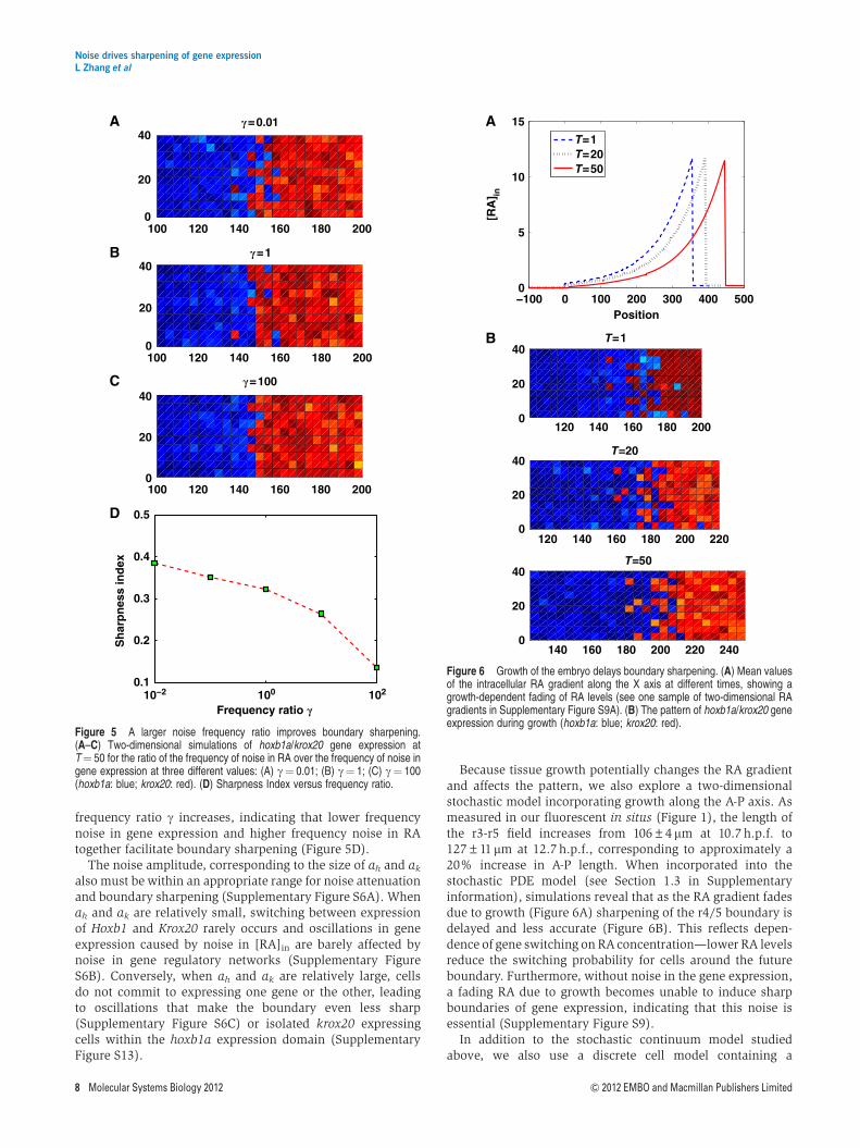
frequency ratio g increases, indicating that lower frequencynoise in gene expression and higher frequency noise in RAtogether facilitate boundary sharpening (Figure 5D).
The noise amplitude, corresponding to the size of ah and ak
also must be within an appropriate range for noise attenuationand boundary sharpening (Supplementary Figure S6A). Whenah and ak are relatively small, switching between expressionof Hoxb1 and Krox20 rarely occurs and oscillations in geneexpression caused by noise in [RA]in are barely affected bynoise in gene regulatory networks (Supplementary FigureS6B). Conversely, when ah and ak are relatively large, cellsdo not commit to expressing one gene or the other, leadingto oscillations that make the boundary even less sharp(Supplementary Figure S6C) or isolated krox20 expressingcells within the hoxb1a expression domain (SupplementaryFigure S13).
Because tissue growth potentially changes the RA gradientand affects the pattern, we also explore a two-dimensionalstochastic model incorporating growth along the A-P axis. Asmeasured in our fluorescent in situs (Figure 1), the length ofthe r3-r5 field increases from 106±4mm at 10.7 h.p.f. to127±11 mm at 12.7 h.p.f., corresponding to approximately a20% increase in A-P length. When incorporated into thestochastic PDE model (see Section 1.3 in Supplementaryinformation), simulations reveal that as the RA gradient fadesdue to growth (Figure 6A) sharpening of the r4/5 boundary isdelayed and less accurate (Figure 6B). This reflects depen-dence of gene switching on RA concentration—lower RA levelsreduce the switching probability for cells around the futureboundary. Furthermore, without noise in the gene expression,a fading RA due to growth becomes unable to induce sharpboundaries of gene expression, indicating that this noise isessential (Supplementary Figure S9).
In addition to the stochastic continuum model studiedabove, we also use a discrete cell model containing a
−100 0 100 200 300 400 5000
5
10
15
Position
[RA
] in
T=1T=20T=50
B
A
140 160 180 200 220 2400
20
40
T=1
T=20
T=50
120 140 160 180 2000
20
40
120 140 160 180 200 2200
20
40
Figure 6 Growth of the embryo delays boundary sharpening. (A) Mean valuesof the intracellular RA gradient along the X axis at different times, showing agrowth-dependent fading of RA levels (see one sample of two-dimensional RAgradients in Supplementary Figure S9A). (B) The pattern of hoxb1a/krox20 geneexpression during growth (hoxb1a: blue; krox20: red).
A
B
C
D
10−2 100 1020.1
0.2
0.3
0.4
0.5
Frequency ratio �
Sh
arp
nes
s in
dex
100 120 140 160 180 2000
20
40
100 120 140 160 180 2000
20
40
100 120 140 160 180 2000
20
40
�=0.01
�=1
�=100
Figure 5 A larger noise frequency ratio improves boundary sharpening.(A–C) Two-dimensional simulations of hoxb1a/krox20 gene expression atT¼ 50 for the ratio of the frequency of noise in RA over the frequency of noise ingene expression at three different values: (A) g¼ 0.01; (B) g¼ 1; (C) g¼ 100(hoxb1a: blue; krox20: red). (D) Sharpness Index versus frequency ratio.
Noise drives sharpening of gene expressionL Zhang et al
8 Molecular Systems Biology 2012 & 2012 EMBO and Macmillan Publishers Limited
one-dimensional array of cells within which the reactionsand regulatory interactions (Figure 2A) are calculatedusing a Stochastic Simulation Algorithm (Gillespie, 1977).The number of RA molecules is obtained either by thecontinuum approach shown above or through a stochasticreaction-diffusion process (Kang et al, 2012; see Section 3 inSupplementary information for more details). We found thatwhen the number of RA molecules is large, noise in geneexpression can drive boundary sharpening (SupplementaryFigures S7 and S8), while when the number of RA moleculesis very small, leading to large fluctuations in the RA gradient,the rough boundary remains. Both results are consistent withthe stochastic continuum approach.
Time delays, such as those that occur during transcriptionalregulation, often affect the dynamics of gene switching andsharpening (Kepler and Elston, 2001; Bratsun et al, 2005).By incorporating a constant time delay in the expressionof hoxb1a and krox20 in our model (see Section 1.4 inSupplementary information; Kuang, 1993), we find additionalstochastic oscillations that reduce the speed and efficiencyof boundary sharpening (Supplementary Figure S10). Onepossible explanation is that the time delay averages (or filters)noise such that gene switching becomes difficult, particularlywhen the time delay is long relative to the dynamics of geneexpression.
Boundaries may also sharpen through cell movementsand differential adhesion that can lead to cell sorting(Xu et al, 1999; Firtel and Meili, 2000; Dormann and Weijer,2003, 2006). Previous studies in the zebrafish hindbrainhave demonstrated some limited sorting at rhombomereboundaries (Cooke et al, 2005; Kemp et al, 2009). Simulationswith two-dimensional stochastic discrete cell models, whichincorporate both directional cell movements and stochasticreaction diffusion (see Section 4 in Supplementary informa-tion), suggest that the speed of cell sorting must be stronglyregulated to facilitate sharpening (Supplementary Figure S11).Moreover, directed cell movements without noise in geneexpression lead to variability in the location of the r4/5boundary (Supplementary Figure S12).
Discussion
Morphogen gradients activate target genes in a concentration-dependent manner to generate distinct spatial domains ofexpression in developing tissues. Extrinsic noise in morphogenconcentration and tissue geometry, as well as intrinsic noisein signal transduction and gene expression, reduces precisionof patterning (Bollenbach et al, 2008; Balazsi et al, 2011;Kang et al, 2012). Here we show, surprisingly, that noise inintracellular signal transduction actually improves precisionand robustness of patterning. In particular, we find that theinitially rough expression boundaries of hoxb1a and krox20 inthe developing zebrafish hindbrain are due to extrinsic noise inthe morphogen (RA) that induces them, but that intracellularfluctuations in their expression lead to gene switching thatenables sharpening. The resulting noise attenuation progres-sively narrows the transition zone between the two geneexpression domains, similar to the sharpening that occursin vivo. Within this transition zone cells transiently co-express
both genes before adopting one segmental fate or the other.This result is consistent with experimental evidence showingthat cells at these stages can upregulate or downregulate Hoxexpression, rather than having to migrate to sort themselvesout at the future boundary (Trainor and Krumlauf, 2000;Schilling et al, 2001) and that rhombomeres form lineallyrestricted compartments at early embryonic stages (Fraseret al, 1990; Jimenez-Guri et al, 2010). Similar rules may alsoapply for other RA target genes (e.g., Vhnf1) and other geneexpression boundaries where cross-inhibition and/or auto-activation between target genes occur in morphogen systems(Rivera-Pomar and Jackle, 1996; Perkins et al, 2006; Balaskaset al, 2012). Notably, this property of the system does notdepend on the genes being direct transcriptional targets of themorphogen signal—Hoxb1 is a direct target while Krox20 isinduced indirectly through Vhnf1, Mafb and other transcrip-tion factors (Barrow et al, 2000; Giudicelli et al, 2001;Hernandez et al, 2004; Alexander et al, 2009).
Our deterministic model shows that an initial low level ofHoxb1 is required to generate the alternating striped patternof Hoxb1 and Krox20 expression in response to a spatiallymonotonic RA gradient (see Figure 2). During gastrulationin zebrafish, hoxb1a expression is initiated several hoursbefore krox20, and induced by the even earlier expressionof hoxb1b in response to RA (McClintock et al, 2001; Mavesand Kimmel, 2005). This early onset is important genetically(it fine-tunes target genes within its expression domain;Labalette et al, 2011) and computationally (i.e., pre-steady-state decoding) during embryonic patterning (Bergmann et al,2007; Saunders and Howard, 2009). This helps explain howalternating, mutually exclusive domains of target gene expres-sion can be induced by the same signal, a common feature ofmany boundary-forming morphogen systems (Lander, 2011).Another major assumption of the model is the irreversibility ofcell switching such that gene expression remains stable whenthe morphogen gradient decreases or disappears (Gould et al,1998; Grapin-Botton et al, 1998). This irreversibility has alsobeen pointed out in previous modeling studies (Meinhardt,1978, 1982).
Intracellular noise may arise from two sources: an intrinsicone due to small numbers of molecules or stochastic fluctua-tions inherent in biochemical reactions and an extrinsic onedriven by fluctuations in cellular environment (Swain et al,2002). Gene expression noise in some morphogen systemsdepends predominantly on fluctuations in transcription andtranslation (Holloway et al, 2011). From our combinationof spatial SSA simulations, which take into considerationthe number of molecules, and stochastic continuum PDEmodels, both types of intracellular noise can be utilized toinduce switching between gene expression states (e.g., theswitching from Hoxb1 to Krox20 is dominant in the transitionregion between r4 and r5), leading to boundary sharpening.Of course, the level of noise in the morphogen signal must bewithin a range that allows switching in the transition regionthrough this system of intracellular noise.
Our models also suggest that the cells in the transition regionnear the r3/r4 boundary utilize somewhat different noiseattenuation mechanisms despite undergoing similar boundarysharpening. Unlike the case at higher levels of RA (i.e., at ther4/r5 boundary) where the switching probability from Hoxb1
Noise drives sharpening of gene expressionL Zhang et al
& 2012 EMBO and Macmillan Publishers Limited Molecular Systems Biology 2012 9
to Krox20 is significantly higher than the one from Krox20to Hoxb1 (Figure 4B), near the r3/r4 boundary where RAlevels are lower, switching probabilities are also lower. Sincewe can detect cells co-expressing hoxb1a and krox20 at ther3/4 boundary during sharpening (Figure 1J–L), it seemslikely that differences lie upstream, at the level of theinductive signals. Cells at this boundary may have intrinsicdifferences in their RA responses. Alternatively, these cellsintegrate responses to additional morphogens such as Fgfs(e.g., Fgf3 and Fgf8), which are produced in the anteriorhindbrain during sharpening and influence both RA degrada-tion and expression of hoxb1a and krox20 (Hernandez et al,2004; Labalette et al, 2011).
In particular, our simulations suggest that both time delaysand cell movements can affect rhombomere boundarysharpening. We find that time delay in the expression ofhoxb1a and krox20 may introduce additional stochasticeffects, leading to reduced speed and efficiency in boundarysharpening. Likewise, cell movements and differential adhe-sion can lead to cell sorting (Xu et al, 1999; Firtel and Meili,2000; Dormann and Weijer, 2003, 2006) and some limitedsorting has been observed at rhombomere boundaries in thehindbrain (Fraser et al, 1990; Cooke et al, 2005; Kemp et al,2009). However, clones derived from single progenitors in theneural plate are for the most part lineally restricted toindividual rhombomeres (Fraser et al, 1990; Jimenez-Guriet al, 2010) and do not move across boundaries. Hindbraincells at these stages are also capable of upregulating ordownregulating Hox expression if they find themselves on thewrong side of a boundary (Trainor and Krumlauf, 2000;Schilling et al, 2001). Our simulations also reveal that: (1) thespeed of cell sorting is critical for sharpening and (2) directedcell movements without noise in gene expression disrupt thelocation of the r4/5 boundary. Our simulation data suggestthat cell movements alone are unlikely to account forrhombomere boundary sharpening, and that noise in geneexpression is critical for this process. However, morecomprehensive experiments are needed to quantify theamount of sorting that occurs and modeling to understandthe roles of this sorting in the establishment and maintenanceof precise boundaries.
Our model is limited to two spatial dimensions. Interest-ingly, in other systems the precision of responses to morpho-gen gradients rapidly increases when considered in threedimensions (Bollenbach et al, 2008). The zebrafish hindbrainis several cell diameters thick during the sharpening periodconsidered here. Therefore, incorporating more accurate tissuegeometries in our stochastic model will undoubtedly revealnew features of the system—most likely including more robustpatterning and boundary sharpening that can resist largeramplitude fluctuations. There exist other potential mechan-isms that may facilitate boundary sharpening and noiseattenuation, including cell-to-cell communication (e.g., Notchsignaling) (Louvi and Artavanis-Tsakonas, 2006; Ozbudakand Lewis, 2008; Koseska et al, 2009; VanHook, 2011) andaveraging (Tanouchi et al, 2008).
While molecular noise often introduces fluctuations inbiochemical reactions and increases uncertainty in cellulardecisions, it also can improve performance objectives ofbiological systems in surprising ways. For example, noise can:
(1) help create synchronous oscillations in cell–cell signalingsystems (Zhou et al, 2005; Springer and Paulsson, 2006); (2)enhance sensitivity in intracellular regulation (e.g., stochasticfocusing; Paulsson et al, 2000), and (3) through reversibleprogression, help reliable cellular decision making (Kuchinaet al, 2011). Our results add to the growing body of evidencethat points to important roles for molecular noise in cell fatedecisions, and reveals a novel mechanism by which intracel-lular molecular noise reduces uncertainty in the ability of afluctuating morphogen to induce precise domains of targetgenes.
Materials and methods
Numerical methods of stochastic PDE model
The stochastic PDEs (1) were solved with a finite-difference approxi-mation in both one- and two-dimensional spaces. The stochasticsystem (2) was solved with Milstein’s method (Higham, 2001). Noisewas added at each grid point in the space at a specified time interval.We used spatial resolutions of 100 grid points in the one-dimensionalmodel or 100�20 grid points in the two-dimensional model andtemporal resolution of h¼ 0.1s. Numerical tests were conducted toensure sufficient resolution. The solutions were typically observed atthe scale time T¼ 1.25 and 50, which is the steady state for all of thevariables at each spatial point to reach an approximately invariantdistribution.
To estimate the stationary distribution from one realization, weperformed Monte Carlo simulations, which are repeated computationsof the stochastic models. We took 1000 samplings in the Monte Carlosimulations to calculate the Sharpness Index. We explored a range ofsample numbers from 50 to 5000 to ensure consistent results. Pleasesee Section 1 in Supplementary information for more details andDataset 2 in Supplementary information for the simulation codes.
Numerical methods for finding MAPs
We followed the algorithm in E et al (2004) to find the MAPs for thegene switch. First, a discrete time interval was used to form a mesh andthe path j is approximated on the mesh. Next, the action S[j] of thispath was approximated according to the midpoint rule. The steepestdescent method was then applied to minimize the discrete action S[j].Please see Section 2 in Supplementary information for the equationsand Dataset 2 in Supplementary information for the simulation codes.
Numerical methods for spatial SSA
We partitioned the one-dimensional or the two-dimensional space intoidentical compartments based on a computational strategy previouslydeveloped (Kang et al, 2012), and applied the Gillespie algorithm to thestochastic reaction-diffusion simulations (Gillespie, 1976). Please seeSections 3 and 4 in Supplementary information for more details andDataset 2 in Supplementary information for the simulation codes.
Gene expression analysis in zebrafish
Wild-type zebrafish embryos (TL) were collected from natural matingsand staged as previously described (Kimmel et al, 1995). Fluorescentwhole mount in situ hybridization was performed as previouslydescribed for Fast Red alone (Thisse et al, 2004) or with two colorsusing tyramide amplification (Zuniga et al, 2010). Probes were synthe-sized from cDNA clones of krox20 (Oxtoby and Jowett, 1993) andhoxb1a (McClintock et al, 2001). Embryos were imaged on an OlympusFluoview FV1000 confocal microscope, processed in Image J, and post-processed on Adobe Photoshop CS3. Fast Red in situ images for krox20were processed and analyzed by the Matlab Image Processing Toolbox(The MathWorks, Natick, MA, USA). Graphs of cell location were
Noise drives sharpening of gene expressionL Zhang et al
10 Molecular Systems Biology 2012 & 2012 EMBO and Macmillan Publishers Limited
made in Microsoft Excel based on confocal stacks analyzed in Image J.Please see Dataset 1 in Supplementary information for the processedcell location data. Full image data are available from the authors uponrequest.
Supplementary information
Supplementary information is available at the Molecular SystemsBiology website (www.nature.com/msb).
AcknowledgementsWe would like to thank Arthur Lander for initial development of thedeterministic model and advice throughout this project, as well as InesGehring for zebrafish care. This study was supported by NIH P50 GM-76516 (QN and TS), NIH R01 GM-67247 (QN), NSF DMS-0917492(QN), and NIH R01 NS-41353 (TS).
Author contributions: LeiZ, LikunZ, AC, and QN performed thecomputational research; KR and TS performed the experimentalresearch with zebrafish; LeiZ, QN, KR and TS wrote the paper.
Conflict of InterestThe authors declare that they have no conflict of interest.
References
Alexander T, Nolte C, Krumlauf R (2009) Hox genes and segmentationof the hindbrain and axial skeleton. Annu Rev Cell Dev Biol 25:431–456
Balaskas N, Ribeiro A, Panovska J, Dessaud E, Sasai N, Page KM,Briscoe J, Ribes V (2012) Gene regulatory logic for reading the sonichedgehog signaling gradient in the vertebrate neural tube. Cell 148:273–284
Balazsi G, van Oudenaarden A, Collins JJ (2011) Cellular decisionmaking and biological noise: from microbes to mammals. Cell 144:910–925
Barrow JR, Stadler HS, Capecchi MR (2000) Roles of Hoxa1 and Hoxa2in patterning the early hindbrain of the mouse. Development 127:933–944
Begemann G, Schilling TF, Rauch GJ, Geisler R, Ingham PW (2001) Thezebrafish neckless mutation reveals a requirement for raldh2 inmesodermal signals that pattern the hindbrain. Development 128:3081–3094
Bergmann S, Sandler O, Sberro H, Shnider S, Schejter E, Shilo BZ,Barkai N (2007) Pre-steady-state decoding of the Bicoid morphogengradient. PLoS Biol 5: e46
Bollenbach T, Pantazis P, Kicheva A, Bokel C, Gonzalez-Gaitan M,Julicher F (2008) Precision of the Dpp gradient. Development 135:1137–1146
Bratsun D, Volfson D, Tsimring LS, Hasty J (2005) Delay-inducedstochastic oscillations in gene regulation. Proc Natl Acad Sci USA102: 14593–14598
Cooke JE, Kemp HA, Moens CB (2005) EphA4 is required for celladhesion and rhombomere-boundary formation in the zebrafishhindbrain. Curr Biol 15: 536–542
Cooke JE, Moens CB (2002) Boundary formation in the hindbrain: Ephonly it were simple. Trends Neurosci 25: 260–267
Dormann D, Weijer CJ (2003) Chemotactic cell movement duringdevelopment. Curr Opin Genet Dev 13: 358–364
Dormann D, Weijer CJ (2006) Chemotactic cell movement duringDictyostelium development and gastrulation. Curr Opin Genet Dev16: 367–373
E W, Ren W, Vanden-Eijnden E (2004) Minimum action method for thestudy of rare events. Commun Pure Appl Math 57: 637–656
Eldar A, Rosin D, Shilo BZ, Barkai N (2003) Self-enhanced ligand degrada-tion underlies robustness of morphogen gradients. Dev Cell 5: 635–646
Elowitz MB, Levine AJ, Siggia ED, Swain PS (2002) Stochastic geneexpression in a single cell. Science 297: 1183–1186
Ferrell Jr JE (2002) Self-perpetuating states in signal transduction:positive feedback, double-negative feedback and bistability. CurrOpin Cell Biol 14: 140–148
Firtel RA, Meili R (2000) Dictyostelium: a model for regulated cellmovement during morphogenesis. Curr Opin Genet Dev 10:421–427
Fraser S, Keynes R, Lumsden A (1990) Segmentation in the chickembryo hindbrain is defined by cell lineage restrictions. Nature344: 431–435
Freidlin MI, Wentzell AD (1998) Random Perturbations of DynamicalSystems. 2nd edn, New York: Springer
Gillespie DT (1976) A general method for numerically simulating thestochastic time evolution of coupled chemical reactions. J ComputPhys 22: 403–434
Gillespie DT (1977) Exact stochastic simulation of coupled chemicalreactions. J Phys Chem 81: 2340–2361
Giudicelli F, Taillebourg E, Charnay P, Gilardi-Hebenstreit P (2001)Krox-20 patterns the hindbrain through both cell-autonomous andnon cell-autonomous mechanisms. Genes Dev 15: 567
Gould A, Itasaki N, Krumlauf R (1998) Initiation of rhombomericHoxb4 expression requires induction by somites and a retinoidpathway. Neuron 21: 39–51
Grapin-Botton A, Bonnin MA, Sieweke M, Le Douarin NM (1998)Defined concentrations of a posteriorizing signal are critical forMafB/Kreisler segmental expression in the hindbrain. Development125: 1173–1181
Hasty J, Pradines J, Dolnik M, Collins JJ (2000) Noise-based switchesand amplifiers for gene expression. Proc Natl Acad Sci USA 97:2075–2080
He F, Ren J, Wang W, Ma J (2012) Evaluating the Drosophila Bicoidmorphogen gradient system through dissecting the noise intranscriptional bursts. Bioinformatics 28: 970–975
Hernandez RE, Rikhof HA, Bachmann R, Moens CB (2004) vhnf1integrates global RA patterning and local FGF signals to directposterior hindbrain development in zebrafish. Development 131:4511–4520
Higham DJ (2001) An algorithmic introduction to numerical simula-tion of stochastic differential equations. SIAM Rev 43: 525–546
Holloway DM, Lopes FJP, LdF Costa, Travencolo BAN, Golyandina N,Usevich K, Spirov AV (2011) Gene expression noise in spatialpatterning: hunchback promoter structure affects noise amplitudeand distribution in Drosophila segmentation. PLoS Comput Biol 7:e1001069
Jaeger J, Surkova S, Blagov M, Janssens H, Kosman D, Kozlov KN,Manu Myasnikova E, Vanario-Alonso CE, Samsonova M, Sharp DH,Reinitz J (2004) Dynamic control of positional information in theearly Drosophila embryo. Nature 430: 368–371
Jimenez-Guri E, Udina F, Colas JF, Sharpe J, Padron-Barthe L,Torres M, Pujades C (2010) Clonal analysis in mice underlinesthe importance of rhombomeric boundaries in cell movementrestriction during hindbrain segmentation. PLoS ONE 5: e10112
Kaern M, Elston TC, Blake WJ, Collins JJ (2005) Stochasticity ingene expression: from theories to phenotypes. Nat Rev Genet 6:451–464
Kang HW, Zheng L, Othmer HG (2012) A new method for choosing thecomputational cell in stochastic reaction-diffusion systems. J MathBiol (doi:10.1007/s00285-011-0469-6)
Kemp HA, Cooke JE, Moens CB (2009) EphA4 and EfnB2a maintainrhombomere coherence by independently regulating intercala-tion of progenitor cells in the zebrafish neural keel. Dev Biol 327:313–326
Kepler TB, Elston TC (2001) Stochasticity in transcriptional regulation:origins, consequences, and mathematical representations. BiophysJ 81: 3116–3136
Kimmel CB, Ballard WW, Kimmel SR, Ullmann B, Schilling TF (1995)Stages of embryonic development of the zebrafish. Dev Dyn 203:253–310
Noise drives sharpening of gene expressionL Zhang et al
& 2012 EMBO and Macmillan Publishers Limited Molecular Systems Biology 2012 11

Koseska A, Zaikin A, Kurths J, Garcia-Ojalvo J (2009) Timing cellulardecision making under noise via cell-cell communication. PLoSONE 4: e4872
Kuang Y (1993) Delay Differential Equations: With Applications inPopulation Dynamics. Academic Press, Boston, USA
Kuchina A, Espinar L, Garcia-Ojalvo J, Suel GM (2011) Reversibleand noisy progression towards a commitment point enablesadaptable and reliable cellular decision-making. PLoS ComputBiol 7: e1002273
Labalette C, Bouchoucha YX, Wassef MA, Gongal PA, Le Men J, BeckerT, Gilardi-Hebenstreit P, Charnay P (2011) Hindbrain patterningrequires fine-tuning of early krox20 transcription by Sprouty 4.Development 138: 317–326
Lander AD (2011) Pattern, growth, and control. Cell 144: 955–969Lopes FJ, Vieira FM, Holloway DM, Bisch PM, Spirov AV (2008) Spatial
bistability generates hunchback expression sharpness in theDrosophila embryo. PLoS Comput Biol 4: e1000184
Louvi A, Artavanis-Tsakonas S (2006) Notch signalling in vertebrateneural development. Nat Rev Neurosci 7: 93–102
Maves L, Kimmel CB (2005) Dynamic and sequential patterning ofthe zebrafish posterior hindbrain by retinoic acid. Dev Biol 285:593–605
McClintock JM, Carlson R, Mann DM, Prince VE (2001) Consequencesof Hox gene duplication in the vertebrates: an investigation ofthe zebrafish Hox paralogue group 1 genes. Development 128:2471–2484
Meinhardt H (1978) Space-dependent cell determination under thecontrol of a morphogen gradient. J Theor Biol 74: 307–321
Meinhardt H (1982) Models of Biological Pattern Formation. London:Academic Press
Meinhardt H (2009) Models for the generation and interpretation ofgradients. Cold Spring Harb Perspect Biol 1: a001362
Niederreither K, Vermot J, Schuhbaur B, Chambon P, Dolle P (2000)Retinoic acid synthesis and hindbrain patterning in the mouseembryo. Development 127: 75–85
Oxtoby E, Jowett T (1993) Cloning of the zebrafish krox-20 gene (krx-20) and its expression during hindbrain development. Nucleic AcidsRes 21: 1087–1095
Ozbudak EM, Lewis J (2008) Notch signalling synchronizes thezebrafish segmentation clock but is not needed to create somiteboundaries. PLoS Genet 4: e15
Paulsson J, Berg OG, Ehrenberg M (2000) Stochastic focusing:fluctuation-enhanced sensitivity of intracellular regulation. ProcNatl Acad Sci USA 97: 7148–7153
Perkins TJ, Jaeger J, Reinitz J, Glass L (2006) Reverse engineeringthe gap gene network of Drosophila melanogaster. PLoS ComputBiol 2: e51
Rivera-Pomar R, Jackle H (1996) From gradients to stripes inDrosophila embryogenesis: filling in the gaps. Trends Genet 12:478–483
Saunders T, Howard M (2009) When it pays to rush: interpretingmorphogen gradients prior to steady-state. Phys Biol 6: 046020
Schilling T, Prince V, Ingham P (2001) Plasticity in zebrafish hoxexpression in the hindbrain and cranial neural crest. Dev Biol 231:201–216
Springer M, Paulsson J (2006) Biological physics: harmonies fromnoise. Nature 439: 27–28
Swain PS, Elowitz MB, Siggia ED (2002) Intrinsic and extrinsiccontributions to stochasticity in gene expression. Proc Natl Acad SciUSA 99: 12795–12800
Tanouchi Y, Tu D, Kim J, You L (2008) Noise reduction by diffusionaldissipation in a minimal quorum sensing motif. PLoS Comput Biol4: e1000167
Thattai M, van Oudenaarden A (2002) Attenuation of noise inultrasensitive signaling cascades. Biophys J 82: 2943–2950
Thisse B, Heyer V, Lux A, Alunni V, Degrave A, Seiliez I, Kirchner J,Parkhill JP, Thisse C (2004) Spatial and temporal expression of thezebrafish genome by large-scale in situ hybridization screening.Methods Cell Biol 77: 505–519
To T-L, Maheshri N (2010) Noise can induce bimodality in positivetranscriptional feedback loops without bistability. Science 317:1142–1145
Trainor PA, Krumlauf R (2000) Patterning the cranial neural crest:hindbrain segmentation and Hox gene plasticity. Nat Rev Neurosci1: 116–124
VanHook AM (2011) Defining Boundaries with Notch. Sci Signal 4:ec229
Wartlick O, Kicheva A, Gonzalez-Gaitan M (2009) Morphogen gradientformation. Cold Spring Harb Perspect Biol 1: a001255
White R, Nie Q, Lander A, Schilling T (2007) Complex regulation ofcyp26a1 creates a robust retinoic acid gradient in the zebrafishembryo. PLoS Biol 5: e304
White RJ, Schilling TF (2008) How degrading: Cyp26s in hindbraindevelopment. Dev Dyn 237: 2775–2790
Xu Q, Mellitzer G, Robinson V, Wilkinson DG (1999) In vivo cell sortingin complementary segmental domains mediated by Eph receptorsand ephrins. Nature 399: 267–271
Zhou T, Chen L, Aihara K (2005) Molecular communication throughstochastic synchronization induced by extracellular fluctuations.Phys Rev Lett 95: 178103
Zuniga E, Stellabotte F, Crump JG (2010) Jagged-Notch signalingensures dorsal skeletal identity in the vertebrate face. Development137: 1843–1852
Molecular Systems Biology is an open-access journalpublished by European Molecular Biology Organiza-
tion and Nature Publishing Group. This work is licensed under aCreative Commons Attribution-Noncommercial-Share Alike 3.0Unported License.
Noise drives sharpening of gene expressionL Zhang et al
12 Molecular Systems Biology 2012 & 2012 EMBO and Macmillan Publishers Limited