No Job Name - University Of Maryland6)_2881-94.pdf · electrical signals, ... Importantly,...
14
Reviews Biofabrication with Chitosan Hyunmin Yi, ²,‡ Li-Qun Wu, ² William E. Bentley, ²,§ Reza Ghodssi, |,⊥ Gary W. Rubloff, ‡,| James N. Culver, ² and Gregory F. Payne* ,² Center for Biosystems Research, University of Maryland Biotechnology Institute, 5115 Plant Sciences Building, College Park, Maryland 20742, Department of Materials Science and Engineering, University of Maryland at College Park, College Park, Maryland 20742, Department of Chemical Engineering, University of Maryland at College Park, College Park, Maryland 20742, The Institute for Systems Research, University of Maryland at College Park, College Park, Maryland 20742, and Department of Electrical and Computer Engineering, University of Maryland at College Park, College Park, Maryland 20742 Received June 15, 2005; Revised Manuscript Received July 27, 2005 The traditional motivation for integrating biological components into microfabricated devices has been to create biosensors that meld the molecular recognition capabilities of biology with the signal processing capabilities of electronic devices. However, a different motivation is emerging; biological components are being explored to radically change how fabrication is achieved at the micro- and nanoscales. Here we review biofabrication, the use of biological materials for fabrication, and focus on three specific biofabrication approaches: directed assembly, where localized external stimuli are employed to guide assembly; enzymatic assembly, where selective biocatalysts are enlisted to build macromolecular structure; and self-assembly, where information internal to the biological material guides its own assembly. Also reviewed are recent results with the aminopolysaccharide chitosan, a material that offers a combination of properties uniquely suited for biofabrication. In particular, chitosan can be directed to assemble in response to locally applied electrical signals, and the chitosan backbone provides sites that can be employed for the assembly of proteins, nucleic acids, and virus particles. Introduction In the past, the primary reason for integrating biological components into microfabricated devices was to enlist the molecular recognition capabilities of nucleic acids, enzymes, and antibodies to perform biosensing functions. 1-3 The coupling of these biosensing components with the signal processing capabilities of microfabricated devices allowed the creation of rapid and sensitive biosensors for detection and quantification (e.g., to diagnose disease). More recently, different biological components are being examined to perform a different function: to facilitate fabrication. Interest in biofabrication (the use of biological materials for fabrica- tion) is driven by the opportunity to access a wider range of fabrication options for construction at the micro- and nanoscale. 4 Potentially, biofabrication may facilitate the fabrication of devices with reduced “minimum feature sizes”, the integration of labile biological components into high throughput testing instruments, and the generation of bio- compatible systems for implantation. Traditionally, there are three general approaches to fab- ricate micro- and nanoscale features into materials. Photo- * To whom correspondence should be addressed. Phone: (301) 405- 8389. Fax: (301) 314-9075. E-mail: [email protected]. ² University of Maryland Biotechnology Institute. ‡ Department of Materials Science and Engineering, University of Maryland at College Park. § Department of Chemical Engineering, University of Maryland at College Park. | The Institute for Systems Research, University of Maryland at College Park. ⊥ Department of Electrical and Computer Engineering, University of Maryland at College Park. © Copyright 2005 by the American Chemical Society November/December 2005 Published by the American Chemical Society Volume 6, Number 6 10.1021/bm050410l CCC: $30.25 © 2005 American Chemical Society Published on Web 09/03/2005
Transcript of No Job Name - University Of Maryland6)_2881-94.pdf · electrical signals, ... Importantly,...

Reviews
Biofabrication with Chitosan
Hyunmin Yi,†,‡ Li-Qun Wu,† William E. Bentley,†,§ Reza Ghodssi,|,⊥ Gary W. Rubloff,‡,|
James N. Culver,† and Gregory F. Payne*,†
Center for Biosystems Research, University of Maryland Biotechnology Institute, 5115 Plant SciencesBuilding, College Park, Maryland 20742, Department of Materials Science and Engineering, University of
Maryland at College Park, College Park, Maryland 20742, Department of Chemical Engineering, Universityof Maryland at College Park, College Park, Maryland 20742, The Institute for Systems Research,
University of Maryland at College Park, College Park, Maryland 20742, and Department of Electrical andComputer Engineering, University of Maryland at College Park, College Park, Maryland 20742
Received June 15, 2005; Revised Manuscript Received July 27, 2005
The traditional motivation for integrating biological components into microfabricated devices has been tocreate biosensors that meld the molecular recognition capabilities of biology with the signal processingcapabilities of electronic devices. However, a different motivation is emerging; biological components arebeing explored to radically change how fabrication is achieved at the micro- and nanoscales. Here we reviewbiofabrication, the use of biological materials for fabrication, and focus on three specific biofabricationapproaches: directed assembly, where localized external stimuli are employed to guide assembly; enzymaticassembly, where selective biocatalysts are enlisted to build macromolecular structure; and self-assembly,where information internal to the biological material guides its own assembly. Also reviewed are recentresults with the aminopolysaccharide chitosan, a material that offers a combination of properties uniquelysuited for biofabrication. In particular, chitosan can be directed to assemble in response to locally appliedelectrical signals, and the chitosan backbone provides sites that can be employed for the assembly of proteins,nucleic acids, and virus particles.
Introduction
In the past, the primary reason for integrating biologicalcomponents into microfabricated devices was to enlist themolecular recognition capabilities of nucleic acids, enzymes,and antibodies to perform biosensing functions.1-3 Thecoupling of these biosensing components with the signal
processing capabilities of microfabricated devices allowedthe creation of rapid and sensitive biosensors for detectionand quantification (e.g., to diagnose disease). More recently,different biological components are being examined toperform a different function: to facilitate fabrication. Interestin biofabrication (the use of biological materials for fabrica-tion) is driven by the opportunity to access a wider range offabrication options for construction at the micro- andnanoscale.4 Potentially, biofabrication may facilitate thefabrication of devices with reduced “minimum feature sizes”,the integration of labile biological components into highthroughput testing instruments, and the generation of bio-compatible systems for implantation.
Traditionally, there are three general approaches to fab-ricate micro- and nanoscale features into materials. Photo-
* To whom correspondence should be addressed. Phone: (301) 405-8389. Fax: (301) 314-9075. E-mail: [email protected].
† University of Maryland Biotechnology Institute.‡ Department of Materials Science and Engineering, University of
Maryland at College Park.§ Department of Chemical Engineering, University of Maryland at
College Park.| The Institute for Systems Research, University of Maryland at College
Park.⊥ Department of Electrical and Computer Engineering, University of
Maryland at College Park.
© Copyright 2005 by the American Chemical Society
November/December 2005 Published by the American Chemical Society Volume 6, Number 6
10.1021/bm050410l CCC: $30.25 © 2005 American Chemical SocietyPublished on Web 09/03/2005

lithography is the primary approach used to fabricate highlyorganized surfaces in the microelectronics industry, andphotolithographic patterning is based on applying localizedoptical stimuli to initiate selective photochemistries. Analternative patterning approach is soft lithography thatincludes a variety of techniques, one of which is microcontactprinting.5,6 In microcontact printing, a “molecular ink” ismechanically “stamped” onto a substrate, and a specificsurface chemistry is often used to ensure the ink is retainedat the stamped locations. Finally, dip-pen and related printingapproaches emerged to mechanically deliver (i.e., “spot”)biological probe molecules (particularly nucleic acids) ontoslides for genome analysis. Once these biological probemolecules are spotted, coupling chemistries are often usedto retain the probe at its specific address. A broad (admittedlyoversimplified) generalization of these three traditionalfabrication approaches is that they confer patterns andstructures to materials by couplinglocalized external stimuli(optical or mechanical) withspecific chemistries.
Interest in biofabrication is driven by the potential that abroader range of fabrication options can be accessed whenbiological materials are employed.7-10 Well-recognized is thepotential of biological materials to self-assemble. Informationwithin their structure enables many biological materials toassemble over a hierarchy of length scales without the needfor externally applied stimuli. However, biological materialsoffer additional opportunities for biofabrication. We reviewthree fabrication options that are enabled by the use ofbiological materials: directed assembly, enzymatic assembly,and self-assembly. Also, we summarize recent results sug-gesting the aminopolysaccharide chitosan may be a keyenabling material for biofabrication.
Unique Properties of Chitosan for Biofabrication
In this section, we briefly summarize chitosan’s uniqueproperties for biofabrication (readers interested in chitosan’suse in food, medical, and textile applications are referred torecent reviews11-15). Chitosan is a linearâ-1,4-linkedpolysaccharide (similar to cellulose) that is obtained by thepartial deacetylation of chitin. Because chitin deacetylationis incomplete, chitosan is formally a copolymer composed
of glucosamine andN-acetylglucosamine. It is important tonote that the term “chitosan” does not refer to a single well-defined structure, and chitosans can differ in molecularweight, degree of acetylation, and sequence (i.e., whetherthe acetylated residues are distributed along the backbonein a random or blocky manner). As a result of these structuraldifferences, the properties of chitosan (e.g., the pKa) can alsovary somewhat. In the following, we consider the behaviorof a typical chitosan with a degree of acetylation of 20% orless and a molecular weight on the order of 200 kDa. Theunique structural feature of chitosan is the presence of theprimary amine at the C-2 position of the glucosamineresidues. Few biological polymers have such a high contentof primary amines, and these amines confer importantfunctional properties to chitosan that can be exploited forbiofabrication.
The inner region of Figure 1 indicates that pH substantiallyalters the charged state and properties of chitosan. At lowpH, these amines are protonated and positively charged, andchitosan is a water-soluble cationic polyelectrolyte. At highpH, chitosan’s amines become deprotonated and the polymerloses its charge and becomes insoluble. Importantly, chito-san’s pKa is near neutrality,16-20 and the soluble-insolubletransition occurs at pHs between 6 and 6.5 which is aparticularly convenient range for biological applications. Incontrast, polylysine has a considerably higher pKa (≈10) andexists only as a polycation at pHs where biological systemsare stable. At high pH, chitosan’s electrostatic repulsions arereduced allowing the formation of inter-polymer associations(e.g., liquid crystalline domains or network junctions) thatcan yield fibers, films, or hydrogels, depending on theconditions used to initiate the soluble-insoluble transition.21
Finally, Figure 1 indicates that chitosan’s amines are reactiveallowing a range of chemistries to be employed to graftsubstituents to functionalize chitosan or to cross-link thechitosan backbone to confer elasticity.22
Fabricating Chitosan Membranes, Films, and Three-Dimensional Structure. The outer region in Figure 1illustrates that chitosan’s properties allow various methodsto be used to fabricate membranes, thin films, and three-dimensional structures. Because chitosan can be dissolvedunder mildly acidic aqueous conditions, it can be readily castinto membranes and films that can be converted intoinsoluble networks by neutralization, as suggested by thedotted line in Figure 1. In addition to generating films andmembranes, chitosan’s pH-responsive solubility has also beenused to fabricate three-dimensional scaffolds using rapidprototyping robotic dispensing.23 Alternatively, cast chitosanmembranes/films can be made insoluble over the entire pH-range by covalently cross-linking the chitosan chains, aprocedure that is facilitated by the abundance of chitosan’sreactive amines. In addition to casting films, microcontactprinting methods have been used to stamp materials onto
Figure 1. Schematic illustrating chitosan’s versatility for fabrication.At low pH (less than about 6), chitosan’s amines are protonatedconferring polycationic behavior to chitosan. At higher pH (aboveabout 6.5), chitosan’s amines are deprontonated and reactive. Alsoat higher pH, chitosan can undergo interpolymer associations thatcan lead to fiber and network (i.e., film and gel) formation.
2882 Biomacromolecules, Vol. 6, No. 6, 2005 Yi et al.

chitosan films24,25 or to stamp chitosan onto activated glasssurfaces.26 Finally, chitosan’s polyelectrolyte behavior allowscomplexation with anionic polyelectrolytes (e.g., to createchitosan-DNA complexes), and layer-by-layer assembly(e.g., to create chitosan-based multilayer films).27-31
The ease of forming chitosan films, along with some ofthe unique properties conferred by chitosan has led to anexplosive growth in the literature as highlighted in Table 1.In some instances, chitosan serves simply as a matrix toentrap components within the film’s network. Examplesinclude the entrapment of nanoparticles (e.g., quantum dots32
and carbon nanotubes33,34) and biologically active compo-nents (e.g., enzymes).35 In other instances, chitosan confersunique functional properties to the films, and the versatility
of this aminopolysaccharide is illustrated by a few suchexamples. Chitosan’s metal binding properties allow thinfilms to be exploited for metal detection.36,37Layer-by-layer(LbL) assembly of chitosan with dextran sulfate allows theconstruction of multilayer films with alternating procoagu-lation (based on chitosan) and anti-coagulation (based ondextran sulfate) properties.38,39Chitosan-based polyelectrolytefilms can also be “dis-assembled” (i.e., degraded) bychitosan-hydrolyzing enzymes.40,41 The ease with whichchitosan’s amines undergo reaction allows flexible films tobe electrochemically micropatterned.42 Langmuir-Blodgettmethods have been utilized to assemble supported lipidbilayers onto chitosan films.43 To summarize, chitosan’sversatility is attracting growing attention in thin film ap-plications for environmental, medical and consumer purposes.
Directed Assembly
As mentioned in the Introduction, photolithography, softlithography, and printing approaches rely on localizedexternal stimuli to create spatial order. In these conventionalfabrication approaches, the external stimulus is either optical(for photolithography) or mechanical (for soft lithographyor printing). There is an emerging trend to use a broaderrange of external stimuli to direct components to assembleand to guide spatial ordering. As suggested in Figure 2, thistrend is emerging because of the increasing capabilities toconstruct hybrid materials that consist of one component(typically an inorganic nanoparticle) that confers responsive-ness to a specific stimulus (e.g., magnetic).
Currently, hybrid materials composed of nanoparticles andbiological components are under intense study for variousapplications (e.g., medical imaging or therapeutic interven-tion).10,44We cite only two examples to illustrate the potentialof hybrid materials to direct or detect assembly. First, Lvovand co-workers used layer-by-layer (LbL) assembly to createcore-shell particles with a co-immobilized enzyme andmagnetic nanoparticle. These hybrid particles retained boththeir biocatalytic activity and their responsiveness to locallyapplied magnetic fields.45 Second, Willner and co-workerscreated a hybrid material from a biological sensing compo-nent and a magnetic particle. This hybrid allowed bio-molecular recognition to be detected by the magnetome-
Table 1. Recent Reports on Chitosan’s Fabrication intoMembranes, Films, and Three-Dimensional Structures
fabrication approach and results ref
solutions of chitosan and gold nanoparticleswere cast into composite films for potentialuses in trace analyses
32
colloidal solutions of carbon nanotubes andchitosan were cast onto an electrode, and thenan enzyme was covalently tethered to the chitosan(using glutaraldehyde) for biosensing applications
33
solutions containing carbon nanotubes and a redoxmediator (the mediator was covalently attached tothe chitosan backbone using glutaraldehyde)were cast into films that permit NADH oxidation forbiosensors, bioreactors or biological fuel cells
34
chitosan, an enzyme, and CdSe quantum dots wereassembled layer-by-layer for biosensor construction
35
chitosan was spin-coated and covalently cross-linkedinto films for metal ion detection
37
chitosan and dextran sulfate were assembledlayer-by-layer to create multilayer films with alternatingpro-coagulant and anti-coagulant properties
39
chitosan and an anionic polysaccharide wereassembled layer-by-layer to generate films thatwere able to undergo enzymatic hydrolysis(e.g. degradation)
40, 41
chitosan solutions were cast into films that wereelectrochemically patterned to alter optical, mechanical,and conducting properties
42
chitosan solutions were spin-coated and thenstabilized lipid bilayers were deposited using theLangmuir-Blodgett trough method
43
chitosan and an azo dye were assembled layer-by-layer for light-induced storage
177
chitosan solutions were used for rapid prototyping roboticdispensing to construct a three-dimensional scaffold witha fully interconnected channel architecture
23
chitosan solutions were spin-coated and thenplasticizer-assisted imprinting was used to createnanoscale topographical features
178
chitosan was microcontact printed onto glutaraldehyde-activated glass to provide patterned regionsfor cell binding
26
an anionic copolymer was microcontact printed ontoa thin chitosan film to pattern regions that promotecell-adhesion (exposed chitosan) or resistcell-adhesion (exposed copolymer)
24
Figure 2. Hybrid materials can be directed to assemble if onecomponent (e.g., the nano-component) can respond to an externalstimulus. Microfabrication allows devices to be constructed that canapply a variety of external stimuli with spatial and temporal control.
Biofabrication with Chitosan Biomacromolecules, Vol. 6, No. 6, 2005 2883

chanical deflection of a cantilever.46 Although these examplesillustrate the potential of directing the assembly (or delivery)of bio-nano hybrids, there are also exciting advances in thesynthesis of such bio-nano hydrids. In particular, molecularbiological methods are being applied to discover/evolvepeptide sequences that allow biological components (e.g.,proteins, virus particles, or cells) to selectively recognize andcouple to nanoparticles.47-50
In the above paragraph, we cited two examples in whichexternally applied magnetic stimuli were used to guideassembly. Even more convenient are electrical stimuli thatcan also be applied with high spatial and temporal control.Further, biological components predictably respond to local-ized electrical stimuli. Nucleic acids are anionic polyelec-trolytes and electric fields are routinely used to guide theirmigration for electrophoretic separations. Proteins arezwitterionic and electric fields can be used to “focus” themat their isoelectric point.
Electrodeposition: Chitosan’s Directed Assembly inResponse to Electrical Stimuli.One of chitosan’s key assetsfor microfabrication is its unique response to appliedelectrical stimuli. Specifically, chitosan can be electrode-posited at a cathode surface by the mechanism illustrated inFigure 3a. When the applied voltage is sufficient for protonsto be reduced at the cathode surface, then a localized pHgradient is generated.51 If this localized gradient is createdin the presence of chitosan (i.e., if the electrodes areimmersed and biased in a slightly acidic chitosan solution),then chitosan chains that experience the high localized pHat the cathode surface can deposit as a thin film. To ourknowledge, there are no reports of the nano- and microscalemorphologies of the electrodeposited chitosan.
Figure 3b shows representative results indicating that thethickness of the electrodeposited chitosan film can be varieddepending on deposition conditions.52 Interestingly, whenhigh current densities are imposed to generate a highlocalized pH that is expected to extend further from thecathode surface, chitosan deposits as a thick (several mm)hydrogel rather than a thin film.53 In summary, chitosan’spH-responsive properties allow it to be directed to assemble(i.e., to electrodeposit) in response to locally applied electricalstimuli.
Whereas Figure 3b shows that chitosan can be controllablyelectrodeposited normal to the cathode surface (i.e., in thezdirection), it is also possible to control chitosan’s depositionlaterally (i.e., in thex-y directions). In particular, chitosancan be deposited onto electrodes that have been microfab-ricated onto standard substrates (i.e., silicon wafers) and thathave arbitrarily complex surface patterns. This microscaleelectrodeposition that is illustrated in Figure 3c, which showsthe deposition of a fluorescently labeled chitosan onto 20µm gold electrodes, is achieved with high lateral resolution.54
The potential utility of chitosan’s electrodeposition canbe illustrated by a couple of examples. First, Figure 4aillustrates that chitosan can mediate the spatially selectiveassembly of nanoparticles that are suspended in a chitosansolution. To demonstrate this capability, 100 nm fluorescentlatex spheres were suspended in a 1% chitosan solution atpH 5. Figure 4b shows the silicon wafer fabricated with 20
µm patterned gold lines that was immersed in the chitosan-containing suspension and biased to serve as the cathode (theanode in this experiment was an unpatterned gold-coatedwafer). The fluorescence photomicrograph and image analy-sis in Figure 4c show that the 100 nm particles wereassembled onto the cathode surface with high lateral resolu-tion (i.e., in the x-y directions). Control experimentsdemonstrate that chitosan is required for nanoparticle as-sembly, whereas further analysis indicated that the nano-particles are entrapped throughout the chitosan matrix (i.e.,in thez direction). Potentially chitosan-mediated electrodepo-sition provides a means to assemble nanoscale particles intohigher-order structures, a requirement that is necessary toexploit many of the unique properties of nanoparticles.55
Figure 3. Directed assembly of chitosan in response to locally appliedelectrical stimuli. (a) Mechanism for chitosan’s directed assembly (i.e.,electrodeposition).53 (b) Thickness of deposited chitosan can becontrolled by deposition conditions (results for deposition at an appliedvoltage of 2.5 V from a 1% chitosan solution with a bulk pH of 552).(c) Deposition is spatially selective as evidenced by deposition offluorescently labeled chitosan onto micropatterned 20 µm goldelectrodes (results for 2 min deposition at current density of 1 A/m2
from a 0.8% labeled-chitosan solution with a bulk pH of 5.654).
2884 Biomacromolecules, Vol. 6, No. 6, 2005 Yi et al.

In a second example, Chen and co-workers used chitosan-mediated electrodeposition to provide a simple means toassemble components for a glucose biosensor. For onebiosensor, they dispersed gold nanoparticles (17 nm) in asolution containing chitosan (0.5%) and the enzyme glucoseoxidase (5 gm/L) and then electrodeposited to form ahydrogel-based nanoparticle-enzyme “biocomposite”. Theauthors report that the gold nanoparticles enhanced theenzyme’s stability and facilitated the electrochemical detec-tion of the H2O2 reaction product.56 The authors assembleda second glucose biosensor by dispersing carbon nanotubes(0.5 g/L) into a chitosan solution (1%; pH 5), adding glucoseoxidase (5 gm/L) to the solution, and then electrodepositing(10 min at 3 V) to generate a chitosan film containing bothcarbon nanotubes and the enzyme. The authors report thecarbon nanotubes were homogeneously distributed through-out the film and catalyzed H2O2 reactions.57 In both cases,chitosan’s electrodeposition provided a simple one-stepmethod to assemble the components, and the method wassufficiently mild to ensure retention of enzyme activity.56,57
In summary, the application of localized signals to directassembly is particularly promising because (i) microfabri-cation allows devices to be created that can impose a rangeof stimuli (e.g., electrical, optical, mechanical, and magnetic)with high spatial and temporal control and (ii) variousmaterials (e.g., nanoparticle hybrids) can now be synthesizedto respond to these stimuli. Chitosan is a particularlypromising material because it responds to locally imposedelectrical signals by depositing as a thin film (or thickerhydrogel), and this directed assembly can be controlledspatially and temporally. Further, the above examples il-
lustrate that chitosan deposition can be used to mediate theassembly of additional components (e.g., nanoparticles andenzymes). Finally, it is important to note that chitosan’sdeposition is not confined to two-dimensional surfaces, asrecent results show that chitosan’s deposition can be extendedto the base and sidewalls of microfluidic channels.58,59
Enzymatic Assembly
As mentioned in the Introduction, traditional microfabri-cation methods create patterns and structure by employingexternal stimuli and specific chemistries. In principle,enzymes should be well-suited for microfabrication becausethey catalyze chemical reactions with exquisite selectivityand they impose less-burdensome requirements on fabrication(enzymes function in aqueous solution and do not requirethe extreme purities or extensive clean-room facilitiescommon to traditional microfabrication). However, theselectivity of enzymes limits their use: many syntheticmaterials are not acted-upon by enzymes (that is why manysynthetic polymers are not biodegradable). The most obviousway to enlist enzymes for biofabrication is to use biologicalmaterials that are substrates for enzymes.
As illustrated in Table 2, there have been some efforts toexploit biological materials and enzymes for fabrication, andmany of these efforts have focused on hydrolytic enzymes.60
For instance, peptides and proteins have been used astemporary fabrication aids (e.g., as scaffolds61 or resists62)because proteases can chemoselectively remove these materi-als without damaging the desired, fabricated structure. Therehave also been efforts to combine the chemical selectivityof enzymes with the spatial resolution of the atomic forcemicroscope (AFM) for nanolithographic patterning of lipidbilayers,63 surface-bound peptides,64 and a self-assembledmonolayer (SAM) of oligonucleotides.65
A second general approach for enlisting enzymes forfabrication is to access the extensive “toolbox” available formanipulating nucleic acids. In addition to the ability ofnucleic acids to self-assemble (i.e., to undergo programmablehybridization), they can be manipulated by a variety ofproteins. Table 2 lists some unique fabrication capabilitiesthat are enabled by these enzymes and binding proteins.Probably the most thoroughly developed example enlistedhomologous recombination with the RecA protein fromE.coli.66 In these studies, RecA polymerization on a single-stranded DNA provides a nucleoprotein filament that canrecognize and bind to a homologous sequence (i.e., a specificaddress) on a double-stranded DNA (dsDNA) scaffold. Thebound nucleoprotein filament acts as a sequence-specificresist that protects the underlying dsDNA scaffold fromsubsequent metallization steps that assemble gold onto theDNA scaffold. Homologous recombination thus provides ameans to pattern insulating gaps into conducting nanowires.67
In additional studies, the nucleoprotein filament was usedto localize a single-walled carbon nanotube to fill theinsulating gap and ultimately to yield a field-effect transis-tor.68
The final entry in Table 2 is an example in which a motorprotein was integrated into a microfabricated device. Al-though this protein is not being used for fabrication (the topic
Figure 4. Chitosan’s electrodeposition can mediate nanoparticleassembly. (a) Mechanism of chitosan-mediated nanoparticle deposi-tion at the cathode. (b) Photomicrograph of silicon wafer patternedwith 20 µm gold lines that were biased for deposition. (c) Fluorescencephotomicrograph showing spatially localized deposition of 100 nmfluorescent latex spheres from a 1% chitosan solution (pH ) 5).55
Biofabrication with Chitosan Biomacromolecules, Vol. 6, No. 6, 2005 2885

of this review), its use represents an exciting effort to exploitbiological systems to transduce chemical energy (in the formof ATP) into mechanical function.69 This example doeshowever illustrate the potential of harnessing molecularbiology to facilitate assembly. Specifically, the F1-ATPasewas engineered to have a histidine tag70 that allowed theprotein to be oriented onto the fabricated nickel posts of theirdevice.71,72 Further, an allosteric site based on zinc bindingwas incorporated to regulate motor function.73
In most of the examples in Table 2, the enzymes eithercatalyze hydrolytic reactions that cleave structure (i.e., reducemolecular weight) or require complex cofactors (e.g., ATP)
or activated substrates (S-adenosylmethionine) to buildstructure. It would be desirable if simple, noncofactor-requiring enzymes were available to build macromolecularstructure; however, few candidate enzymes are known. Onecandidate is transglutaminase that catalyzes transamidationreactions between the glutamine and lysine residues ofproteins.
This single enzymatic activity has been used to site-selectively modify proteins74-78 and peptides,79 and thusenables control of macromolecular architecture. Also, trans-glutaminases have been used to catalyze covalent cross-linking reactions80-87 that confer mechanical properties (i.e.,elasticity). Since these networks are constructed under mildconditions, labile biological components (viable cells88 andbiologically active peptides89-94) can be incorporated intothese matrixes without loss of biological function. Interest-ingly, while transglutaminase is quite effective in cross-linking open chain proteins (when Lys and Gln residues are
Table 2. Examples of the Use of Enzymes for Fabrication
enzyme use ref
Hydrolytic Enzymesprotease to digest gelatin resist and to selectively etch 60protease to digest a peptide scaffold after it had been
used as a template for the construction of a silver nanowire61
protease to digest a gelatin resist without destruction of an assemblednucleic acid probe or polysaccharide sublayer
62
phospholipase A2 to pattern supported lipid membrane gel for“enzyme-assisted nanoscale lithography”
63
protease to pattern surface-bound peptides with a protease tethered toan AFM tip for “enzymatic lithography”
64
DNase I to pattern a self-assembled monolayer of oligonucleotideusing an AFM tip inked with DNase and then activating the writtenDNase with Mg2+ for “enzymatic nanolithography”
65
amylase to destabilize and precipitate starch-stabilized Au nanoparticles 179acetylcholinesterase to convert acetylthiolcholine into thiolcholine that can self-assemble
onto micropatterned gold surface180
Enzymes/Proteins that Act on Nucleic AcidsRecA to provide a sequence-specific resist for spatially selective DNA
metallization (“sequence-specific molecular lithography”)67
RecA to create a field-effect transistor using homologous recombinationto provide a sequence-specific resist and a means to localizea single walled carbon nanotube
68
DNA methyltransferase to introduce sequence-specific conformational perturbations to DNA 181restriction endonucleaseand DNA ligase
to program DNA self-assembly using a restriction endonucleaseto de-protect and create cohesive ends for hybridization, and thenDNA ligase to covalently connect the strands(“programmed enzymatic self-assembly of DNA”)
182
Taq DNA polymerase to tether oligo d(A-T)s to surfaces by polymerization of dATPand dTTP in the absence of added primer/template
183
Oxidation/Reduction Enzymeshorseradish peroxidase to pattern conducting polymers by combining dip pen
nanolithography (to write the monomer onto a surface)with enzyme-catalyzed polymerization
184, 185
horseradish peroxidase to polymerize aniline onto a DNA template 186alcohol dehydrogenase to enzymatically generate NADH that mediates the
shape-selective growth of nanoparticles187
Motor ProteinsF1-ATPase to power a nano-device using biomolecular motor
(the motor was engineered through rDNA to selectivelyinterface with the device through His fusion tags)
71
2886 Biomacromolecules, Vol. 6, No. 6, 2005 Yi et al.

accessible), it is less able (or unable) to react with globularproteins95 unless their native structures are partially un-folded.96-98 This apparent limitation allowed Nagamune andco-workers to engineer proteins to have N-terminal fusiontags with accessible residues for transglutaminase-catalyzedreactions. Proteins with these fusion tags were enzymaticallyconjugated to yield homo- and heterodimeric proteins99,100
and functional heterodimers.101,102Although the above studieswere not focused on microfabrication, they demonstrate thatthis single noncofactor-requiring enzymatic activity allowscomplex macromolecular architectures to be constructed,important mechanical properties to be conferred, and labilebiological functions to be added. Further, molecular biologi-cal methods could be employed to construct specific sitesfor transglutaminase catalysis. Not surprisingly, transglutami-nase’s potential is beginning to attract attention for micro-fabrication applications.
A second, noncofactor-requiring enzymatic approach forbuilding macromolecular structure is based on the enzymetyrosinase. Tyrosinases (and related phenol oxidases) areoxidative enzymes that convert a broad range of phenols intoo-quinones. Tyrosinases are simple to use because molecularoxygen is the oxidant and no complex cofactors are required.Theo-quinones that are produced are quite reactive and afterdiffusing from the enzyme’s active site can undergo a cascadeof uncatalyzed reactions. In nature, this enzyme is responsiblefor the enzymatic browning of foods, the hardening of insectcuticles, and the setting of the mussel’s water resistantadhesive protein.103-106 Previous studies have also shown thattyrosinase-generatedo-quinones can react with the reactiveamines of chitosan to create grafted and cross-linked chitosanderivatives.107-114
Conjugation: Enzymatic Assembly of Proteins ontoChitosan. Figure 5 shows two important features of thetyrosinase-initiated grafting to chitosan. The scheme in Figure5a illustrates that tyrosinase’s substrate range is not limitedto low molecular weight phenols, but rather this enzyme canoxidize accessible tyrosine residues of proteins. Thus,tyrosinase serves to “activate” proteins for their assembly(i.e., conjugation) onto chitosan.115
The second feature of tyrosinase-initiated conjugation isthat it offers the potential of conferring chitosan’s pH-responsive properties to proteins.116 This potential is dem-onstrated by studies with a green fluorescent protein (GFP)that was constructed to have an N-terminal hexa-histidinetag (to facilitate purification from cell extracts) and aC-terminal penta-tyrosine tag (to provide additional tyrosineresidues for tyrosinase-initiated conjugation). Figure 5bshows that, when this His-GFP-Tyr protein was studied as acontrol, it was observed to be soluble at varying pHs and itsfluorescence was independent of pH. When the His-GFP-Tyr protein was conjugated to chitosan using tyrosinase, the
GFP-chitosan conjugate was observed to be soluble at lowpH but to precipitate as the pH was raised near chitosan’spKa. The conjugate’s pH-responsive behavior is illustratedin Figure 5b by the series of fluorescence photographs andthe plot of the supernatant’s fluorescence versus pH.117
Coupling Chitosan’s Enzymatic and Directed As-semblies.Tyrosinase provides the opportunity to assembleproteins onto the chitosan backbone, and the protein-chitosan conjugates can offer the pH-responsive propertiesthat allow for their electrodeposition. This coupling ofenzymatic and directed assembly is illustrated in Figure 6which shows that chitosan is uniquely suited to “interface”
Figure 5. Enzymatic assembly of proteins onto the chitosan back-bone. (a) Schematic of the tyrosinase-initiated enzymatic assemblyof proteins onto chitosan (i.e., conjugation). (b) Conjugation can conferchitosan’s pH-responsive solubility to the model green fluorescentprotein (GFP). The unconjugated control (designated “His-GFP-Tyr”)is soluble over the entire pH range, whereas the conjugated proteinis soluble at low pH but becomes insoluble at higher pHs. Insert showsthat increasing pH leads to the formation of a fluorescent precipitatefor the conjugate of His-GFP-Tyr and chitosan.117
Figure 6. Coupling directed assembly with enzymatic assembly toselectively assemble a protein at a microfabricated address. Sche-matic showing that conjugation requires enzymatic activation, elec-trodeposition requires an imposed electrical signal, both provideadded control to protein assembly. Fluorescence photomicrographsshow the assembly of the GFP-chitosan conjugates onto micropat-terned cathodes of varying sizes and spacing.
Biofabrication with Chitosan Biomacromolecules, Vol. 6, No. 6, 2005 2887
proteins to microfabricated devices. The left side of theschematic of Figure 6 shows that chitosan’s reactivity allowsconjugation through the protein’s activatedo-quinone residue.Activation is required for this “enzymatic assembly” as GFPis not conjugated to chitosan in the absence of tyrosinase.Chitosan confers stimuli-responsive properties to the con-jugate, thus allowing localized electrical stimuli to direct theconjugate’s assembly onto microfabricated surfaces. Thefluorescence photomicrographs of Figure 6 show that theGFP-chitosan conjugate can be assembled onto gold-patterned cathodes of varying widths and spacing. Addition-ally, the fact that GFP retains its fluorescence providesevidence that this protein’s tertiary structure is unaffectedby the combination of tyrosinase-mediated conjugation andelectrodeposition. This combination of enzymatic and di-rected assembly provides a simple and potentially genericmeans to guide the spatially selective assembly of proteinsin response to locally applied external signals.117
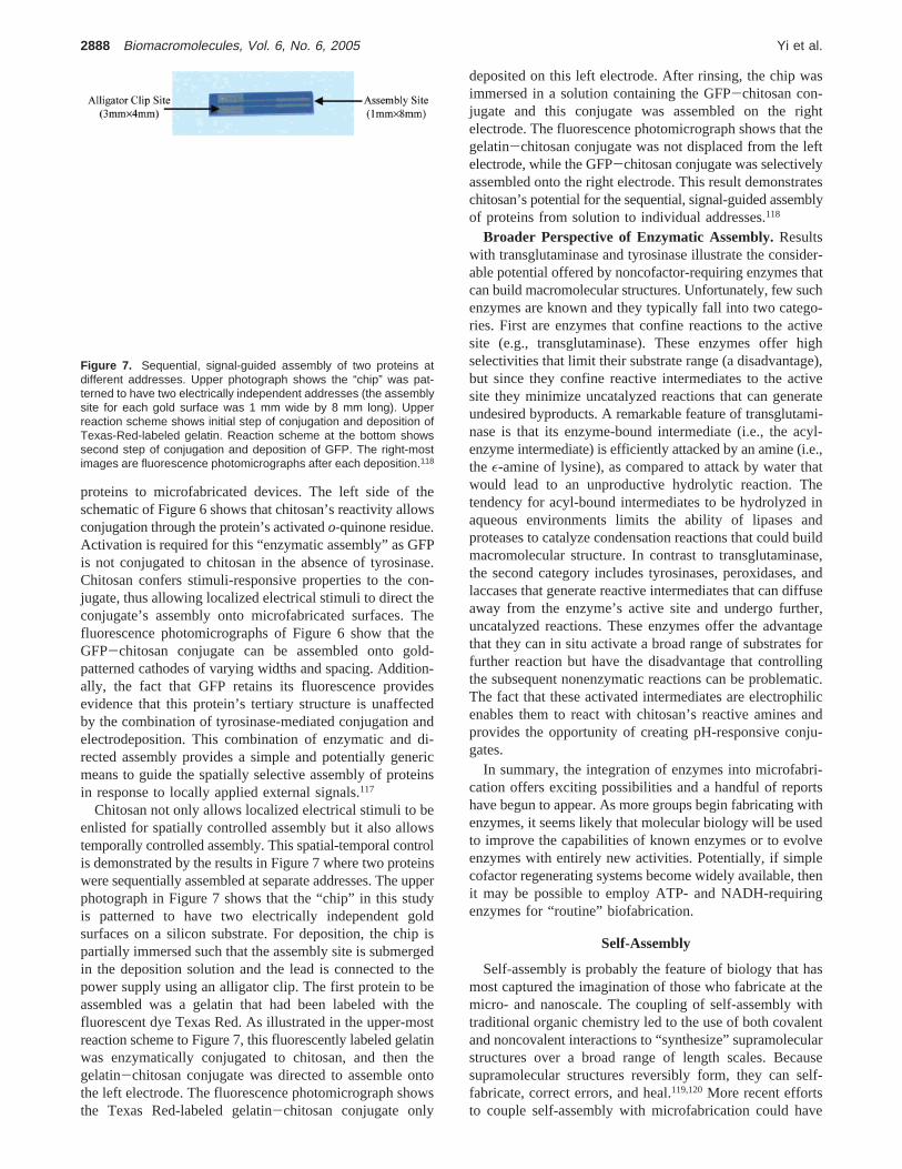
Chitosan not only allows localized electrical stimuli to beenlisted for spatially controlled assembly but it also allowstemporally controlled assembly. This spatial-temporal controlis demonstrated by the results in Figure 7 where two proteinswere sequentially assembled at separate addresses. The upperphotograph in Figure 7 shows that the “chip” in this studyis patterned to have two electrically independent goldsurfaces on a silicon substrate. For deposition, the chip ispartially immersed such that the assembly site is submergedin the deposition solution and the lead is connected to thepower supply using an alligator clip. The first protein to beassembled was a gelatin that had been labeled with thefluorescent dye Texas Red. As illustrated in the upper-mostreaction scheme to Figure 7, this fluorescently labeled gelatinwas enzymatically conjugated to chitosan, and then thegelatin-chitosan conjugate was directed to assemble ontothe left electrode. The fluorescence photomicrograph showsthe Texas Red-labeled gelatin-chitosan conjugate only
deposited on this left electrode. After rinsing, the chip wasimmersed in a solution containing the GFP-chitosan con-jugate and this conjugate was assembled on the rightelectrode. The fluorescence photomicrograph shows that thegelatin-chitosan conjugate was not displaced from the leftelectrode, while the GFP-chitosan conjugate was selectivelyassembled onto the right electrode. This result demonstrateschitosan’s potential for the sequential, signal-guided assemblyof proteins from solution to individual addresses.118
Broader Perspective of Enzymatic Assembly.Resultswith transglutaminase and tyrosinase illustrate the consider-able potential offered by noncofactor-requiring enzymes thatcan build macromolecular structures. Unfortunately, few suchenzymes are known and they typically fall into two catego-ries. First are enzymes that confine reactions to the activesite (e.g., transglutaminase). These enzymes offer highselectivities that limit their substrate range (a disadvantage),but since they confine reactive intermediates to the activesite they minimize uncatalyzed reactions that can generateundesired byproducts. A remarkable feature of transglutami-nase is that its enzyme-bound intermediate (i.e., the acyl-enzyme intermediate) is efficiently attacked by an amine (i.e.,the ε-amine of lysine), as compared to attack by water thatwould lead to an unproductive hydrolytic reaction. Thetendency for acyl-bound intermediates to be hydrolyzed inaqueous environments limits the ability of lipases andproteases to catalyze condensation reactions that could buildmacromolecular structure. In contrast to transglutaminase,the second category includes tyrosinases, peroxidases, andlaccases that generate reactive intermediates that can diffuseaway from the enzyme’s active site and undergo further,uncatalyzed reactions. These enzymes offer the advantagethat they can in situ activate a broad range of substrates forfurther reaction but have the disadvantage that controllingthe subsequent nonenzymatic reactions can be problematic.The fact that these activated intermediates are electrophilicenables them to react with chitosan’s reactive amines andprovides the opportunity of creating pH-responsive conju-gates.
In summary, the integration of enzymes into microfabri-cation offers exciting possibilities and a handful of reportshave begun to appear. As more groups begin fabricating withenzymes, it seems likely that molecular biology will be usedto improve the capabilities of known enzymes or to evolveenzymes with entirely new activities. Potentially, if simplecofactor regenerating systems become widely available, thenit may be possible to employ ATP- and NADH-requiringenzymes for “routine” biofabrication.
Self-Assembly
Self-assembly is probably the feature of biology that hasmost captured the imagination of those who fabricate at themicro- and nanoscale. The coupling of self-assembly withtraditional organic chemistry led to the use of both covalentand noncovalent interactions to “synthesize” supramolecularstructures over a broad range of length scales. Becausesupramolecular structures reversibly form, they can self-fabricate, correct errors, and heal.119,120More recent effortsto couple self-assembly with microfabrication could have
Figure 7. Sequential, signal-guided assembly of two proteins atdifferent addresses. Upper photograph shows the “chip” was pat-terned to have two electrically independent addresses (the assemblysite for each gold surface was 1 mm wide by 8 mm long). Upperreaction scheme shows initial step of conjugation and deposition ofTexas-Red-labeled gelatin. Reaction scheme at the bottom showssecond step of conjugation and deposition of GFP. The right-mostimages are fluorescence photomicrographs after each deposition.118
2888 Biomacromolecules, Vol. 6, No. 6, 2005 Yi et al.
revolutionary technological effects. Conventional top-downmicrofabrication is having increasing difficulty keeping pacewith the relentless reductions in feature sizes required to meetthe insatiable demand for greater computational power.121,122
Many biological structures self-assemble at attractive sizescales, and these materials could serve as building blocks ortemplates for fabrication,123,124or as models to mimic.125
Lipid bilayers are probably the simplest self-assembledstructures in biology, and they have sparked various technicalapplications. They have been used to template126,127 or tolocalize components either within the membrane bilayer128,129
or within an internal aqueous compartment(s).130-133 Thereare even efforts to use lipids to construct soft microfluidicnetworks.134 Lessons from lipid self-assembly are also beingmimicked in the construction of block polymers that canphase separate to form domains of various thickness, thatform stronger interfaces, and that confer long range order.135
Nucleic acids offer a range of capabilities useful forfabrication. As mentioned earlier, nucleic acids have beenused as templates for metallization to create nanoscale wiresand field effect transistors.66,136,137DNA self-assembly (i.e.,base-pairing) is programmable and predictable allowingDNA-based materials to be engineered to assemble intocomplex architectures (e.g., dendrimers) over a range oflength scales and to perform mechanical actions.138-141
Finally, DNA has been conjugated to nanoparticles to allowDNA’s programmable assembly to be employed for thethermally reversible formation of nanoparticle networks.142-144
Protein self-assembly also offers a remarkable array ofopportunities even in the absence of a solution to the“protein-folding problem”.8 Despite an inability to predictthe self-assembly of large polypeptides, there are growingefforts to exploit predictable secondary structural elements(e.g., R-helices andâ-sheets) to generate self-assemblingpeptide-based materials.145-158 Further, there are importantexamples of known protein systems that self-assemble intouseful structures, eliminating the requirement for de novopolypeptide design for these cases. Examples include thefilament-forming proteins actin and tubulin that have eachbeen used as templates for nanowire synthesis.159,160 Ad-ditionally, the self-assembling coat proteins of virus particlesprovide a rich source of nanostructures123 that can be enlistedto compartmentalize161,162or to template.163-168 A significantadvantage of protein-based materials is that biotechnologicalmethods can extend their capabilities in two important ways.First, known sequences can be genetically fused to proteinsto confer added functionality (e.g., fusion tags of elastin-like peptides confer thermally reversible surface assemblycapabilities169). Second, unknown sequences can be “dis-covered” to confer desired functionality (e.g., peptides thatbind to inorganics47,49,50,163).
Coupling Self-Assembly with Chitosan’s Directed As-sembly. Although self-assembly offers the potential forbottom-up self-fabrication of nanoscale structures, it is notyet obvious how to interface these biological or biomimeticstructures to specific addresses of microfabricated surfaces.170
Potentially, chitosan can serve as the device-biologyinterface by integrating chitosan’s ability to selectivelyelectrodeposit at the device surface with its ability to be
readily functionalized with biological components. Specif-ically, the reactivity of chitosan’s amines allows previouslydeposited films to be functionalized using standard couplingchemistries based on homobifunctional (e.g., glutaraldehyde),heterobifunctional (e.g.,γ-maleimido butyric acid succin-imidyl ester), or biological (e.g., biotin) agents. In our studies,we used glutaraldehyde as a model to covalently tether anamine-terminated single stranded DNA oligonucleotide171
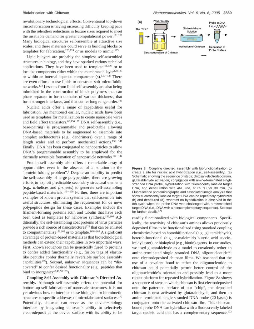
onto electrodeposited chitosan films. We reasoned that theuse of a covalent bond to tether the oligonucleotide tochitosan could potentially permit better control of theoligonucleotide’s orientation and possibly lead to a morerobust platform for repeated hybridization. Figure 8a showsa sequence of steps in which chitosan is first electrodepositedonto the patterned surface of our “chip”, the depositedchitosan is next activated by glutaraldehyde, and then anamine-terminated single stranded DNA probe (20 bases) isconjugated onto the activated chitosan film. This chitosan-bound probe DNA can hybridize with a fluorescently labeledtarget nucleic acid that has a complementary sequence.172
Figure 8. Coupling directed assembly with biofunctionalization tocreate a site for nucleic acid hybridization (i.e., self-assembly). (a)Schematic showing the sequence of steps; chitosan electrodeposition,glutaraldehyde activation, conjugation with amine-terminated singlestranded DNA probe, hybridization with fluorescently labeled targetDNA, and denaturation with 4M urea, at 65 °C for 30 min. (b)Fluorescence photomicrographs and associated image analysis thatshow fluorescently labeled target DNA can be repeatedly hybridized(h) and denatured (d), whereas no hybridization is observed in the8th cycle when the probe DNA was challenged with a mismatchedtarget DNA (i.e., DNA with a noncomplementary sequence). See textfor further details.172
Biofabrication with Chitosan Biomacromolecules, Vol. 6, No. 6, 2005 2889

In addition, this target can be reversibly “dis-assembled” bysubjecting the chip to denaturing conditions (4 M urea and65 °C).
Results from a sequence of hybridization (h) and dena-turation (d) steps are shown in Figure 8b as a series offluorescence photomicrographs and their associated imageanalysis. These results demonstrate that the deposited chi-tosan provides a robust platform for tethering probe nucleicacids that can serve as a nucleation site for reversible self-assembly (i.e., hybridization). Figure 8b also indicates thathybridization to the chitosan-bound probe relies on themolecular recognition capabilities inherent to DNA base-pairing. Specifically, during the 8th hybridization attemptin Figure 8b, the chitosan-bound probe was challenged withhigh concentrations of a fluorescently labeled target DNAthat had a mismatched (i.e., noncomplementary) sequence.After observing that this mismatched target did not hybridizeto the chitosan-bound probe (designated “8mismatch”), weconfirmed that the probe could still hybridize to the fluo-rescently labeled target DNA of complementary sequence(designated “9h”).172
To further illustrate the potential of chitosan to serve as adevice-biology interface, we used surface-bound ssDNA totether nanotubes of the tobacco mosaic virus (TMV). EachTMV particle consists of a single strand of genomic RNAencapsidated within a protein nanotube (inner cavity andouter diameter of 4 and 18 nm, respectively) that is composedof 2130 identical capsid protein subunits (subunit molecularweight of 17.5 kDa). Figure 9a illustrates TMV self-
assembly, whereas Figure 9b is a transmission electronmicrograph showing these nanotubes. In our experiments,we labeled the virus particles with fluorescein to facilitatevisualization. To assemble the TMV nanotubes onto elec-trodeposited chitosan, we began by partially disassemblingthe fluorescein-labeled virus particles to expose the 5′ endof the viral RNA. As illustrated in Figure 9c, the exposedviral RNA can hybridize to a complementary ssDNA 25-mer that had been previously anchored to electrodepositedchitosan. Thus, the chitosan-bound DNA serves as a “con-nection” site to capture the TMV particles. Experimentalevidence for this hybridization-based capture is shown inFigure 9d that shows the fluorescently labeled TMV is“targeted” to the left electrode that had the complementaryDNA strand. No TMV assembly was observed on theuntreated gold surface of the right electrode.173 This resultillustrates the coupling of electrodeposition and functional-ization and further demonstrates the versatility of chitosanas a device-biology interface.
More broadly, the results in Figure 9 suggest chitosan mayprovide a key interconnect between conventional micro-fabrication and molecular-level self-assembly. Microfabri-cation allows devices to be created that can impose controlledelectrical stimuli to direct chitosan’s assembly. Biologicalcomponents (e.g., ssDNA) can be readily conjugated to theelectrodeposited chitosan using facile methods (e.g., glut-araldehyde) to create sites to either “connect” the self-assembled system or to “nucleate” self-assembly. Finally,molecular biological techniques can be integrated to conferdesired functionality to the self-assembled structure. For thecase of the self-assembled TMV, the coat protein can beengineered to enhance its role as a template for metalassembly.168,174-176
Conclusions
Nature employs a small number of organic materials asbuilding blocks to construct a diversity of structures thatperform a range of functions. These capabilities have sparkedinterest in fabricating devices using these same biologicalbuilding blocks or their mimics. Self-assembly is the featurethat has attracted the most attention, and several studies havedemonstrated the potential of biological components to self-fabricate nanoscale structures of controlled size and shape.However, the use of biological materials also allows accessto enzymes for selective catalysis under mild conditions.Enzymatic assembly offers exciting opportunities that willbe further broadened if the tools of modern biology can beenlisted to engineer/evolve/design enzymes to build macro-molecular structure or catalyze surface assembly. Finally, arange of localized external stimuli are beginning to be usedto guide the assembly of nano-bio hybrids. Directed assemblyoften employs the stimuli-responsive character of the nanocomponent, whereas molecular biological methods areproviding the means to link the bio to the nano. Directedassembly also allows the strengths of microfabrication to beaccessed by creating devices that can impose stimuli withhigh spatial and temporal control.
Chitosan is an aminopolysaccharide that offers pH-responsive-solubility, forms films and hydrogels, and con-
Figure 9. Coupling chitosan’s directed assembly with TMV self-assembly. (a) TMV nanotubes are formed from thousands of coatprotein subunits that self-assemble around the virus’ single strandedRNA genome (Adapted from ref 188 with permission from Namba etal. Science 1985, 227 (4688), 773-6. Copyright 1985 AAAS). (b)Transmission electron micrograph of TMV nanotubes. (c) Schematicshowing that partial disassembly of the virus particle exposes the 5′end of the viral RNA allowing hybridization to the surface-bound DNAprobe. (d) Fluorescence photomicrograph showing capture of fluo-rescein-labeled TMV nanotubes on the left electrode containing TMV-specific probe DNA.173
2890 Biomacromolecules, Vol. 6, No. 6, 2005 Yi et al.

tains readily modifiable primary amine substituents. Chito-san’s pH-responsive solubility allows it to “recognize”localized electrical stimuli and respond by assembling (i.e.,depositing) as a thin film. Chitosan’s reactivity allows it tobe readily functionalized: proteins can be enzymaticallyassembled onto the stimuli-responsive backbone, whereasnucleic acids can be tethered to electrodeposited films toserve as sites for self-assembly. These capabilities conferconsiderable versatility to chitosan and suggest this biopoly-mer has a particularly bright future for biofabrication of thedevice-biology interface.
Acknowledgment. Financial support was provided by theNational Science Foundation (Grants BES-0114790, DMR-08008, and DMR0231291) and the Department of Energy(DE-FG02-02ER45975 and DE-FG02-01ER63019).
References and Notes
(1) Cote, G. L.; Lec, R. M.; Pishko, M. V. Emerging biomedical sensingtechnologies and their applications.IEEE Sens. J.2003, 3 (3), 251-266.
(2) Grayson, A. C. R.; Shawgo, R. S.; Johnson, A. M.; Flynn, N. T.; Li,Y. W.; Cima, M. J.; Langer, R. A BioMEMS review: MEMStechnology for physiologically integrated devices.Proc. IEEE2004,92 (1), 6-21.
(3) Baeumner, A. J. Biosensors for environmental pollutants and foodcontaminants.Anal. Bioanal. Chem.2003, 377 (3), 434-445.
(4) Wu, L. Q.; Payne, G. F. Biofabrication: using biological materialsand biocatalysts to construct nanostructured assemblies.TrendsBiotechnol.2004, 22 (11), 593-9.
(5) Xia, Y.; Whitesides, G. M. Soft Lithography.Angew. Chem. Int. Ed.Engl. 1998, 37, 550-575.
(6) Gates, B. D.; Xu, Q.; Love, C.; Wolfe, B. D.; Whitesides, G. M.Unconventional Nanofabrication.Annu. ReV. Mater. Res.2004, 34,339-372.
(7) Ball, P. Natural strategies for the molecular engineer.Nanotechnology2002, 13 (5), R15-R28.
(8) Goodsell, D.Bionanotechnology; Wiley-Liss: Hoboken, NJ, 2004.(9) Jones, R.Soft Machines. Nanotechnology and Life; Oxford University
Press: Oxford, 2004.(10) Niemeyer, C. M. Nanoparticles, proteins, and nucleic acids: Bio-
technology meets materials science.Angew. Chem. Int. Ed.2001,40 (22), 4128-4158.
(11) Kumar, M. N.; Muzzarelli, R. A.; Muzzarelli, C.; Sashiwa, H.; Domb,A. J. Chitosan chemistry and pharmaceutical perspectives.Chem.ReV. 2004, 104 (12), 6017-84.
(12) Berger, J.; Reist, M.; Mayer, J. M.; Felt, O.; Peppas, N. A.; Gurny,R. Structure and interactions in covalently and ionically cross-linkedchitosan hydrogels for biomedical applications.Eur. J. Pharm.Biopharm.2004, 57 (1), 19-34.
(13) Kumar, M. N. V. R. A review of chitin and chitosan applications.React. Funct. Polym.2000, 46 (1), 1-27.
(14) Shahidi, F.; Arachchi, J. K. V.; Jeon, Y. J. Food applications of chitinand chitosans.Trends Food Sci. Technol.1999, 10 (2), 37-51.
(15) Lim, S. H.; Hudson, S. M. Review of chitosan and its derivatives asantimicrobial agents and their uses as textile chemicals.J. Macromol.Sci. Polym. ReV. 2003, C43 (2), 223-269.
(16) Rinaudo, M.; Pavlov, G.; Desbrieres, J. Influence of acetic acidconcentration on the solubilization of chitosan.Polymer1999, 40,7029-7032.
(17) Sorlier, P.; Denuziere, A.; Viton, C.; Domard, A. Relation betweenthe Degree of Acetylation and the Electrostatic Properties of Chitinand Chitosan.Biomacromolecules2001, 2 (3), 765-772.
(18) Strand, S. P.; Tommeraas, K.; Varum, K. M.; Ostgaard, K. Electro-phoretic light scattering studies of chitosans with different degreesof N-acetylation.Biomacromolecules2001, 2 (4), 1310-1314.
(19) Anthonsen, M. W.; Smidsrod, O. Hydrogen-Ion Titration of Chitosanswith Varying Degrees of N-Acetylation by Monitoring Induced H-1-Nmr Chemical-Shifts.Carbohydr. Polym.1995, 26 (4), 303-305.
(20) Varum, K. M.; Ottoy, M. H.; Smidsrod, O. Water-solubility ofpartially N-acetylated chitosans as a function of pH: Effect ofchemical composition and depolymerization.Carbohydr. Polym.1994, 25, 65-70.
(21) Montembault, A.; Viton, C.; Domard, A. Rheometric study of thegelation of chitosan in aqueous solution without cross-linking agent.Biomacromolecules2005, 6 (2), 653-62.
(22) Kurita, A. Controlled functionalization of the polysaccharide chitin.Prog. Polym. Sci.2001, 26, 1921-1971.
(23) Ang, T. H.; Sultana, F. S. A.; Hutmacher, D. W.; Wong, Y. S.; Fuh,J. Y. H.; Mo, X. M.; Loh, H. T.; Burdet, E.; Teoh, S. H. Fabricationof 3D chitosan-hydroxyapatite scaffolds using a robotic dispensingsystem.Mat. Sci. Eng. C2002, 20 (1-2), 35-42.
(24) Co, C. C.; Wang, Y. C.; Ho, C. C. Biocompatible micropatterningof two different cell types.J. Am. Chem. Soc.2005, 127 (6), 1598-9.
(25) Kumar, G.; Wang, Y. C.; Co, C.; Ho, C. C. Spatially controlled cellengineering on biomaterials using polyelectrolytes.Langmuir2003,19 (25), 10550-10556.
(26) Feng, J.; Gao, C. Y.; Wang, B.; Shen, J. C. Co-patterning chitosanand bovine serum albumin on an aldehyde-enriched glass substrateby microcontact printing.Thin Solid Films2004, 460 (1-2), 286-290.
(27) Serizawa, T.; Goto, H.; Kishida, A.; Endo, T.; Akashi, M. Improvedalternate deposition of biodegradable naturally occurring polymersonto a quartz crystal microbalance.J. Polym. Sci. A Polym. Chem.1999, 37 (6), 801-804.
(28) Huang, H. Z.; Yang, X. R. Chitosan mediated assembly of goldnanoparticles multilayer.Colloids Surf., A2003, 226(1-3), 77-86.
(29) Lvov, Y.; Onda, M.; Ariga, K.; Kunitake, T. Ultrathin films ofcharged polysaccharides assembled alternately with linear polyions.J. Biomater. Sci. Polym. Ed.1998, 9 (4), 345-355.
(30) Richert, L.; Lavalle, P.; Payan, E.; Shu, X. Z.; Prestwich, G. D.;Stoltz, J. F.; Schaaf, P.; Voegel, J. C.; Picart, C. Layer by layerbuildup of polysaccharide films: Physical chemistry and cellularadhesion aspects.Langmuir2004, 20 (2), 448-458.
(31) Thierry, B.; Winnik, F. M.; Merhi, Y.; Silver, J.; Tabrizian, M.Bioactive coatings of endovascular stents based on polyelectrolytemultilayers.Biomacromolecules2003, 4 (6), 1564-1571.
(32) dos Santos, D. S.; Goulet, P. J. G.; Pieczonka, N. P. W.; Oliveira,O. N.; Aroca, R. F. Gold nanoparticle embedded, self-sustainedchitosan films as substrates for surface-enhanced Raman scattering.Langmuir2004, 20 (23), 10273-10277.
(33) Zhang, M. G.; Smith, A.; Gorski, W. Carbon nanotube-chitosansystem for electrochemical sensing based on dehydrogenase enzymes.Anal. Chem.2004, 76 (17), 5045-5050.
(34) Zhang, M. G.; Gorski, W. Electrochemical sensing platform basedon the carbon nanotubes/redox mediators-biopolymer system.J. Am.Chem. Soc.2005, 127 (7), 2058-2059.
(35) Constantine, C. A.; Gattas-Asfura, K. M.; Mello, S. V.; Crespo, G.;Rastogi, V.; Cheng, T. C.; DeFrank, J. J.; Leblanc, R. M. Layer-by-layer biosensor assembly incorporating functionalized quantum dots.Langmuir2003, 19 (23), 9863-9867.
(36) Schauer, C. L.; Chen, M. S.; Chatterley, M.; Eisemann, K.; Welsh,E. R.; Price, R. R.; Schoen, P. E.; Ligler, F. S. Color changes inchitosan and poly(allylamine) films upon metal binding.Thin SolidFilms 2003, 434 (1-2), 250-257.
(37) Schauer, C. L.; Chen, M. S.; Price, R. R.; Schoen, P. E.; Ligler, F.S. Colored thin films for specific metal ion detection.EnViron. Sci.Technol.2004, 38 (16), 4409-13.
(38) Serizawa, T.; Yamaguchi, M.; Matsuyama, T.; Akashi, M. Alternatingbioactivity of polymeric layer-by-layer assemblies: Anti- vs proco-agulation of human blood on chitosan and dextran sulfate layers.Biomacromolecules2000, 1 (3), 306-309.
(39) Serizawa, T.; Yamaguchi, M.; Akashi, M. Alternating bioactivity ofpolymeric layer-by-layer assemblies: Anticoagulation vs procoagu-lation of human blood.Biomacromolecules2002, 3 (4), 724-731.
(40) Serizawa, T.; Yamaguchi, M.; Akashi, M. Enzymatic hydrolysis ofa layer-by-layer assembly prepared from chitosan and dextran sulfate.Macromolecules2002, 35 (23), 8656-8658.
(41) Etienne, O.; Schneider, A.; Taddei, C.; Richert, L.; Schaaf, P.; Voegel,J. C.; Egles, C.; Picart, C. Degradability of polysaccharides multilayerfilms in the oral environment: an in vitro and in vivo study.Biomacromolecules2005, 6 (2), 726-733.
(42) Wu, L. Q.; Ghodssi, R.; Elabd, Y. A.; Payne, G. F. BiomimeticPattern Transfer.AdV. Funct. Mater.2005, 15, 189-195.
(43) Baumgart, T.; Offenhausser, A. Polysaccharide-supported planarbilayer lipid model membranes.Langmuir2003, 19 (5), 1730-1737.
(44) Katz, E.; Willner, I. Integrated nanoparticle-biomolecule hybridsystems: synthesis, properties, and applications.Angew. Chem. Int.Ed. Engl.2004, 43 (45), 6042-108.
(45) Fang, M.; Grant, P. S.; McShane, M. J.; Sukhorukov, G. B.; Golub,V. O.; Lvov, Y. M. Magnetic bio/nanoreactor with multilayer shells
Biofabrication with Chitosan Biomacromolecules, Vol. 6, No. 6, 2005 2891
of glucose oxidase and inorganic nanoparticles.Langmuir2002, 18(16), 6338-6344.
(46) Weizmann, Y.; Patolsky, F.; Lioubashevski, O.; Willner, I. Magneto-mechanical detection of nucleic acids and telomerase activity incancer cells.J. Am. Chem. Soc.2004, 126 (4), 1073-80.
(47) Sarikaya, M.; Tamerler, C.; Schwartz, D. T.; Baneyx, F. O. Materialsassembly and formation using engineered polypeptides.Annu. ReV.Mater. Res.2004, 34, 373-408.
(48) Sarikaya, M.; Tamerler, C.; Jen, A. K.; Schulten, K.; Baneyx, F.Molecular biomimetics: nanotechnology through biology.Nat. Mater.2003, 2 (9), 577-85.
(49) Naik, R. R.; Stringer, S. J.; Agarwal, G.; Jones, S. E.; Stone, M. O.Biomimetic synthesis and patterning of silver nanoparticles.Nat.Mater. 2002, 1 (3), 169-172.
(50) Whaley, S. R.; English, D. S.; Hu, E. L.; Barbara, P. F.; Belcher, A.M. Selection of peptides with semiconductor binding specificity fordirected nanocrystal assembly.Nature2000, 405 (6787), 665-668.
(51) Shacham, R.; Avnir, D.; Mandler, D. Electrodeposition of methylatedsol-gel films on conducting surfaces.AdV. Mater.1999, 11 (5), 384-388.
(52) Wu, L. Q.; Gadre, A. P.; Yi, H. M.; Kastantin, M. J.; Rubloff, G.W.; Bentley, W. E.; Payne, G. F.; Ghodssi, R. Voltage-dependentassembly of the polysaccharide chitosan onto an electrode surface.Langmuir2002, 18 (22), 8620-8625.
(53) Fernandes, R.; Wu, L. Q.; Chen, T. H.; Yi, H. M.; Rubloff, G. W.;Ghodssi, R.; Bentley, W. E.; Payne, G. F. Electrochemically induceddeposition of a polysaccharide hydrogel onto a patterned surface.Langmuir2003, 19 (10), 4058-4062.
(54) Wu, L.-Q.; Yi, H.; Li, S.; Rubloff, G. W.; Bentley, W. E.; Ghodssi,R.; Payne, G. F. Spatially selective Deposition of a ReactivePolysaccharide Layer onto a Patterned Template.Langmuir 2003,19, 519-524.
(55) Wu, L. Q.; Lee, K.; Wang, X.; English, D. S.; Losert, W.; Payne, G.F. Chitosan-Mediated and Spatially Selective Electrodeposition ofNanoscale Particles.Langmuir2005, 21 (8), 3641-3646.
(56) Luo, X. L.; Xu, J. J.; Du, Y.; Chen, H. Y. A glucose biosensor basedon chitosan-glucose oxidase-gold nanoparticles biocomposite formedby one-step electrodeposition.Anal. Biochem.2004, 334 (2), 284-289.
(57) Luo, X. L.; Xu, J. J.; Wang, J. L.; Chen, H. Y. Electrochemicallydeposited nanocomposite of chitosan and carbon nanotubes forbiosensor application.Chem. Comm.2005 (16), 2169-2171.
(58) Kastantin, M. J.; Li, S.; Gadre, A. P.; Wu, L. Q.; Bentley, W. E.;Payne, G. F.; Rubloff, G. W.; Ghodssi, R. Integrated fabrication ofpolymeric devices for biological applications.Sensors Mater.2003,15 (6), 295-311.
(59) Powers, M. A.; Koev, S. T.; Schleunitz, A.; Yi, H.; Hodzic, V.;Bentley, W. E.; Payne, G. F.; Rubloff, G. W.; Ghodssi, R. Afabrication platform for electrically mediated optically active bio-functionalized sites in BioMEMS.Lab Chip2005, 5, 583-586.
(60) Yang, L. J.; Lin, W. Z.; J, Y. T.; Tai, Y. C. Photopatternable gelatinas protection layers in low-temperature surface micromachinings.Sens. Actuators A2003, 103, 284-290.
(61) Reches, M.; Gazit, E. Casting metal nanowires within discrete self-assembled peptide nanotubes.Science2003, 300 (5619), 625-7.
(62) Fernandes, R.; Yi, H. M.; Wu, L. Q.; Rubloff, G. W.; Ghodssi, R.;Bentley, W. E.; Payne, G. F. Thermo-biolithography: A techniquefor patterning nucleic acids and proteins.Langmuir 2004, 20 (3),906-913.
(63) Clausen-Schaumann, H.; Grandbois, M.; Gaub, H. Enzyme-assistedNanoscale Lithography in Lipid Membranes.AdV. Mater. 1998, 10(12), 949-952.
(64) Takeda, S.; Nakamura, C.; Miyamoto, C.; Nakamura, N.; Kageshima,M.; Tokumoto, H.; Miyake, J. Lithographing of Biomolecules on aSubstrate Surface Using an Enzyme-Immobilized AFM Tip.NanoLett. 2003, 3 (11), 1471-1474.
(65) Hyun, J.; Kim, J.; Craig, S. L.; Chilkoti, A. Enzymatic Nanolithog-raphy of a Self-Assembled Oligonucleotide Monolayer on Gold.J.Am. Chem. Soc.2004, 126 (15), 4770-1.
(66) Braun, E.; Keren, K. From DNA to Transistors.AdV. Phys.2004, 53(4), 441-496.
(67) Keren, K.; Krueger, M.; Gilad, R.; Ben-Yoseph, G.; Sivan, U.; Braun,E. Sequence-specific molecular lithography on single DNA mol-ecules.Science2002, 297 (5578), 72-5.
(68) Keren, K.; Berman, R. S.; Buchstab, E.; Sivan, U.; Braun, E. DNA-templated carbon nanotube field-effect transistor.Science2003, 302(5649), 1380-1382.
(69) Mavroidis, C.; Dubey, A.; Yarmush, M. L. Molecular machines.Annu. ReV. Biomed. Eng.2004, 6, 363-95.
(70) Noji, H.; Yasuda, R.; Yoshida, M.; Kinosita, K., Jr. Direct observationof the rotation of F1-ATPase.Nature1997, 386 (6622), 299-302.
(71) Soong, R. K.; Bachand, G. D.; Neves, H. P.; Olkhovets, A. G.;Craighead, H. G.; Montemagno, C. D. Powering an inorganicnanodevice with a biomolecular motor.Science2000, 290 (5496),1555-1558.
(72) Montemagno, C.; Bachand, G. Constructing nanomechanical devicespowered by biomolecular motors.Nanotechnology1999, 10 (3), 225-231.
(73) Liu, H.; Schmidt, J. J.; Bachand, G. D.; Rizk, S. S.; Looger, L. L.;Hellinga, H. W.; Montemagno, C. D. Control of a biomolecularmotor-powered nanodevice with an engineered chemical switch.Nat.Mater. 2002, 1 (3), 173-7.
(74) Ohtsuka, T.; Sawa, A.; Kawabata, R.; Nio, N.; Motoki, M. Substratespecificities of microbial transglutaminase for primary amines.J.Agric. Food Chem.2000, 48 (12), 6230-6233.
(75) Sato, H.; Hayashi, E.; Yamada, N.; Yatagai, M.; Takahara, Y. Furtherstudies on the site-specific protein modification by microbial trans-glutaminase.Bioconj. Chem.2001, 12 (5), 701-710.
(76) Villalonga, R.; Fernandez, M.; Fragoso, A.; Cao, R.; Di Pierro, P.;Mariniello, L.; Porta, R. Transglutaminase-catalyzed synthesis oftrypsin-cyclodextrin conjugates: kinetics and stability properties.Biotechnol. Bioeng.2003, 81 (6), 732-7.
(77) Villalonga, R.; Fernandez, M.; Fragoso, A.; Cao, R.; Mariniello, L.;Porta, R. Thermal stabilization of trypsin by enzymic modificationwith beta-cyclodextrin derivatives.Biotechnol. Appl. Biochem.2003,38, 53-59.
(78) Josten, A.; Meusel, M.; Spener, F. Microbial transglutaminase-mediated synthesis of hapten-protein conjugates for immunoassays.Anal. Biochem.1998, 258 (2), 202-208.
(79) Collier, J. H.; Messersmith, P. B. Enzymatic modification of self-assembled peptide structures with tissue transglutaminase.Biocon-jugate Chem.2003, 14 (4), 748-55.
(80) Babin, H.; Dickinson, E. Influence of transglutaminase treatment onthe thermoreversible gelation of gelatin.Food Hydrocolloids2001,15, 271-276.
(81) Lim, L. T.; Mine, Y.; Tung, M. A. Barrier and tensile properties oftransglutaminase cross-linked gelatin films as affected by relativehumidity, temperature, and glycerol content.J. Food Sci.1999, 64(4), 616-622.
(82) Crescenzi, V.; Francescangeli, A.; Taglienti, A. New Gelatin-BasedHydrogels via Enzymatic Networking.Biomacromolecules2002, 3(6), 1384-1391.
(83) Fuchsbauer, H. L.; Gerber, U.; Engelmann, J.; Seeger, T.; Sinks, C.;Hecht, T. Influence of gelatin matrixes cross-linked with trans-glutaminase on the properties of an enclosed bioactive material usingbeta-galactosidase as model system.Biomaterials1996, 17 (15),1481-1488.
(84) Chen, T. H.; Embree, H. D.; Brown, E. M.; Taylor, M. M.; Payne,G. F. Enzyme-catalyzed gel formation of gelatin and chitosan:potential for in situ applications.Biomaterials2003, 24 (17), 2831-2841.
(85) Sperinde, J.; Griffith, L. Synthesis and Characterization of Enzymati-cally-Cross-Linked Poly(ethylene glycol) Hydrogels.Macromolecules1997, 30, 5255-5264.
(86) Sperinde, J.; Griffith, L. Control and Prediction of Gelation Kineticsin Enzymatically Cross-Linked Poly(ethylene glycol) Hydrogels.Macromolecules2000, 33, 5476-5480.
(87) Hu, B. H.; Messersmith, P. B. Rational design of transglutaminasesubstrate peptides for rapid enzymatic formation of hydrogels.J. Am.Chem. Soc.2003, 125 (47), 14298-9.
(88) Chen, T.; Small, D. A.; McDermott, M. K.; Bentley, W. E.; Payne,G. F. Enzymatic Methods for In situ Cell Entrapment and CellRelease.Biomacromolecules2003, 4, 1558-1563.
(89) Schense, J. C.; Hubbell, J. A. Cross-linking exogenous bifunctionalpeptides into fibrin gels with factor XIIIa.Bioconjug. Chem.1999,10 (1), 75-81.
(90) Schense, J. C.; Bloch, J.; Aebischer, P.; Hubbell, J. A. Enzymaticincorporation of bioactive peptides into fibrin matrixes enhancesneurite extension.Nat. Biotechnol.2000, 18 (4), 415-9.
(91) Schense, J. C.; Hubbell, J. A. Three-dimensional migration of neuritesis mediated by adhesion site density and affinity.J. Biol. Chem.2000,275 (10), 6813-8.
(92) Sakiyama, S. E.; Schense, J. C.; Hubbell, J. A. Incorporation ofheparin-binding peptides into fibrin gels enhances neurite exten-sion: an example of designer matrixes in tissue engineering.FASEBJ. 1999, 13 (15), 2214-24.
2892 Biomacromolecules, Vol. 6, No. 6, 2005 Yi et al.

(93) Sakiyama-Elbert, S. E.; Hubbell, J. A. Controlled release of nervegrowth factor from a heparin-containing fibrin-based cell ingrowthmatrix. J. Controlled Release2000, 69 (1), 149-58.
(94) Sakiyama-Elbert, S. E.; Hubbell, J. A. Development of fibrinderivatives for controlled release of heparin-binding growth factors.J. Controlled Release2000, 65 (3), 389-402.
(95) Seitz, A.; Schneider, F.; Pasternack, R.; Fuchsbauer, H. L.; Hampp,N. Enzymatic cross-linking of purple membranes catalyzed bybacterial transglutaminase.Biomacromolecules2001, 2 (1), 233-8.
(96) Nieuwenhuizen, W. F.; Dekker, H. L.; De Koning, L. J.; Groneveld,T.; De Koster, C. G.; De Jong, G. A. H. Modification of glutamineand lysine residues in holo and apo alpha-lactalbumin with microbialtransglutaminase.J. Agric. Food Chem.2003, 51 (24), 7132-7139.
(97) Nieuwenhuizen, W. F.; Dekker, H. L.; Groneveld, T.; de Koster, C.G.; de Jong, G. A. H. Transglutaminase-mediated modification ofglutamine and lysine residues in native bovine beta-lactoglobulin.Biotechnol. Bioeng.2004, 85 (3), 248-258.
(98) Matsumura, Y.; Chanyongvorakul, Y.; Kumazawa, Y.; Ohtsuka, T.;Mori, T. Enhanced susceptibility to transglutaminase reaction ofalpha-lactalbumin in the molten globule state.Biochim. Biophys. Acta-Protein Struct. Mol. Enzymol.1996, 1292 (1), 69-76.
(99) Kamiya, N.; Tanaka, T.; Suzuki, T.; Takazawa, T.; Takeda, S.;Watanabe, K.; Nagamune, T. S-peptide as a potent peptidyl linkerfor protein cross-linking by microbial transglutaminase from Strep-tomyces mobaraensis.Bioconjugate Chem.2003, 14 (2), 351-357.
(100) Tanaka, T.; Kamiya, N.; Nagamune, T. Peptidyl linkers for proteinheterodimerization catalyzed by microbial transglutaminase.Biocon-jugate Chem.2004, 15 (3), 491-497.
(101) Kamiya, N.; Takazawa, T.; Tanaka, T.; Ueda, H.; Nagamune, T. Site-specific cross-linking of functional proteins by transglutamination.Enzyme Microb. Technol.2003, 33 (4), 492-496.
(102) Takazawa, T.; Kamiya, N.; Ueda, H.; Nagamune, T. Enzymaticlabeling of a single chain variable fragment of an antibody withalkaline phosphatase by mircobial transglutaminase.Biotechnol.Bioeng.2004, 86 (4), 399-404.
(103) Burzio, L. A.; Waite, J. H. Reactivity of peptidyl-tyrosine tohydroxylation and cross-linking.Protein Sci.2001, 10, 735-740.
(104) Burzio, L. A.; Waite, J. H. Cross-linking in adhesive quinoproteins:studies with model decapeptides.Biochemistry2000, 39 (36), 11147-53.
(105) Sugumaran, M. Molecular mechanisms for cuticular sclerotization.AdV. Insect Physiol.1988, 21, 179-231.
(106) Sugumaran, M. Oxidation chemistry of 1,2-dehydro-N-acetyl-dopamines: Direct evidence for the formation of 1,2-dehydro-N-acetyldopamine quinone.Arch. Biochem. Biophys.2000, 378 (2),404-410.
(107) Kerwin, J. L.; Whitney, D. L.; Sheikh, A. Mass spectrometric profilingof glucosamine, glucosamine polymers and their catecholamineadducts- Model reactions and cuticular hydrolysates of Toxorhyn-chites amboinensis (Culicidae) pupae.Insect Biochem. Mol. Biol.1999, 29 (7), 599-607.
(108) Aberg, C. M.; Chen, T. H.; Olumide, A.; Raghavan, S. R.; Payne,G. F. Enzymatic Grafting of Peptides from Casein Hydrolysate toChitosan. Potential for Value-Added Byproducts from Food-Process-ing Wastes.J. Agric. Food Chem.2004, 52 (4), 788-793.
(109) Wu, L. Q.; Embree, H. D.; Balgley, B. M.; Smith, P. J.; Payne, G.F. Utilizing renewable resources to create functional polymers:Chitosan-based associative thickener.EnViron. Sci. Technol.2002,36 (15), 3446-3454.
(110) Muzzarelli, C.; Muzzarelli, R. A. A. Reactivity of quinones towardchitosan.Trends Glycosci. Glycotechnol.2002, 14, 223-229.
(111) Payne, G. F.; Chaubal, M. V.; Barbari, T. A. Enzyme-catalyzedpolymer modification: Reaction of phenolic compounds with chitosanfilms. Polymer1996, 37 (20), 4643-4648.
(112) Yamada, K.; Chen, T.; Kumar, G.; Vesnovsky, O.; Topoleski, L.D.; Payne, G. F. Chitosan Based Water-Resistant Adhesive. Analogyto Mussel Glue.Biomacromolecules2000, 1 (2), 252-8.
(113) Sun, W. Q.; Payne, G. F.; Moas, M. S. G. L.; Chu, J. H.; Wallace,K. K. Tyrosinase Reaction Chitosan Adsorption for RemovingPhenols from Wastewater.Biotechnol. Prog.1992, 8 (3), 179-186.
(114) Muzzarelli, R. A. A.; Ilari, P.; Xia, W. S.; Pinotti, M.; Tomasetti,M. Tyrosinase-Mediated Quinone Tanning of Chitinous Materials.Carbohydr. Polym.1994, 24 (4), 295-300.
(115) Chen, T. H.; Embree, H. D.; Wu, L. Q.; Payne, G. F. In vitro protein-polysaccharide conjugation: Tyrosinase-catalyzed conjugation ofgelatin and chitosan.Biopolymers2002, 64 (6), 292-302.
(116) Vazquez-Duhalt, R.; Tinoco, R.; D’Antonio, P.; Topoleski, L. D.T.; Payne, G. F. Enzyme conjugation to the polysaccharide chito-
san: Smart biocatalysts and biocatalytic hydrogels.BioconjugateChem.2001, 12 (2), 301-306.
(117) Chen, T.; Small, D. A.; Wu, L.-Q.; Rubloff, G. W.; Ghodssi, R.;Vazquez-Duhalt, R.; Bentley, W. E.; Payne, G. F. Nature-InspiredCreation of Protein-Polysaccharide Conjugate and its SubsequentAssembly onto a Patterned Surface.Langmuir2003, 19, 9382-9386.
(118) Yi, H.; Wu, L. Q.; Ghodssi, R.; Rubloff, G. W.; Payne, G. F.; Bentley,W. E. Signal-Directed Sequential Assembly of Biomolecules onPatterned Surfaces.Langmuir2005, 21 (6), 2104-7.
(119) Lehn, J. M. Toward self-organization and complex matter.Science2002, 295 (5564), 2400-3.
(120) Reinhoudt, D. N.; Crego-Calama, M. Synthesis beyond the molecule.Science2002, 295 (5564), 2403-2407.
(121) Whitesides, G. M.; Grzybowski, B. Self-assembly at all scales.Science2002, 295 (5564), 2418-21.
(122) Whitesides, G. M. The ‘right’ size in nanobiotechnology.Nat.Biotechnol.2003, 21 (10), 1161-1165.
(123) Bittner, A. M. Biomolecular rods and tubes in nanotechnology.Naturwissenschaften2005, 92 (2), 51-64.
(124) Seeman, N. C.; Belcher, A. M. Emulating biology: buildingnanostructures from the bottom up.Proc. Natl. Acad. Sci. U.S.A.2002, 99 Suppl 2, 6451-5.
(125) Ball, P. Life’s lessons in design.Nature2001, 409 (6818), 413-6.(126) Shimizu, T.; Masuda, M.; Minamikawa, H. Supramolecular Nanotube
Architectures based on Amphiphilic Molecules.Chem. ReV. 2005,105 (4), 1401-43.
(127) Hatano, T.; Bae, A. H.; Takeuchi, M.; Fujita, N.; Kaneko, K.; Ihara,H.; Takafuji, M.; Shinkai, S. Helical superstructure of conductivepolymers as created by electrochemical polymerization by usingsynthetic lipid assemblies as a template.Angew. Chem. Int. Ed. Engl.2004, 43 (4), 465-9.
(128) Steinberg-Yfrach, G.; Rigaud, J. L.; Durantini, E. N.; Moore, A. L.;Gust, D.; Moore, T. A. Light-driven production of ATP catalyzedby F0F1-ATP synthase in an artificial photosynthetic membrane.Nature1998, 392 (6675), 479-82.
(129) Fang, Y.; Frutos, A. G.; Lahiri, J. Membrane protein microarrays.J.Am. Chem. Soc.2002, 124 (11), 2394-5.
(130) Karlsson, M.; Davidson, M.; Karlsson, R.; Karlsson, A.; Bergenholtz,J.; Konkoli, Z.; Jesorka, A.; Lobovkina, T.; Hurtig, J.; Voinova, M.;Orwar, O. Biomimetic nanoscale reactors and networks.Annu. ReV.Phys. Chem.2004, 55, 613-649.
(131) Karlsson, A.; Sott, K.; Markstrom, M.; Davidson, M.; Konkoli, Z.;Orwar, O. Controlled Initiation of Enzymatic Reactions in Micrometer-Sized Biomimetic Compartments.J. Phys. Chem. B2005, 109 (4),1609-1617.
(132) Graff, A.; Winterhalter, M.; Meier, W. Nanoreactors from Polymer-Stabilized Liposomes.Langmuir2001, 17 (3), 919-923.
(133) Andresen, T. L.; Jensen, S. S.; Jorgensen, K. Advanced strategies inliposomal cancer therapy: Problems and prospects of active andtumor specific drug release.Prog. Lipid Res.2005, 44 (1), 68-97.
(134) Karlsson, M.; Sott, K.; Davidson, M.; Cans, A. S.; Linderholm, P.;Chiu, D.; Orwar, O. Formation of geometrically complex lipidnanotube-vesicle networks of higher-order topologies.Proc. Natl.Acad. Sci. U.S.A.2002, 99 (18), 11573-8.
(135) Discher, B. M.; Hammer, D. A.; Bates, F. S.; Discher, D. E. Polymervesicles in various media.Curr. Opin. Colloid Interface Sci.2000,5 (1-2), 125-131.
(136) Braun, E.; Eichen, Y.; Sivan, U.; Ben-Yoseph, G. DNA-templatedassembly and electrode attachment of a conducting silver wire.Nature1998, 391 (6669), 775-778.
(137) Keren, K.; Berman, R. S.; Braun, E. Patterned DNA Metallizationby Sequence-Specific Localization of a Reducing Agent.Nano Lett.2004, 4 (2), 323-326.
(138) Seeman, N. C. DNA engineering and its application to nanotech-nology.Trends Biotechnol.1999, 17 (11), 437-43.
(139) Seeman, N. C. At the Crossroads of Chemistry, Biology, andMaterials: Structural DNA Nanotechnology.Chem. Biol.2003, 10,1151-1159.
(140) Li, Y.; Tseng, Y. D.; Kwon, S. Y.; D’Espaux, L.; Bunch, J. S.;McEuen, P. L.; Luo, D. Controlled assembly of dendrimer-like DNA.Nat. Mater.2004, 3 (1), 38-42.
(141) Yan, H.; Zhang, X.; Shen, Z.; Seeman, N. C. A robust DNAmechanical device controlled by hybridization topology.Nature2002,415 (6867), 62-5.
(142) Mirkin, C. A.; Letsinger, R. L.; Mucic, R. C.; Storhoff, J. J. A DNA-based method for rationally assembling nanoparticles into macro-scopic materials.Nature1996, 382 (6592), 607-609.
Biofabrication with Chitosan Biomacromolecules, Vol. 6, No. 6, 2005 2893

(143) Mitchell, G. P.; Mirkin, C. A.; Letsinger, R. L. Programmed Assemblyof DNA Functionalized Quantum Dots.J. Am. Chem. Soc.1999,121 (35), 8122-8123.
(144) Storhoff, J. J.; Mirkin, C. A. Programmed materials synthesis withDNA. Chem. ReV. 1999, 99 (7), 1849-1862.
(145) Schneider, J. P.; Pochan, D. J.; Ozbas, B.; Rajagopal, K.; Pakstis,L.; Kretsinger, J. Responsive Hydrogels from the IntramolecularFolding and Self-Assembly of a Designed Peptide.J. Am. Chem.Soc.2002, 124 (50), 15030-15037.
(146) Nowak, A. P.; Breedveld, V.; Pakstis, L.; Ozbas, B.; Pine, D. J.;Pochan, D.; Deming, T. J. Rapidly recovering hydrogel scaffoldsfrom self-assembling diblock copolypeptide amphiphiles.Nature2002, 417, 424-428.
(147) Bellomo, E. G.; Wyrsta, M. D.; Pakstis, L.; Pochan, D. J.; Deming,T. J. Stimuli-responsive polypeptide vesicles by conformation-specificassembly.Nat. Mater.2004, 3 (4), 244-8.
(148) Zhao, X.; Zhang, S. Fabrication of molecular materials using peptideconstruction motifs.Trends. Biotechnol.2004, 22 (9), 470-6.
(149) Rajagopal, K.; Schneider, J. P. Self-assembling peptides and proteinsfor nanotechnological applications.Curr. Opin. Struct. Biol.2004,14 (4), 480-6.
(150) Urry, D. W. Five axioms for the functional design of peptide-basedpolymers as molecular machines and materials: Principle formacromolecular assemblies.Biopolymers1998, 47, 167-178.
(151) Soldatov, D. V.; Moudrakovski, I. L.; Ripmeester, J. A. Dipeptidesas microporous materials.Angew. Chem. Int. Ed. Engl.2004, 43 (46),6308-11.
(152) Zhang, S. G. Fabrication of novel biomaterials through molecularself-assembly.Nat. Biotechnol.2003, 21 (10), 1171-1178.
(153) Collier, J. H.; Messersmith, P. B. Self-assembling polymer-peptideconjugates: Nanostructural tailoring.AdV. Mat.2004, 16 (11), 907-910.
(154) Yemini, M.; Reches, M.; Rishpon, J.; Gazit, E. Novel electrochemicalbiosensing platform using self-assembled peptide nanotubes.NanoLett. 2005, 5 (1), 183-186.
(155) Farmer, R. S.; Kiick, K. L. Conformational Behavior of ChemicallyReactive Alanine-Rich Repetitive Protein Polymers.Biomacro-molecules2005, 6 (3), 1531-1539.
(156) Stevens, M. M.; Allen, S.; Davies, M. C.; Roberts, C. J.; Sakata, J.K.; Tendler, S. J. B.; Tirrell, D. A.; Williams, P. M. Molecular LevelInvestigations of the Inter- and Intramolecular Interactions of pH-Responsive Artificial Triblock Proteins.Biomacromolecules2005,6 (3), 1266-1271.
(157) Ramachandran, S.; Tseng, Y.; Yu, Y. B. Repeated Rapid Shear-Responsiveness of Peptide Hydrogels with Tunable Shear Modulus.Biomacromolecules2005, 6 (3), 1316-1321.
(158) Checot, F.; Brulet, A.; Oberdisse, J.; Gnanou, Y.; Mondain-Monval,O.; Lecommandoux, S. Structure of Polypeptide-Based DiblockCopolymers in Solution: Stimuli-Responsive Vesicles and Micelles.Langmuir2005, 21 (10), 4308-4315.
(159) Patolsky, F.; Weizmann, Y.; Willner, I. Actin-based metallic nano-wires as bio-nanotransporters.Nat. Mater.2004, 3 (10), 692-695.
(160) Fritzsche, W.; Bohm, K.; Unger, E.; Kohler, J. Metallic nanowirescreated by biopolymer masking.Appl. Phys. Lett.1999, 75 (18),2854-2856.
(161) Douglas, T.; Young, M. Host-guest encapsulation of materials byassembled virus protein cages.Nature1998, 393, 152-155.
(162) Hooker, J. M.; Kovacs, E. W.; Francis, M. B. Interior SurfaceModification of Bacteriophage MS2.J. Am. Chem. Soc.2004, 126(12), 3718-3719.
(163) Mao, C.; Flynn, C. E.; Hayhurst, A.; Sweeney, R.; Qi, J.; Georgiou,G.; Iverson, B.; Belcher, A. M. Viral assembly of oriented quantumdot nanowires.Proc. Natl. Acad. Sci. U.S.A.2003, 100 (12), 6946-51.
(164) Mao, C.; Solis, D. J.; Reiss, B. D.; Kottmann, S. T.; Sweeney, R.Y.; Hayhurst, A.; Georgiou, G.; Iverson, B.; Belcher, A. M. Virus-based toolkit for the directed synthesis of magnetic and semiconduct-ing nanowires.Science2004, 303 (5655), 213-7.
(165) Lee, S. W.; Mao, C.; Flynn, C. E.; Belcher, A. M. Ordering ofquantum dots using genetically engineered viruses.Science2002,296 (5569), 892-5.
(166) Schlick, T. L.; Ding, Z. B.; Kovacs, E. W.; Francis, M. B. Dual-Surface Modification of the Tobacco Mosaic Virus.J. Am. Chem.Soc.2005, 127 (11), 3718-3723.
(167) Chatterji, A.; Ochoa, W. F.; Ueno, T.; Lin, T. W.; Johnson, J. E. AVirus-Based Nanoblock with Tunable Electrostatic Properties.NanoLett. 2005, 5 (4), 597-602.
(168) Nam, K. T.; Peelle, B. R.; Lee, S. W.; Belcher, A. M. GeneticallyDriven Assembly of Nanorings based on the M13 Virus.Nano Lett.2004, 4 (1), 23-27.
(169) Hyun, J.; Lee, W. K.; Nath, N.; Chilkoti, A.; Zauscher, S. Captureand Release of Proteins on the Nanoscale by Stimuli-ResponsiveElastin-Like Polypeptide “Switches”.J. Am. Chem. Soc.2004, 126(23), 7330-5.
(170) Ball, P. Synthetic biology for nanotechnology.Nanotechnology2005,16 (1), R1-R8.
(171) Yi, H. M.; Wu, L. Q.; Sumner, J. J.; Gillespie, J. B.; Payne, G. F.;Bentley, W. E. Chitosan scaffolds for biomolecular assembly:Coupling nucleic acid probes for detecting hybridization.Biotechnol.Bioeng.2003, 83 (6), 646-652.
(172) Yi, H. M.; Wu, L. Q.; Ghodssi, R.; Rubloff, G. W.; Payne, G. F.;Bentley, W. E. Robust Technique for Assembly of Nucleic AcidHybridization Chips based on Electrochemically Templated Chitosan.Anal. Chem.2004, 76 (2), 365-372.
(173) Yi, H.; Nisar, S.; Lee, S.-Y.; Powers, M. A.; Bentley, W. E.; Payne,G. F.; Ghodssi, R.; Rubloff, G. W.; Harris, M. T.; Culver, J. N.Patterned Assembly of Genetically Modified Viral Nanotemplatesvia Nucleic Acid Hybridization.Nano Lett.2005, in press.
(174) Lee, S.-Y.; Royston, E.; Culver, J. N.; Harris, M. T. Improved metalcluster deposition on a genetically engineered tobacco mosaic virustemplate.Nanotechnology2005, 16, S435-S441.
(175) Knez, M.; Sumser, M.; Bittner, A. M.; Wege, C.; Jeske, H.; Martin,T. P.; Kern, K. Spatially selective nucleation of metal clusters onthe tobacco mosaic virus.AdV. Funct. Mater.2004, 14 (2), 116-124.
(176) Knez, M.; Bittner, A. M.; Boes, F.; Wege, C.; Jeske, H.; Maiss, E.;Kern, K. Biotemplate Synthesis of 3-nm Nickel and Cobalt Nano-wires.Nano Lett.2003, 3 (8), 1079-1082.
(177) dos Santos, D. S.; Bassi, A.; Rodrigues, J. J.; Misoguti, L.; Oliveira,O. N.; Mendonca, C. R. Light-Induced Storage in Layer-by-LayerFilms of Chitosan and an Azo Dye.Biomacromolecules2003, 4 (6),1502-1505.
(178) Tan, L.; Kong, Y. P.; Pang, S. W.; Yee, A. F. Imprinting of polymerat low temperature and pressure.J. Vac. Sci. Technol. B2004, 22(5), 2486-2492.
(179) Sarma, T. K.; Chattopadhyay, A. Starch-Mediated Shape-SelectiveSynthesis of Au Nanoparticles with Tunable Longitudinal PlasmonResonance.Langmuir2004, 20 (9), 3520-3524.
(180) Jang, C. H.; Stevens, B. D.; Carlier, P. R.; Calter, M. A.; Ducker,W. A. Immobilized Enzymes as Catalytically Active Tools forNanofabrication.J. Am. Chem. Soc.2002, 124 (41), 12114-12115.
(181) Yun, C. S.; Khitrov, G. A.; Vergona, D. E.; Reich, N. O.; Strouse,G. F. Enzymatic Manipulation of DNA-Nanomaterial Constructs.J. Am. Chem. Soc.2002, 124 (26), 7644-7645.
(182) Kanaras, A. G.; Wang, Z. X.; Bates, A. D.; Cosstick, R.; Brust, M.Toward multistep nanostructure synthesis: Programmed enzymaticself-assembly of DNA/gold systems.Angew. Chem. Int. Ed.2003,42 (2), 191-+.
(183) Chi, Y. S.; Jung, Y. H.; Choi, I. S.; Kim, Y. G. Surface-InitiatedGrowth of poly d(A-T) by Taq DNA Polymerase.Langmuir2005,21 (10), 4669-4673.
(184) Xu, P.; Kaplan, D. L. Horseradish peroxidase catalyzed polymeri-zation of tyrosine derivatives for nanoscale surface patterning.J.Macromol. Sci. Pure Appl. Chem.2004, A41 (12), 1437-1445.
(185) Xu, P.; Kaplan, D. L. Nanoscale surface patterning of enzyme-catalyzed polymeric conducting wires.AdV. Mat. 2004, 16 (7),628-+.
(186) Nagarajan, R.; Liu, W.; Kumar, J.; Tripathy, S. K.; Bruno, F. F.;Samuelson, L. A. Manipulating DNA Conformation using IntertwinedConducting Polymer Chains.Macromolecules2001, 34 (12), 3921-3927.
(187) Xiao, Y.; Shlyahovsky, B.; Popov, I.; Pavlov, V.; Willner, I. Shapeand Color of Au Nanoparticles Follow Biocatalytic Processes.Langmuir2005, 21 (13), 5659-5662.
(188) Namba, K.; Caspar, D. L.; Stubbs, G. J. Computer graphicsrepresentation of levels of organization in tobacco mosaic virusstructure.Science1985, 227 (4688), 773-6.
BM050410L
2894 Biomacromolecules, Vol. 6, No. 6, 2005 Yi et al.