No financial disclosures 5097ba24883401053619f88f970b-800wi.
77
Esophageal Manometry Kevin Kolendich, MD Gastroenterology and Hepatology Missoula Medical Conference October 24 th , 2014
-
Upload
sydni-crenshaw -
Category
Documents
-
view
217 -
download
3
Transcript of No financial disclosures 5097ba24883401053619f88f970b-800wi.

Esophageal Manometry
Kevin Kolendich, MDGastroenterology and Hepatology
Missoula Medical ConferenceOctober 24th , 2014

Financial Disclosures
No financial disclosures
http://monkeyartawards.typepad.com/.a/6a00e55097ba24883401053619f88f970b-800wi

A Little Background on Me

St. Patrick Hospital
St. Patrick Hospital is a 213-bed hospital with a level 2 trauma center. St. Patrick currently has 1,400 employees and 266 physicians.

St. Patrick Hospital
St. Patrick Hospital Motility Team. Jenifer Alsbury, RN Tamara Keogh, RN Christi Brinda, RN
High volume motility center.

Esophageal Manometry

Esophageal ManometryDefinition
A diagnostic test in which a thin tube is passed into the esophagus to measure the pressures exerted by the muscles of the esophagus over time during a swallow.

Is this how esophageal manometry appears to you?
http://timothystotz.com/wp-content/uploads/2013/05/flowering-staircase-timothy-stotz.jpg

Once understood, manometry is so simple that anyone can do it
http://2.bp.blogspot.com/_BrhJY2UGYrE/S8zk98lUGOI/AAAAAAAAADU/jWC4zHes7LY/s400/Farside+-+pull.jpg
http://weaponsman.com/wp-content/uploads/2013/12/DogOpeningDoor.jpg

What are the learning objectives from today’s
lecture?

Learning Objectives
Describe normal esophageal anatomy.
Understand the difference between water perfused manometry and solid state esophageal manometry.

Learning Objectives
Be able to properly identify and mark the following anatomic landmarks using high resolution manometry. The upper esophageal sphincter (UES) The esophageal body The esophago-gastric junction (EGJ)
Be able to describe patient preparation for esophageal manometry.

Learning Objectives
Be able to describe how an esophageal motility catheter is placed.
Understand when to refer patients for esophageal manometry.
Know where to find resources to further your understanding of manometry.

Describe normal esophageal anatomy

Normal Esophageal Anatomy
http://www.webmd.com/digestive-disorders/picture-of-the-esophagus

Normal Esophageal Anatomy
Upper Esophageal Sphincter (UES) Cervical esophagus Cricopharyngeus Inferior pharyngeal
constrictor
http://classconnection.s3.amazonaws.com/231/flashcards/1157231/jpg/pharyngeal-muscles1328499814983.jpg

Normal Esophageal Anatomy
Esophageal body The proximal 5% is
striated muscle.
The middle 35%-40% is mixed (transition zone).
The distal 50%-60% is entirely smooth muscle.
http://www.christinas-home-remedies.com/image-files/esophagus-anatomy.jpg

Normal Esophageal Anatomy
Muscular composition
Outer layer (longitudinal).
Inner layer (circular).▪ more precisely helical
muscle.
http://www.nature.com/gimo/contents/pt1/full/gimo6.html

Clinical Correlation
Clinical Correlation: Presbyesophagus
also known as tertiary contractions
http://upload.wikimedia.org/wikipedia/commons/f/ff/Korkenzieher-%C3%96sophagus.jpg

Normal Esophageal Anatomy
There are three major contributors to the EGJ high pressure zone.
1. The LES
2. The crural diaphragm
3. The muscular architecture of the gastric cardia
http://www.nature.com/gimo/contents/pt1/images/gimo14-f1.jpg

Understand the difference between water perfused
manometry and solid state esophageal manometry

Manometry
Water Perfusion Manometry
(Conventional). Every 5 cm
Solid State Manometry (High Resolution, 3D). Every 1 cm
http://www.upmc.com/patients-visitors/education/PublishingImages/G-L/EsophManometry-1.jpg

Equipment – Conventional Manometry
8 channels, 4 are located 5 cm from the tip of the catheter with 4 other more proximal sensors spaced 5 cm apart.
3.9 mm diameter.
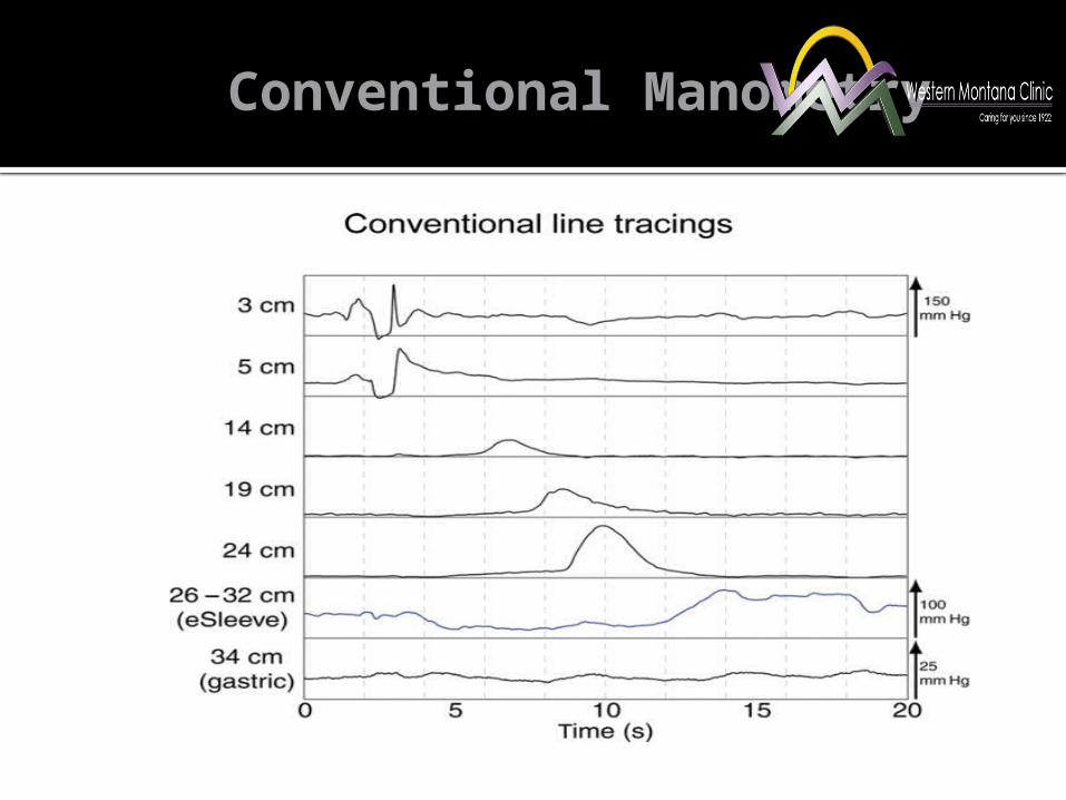
Conventional Manometry

Equipment – High Resolution Manometry
All sensors are truly circumferential .
36 channels spaced 1 cm apart 12 pressure sensing points at each channel (432 data points) .
Small diameter 2.75 mm.
Source: http://www.sierrainst.com/manoscan360.html

High Resolution Manometry
Magenta end of color spectrum (hot colors) = highest pressure.
Blue end of color spectrum (cool colors) = lowest pressure.

High Resolution Manometry

Discuss the advantages high resolution manometry has over conventional
esophageal manometry.
High Resolution Manometryvs. Conventional Manometry
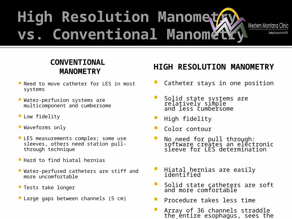
CONVENTIONAL MANOMETRY
Need to move catheter for LES in most systems
Water-perfusion systems are multicomponent and cumbersome
Low fidelity
Waveforms only
LES measurements complex; some use sleeves, others need station pull-through technique
Hard to find hiatal hernias
Water-perfused catheters are stiff and more uncomfortable
Tests take longer
Large gaps between channels (5 cm)
HIGH RESOLUTION MANOMETRY
Catheter stays in one position
Solid state systems are relatively simpleand less cumbersome
High fidelity Color contour No need for pull through: software
creates an electronic sleeve for LES determination
Hiatal hernias are easily identified Solid state catheters are soft and
more comfortable Procedure takes less time Array of 36 channels straddle the
entire esophagus, sees the entire organ

High Resolution Manometryvs. Conventional Manometry

Patient Preparationfor
High Resolution Esophageal Manometry

Pre-Pr0cedure Counseling
How do you describe esophageal manometry to a patient?
During esophageal manometry, a thin, pressure-sensitive, flexible tube is passed through your nose and into your stomach.
When the tube is in your esophagus, you will be asked to swallow. The pressure of the muscle contractions will be measured along the length of your esophagus.
The tube is removed after the test is completed. The test takes about 1 hour.

Pre-Pr0cedure Counseling
How do you tell patients to prepare for a manometry? Patients should not have anything to eat
or drink for 4-6 hours before the test (varies by center).
There is no need for bowel preparation. Take all prescribed medications as usual. ▪ This includes anticoagulants, aspirin, and
NSAIDs, acid suppressive therapy.

Pre-Pr0cedure Counseling
How will the test feel? Typically, the test is not uncomfortable. Some patients may experience a
gagging sensation when the tube is being placed.

How is an Esophageal Manometry Probe
Placed?

Catheter Placement
Before bringing the patient into the room an RN performs a focused H&P and chart review.▪ Indication (dysphagia, chest pain, pre-operative
evaluation, etc.)▪ Allergies (assure the patient isn’t allergic to
lidocaine)▪ If they are use sterile lubricant jelly
▪ Pertinent past surgeries (Nasal, esophageal, bariatric surgery etc.)
Make sure the patient did not eat or drink anything for 4 to 6 hours prior to test (this varies by center).

Catheter Placement
The patient is brought into the procedure room.
A gown is placed over their upper body and they sit on the edge of a gurney.
The patient occludes each nostril and sniffs to determine if their right or left nostril is more patent.
http://classconnection.s3.amazonaws.com/369/flashcards/1414369/png/assess_patency_of_nostrils1333807384492.png

Catheter Placement
The nostril is numbed with 2% lidocaine jelly using a 6-inch cotton tip applicator.
The manometric catheter is lubricated with 2% lidocaine.
http://jan.ucc.nau.edu/daa/woundproducts/curasolgel3.jpg

Catheter Placement
The patient brings their chin down to their chest.
The catheter is advanced through the medicated nostril into the esophagus while the patient swallows.
http://www.bartleby.com/107/Images/large/image855.gif
https://myhealth.alberta.ca/health/_layouts/healthwise/media/medical/hw/h9991890_001.jpg

Catheter Placement
The manometric catheter is advanced until it crosses the lower esophageal sphincter and its distal tip is in the stomach.
The catheter is secured in place with tape.
The patient then lies supine on a gurney.
http://www.upmc.com/patients-visitors/education/PublishingImages/G-L/EsophManometry-1.jpg

Catheter Placement
5 ml of water (or saline) is placed into the patient’s mouth using a syringe.
The patient holds the liquid in their mouth then swallows once.
30 seconds later this is repeated.
10 wet swallows are performed.
The catheter is removed. http://www.robertsewellmd.com/Portals/2/Motility%20HD.jpg
Kevin Kolendich

Describe the manometric findings present during a normal swallow.
1. LES relaxation2. Normal esophageal peristalsis
3. UES relaxation

The Esophagus at Rest

Manometry Tracing
http://www.nature.com/ajg/journal/v105/n5/images/ajg2010165i1.gif

Name the indications for esophageal manometry

Indications
Dysphagia.
Non-cardiac chest pain.
Placement of intraluminal devices (e.g. pH probes).
Preoperative assessment of patients being considered for anti-reflux surgery and bariatric surgery.
Detecting esophageal motor abnormalities associated with systemic diseases (e.g. connective tissue diseases).
American Gastroenterological Association Patient Care Committee on May 15, 1994

Dysphagia
The first step is to distinguish between oropharyngeal dysphagia and esophageal dysphagia.
Oropharyngeal dysphagia: Arises from dysfunction of the pharynx and
upper esophageal sphincter.
Esophageal dysphagia: Arises from disorders of the esophageal body
and lower esophageal sphincter.

Dysphagia
OROPHARYNGEAL
Have difficulty initiating a swallow.
Localize symptoms to the cervical region.
Frequently associated with coughing, choking, nasal regurgitation, and dysphonia.
ESOPHAGEAL
Have difficulty swallowing several seconds after initiating a swallow.
Localize symptoms to the suprasternal notch or behind the sternum.
May be associated with a history of food impaction or food “sticking” in the chest.

What are some the appropriate questions to ask a patient with
dysphagia in the office?

Dysphagia
Do you have problems initiating a swallow or do you feel food getting stuck a few seconds after swallowing?
Do you cough or choke or is food coming back through your nose after swallowing?
Do you have problem swallowing solids, liquids, or both?
How long have you had problems swallowing and have your symptoms progressed, remained stable, or are they intermittent?

Dysphagia
Could you point to where you feel food is getting stuck?
What medications are you using now? potassium chloride alendronate ferrous sulfate quinidine ascorbic acid tetracycline aspirin NSAIDs

Dysphagia
Upper Endoscopy: Patients with suspected
esophageal dysphagia should be referred for an upper endoscopy as the initial test.
Structural assessment.
Has the advantage that biopsies can be obtained and intervention performed.
http://endoscopycenterofdelaware.com/assets/uploaded/images/all/UpperEndoscopy.jpg

Dysphagia
Barium swallow: This is a good second test
following a negative upper endoscopy if a mechanical obstruction is still suspected.▪ External compression.▪ B rings (Schatzki ring) can be
missed. ▪ Zenker’s diverticulum ▪ Cricopharyngeal bar
Structural and functional assessment.
Can assess the UES and pharynx more reliably than upper endoscopy.
http://img.webmd.com/dtmcms/live/webmd/consumer_assets/site_images/media/medical/hw/h9991108.jpg

Dysphagia
Esophageal Manometry: Motility testing should
be performed in patients with dysphagia in whom upper endoscopy is unrevealing and/or an esophageal motility disorder is suspected.
Functional assessment. http://gastrocure.com/images/Esophageal_Manometry_img_6.jpg

Case #1
47 year old woman with 2 years progressive dysphagia to solids and liquids. After meals she has a sensation of fullness in her chest . She often drinks water to make solid food pass.
A recent EGD was normal thus she was referred for esophageal manometry.

Achalasia - Type II
Normal
1. Mean IRP > upper limits of normal (incomplete LES relaxation)
2. Absence of esophageal peristalsis (note: specifics vary for type I, II, and III achalasia)

Case #1
The patient underwent pneumatic dilation of her LES to 30 mm with marked improvement in her symptoms.
Therapeutic options Heller myotomy Pneumatic dilation Botox injection into the LES POEM (Per Oral Endoscopic Myotomy)
Chest Pain
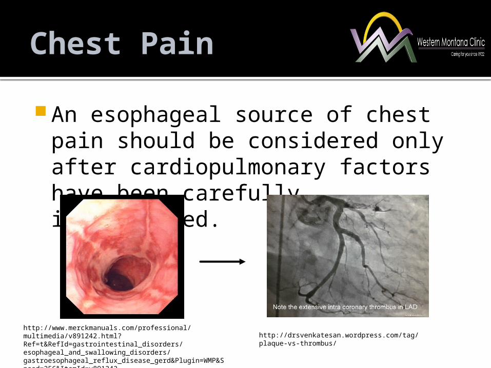
An esophageal source of chest pain should be considered only after cardiopulmonary factors have been carefully investigated.
http://drsvenkatesan.wordpress.com/tag/plaque-vs-thrombus/
http://www.merckmanuals.com/professional/multimedia/v891242.html?Ref=t&RefId=gastrointestinal_disorders/esophageal_and_swallowing_disorders/gastroesophageal_reflux_disease_gerd&Plugin=WMP&Speed=256&ItemId=v891242

Chest Pain
A patient should first undergo an upper endoscopy and exclusion of GERD.
GERD is the most common cause of non-cardiac chest pain.
GERD is much more common than an esophageal motility disorder. http://trustinfood.org/chest-pain/wp-content/
uploads/2012/10/Chest-Pain-After-Eating.jpg

Case #2
A 84 year old male complains of severe chest pain which onsets during meals.
EGD, CT abd/pelvis, barium esophagram, and 24 hour ph study are all normal. A cardiac work-up and which includes a nuclear perfusion test is normal. He is told by an ER physician that his symptoms are stress induced and “in his head”.
The patient is referred by his PCP for esophageal manometry.

Jackhammer Esophagus
1. DCI > 8,000 mmHg-cm-s (hypercontractile esophagus)
2. Normal mean IRP (the EGJ relaxes normally)

Case #2
He initially is treated with diltiazem which causes intolerable hypotension and orthostasis. This medication is stopped and he undergoes EGD with botox injection into the LES with complete resolution of his symptoms.
Therapy Rule out EGJ outflow obstruction causing reactive
hypercontractile peristalsis Treat GERD if present Calcium Channel blockers (Diltiazem) Nitrates (Isosorbide dinitrate) Sildenafil Botox injection into the LES

Prior to Anti-reflux Surgery
The most important role of esophageal manometry in patients with GERD has traditionally been for evaluation prior to antireflux surgery. http://en.wikipedia.org/wiki/Nissen_fundoplication

Prior to Anti-reflux Surgery
Why is esophageal manometry done prior to esophageal or gastric surgery?

Prior to Anti-reflux Surgery
Manometry may lead to a modification of the surgical approach.
www.wehealny.org

Prior to Anti-reflux Surgery
Esopahgeal manometry may lead to an alternative diagnosis such as scleroderma or achalasia.
http://www.nature.com/gimo/contents/pt1/images/gimo20-f4.jpg

Prior to Anti-reflux Surgery
Evaluation of post operative symptoms.
The best way to determine if a surgery is causal of a manometric abnormality is comparison of a patient’s pre-operative manometry study to their post-operative manometry study.

Case #3
42 year old woman with long standing heartburn which has had relatively little improvement despite trials of omeprazole, esomeprazole, and dexlansoprazole.
Upper endoscopy reveals a small sliding hiatal hernia with LA class A erosive esophagitis.
She is referred to a surgeon for consideration of fundoplication. Her surgeon orders pre-operative esophageal manometry and pH testing.

Jackhammer Esophagus
1. DCI > 8,000 mmHg-cm-s (hypercontractile esophagus)
2. Normal mean IRP (the EGJ relaxes normally)

Case #3
The patient was treated for a hypercontractile esophageal disorder with a calcium channel blocker with a significant reduction in symptoms.

Case #4
A 49 year old woman with GERD complains of severe heartburn. This improves with twice daily PPI therapy and lifestyle modification, however severe nocturnal symptoms persist.
EGD reveals a hiatal hernia and LA class C esophagitis despite compliance with BID PPI.
She is referred to a surgeon for consideration of fundoplication. Her surgeon orders pre-operative esophageal manometry and pH testing.

Weak Peristalsis with LargePeristaltic Defects
1. Normal mean IRP (normal EGJ relaxation)
2. > 20% of swallows with large (> 5 cm) breaks in the 20 mmHg isobaric contour.
Normal

Case #4
Ambulatory reflux monitoring revealed an 12% incidence of acid reflux despite compliance with PPI therapy with an elevated DeMeester score.
The patient underwent Toupet fundoplication. Her symptom of heartburn has resolved and she is now off PPI therapy.
Her only concern post-operatively bloating and an inability to belch.

Know where to find resources to further your understanding of
manometry

Further Education
If you are interested in further education consider reading the following text.
American Neurogastroenterology and Motility Society.

References
1. Conklin, J., Pimentel, M., Soffer, E. Color Atlas of High Resolution Manometry. Springer (2009)
2. Kahrilas, Peter J. et al. Esophageal Motility Disorders in Terms of Pressure Topography. J Clin Gastroenterol 2008;42:627-635
3. Lin, Henry C. High Resolution Esophageal Manometry. Core Curriculum Conference, The University of New Mexico. 2008, 2011.

Thank YouContact Information: Kevin Kolendich, MD
Western Montana Clinic(406) 329-7169