NLRP12 promotes host resistance against Pseudomonas ... · verity of P. aeruginosa keratitis,...
13
8063 Abstract. – OBJECTIVE: To investigate the role of NLRP12 in regulating Pseudomonas aeru- ginosa (P. aeruginosa) keratitis. MATERIALS AND METHODS: Real-Time-PCR and Western blot were performed to measure the NLRP12 level in corneas and bone marrow-derived macrophages (BMDMs) of C57BL/6 (B6) mice. B6 mice received a subconjunctival injection of len- tivirus expressing active NLRP12 (NLRP12-lentivi- rus) or Ctl-lentivirus (as control), followed by infec- tion of P. aeruginosa. The clinical score, slit lamp and bacterial plate count of mice were evaluated. In addition, myeloperoxidase (MPO) was detect- ed to assess the infiltration of polymorphonuclear neutrophil (PMN). Cytokine levels were measured by Real Time-PCR and ELISA. Meanwhile, the bac- terial burden was also evaluated. The activation of NF-κB signaling was determined by pIκBα/IκBα levels based on Western blot and NF-κB-depen- dent Luciferase activity on the basis of Luciferase assays using 293T cells. RESULTS: NLRP12 mRNA and protein levels were decreased in B6 corneas and BMDMs af- ter P. aeruginosa infection. The over-expression of NLRP12 in B6 corneas significantly amelio- rated the severity of corneal disease, bacterial burden, PMN infiltration and pro-inflammatory cytokine expression. In vitro analysis demon- strated that the up-regulation of NLRP12 sup- pressed pro-inflammatory cytokine production and enhanced bacterial clearance in RAW264.7 cells. The protein levels of pIκBα and IκBα were significantly decreased after NLRP12-lentivirus treatment compared with that of Ctl-lentivirus. NF-κB-dependent Luciferase activity was po- tently inhibited by NLRP12 infected with P. aeru- ginosa or cotransfected with the downstream signaling molecules including IKKα and IKKβ in 293T cells. CONCLUSIONS: NLRP12 decreases the se- verity of P. aeruginosa keratitis, reduces corneal inflammation and bacterial burden through the down-regulation of the NF-κB signaling pathway. Key Words: NLRP12, Pseudomonas aeruginosa, Keratitis, Regu- latory mechanism. Introduction As a common type of Gram-negative bacte- ria, Pseudomonas aeruginosa (P. aeruginosa) is associated with microbial keratitis, especially in individuals with soft contact lenses 1 . P. aerugi- nosa keratitis represents a progressive corneal disease which might lead to inflammatory epi- thelial edema, stromal infiltration, corneal ulce- ration, tissue destruction, as well as blindness 2 . The pathogenesis of P. aeruginosa keratitis is complicated and involves interactions between host and invading pathogens. Beyond bacterial virulence factors, host inflammation-mediated immunopathologic damage is reported to be another major contributing factor to the patho- genesis of P. aeruginosa keratitis 3 . The host in- flammatory response is a self-protective respon- se to bacterial invasion, and is characterized by an accumulation of inflammatory cells and se- cretion of cytokines. In the process of infection, inflammatory cells such as polymorphonuclear neutrophils (PMNs) and monocytes/macropha- ges are activated and recruited to the infected cornea to eliminate invading pathogens 4 . Once activated, inflammatory cells also secret va- European Review for Medical and Pharmacological Sciences 2018; 22: 8063-8075 Q.-C. DENG 1 , C.-T. DENG 2 , W.-S. LI 3 , S.-W. SHU 1 , M.-R. ZHOU 1 W.-B. KUANG 1 1 Department of Laboratory, Longhua District Central Hospital, Longhua District, Shenzhen, China 2 Department of Infection, He Yuan People’s Hospital, He Yuan, China 3 Department of Neurosurgery, He Yuan People’s Hospital, He Yuan, China Qiuchan Deng and Chuntao Deng contributed equally to this work Corresponding Author: Wenbin Kuang, MD; e-mail: [email protected] NLRP12 promotes host resistance against Pseudomonas aeruginosa keratitis inflammatory responses through the negative regulation of NF- κB signaling
Transcript of NLRP12 promotes host resistance against Pseudomonas ... · verity of P. aeruginosa keratitis,...
8063
Abstract. – OBJECTIVE: To investigate the role of NLRP12 in regulating Pseudomonas aeru-ginosa (P. aeruginosa) keratitis.
MATERIALS AND METHODS: Real-Time-PCR and Western blot were performed to measure the NLRP12 level in corneas and bone marrow-derived macrophages (BMDMs) of C57BL/6 (B6) mice. B6 mice received a subconjunctival injection of len-tivirus expressing active NLRP12 (NLRP12-lentivi-rus) or Ctl-lentivirus (as control), followed by infec-tion of P. aeruginosa. The clinical score, slit lamp and bacterial plate count of mice were evaluated. In addition, myeloperoxidase (MPO) was detect-ed to assess the infiltration of polymorphonuclear neutrophil (PMN). Cytokine levels were measured by Real Time-PCR and ELISA. Meanwhile, the bac-terial burden was also evaluated. The activation of NF-κB signaling was determined by pIκBα/IκBα levels based on Western blot and NF-κB-depen-dent Luciferase activity on the basis of Luciferase assays using 293T cells.
RESULTS: NLRP12 mRNA and protein levels were decreased in B6 corneas and BMDMs af-ter P. aeruginosa infection. The over-expression of NLRP12 in B6 corneas significantly amelio-rated the severity of corneal disease, bacterial burden, PMN infiltration and pro-inflammatory cytokine expression. In vitro analysis demon-strated that the up-regulation of NLRP12 sup-pressed pro-inflammatory cytokine production and enhanced bacterial clearance in RAW264.7 cells. The protein levels of pIκBα and IκBα were significantly decreased after NLRP12-lentivirus treatment compared with that of Ctl-lentivirus. NF-κB-dependent Luciferase activity was po-tently inhibited by NLRP12 infected with P. aeru-ginosa or cotransfected with the downstream signaling molecules including IKKα and IKKβ in 293T cells.
CONCLUSIONS: NLRP12 decreases the se-verity of P. aeruginosa keratitis, reduces corneal inflammation and bacterial burden through the down-regulation of the NF-κB signaling pathway.Key Words:
NLRP12, Pseudomonas aeruginosa, Keratitis, Regu-latory mechanism.
Introduction
As a common type of Gram-negative bacte-ria, Pseudomonas aeruginosa (P. aeruginosa) is associated with microbial keratitis, especially in individuals with soft contact lenses1. P. aerugi-nosa keratitis represents a progressive corneal disease which might lead to inflammatory epi-thelial edema, stromal infiltration, corneal ulce-ration, tissue destruction, as well as blindness2. The pathogenesis of P. aeruginosa keratitis is complicated and involves interactions between host and invading pathogens. Beyond bacterial virulence factors, host inflammation-mediated immunopathologic damage is reported to be another major contributing factor to the patho-genesis of P. aeruginosa keratitis3. The host in-flammatory response is a self-protective respon-se to bacterial invasion, and is characterized by an accumulation of inflammatory cells and se-cretion of cytokines. In the process of infection, inflammatory cells such as polymorphonuclear neutrophils (PMNs) and monocytes/macropha-ges are activated and recruited to the infected cornea to eliminate invading pathogens4. Once activated, inflammatory cells also secret va-
European Review for Medical and Pharmacological Sciences 2018; 22: 8063-8075
Q.-C. DENG1, C.-T. DENG2, W.-S. LI3, S.-W. SHU1, M.-R. ZHOU1 W.-B. KUANG1
1Department of Laboratory, Longhua District Central Hospital, Longhua District, Shenzhen, China2Department of Infection, He Yuan People’s Hospital, He Yuan, China3Department of Neurosurgery, He Yuan People’s Hospital, He Yuan, China
Qiuchan Deng and Chuntao Deng contributed equally to this work
Corresponding Author: Wenbin Kuang, MD; e-mail: [email protected]
NLRP12 promotes host resistance against Pseudomonas aeruginosa keratitis inflammatory responses through the negative regulation of NF-κB signaling

Q.-C. Deng, C.-T. Deng, W.-S. Li, S.-W. Shu, M.-R. Zhou W.-B. Kuang
8064
rious pro-inflammatory cytokines such as in-terleukin 6 (IL-6), interleukin 1 beta (IL-1β), tumor necrosis factor α (TNF-α) or macrophage inflammatory protein 2 (MIP-2)5. These inflam-matory cytokines promote bacteria clearance. Nonetheless, the aberrant expressions lead to tissue damage and corneal perforation2. The-refore, the regulation of both bacterial burden and ocular inflammation is important for the treatment of P. aeruginosa keratitis. Nod-like receptors (NLRs) represent a large family of cytoplasmic PRRs which are characterized by a conserved nucleotide-binding and oligomeri-zation domain (NOD) and a leucine-rich repeat (LRR) region6. The function includes pathogen/damage sensing, modulation of inflammatory signaling transduction7-9. NLR proteins, such as NLRP1, NLRP3, and NLRC4, activate caspa-se-1 through the assembly of a complex which is termed as inflammasome10. These inflammaso-me-forming NLRs mediate the processing and maturation of the proinflammatory cytokines pro-IL-1β and pro-IL-18 into IL-1β and IL-18. On the other hand, non-inflammasome-forming members regulate other key inflammatory pa-thways; for example, NOD1 and NOD2 acti-vate NF-κB and MAPK pathways11-14, NLRP6, NLRC3, NLRC5 and NLRX1 have been re-ported involved in the regulation of inflamma-tion15-18. NLRP12 (known as RNO, PYPAF7, and Monarch-1), a member of the family consisting of an N-terminal PYD, an NBD, and C-terminal LRR regions, is one of the first described NLR proteins and its function remains controversial. Because of the typical tripartite domain structu-re similar to NLRP3, NLRP12 was considered to have functions in the inflammasome assem-bly. Recent findings indicate the involvement of NLRP12 in colon cancer19,20. A previous study showed that NLRP12-deficient mice were more susceptible to Yersinia pestis infection, with re-duction of IL-1β and IL-18 levels in serum and growing bacterial load in spleen21. NLRP12 is also reported to suppress colon inflammation and tumorigenesis through the negative regu-lation of NF-κB signaling19,20,22. However, the potential role of NLRP12 in microbial keratitis remains poorly understood. NF-κB family is involved in the regulation of inflammatory and immune responses. It locates in the cytoplasm as an inactive form by the inhibitor IκB. Prote-asome-mediated degradation of IkB causes its phosphorylation by IκB kinase (IKK) complex, IKKα-IKKβ-IKKγ (NEMO). The subsequent
polyubiquitination and degradation of IκB lead to the release and nuclear translocation of NF-κB, which stimulates the transcription and expression of various inflammatory chemoki-nes, cytokines and cell surface proteins. In vi-tro analysis of NLRP12 in human monocytic cell lines THP-1 suggests silencing of NLRP12 promoted NF-κB activation and the secretion of proinflammatory cytokines in response to TLR agonists, TNF-α and Mycobacterium tuberculo-sis23,24. In the present study, we aimed to detect the level of NLRP12 in mouse corneas after P. aeruginosa infection and determine its effect in diseases caused by P. aeruginosa keratitis.
Materials and Methods
Ocular Infection and Clinical EvaluationFemale C57BL/6 (B6) mice aged 8 weeks were
purchased from the Animal Supply Center of Guan-gdong Medical University. The left cornea of B6 mice was infected by P. aeruginosa stain (ATCC 19660), as described previously25-27. Corneal disease was graded in scale as follows: 0, clear or slight opa-city, partially or fully covering the pupil; +1, slight opacity, partially or fully covering the anterior seg-ment; +2, dense opacity, partially or fully covering the pupil; +3, dense opacity, covering the entire an-terior segment; and +4, corneal perforation or phthi-sis. The clinical score was recorded for each mouse after infection and photography with a slit lamp was used to illustrate the disease response. All procedu-res involving animals were performed in complian-ce with the ARVO Statement for the Use of Animals in Ophthalmic and Vision Research. Animal Ethics Committee approval was granted by our institution.
Isolation of Murine Bone Marrow-Derived Macrophages
Bone marrow-derived macrophages (BMDMs) were isolated as described previously28. Then, cel-ls were cultured in Dulbecco’s Modified Eagle’s Medium (DMEM; Invitrogen, Carlsbad, CA, USA) supplemented with 10% fetal bovine serum (FBS; Invitrogen, Carlsbad, CA, USA) and 30% (vol/vol) L-929 conditioned medium which was used as a source of macrophage colony-stimula-ting factor. BMDMs were collected as a homoge-neous population of adherent cells after culturing for 7 days. Cultured cells were stained with Alexa Fluor 488 conjugated anti-F4/80 (Invitrogen, Car-lsbad, CA, USA) to measure the purity, which was more than 95%.
NLRP12 in corneal inflammation
8065
Cell CultureMurine macrophage-like RAW264.7 cells
(ATCC; Manassas, VA, USA) were cultured in DMEM media supplemented with 10% (v/v) FBS, 1% penicillin-streptomycin (Invitrogen, Carlsbad, CA, USA), and 1% L-glutamine (Invitrogen, Car-lsbad, CA, USA) at the permissive temperature of 37°C. Human embryonic kidney (HEK) 293T cel-ls (ATCC, Manassas, VA, USA) were cultured in DMEM media supplemented with 10% (v/v) FBS, 100 units/ml penicillin, 100 µg/ml streptomycin (Invitrogen, Carlsbad, CA, USA) and maintained in 5% CO2 at 37°C.
Recombinant Lentivirus Packaged and Titered
Lentivirus vector encoding NLRP12 was bought from Addgene (Seattle, WA, USA) and prepared as described previously29. In brief, cells were seeded into 10 cm dishes (4×10⁶ cel-ls per dish) 1 day before transfection and cul-tured overnight in DMEM containing 10% (v/v) FBS, followed by fresh media containing 25 mM chloroquine (Sigma-Aldrich, St. Louis, MO, USA) during transfection. For each dish, 10 mg lentivirus vector with 3 mg VSV-G envelo-pe plasmid and 10 mg pCMV∆R8.2 packaging plasmid were adjusted to 450 ml in water fol-lowed by mixing with 50 ml 2.5M CaCl2 (Sig-ma-Aldrich, St Louis, MO, USA). Then, 500 ml of 2×HBS buffered-saline (280 mM NaCl, 10 mM KCl, 1.5 mM Na2HPO4, 12 mM dextrose, 50 mM 4-(2-hydroxyethyl)-1-piperazineethane-sulfonic acid (HEPES), pH 7.05) was added for precipitation. At 12 h after transfection, cells were cultured with complete fresh media. After another 36 h, the conditioned medium was added and subsequent centrifugation was performed at 4000 g for 5 min. Then, lentivirus particles were concentrated through centrifugation at 50000 g for 140 min and the pellet was resuspended in 400 ml phosphate-buffered solution (PBS) with 0.1% bovine serum albumin (BSA). The viral titer was measured using the HIV p24 Antigen enzyme-linked immunosorbent assay (ELISA) kit (ZeptoMetrix, Buffalo, NY, USA).
Subconjunctival Injection of LentivirusLentivirus expressing NLRP12 (referred as
NLRP12-lentivirus below) or control (referred as Ctl-lentivirus below) was administrated sub-conjunctivally into the left eye of mice (5 ml/mouse at a viral titer of 108, n=5/group/time) once a week three times before ocular infection.
Lentivirus TransductionRAW264.7 cells (2×10⁵/well) and 293 T cells
(8×10⁴/well) were seeded into a 12-well plate. To over-express NLRLP12, cells were infected with NLRP12-lentivirus or Ctl-lentivirus at MOI 10 for 6 h in the presence of 8 mg/ml polybrene (Sig-ma-Aldrich, St. Louis, MO, USA). After that, the infected cells were cultured in complete culture media for another 24 h for detections of PCR, ELISA or killing assays.
Real Time-PCRTotal RNA was isolated using TRIzol reagent
(Invitrogen, Carlsbad, CA, USA) according to the manufacturer’s instructions, and quantitated by a NanoDrop 2000C Spectrophotometers (Thermo Scientific, Waltham, MA, USA). Total RNA (1 mg) was reversely transcribed into cDNA, which was used for amplification using SYBR Green Master Mix (TaKaRa Bio, Otsu, Shiga, Japan) in accor-dance with manufacturer’s instructions. Quanti-tative Real Time-PCR reactions were performed using the CFX96 Real Time-PCR System (Bio-Rad, Hercules, CA, USA). The relative mRNA levels were quantified via normalization with β-actin. Primer sequences were listed in Table I.
Western BlotWhole corneas (n=5/group/time) were collected
and pooled from normal uninfected and infected mice eyes at 1, 3, 5 days post-infection followed by being lysed and homogenized in a 1 ml glass tissue homogenizer with lysis buffer containing 1mM phenylmethylsulfonyl fluoride, 1% (v/v) protease inhibitor cocktail, and 1 mM DTT (Sig-ma-Aldrich, St. Louis, MO, USA). Cultured cells were washed three times using ice-cold PBS fol-lowed by treatment with lysis buffer. The protein concentration was determined by Quick Start Bradford protein assay (Bio-Rad, Hercules, CA, USA). 30 mg of each sample was loaded, sepa-rated on 10% sodium dodecyl sulfate-polyacryla-mide gel electrophoresis (SDS-PAGE), and then transferred to a nitrocellulose membrane (Pall Life Sciences, Ann Arbor, MI, USA) followed by blockage with Tris-Buffered Saline and Tween-20 (TBST) containing 5% non-fat dry milk. Then, NLRP12 polyclonal Ab (1:100, Abcam, Cambrid-ge, MA, USA), κBα (44D4)/Phospho-IκBα (Ser32) (14D4) Rabbit mAb (1:1000, Cell Signaling Tech-nology, Danvers, MA, USA) was added and incu-bated overnight at 4°C, respectively, followed by incubation with secondary IRDye 800CW Don-key anti-rabbit IgG (H+L) Ab (1:5000, LI-COR

Q.-C. Deng, C.-T. Deng, W.-S. Li, S.-W. Shu, M.-R. Zhou W.-B. Kuang
8066
Biosciences, Lincoln, NE, USA) for 1 h. After that, blots were detected using Odyssey Infrared Imaging System (LI-COR Biosciences, Lincoln, NE, USA) according to the manufacturer’s proto-col. Semi-quantitative analysis of WB bands was performed using Image J software.
Enzyme-Linked Immunosorbent Assay (ELISA)
Corneas and cell supernatants were collected and analyzed for secreted IL-1β, MIP-2, IL-6 and TNF-α using specific ELISA kits (R&D Systems, Minneapolis, MN, USA) according to manufactu-rer’s instructions. Each sample was assayed in du-plicate of three separate experiments. The repor-ted sensitivity of these assays is <3.0 pg/mL for IL1β, <1.5 pg/mL for MIP-2, 1.3 to 1.8 pg/mL for IL-6, and <5.1 pg/mL for TNF-α.
Myeloperoxidase (MPO) AssayCorneas were collected at 1, 3, 5 days post-in-
fection (n=5/group/time) and homogenized in PBS followed by measuring the MPO concentra-tion using Zen Myeloperoxidase ELISA Kit (In-vitrogen, Carlsbad, CA, USA) according to the manufacturer’s recommendations.
Bacterial Plate CountsCollected individual corneas were homogeni-
zed in sterilized water containing 0.85% (wt/vol) NaCl and 0.25% bovine serum albumin (BSA). Serial 10-fold dilutions of the samples were plated
on Pseudomonas isolation agar (BD Biosciences, Franklin Lakes, NJ, USA) in triplicate followed by incubation at 37°C overnight. The results are reported as 106 colony-forming unite (CFU) per cornea ± SEM.
Bacterial Killing AssayThe intracellular bacterial killing was eva-
luated by plate count as described previou-sly30. Briefly, RAW264.7 cells were treated with NLRP12- or Ctl-lentivirus for 24 h, and challenged with P. aeruginosa at an MOI of 25. After 1 hour, cells were treated with gen-tamicin (Sigma-Aldrich, St. Louis, MO, USA) at 300 g/mL for 30 minutes to eliminate the ex-tracellular bacteria, followed by washing with PBS three times. Afterward, one of the dupli-cate wells was lysed in 0.1% Triton-X, and the other duplicate well was incubated at 37°C for 1 hour followed by lysis in 0.1% Triton-X. Se-rial 10-fold dilutions of each sample were pla-ted on Pseudomonas isolation agar in triplicate and incubated at 37°C overnight. For intracel-lular bacterial killing, the efficiency was calcu-lated as follows: intracellular bacterial killin-g=(CFU[1h]–CFU[2h]) /CFU (1h)×100%.
Luciferase AssaysHEK293 cells were transfected with NF-κB
Luciferase plasmids and exogenous IKKα, IKKβ plasmids used as stimulators. Dual-Luciferase kits (Promega, Madison, WI, USA) were used for
Table I. Nucleotide sequence of the specific primers used in PCR amplification.
Gene Primer Sequence (5’-3’)
b-actin GAT TAC TGC TCT GGC TCC TAG C F GAC TCA TCG TAC TCC TGC TTG C R
NLRP12 AAG ACC GCA ATG CAC GAT TAG F TGG AGC GTT CCC ACT CTA CA R
IL-1b CGC AGC AGC ACA TCA ACA AGA GC F TGT CCT CAT CCT GGA AGG TCC ACG R
MIP-2 TGT CAA TGC CTG AAG ACC CTG CC F AAC TTT TTG ACC GCC CTT GAG AGT GG R
IL-6 CAC AAG TCC GGA GAG GAG AC F CAG AAT TGC CAT TGC ACA AC R
TNF-a CAC AGA AAG CAT GAT CCG CGA C F TGC CAC AAG CAG GAA TGA GAA GAG R
NLRP12 in corneal inflammation
8067
subsequent analysis according to manufacturer’s instructions.
Statistical AnalysisThe difference in clinical score between
NLRP12- or Ctl-lentivirus-treated mice corneas at 1, 3, and 5 days after P. aeruginosa infection was assessed by the Mann-Whitney U test. The significance of other assays were evaluated by unpaired, two-tailed Student’s t-test. Continuous data from multiple groups were analyzed by using one-way ANOVA, with the Tukey’s post-hoc test. p<0.05 was considered statistically significant.
Results
NLRP12 Expression Was Decreased in Mouse Corneas and BMDMs Challenged With P. Aeruginosa
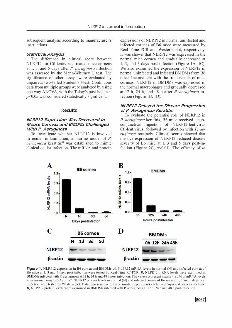
To investigate whether NLRP12 is involved in ocular inflammation, a murine model of P. aeruginosa keratitis31 was established to mimic clinical ocular infection. The mRNA and protein
expressions of NLRP12 in normal uninfected and infected corneas of B6 mice were measured by Real Time-PCR and Western blot, respectively. It was shown that NLRP12 was expressed in the normal mice cornea and gradually decreased at 1, 3, and 5 days post-infection (Figure 1A, 1C). We also examined the expression of NLRP12 in normal uninfected and infected BMDMs from B6 mice. Inconsistent with the front results of mice corneas, NLRP12 in BMDMs was expressed in the normal macrophages and gradually decreased at 12 h, 24 h, and 48 h after P. aeruginosa in-fection (Figure 1B, 1D).
NLRP12 Delayed the Disease Progression of P. Aeruginosa Keratitis
To evaluate the potential role of NLRP12 in P. aeruginosa keratitis, B6 mice received a sub-conjunctival injection of NLRP12-lentivirus Ctl-lentivirus, followed by infection with P. ae-ruginosa routinely. Clinical scores showed that the overexpression of NLRP12 reduced disease severity of B6 mice at 1, 3 and 5 days post-in-fection (Figure 2C, p<0.01). The efficacy of in
Figure 1. NLRP12 expression in B6 cornea and BMDMs. A, NLPR12 mRNA levels in normal (N) and infected cornea of B6 mice at 1, 3 and 5 days post-infection were tested by Real-Time RT-PCR. B, NLPR12 mRNA levels were examined in BMDMs infected with P. aeruginosa at 12 h, 24 h and 48 h post infection. The values represent means ± SEM of mRNA levels after normalizing to β-Actin. C, NLPR12 protein levels in normal (N) and infected cornea of B6 mice at 1, 3 and 5 days post infection were tested by Western blot. Data represent one of three similar experiments each using 5-pooled corneas per time. D, NLPR12 protein levels were examined in BMDMs infected with P. aeruginosa at 12 h, 24 h and 48 h post-infection.

Q.-C. Deng, C.-T. Deng, W.-S. Li, S.-W. Shu, M.-R. Zhou W.-B. Kuang
8068
of NLRP12-lentivirus significantly enhanced NLRP12 expression (Figure 2B). Treatment with NLRP12-lentivirus resulted in a decrease of cor-neal opacity in P. aeruginosa infected B6 cornea
vivo use of NLRP12-lentivirus was confirmed by Western blot, and showed that P. aeruginosa in-fection reduced NLRP12 expression in B6 corne-as (Figure 2A), but the subconjunctival injection
Figure 2. NLRP12 promoted host resistance against P. aeruginosa keratitis. NLRP12 protein levels were tested by Western blot A-B. Clinical scores C, and slit lamp at 3 days post infection reduced disease severity in control D, or NLRP12-lentivirus treated E, B6 mice. NLRP12-lentivirus led to reduced recruitment of PMNs as detected by MPO activity G, and decreased bacterial counts H, at 3 and 5 days post infection (p<0.01). Data are the means ± SEM and represent two individual experi-ments each with five animals per group/time/assay. PA stands for P. aeruginosa; **p<0.01.

NLRP12 in corneal inflammation
8069
at 3 days post-infection (Figure 2D, grade = +3), while in the control group, the grades of the in-fected corneas at 3 days post infection were +2 (Figure 2E). Since both host inflammation and bacterial virulence contribute to the pathogenesis of P. aeruginosa keratitis, we further investigated the effect of NLRP12-lentivirus on PMN infil-tration and bacterial load. The PMN infiltration was significantly reduced in mice infected by NL-RP12-lentivirus compared with that by Ctl-lenti-virus (p<0.01) (Figure 2G). Of note, NLRP12-len-tivirus infection reduced the bacterial load after 3 and 5 days (p<0.01).
NLRP12 Inhibited Pro-Inflammatory Cytokine Expression in P. Aeruginosa Keratitis
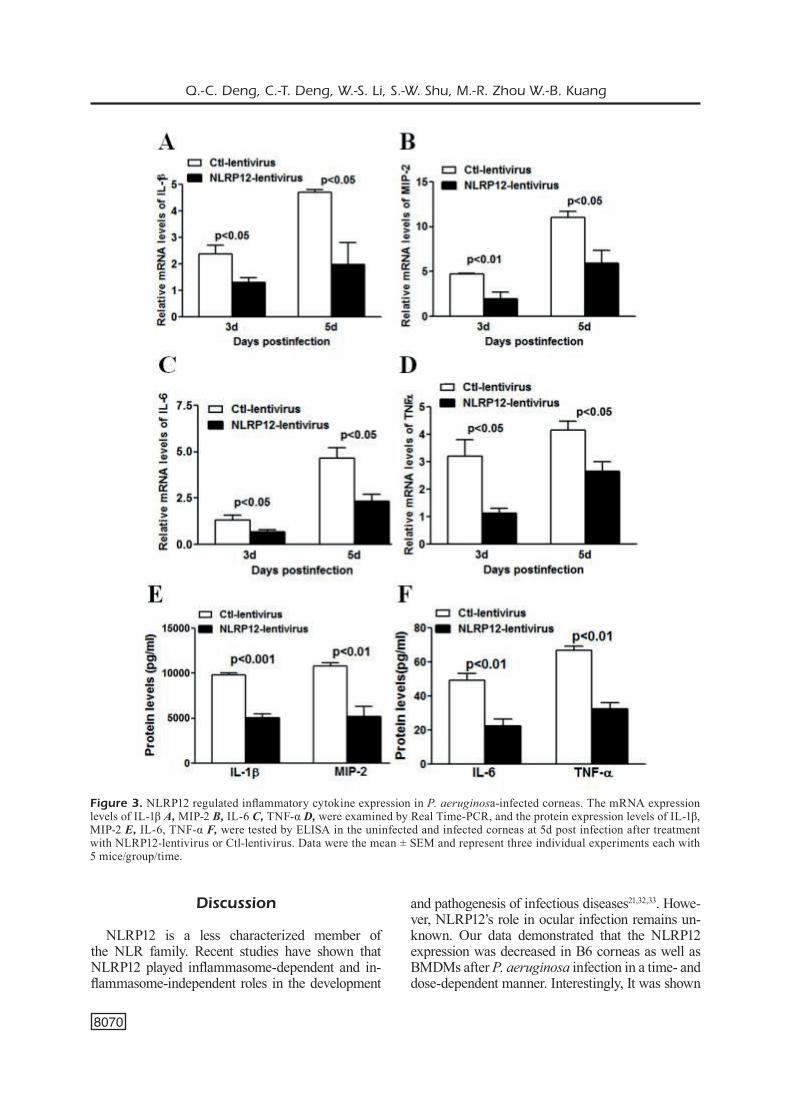
To uncover the mechanism by which NLRP12 delayed the disease progression of P. aerugino-sa keratitis, we first examined the expression of pro-inflammatory cytokines by Real Time-PCR and ELISA in groups of NLRP12-lentivirus and Ctl-lentivirus before and after P. aerugi-nosa infection. The PCR analysis showed that the over-expression of NLRP12 suppressed the mRNA expression of IL-1β (Figure 3A), MIP-2 (Figure 3B), IL-6 (Figure 3C), and TNF-α (Fi-gure 3D) in infected corneas. Consistently, the protein expression levels of IL-1β, MIP-2 (Figure 3E), and IL-6, TNF-α (Figure 3F) measured by ELISA were significantly downregulated due to over-expression of NLRP12. These data sugge-sted that NLRP12 decreased secretion of pro-in-flammatory cytokines induced by P. aeruginosa infection.
NLRP12 Promoted Intracellular Bacterial Killing in P. Aeruginosa-Challenged Macrophages
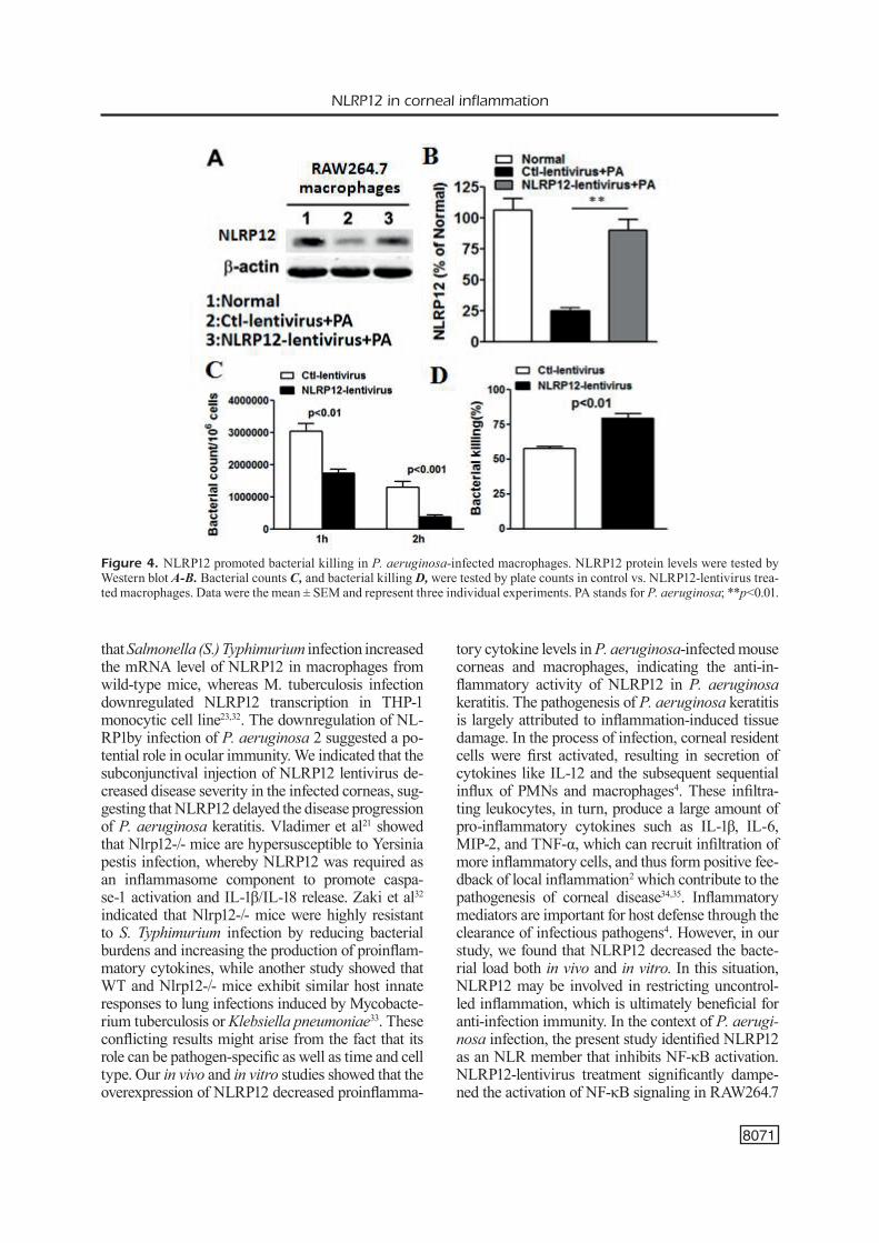
In addition to in vivo murine model, we also evaluated the role of NLRP12 on the inflamma-tory response in vitro. RAW 264.7 cells were in-fected with NLRP12-lentivirus or Ctl-lentivirus, followed by P. aeruginosa challenge at MOI 25. Our result showed that P. aeruginosa infection reduced NLRP12 expression in RAW 264.7 cells, but the infection of NLRP12-lentivirus apparent-ly enhanced NLRP12 expression (Figure 4A, 4B). Plate count analysis showed that the over-expres-sion of NLRP12 significantly reduced the intracel-lular bacterial load in RAW264.7 cells at 1 and 2 h post-infection (Figure 4C, p<0.01 and p<0.001, respectively), compared with that of control. The result of bacterial clearance indicated that the
over-expression of NLRP12 significantly enhan-ced intracellular bacterial killing in RAW264.7 cells (Figure 4D, p<0.01).
NLRP12 Inhibited Inflammatory Cytokine Expression in P. Aeruginosa-Challenged Macrophages
Consistent with in vivo studies, we also found downregulation of inflammatory cytokines in vitro. Macrophages were infected with NL-RP12-lentivirus or Ctl-lentivirus, followed by PA challenge. The levels of inflammatory cytokines IL-1β (Figure 5A), MIP-2 (Figure 5B), IL-6 (Fi-gure 5C) and TNF-α (Figure 5D) were signifi-cantly downregulated due to the overexpression of NLRP12. Meanwhile, ELISA data indicated that the protein levels of IL-1β, MIP-2 (Figure 5E), IL-6 and TNF-α (Figure 5F) were significantly downregulated after overexpression of NLRP12. Both in vivo and in vitro studies indicated that NLRP12 inhibited inflammatory response by suppressing inflammatory cytokines production during P. aeruginosa infection.
NLRP12 Negatively Regulates NF-κB Signaling
Since stringent control of the NF-κB signaling pathways is critical to effective host immune re-sponses, we next assessed the potential role of NLRP12 in regulating the activation of NF-κB signaling in macrophages infected with P. aeru-ginosa. Murine macrophage-like RAW264.7 cells were infected with NLRP12-lentivirus or Ctl-len-tivirus, followed by P. aeruginosa challenge. The level of phospho-IκB was gradually upregulated at different time points after P. aeruginosa in-fection (Figure 6A). However, phospho-IκB levels were significantly decreased after treatment with NLRP12-lentivirus compared with Ctl-lentivirus after 1 and 2 h (Figure 6B, p<0.05 and p<0.01, respectively). To determine whether NLRP12 was involved in P. aeruginosa-mediated NF-kB acti-vation, we transfected human HEK293T (293T) cells with NF-κB-Luciferase reporter DNA, with or without the NLRP12 plasmid, followed by tre-atment with P. aeruginosa. The result of Figure 6C showed that NLRP12 potently inhibited P. ae-ruginosa-induced NF-κB activation. The expres-sion of IKKα (Figure 6D) or IKKβ (Figure 6E) significantly induced NF-κB-Luciferase activity, but this activity was inhibited when NLRP12 was cotransfected. These data suggested that NLRP12 might be a negative regulator of NF-κB activation induced by P. aeruginosa.
Q.-C. Deng, C.-T. Deng, W.-S. Li, S.-W. Shu, M.-R. Zhou W.-B. Kuang
8070
Discussion
NLRP12 is a less characterized member of the NLR family. Recent studies have shown that NLRP12 played inflammasome-dependent and in-flammasome-independent roles in the development
and pathogenesis of infectious diseases21,32,33. Howe-ver, NLRP12’s role in ocular infection remains un-known. Our data demonstrated that the NLRP12 expression was decreased in B6 corneas as well as BMDMs after P. aeruginosa infection in a time- and dose-dependent manner. Interestingly, It was shown
Figure 3. NLRP12 regulated inflammatory cytokine expression in P. aeruginosa-infected corneas. The mRNA expression levels of IL-1β A, MIP-2 B, IL-6 C, TNF-α D, were examined by Real Time-PCR, and the protein expression levels of IL-1β, MIP-2 E, IL-6, TNF-α F, were tested by ELISA in the uninfected and infected corneas at 5d post infection after treatment with NLRP12-lentivirus or Ctl-lentivirus. Data were the mean ± SEM and represent three individual experiments each with 5 mice/group/time.
NLRP12 in corneal inflammation
8071
that Salmonella (S.) Typhimurium infection increased the mRNA level of NLRP12 in macrophages from wild-type mice, whereas M. tuberculosis infection downregulated NLRP12 transcription in THP-1 monocytic cell line23,32. The downregulation of NL-RP1by infection of P. aeruginosa 2 suggested a po-tential role in ocular immunity. We indicated that the subconjunctival injection of NLRP12 lentivirus de-creased disease severity in the infected corneas, sug-gesting that NLRP12 delayed the disease progression of P. aeruginosa keratitis. Vladimer et al21 showed that Nlrp12-/- mice are hypersusceptible to Yersinia pestis infection, whereby NLRP12 was required as an inflammasome component to promote caspa-se-1 activation and IL-1β/IL-18 release. Zaki et al32 indicated that Nlrp12-/- mice were highly resistant to S. Typhimurium infection by reducing bacterial burdens and increasing the production of proinflam-matory cytokines, while another study showed that WT and Nlrp12-/- mice exhibit similar host innate responses to lung infections induced by Mycobacte-rium tuberculosis or Klebsiella pneumoniae33. These conflicting results might arise from the fact that its role can be pathogen-specific as well as time and cell type. Our in vivo and in vitro studies showed that the overexpression of NLRP12 decreased proinflamma-
tory cytokine levels in P. aeruginosa-infected mouse corneas and macrophages, indicating the anti-in-flammatory activity of NLRP12 in P. aeruginosa keratitis. The pathogenesis of P. aeruginosa keratitis is largely attributed to inflammation-induced tissue damage. In the process of infection, corneal resident cells were first activated, resulting in secretion of cytokines like IL-12 and the subsequent sequential influx of PMNs and macrophages4. These infiltra-ting leukocytes, in turn, produce a large amount of pro-inflammatory cytokines such as IL-1β, IL-6, MIP-2, and TNF-α, which can recruit infiltration of more inflammatory cells, and thus form positive fee-dback of local inflammation2 which contribute to the pathogenesis of corneal disease34,35. Inflammatory mediators are important for host defense through the clearance of infectious pathogens4. However, in our study, we found that NLRP12 decreased the bacte-rial load both in vivo and in vitro. In this situation, NLRP12 may be involved in restricting uncontrol-led inflammation, which is ultimately beneficial for anti-infection immunity. In the context of P. aerugi-nosa infection, the present study identified NLRP12 as an NLR member that inhibits NF-κB activation. NLRP12-lentivirus treatment significantly dampe-ned the activation of NF-κB signaling in RAW264.7
Figure 4. NLRP12 promoted bacterial killing in P. aeruginosa-infected macrophages. NLRP12 protein levels were tested by Western blot A-B. Bacterial counts C, and bacterial killing D, were tested by plate counts in control vs. NLRP12-lentivirus trea-ted macrophages. Data were the mean ± SEM and represent three individual experiments. PA stands for P. aeruginosa; **p<0.01.

Q.-C. Deng, C.-T. Deng, W.-S. Li, S.-W. Shu, M.-R. Zhou W.-B. Kuang
8072
cells and Luciferase assays confirmed the inte-raction between NF-κB IKKα/IKKβ and NLRP12. Previous evidence demonstrated that a synthetic analog cord factor, trehalose-6,6-dimycolate (TDP) from Mycobacterium tuberculosis and LPS from
K. pneumoniae significantly increased the levels of TNF-α and IL-6 in Nlrp12−/− bone marrow-derived DCs compared with those in WT cells, indicating that NLRP12 plays a role in suppressing NF-κB si-gnaling in response to these bacterial components33.
Figure 5. NLRP12 regulated inflammatory cytokine expression in P. aeruginosa-infected macrophages. The mRNA expres-sion levels of IL-1β A, MIP-2 B, IL-6 C, TNF-α D, were examined by Real Time-PCR, and the protein expression levels of IL-1β, MIP-2 E, IL-6, TNF-α F, were tested by ELISA in the uninfected and infected macrophages at 24 h post infection after tre-atment with NLRP12-lentivirus versus Ctl-lentivirus. Data were the mean ± SEM and represent three individual experiments.

NLRP12 in corneal inflammation
8073
Similarly, during the efficient control of S. Typhimu-rium infection, NLRP12 was shown to suppress the activation of NF-κB and ERK signaling and to inhi-bit the production of inflammatory cytokines and NO32. However, whether NLRP12 suppresses the NF-κB activation (e.g., Salmonella, Mycobacterium, and Klebsiella infections) or activates caspase-1 (e.g., Yersinia infection) remains unclear and requires fur-ther investigations.
Conclusions
We demonstrated that NRP12 reduced the se-verity of P. aeruginosa keratitis via inhibiting corneal inflammation and enhancing bacterial killing, which provides a potential therapeutic strategy for P. aeruginosa keratitis.
AcknowledgmentsThis study was supported by National Natural Sci-ence Foundation of China (81601382), Guangdong Med-ical Science Foundation (A2016543), Shenzhen Science and Technology Plan Project (JCYJ20160428141830465), (JCYJ20160428135756806) Shenzhen Longhua Dis-trict Science and Technology Innovation Fund Project (20160523A1030137).
Conflict of InterestThe Authors declare that they have no conflict of interest.
References
1) Rattanatam t, Heng WJ, Rapuano CJ, Laibson pR, CoHen eJ. Trends in contact lens-related corneal ulcers. Cornea 2001; 20: 290-294.
Figure 6. NLRP12 negatively regulates NF-κB signaling. A, pIκBα levels was evaluated in macrophages from different groups. B, Bar graphs showed pIκBα and IκBα levels. C, 293T cells were transfected with an NF-κB-Luciferase reporter pla-smid, together with an empty vector or NLRP12 construct. NF-κB-dependent Luciferase activity (fold induction) was detected after treatment with P. aeruginosa. C-D, 293T cells were transfected with IKKα and IKKβ, along with NF-κB-Luciferase. PA stands for P. aeruginosa; *p<0.05; **p<0.01.
Q.-C. Deng, C.-T. Deng, W.-S. Li, S.-W. Shu, M.-R. Zhou W.-B. Kuang
8074
2) HazLett LD. Corneal response to Pseudomonas aeruginosa infection. Prog Retin Eye Res 2004; 23: 1-30.
3) FLeiszig sm, evans DJ. The pathogenesis of bacte-rial keratitis: studies with Pseudomonas aerugino-sa. Clin Exp Optom 2002; 85: 271-278.
4) HazLett LD. Pathogenic mechanisms of P. aerugi-nosa keratitis: a review of the role of T cells, Lan-gerhans cells, PMN, and cytokines. DNA Cell Biol 2002; 21: 383-390.
5) KeRnaCKi Ka, goebeL DJ, poosCH ms, HazLett LD. Early cytokine and chemokine gene expression during Pseudomonas aeruginosa corneal in-fection in mice. Infect Immun 1998; 66: 376-379.
6) RaJWa p, zyCzKoWsKi m, paRaDysz a, buJaK K, bRyniaR-sKi p. Evaluation of the prognostic value of LMR, PLR, NLR, and dNLR in urothelial bladder cancer patients treated with radical cystectomy. Eur Rev Med Pharmacol Sci 2018; 22: 3027-3037.
7) aKiRa s, uematsu s, taKeuCHi o. Pathogen recogni-tion and innate immunity. Cell 2006; 124: 783-801.
8) inoHaRa, CHamaiLLaRD, mCDonaLD C, nunez g. NOD-LRR proteins: role in host-microbial inte-ractions and inflammatory disease. Annu Rev Biochem 2005; 74: 355-383.
9) meyLan e, tsCHopp J, KaRin m. Intracellular pattern recognition receptors in the host response. Natu-re 2006; 442: 39-44.
10) Kanneganti tD, LamKanFi m, nunez g. Intracellular NOD-like receptors in host defense and disease. Immunity 2007; 27: 549-559.
11) giRaRDin se, boneCa ig, CaRneiRo La, antignaC a, JeHanno m, viaLa J, teDin K, taHa mK, Labigne a, zaHRingeR u, CoyLe aJ, DisteFano ps, beRtin J, san-sonetti pJ, pHiLpott DJ. Nod1 detects a unique mu-ropeptide from Gram-negative bacterial peptido-glycan. Science 2003; 300: 1584-1587.
12) CHamaiLLaRD m, HasHimoto m, HoRie y, masumoto J, Qiu s, saab L, oguRa y, KaWasaKi a, FuKase K, Ku-sumoto s, vaLvano ma, FosteR sJ, maK tW, nunez g, inoHaRa n. An essential role for NOD1 in host recognition of bacterial peptidoglycan containing diaminopimelic acid. Nat Immunol 2003; 4: 702-707.
13) inoHaRa n, oguRa y, FontaLba a, gutieRRez o, pons F, CRespo J, FuKase K, inamuRa s, Kusumoto s, HasHi-moto m, FosteR sJ, moRan ap, FeRnanDez-Luna JL, nunez g. Host recognition of bacterial muramyl di-peptide mediated through NOD2. Implications for Crohn’s disease. J Biol Chem 2003; 278: 5509-5512.
14) giRaRDin se, boneCa ig, viaLa J, CHamaiLLaRD m, La-bigne a, tHomas g, pHiLpott DJ, sansonetti pJ. Nod2 is a general sensor of peptidoglycan through mu-ramyl dipeptide (MDP) detection. J Biol Chem 2003; 278: 8869-8872.
15) sCHneiDeR m, zimmeRmann ag, RobeRts Ra, zHang L, sWanson Kv, Wen H, Davis bK, aLLen iC, HoLL eK, ye z, RaHman aH, Conti bJ, eitas tK, KoLLeR bH, ting Jp. The innate immune sensor NLRC3 attenuates toll-like receptor signaling via modification of the
signaling adaptor TRAF6 and transcription factor NF-kappaB. Nat Immunol 2012; 13: 823-831.
16) aLLen iC, mooRe Cb, sCHneiDeR m, Lei y, Davis bK, sCuLL ma, gRis D, Roney Ke, zimmeRmann ag, boW-zaRD Jb, RanJan p, monRoe Km, piCKLes RJ, sambHaRa s, ting Jp. NLRX1 protein attenuates inflammatory responses to infection by interfering with the RIG-I-MAVS and TRAF6-NF-kappaB signaling pa-thways. Immunity 2011; 34: 854-865.
17) ananD pK, maLiReDDi RK, LuKens JR, vogeL p, beRtin J, LamKanFi m, Kanneganti tD. NLRP6 negatively re-gulates innate immunity and host defence against bacterial pathogens. Nature 2012; 488: 389-393.
18) Cui J, zHu L, Xia X, Wang Hy, LegRas X, Hong J, Ji J, sHen p, zHeng s, CHen zJ, Wang RF. NLRC5 nega-tively regulates the NF-kappaB and type I interfe-ron signaling pathways. Cell 2010; 141: 483-496.
19) zaKi mH, vogeL p, maLiReDDi RK, boDy-maLapeL m, ananD pK, beRtin J, gReen DR, LamKanFi m, Kanne-ganti tD. The NOD-like receptor NLRP12 attenua-tes colon inflammation and tumorigenesis. Can-cer Cell 2011; 20: 649-660.
20) aLLen iC, WiLson Je, sCHneiDeR m, LiCH JD, RobeRts Ra, aRtHuR JC, WooDFoRD Rm, Davis bK, uRonis Jm, HeRFaRtH HH, Jobin C, RogeRs ab, ting Jp. NLRP12 suppresses colon inflammation and tumorigenesis through the negative regulation of noncanonical NF-kappaB signaling. Immunity 2012; 36: 742-754.
21) vLaDimeR gi, Weng D, paQuette sW, vanaJa sK, Ra-tHinam va, aune mH, ConLon Je, buRbage JJ, pRouLX mK, Liu Q, ReeD g, meCsas JC, iWaKuRa y, beRtin J, goguen JD, FitzgeRaLD Ka, Lien e. The NLRP12 in-flammasome recognizes Yersinia pestis. Immuni-ty 2012; 37: 96-107.
22) aRtHuR JC, LiCH JD, ye z, aLLen iC, gRis D, WiLson Je, sCHneiDeR m, Roney Ke, o’ConnoR bp, mooRe Cb, moRRison a, sutteRWaLa Fs, beRtin J, KoLLeR bH, Liu z, ting Jp. Cutting edge: NLRP12 controls den-dritic and myeloid cell migration to affect contact hypersensitivity. J Immunol 2010; 185: 4515-4519.
23) WiLLiams KL, LiCH JD, DunCan Ja, ReeD W, RaLLabHan-Di p, mooRe C, KuRtz s, CoFFieLD vm, aCCavitti-Lo-peR ma, su L, vogeL sn, bRaunstein m, ting Jp. The CATERPILLER protein monarch-1 is an antago-nist of toll-like receptor-, tumor necrosis factor alpha-, and Mycobacterium tuberculosis-induced pro-inflammatory signals. J Biol Chem 2005; 280: 39914-39924.
24) LiCH JD, WiLLiams KL, mooRe Cb, aRtHuR JC, Davis bK, taXman DJ, ting Jp. Monarch-1 suppresses non-canonical NF-kappaB activation and p52-de-pendent chemokine expression in monocytes. J Immunol 2007; 178: 1256-1260.
25) Wu m, mCCLeLLan sa, baRRett Rp, HazLett LD. Be-ta-defensin-2 promotes resistance against in-fection with P. aeruginosa. J Immunol 2009; 182: 1609-1616.
26) naKamuRa H, siDDiQui ss, sHen X, maLiK ab, puLiDo Js, KumaR nm, yue by. RNA interference targeting transforming growth factor-beta type II receptor suppresses ocular inflammation and fibrosis. Mol Vis 2004; 10: 703-711.

NLRP12 in corneal inflammation
8075
27) KLeinman me, yamaDa K, taKeDa a, CHanDRaseKaRan v, nozaKi m, baFFi Jz, aLbuQueRQue RJ, yamasaKi s, itaya m, pan y, appuKuttan b, gibbs D, yang z, KaRiKo K, ambati bK, WiLgus ta, DipietRo La, saKu-Rai e, zHang K, smitH JR, tayLoR eW, ambati J. Se-quence- and target-independent angiogenesis suppression by siRNA via TLR3. Nature 2008; 452: 591-597.
28) WeisCHenFeLDt J, poRse b. Bone marrow-derived macrophages (BMM): isolation and applications. CSH Protoc 2008; 2008: pdb prot5080.
29) FueReR C, nusse R. Lentiviral vectors to probe and manipulate the Wnt signaling pathway. PLoS One 2010; 5: e9370.
30) CHen K, yin L, nie X, Deng Q, Wu y, zHu m, Li D, Li m, Wu m, Huang X. Beta-catenin promotes host resistance against Pseudomonas aeruginosa ke-ratitis. J Infect 2013; 67: 584-594.
31) HazLett LD, mCCLeLLan s, KWon b, baRRett R. Incre-ased severity of Pseudomonas aeruginosa corne-al infection in strains of mice designated as Th1
versus Th2 responsive. Invest Ophthalmol Vis Sci 2000; 41: 805-810.
32) zaKi mH, man sm, vogeL p, LamKanFi m, Kanneganti tD. Salmonella exploits NLRP12-dependent innate im-mune signaling to suppress host defenses during in-fection. Proc Natl Acad Sci U S A 2014; 111: 385-390.
33) aLLen iC, mCeLvania-teKippe e, WiLson Je, LiCH JD, aR-tHuR JC, suLLivan Jt, bRaunstein m, ting Jp. Characte-rization of NLRP12 during the in vivo host immune response to Klebsiella pneumoniae and Mycobacte-rium tuberculosis. PLoS One 2013; 8: e60842.
34) CoLe n, bao s, WiLLCoX m, HusbanD aJ. TNF-alpha production in the cornea in response to Pseudo-monas aeruginosa challenge. Immunol Cell Biol 1999; 77: 164-166.
35) RuDneR XL, KeRnaCKi Ka, baRRett Rp, HazLett LD. Prolonged elevation of IL-1 in Pseudomonas ae-ruginosa ocular infection regulates macropha-ge-inflammatory protein-2 production, polymor-phonuclear neutrophil persistence, and corneal perforation. J Immunol 2000; 164: 6576-6582.