NIH Public Access Rheum Dis Clin North Am 1 and Jennifer E. … · Nodular Anterior...
26
Scleritis and Peripheral Ulcerative Keratitis Anat Galor, MD 1 and Jennifer E. Thorne, MD, PhD 1,2 1 Department of Ophthalmology, Wilmer Eye Institute, Johns Hopkins University, School of Medicine, Baltimore, Maryland 2 Department of Epidemiology, Center for Clinical Trials, Johns Hopkins University Bloomberg School of Public Health, Baltimore, Maryland Abstract Scleritis and peripheral ulcerative keratitis (PUK) can present as isolated conditions or as part of a systemic inflammatory or infectious disorder. Both are serious ocular conditions that can result in vision loss and therefore require early diagnosis and treatment. Nearly two-thirds of patients with non-infectious scleritis require systemic glucocorticoid therapy, and one fourth need a glucocorticoid-sparing agent, as well. Essentially all patients with non-infectious PUK require systemic glucocorticoids. Detailed clinical history, thorough physical examination, and thoughtful laboratory evaluations are all important in the exclusion of underlying disorders and extraocular involvement. Keywords Scleritis; peripheral ulcerative keratitis; glucocorticoids; cyclophosphamide; vasculitis; rheumatoid arthritis Scleritis and peripheral ulcerative keratitis (PUK) are two ocular disorders that require urgent attention for the purpose of diagnosis, treatment, and detection of underlying systemic inflammatory diseases. Part 1: Scleritis Anatomy of the Sclera The sclera, which serves as the protective outer layer of the eye (Figure 1), is composed of connective tissue: collagen, elastin, and proteoglycans. The sclera starts at the limbus, where it is continuous with the cornea, and ends at the optic canal, where it is continuous with the dura. The six extraocular muscles insert into the sclera. Scleral tissue receives its sensory innervation from the short posterior and long ciliary nerves [1]. The sclera, an avascular structure, receives its nourishment from adjacent vascularized tissues: the choroidal plexus beneath and the episcleral plexus above. The episclera has two vascular plexi, one superficial whose vessels are arranged radially, and another deep whose vessels adhere to the sclera. Corresponding Author: Jennifer E. Thorne, MD, PhD, Wilmer Eye Institute, 550 North Broadway, Suite 700, Baltimore, Maryland 21205; Phone 410-955-1966; Fax 410-955-0629; email [email protected]. Proprietary Interests: None Publisher's Disclaimer: This is a PDF file of an unedited manuscript that has been accepted for publication. As a service to our customers we are providing this early version of the manuscript. The manuscript will undergo copyediting, typesetting, and review of the resulting proof before it is published in its final citable form. Please note that during the production process errors may be discovered which could affect the content, and all legal disclaimers that apply to the journal pertain. NIH Public Access Author Manuscript Rheum Dis Clin North Am. Author manuscript; available in PMC 2008 November 1. Published in final edited form as: Rheum Dis Clin North Am. 2007 November ; 33(4): 835–854. NIH-PA Author Manuscript NIH-PA Author Manuscript NIH-PA Author Manuscript
Transcript of NIH Public Access Rheum Dis Clin North Am 1 and Jennifer E. … · Nodular Anterior...
Scleritis and Peripheral Ulcerative Keratitis
Anat Galor, MD1 and Jennifer E. Thorne, MD, PhD1,2
1Department of Ophthalmology, Wilmer Eye Institute, Johns Hopkins University, School of Medicine,Baltimore, Maryland 2Department of Epidemiology, Center for Clinical Trials, Johns Hopkins UniversityBloomberg School of Public Health, Baltimore, Maryland
AbstractScleritis and peripheral ulcerative keratitis (PUK) can present as isolated conditions or as part of asystemic inflammatory or infectious disorder. Both are serious ocular conditions that can result invision loss and therefore require early diagnosis and treatment. Nearly two-thirds of patients withnon-infectious scleritis require systemic glucocorticoid therapy, and one fourth need aglucocorticoid-sparing agent, as well. Essentially all patients with non-infectious PUK requiresystemic glucocorticoids. Detailed clinical history, thorough physical examination, and thoughtfullaboratory evaluations are all important in the exclusion of underlying disorders and extraocularinvolvement.
KeywordsScleritis; peripheral ulcerative keratitis; glucocorticoids; cyclophosphamide; vasculitis; rheumatoidarthritis
Scleritis and peripheral ulcerative keratitis (PUK) are two ocular disorders that require urgentattention for the purpose of diagnosis, treatment, and detection of underlying systemicinflammatory diseases.
Part 1: ScleritisAnatomy of the Sclera
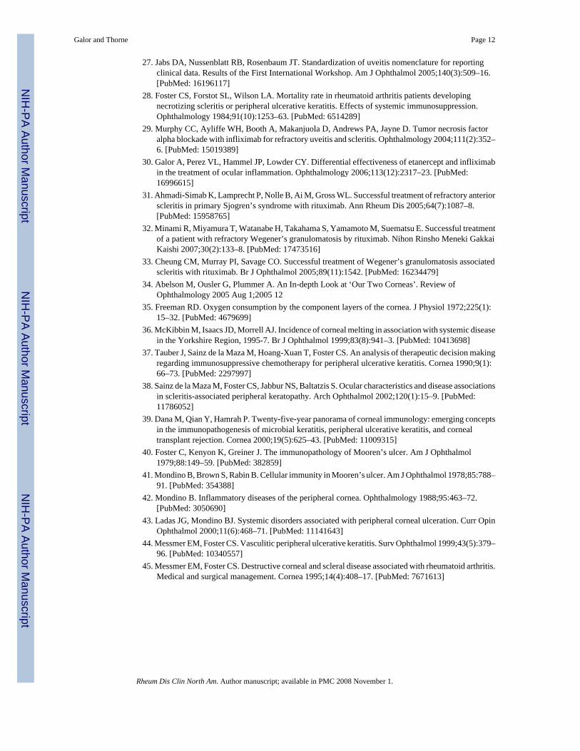
The sclera, which serves as the protective outer layer of the eye (Figure 1), is composed ofconnective tissue: collagen, elastin, and proteoglycans. The sclera starts at the limbus, whereit is continuous with the cornea, and ends at the optic canal, where it is continuous with thedura. The six extraocular muscles insert into the sclera. Scleral tissue receives its sensoryinnervation from the short posterior and long ciliary nerves [1]. The sclera, an avascularstructure, receives its nourishment from adjacent vascularized tissues: the choroidal plexusbeneath and the episcleral plexus above. The episclera has two vascular plexi, one superficialwhose vessels are arranged radially, and another deep whose vessels adhere to the sclera.
Corresponding Author: Jennifer E. Thorne, MD, PhD, Wilmer Eye Institute, 550 North Broadway, Suite 700, Baltimore, Maryland 21205;Phone 410-955-1966; Fax 410-955-0629; email [email protected] Interests: NonePublisher's Disclaimer: This is a PDF file of an unedited manuscript that has been accepted for publication. As a service to our customerswe are providing this early version of the manuscript. The manuscript will undergo copyediting, typesetting, and review of the resultingproof before it is published in its final citable form. Please note that during the production process errors may be discovered which couldaffect the content, and all legal disclaimers that apply to the journal pertain.
NIH Public AccessAuthor ManuscriptRheum Dis Clin North Am. Author manuscript; available in PMC 2008 November 1.
Published in final edited form as:Rheum Dis Clin North Am. 2007 November ; 33(4): 835–854.
NIH
-PA Author Manuscript
NIH
-PA Author Manuscript
NIH
-PA Author Manuscript
Epidemiology of ScleritisScleritis can occur in any age group but usually presents between ages 30 and 50 [2,3]. Womenare affected approximately twice more often than men, and there is no racial or geographicpredilection [2,3]. There is no known HLA association [4]. The prevalence of scleritis in thegeneral population is estimated to be 6 cases per 100,000 peopl, but has been described inbetween 0.2% and 6.3% of patients with RA and up to 7% of those with Wegener’sgranulomatosis [5-7].
Pathogenesis of scleritisStudies on the pathogenesis of scleritis are limited. However, the data available support animportant if not predominant role for T cells in the inflammatory process [8]. Inflammatorycells, mostly T cells and macrophages, are found on biopsy specimens [8,9]. Evidence ofvasculitis with neutrophil invasion and necrosis of the vessel wall was observed in onehistopathologic study but not in a second one [8,10]. Antibody deposition has also beendescribed in one study [8]. One group that divided scleritis into categories by morphologyfound that zonal necrotizing granulomas were more common in patients with associatedsystemic autoimmune conditions and that nonzonal diffuse scleral inflammation was morecommon in patients without an associated systemic condition [11].
Clinical History in ScleritisThe chief complaint of patients with scleritis is pain that responds poorly to analgesics. Thepain is described as dull, aching, or boring and may be severe and constant. Because theextraocular muscles insert into the sclera, the pain typically worsens with eye movement. Thepain often awakens patients from sleep and is typically worse in early morning. The pain canspread to the periorbital region, brow, forehead, temple, ear, or jaw, and may bedisproportionate to the clinical findings.
Up to 20% of patients with scleritis have little or no pain [12]. The absence of pain is observedin three settings: 1) milder disease (e.g., diffuse anterior or nodular scleritis, as opposed to thenecrotizing type [see below]; 2) patients already on immunosuppressive medications at thetime their symptoms begin; and, 3) scleromalacia perforans, a rare complication of advancedRA. Other complaints may include tearing, photophobia, and decreased vision. Patients withanterior scleritis usually notice eye redness. Patients with posterior scleritis may complain ofptosis and swelling of the periorbital tissue.
Clinical Findings in ScleritisPatients with anterior scleritis present with a red eye. However, in some cases, the redness islocated underneath the lid and may be easily overlooked without a focused eye examination.The redness has a violaceous hue that is best seen in natural sunlight. Slit lamp examinationreveals scleral edema and dilatation of both the superficial and deep episcleral vessels. The eyein patients with scleritis is tender to palpation. Ocular erythema persists after the applicationof topical phenylephrine. Approximately 25% of patients have bilateral disease at presentation,but scleritis ultimately becomes bilateral in about 50% of patients [3,12,13].
Classification of ScleritisThe Watson and Hayreh classification of scleritis [12] divides the disorder into anterior andposterior types based upon the anatomic distribution of disease. Anterior scleritis is furthersubdivided into diffuse, nodular, necrotizing with inflammation, and necrotizing withoutinflammation (scleromalacia perforans). Although these forms of scleritis correspond roughlyto different degrees of severity, it is unusual for a case of scleritis to evolve from one type toanother, e.g., from diffuse anterior scleritis to necrotizing scleritis.
Galor and Thorne Page 2
Rheum Dis Clin North Am. Author manuscript; available in PMC 2008 November 1.
NIH
-PA Author Manuscript
NIH
-PA Author Manuscript
NIH
-PA Author Manuscript
Diffuse Anterior Scleritis—Diffuse anterior disease, the most common clinicalpresentation of scleritis, occurs in approximately 60% of patients [3]. Although the onset ofdisease is insidious, ocular inflammation may involve a substantial proportion or all of theanterior sclera at presentation (Figure 2). The normal radial pattern of the episcleral vessels islost and one may see beading and tortuosity of the vessels. Signs of corneal inflammation suchas peripheral corneal infiltrates or mild corneal thinning may be present [13]. Frank cornealulceration is not typical as it is more common in necrotizing scleral disease (described below).Progression to nodular or necrotizing scleritis has been observed, but is uncommon [3,14].Resolution of the ocular inflammation in diffuse anterior scleritis may leave a bluish gray huecaused by rearrangement of the scleral collagen fibers (Figure 3). This does not represent scleralthinning, which is an uncommon sequela of anterior scleritis. Systemic disorders have beendescribed in up to 45% of patients with diffuse anterior scleritis, most commonly RA [13,15].
Nodular Anterior Scleritis—Nodular scleritis is the second most common clinicalpresentation of anterior scleritis (Figure 4), accounting for approximately 20% of scleritis cases[3]. Patients with nodular scleritis present with a firm, immobile, and tender nodule thattypically is found close to the limbus. Similar to diffuse anterior scleritis, progression to otherforms of scleritis is uncommon [3,14]. A systemic disease is diagnosed in 40-50% of patients[13].
Necrotizing Anterior Scleritis with Inflammation—Necrotizing scleritis is the mostserious clinical presentation of anterior scleritis. This condition has an older age of onsetcompared to the other types of scleritis and a higher proportion of patients (50-80%) have anunderlying systemic disease [14]. The two diseases most often associated with necrotizinganterior scleritis are Wegener’s granulomatosis and RA [13,16]. Necrotizing scleritis typicallyrequires therapy with glucocorticoids and/or immunosuppressive drugs to control the disease[3]. Patients present with typical signs of anterior scleritis combined with areas of white sclerasurrounded by edema and congestion (Figure 5). The areas of white sclera represent capillaryclosure of the episcleral vasculature, with infarction and necrosis of the underlying sclera.Involvement of adjacent ocular structures, including the cornea, ciliary body, and trabecularmeshwork is observed with secondary corneal ulceration, uveitis, or increased intraocularpressure [3]. After resolution of scleritis with appropriate treatment, there is thinning of thesclera with translucency, and the choroid often is seen through the sclera (Figure 6). Despitesuch thinning, rupture of the globe is rare.
Scleromalacia Perforans (Necrotizing Anterior Scleritis Without Inflammation)—Scleromalacia perforans, also known as necrotizing anterior scleritis without inflammation,is a rare but severe form of scleritis that tends to involve both eyes and presents without redness,pain, or edema. Often there is thinning and atrophy of the episclera and loss of the episcleralvasculature (Figure 7). Localized areas of yellow-white infarcted tissue are seen. Such areasare demarcated clearly from surrounding tissue. Thinning may become so pronounced that thechoroid is covered by conjunctivae alone. Given the lack of typical symptoms, patients oftenpresent with discolored sclera or blurred vision secondary to astigmatism due to scleralthinning. The classic patient with scleromalacia perforans is an elderly woman withlongstanding RA [13].
Posterior Scleritis—Posterior scleritis is defined as involvement of the sclera byinflammation posterior to the insertion of the medial and lateral rectus muscles. This form ofscleritis is difficult to diagnose because of the secluded nature of its ocular inflammation andits non-specific clinical features [13]. Patients may present with deep-seated headaches, painupon movements of the eye, proptosis, ptosis, double vision, and reduced visual acuity. Ocularexamination may be normal or may disclose localized or generalized exudative retinal
Galor and Thorne Page 3
Rheum Dis Clin North Am. Author manuscript; available in PMC 2008 November 1.
NIH
-PA Author Manuscript
NIH
-PA Author Manuscript
NIH
-PA Author Manuscript
detachment (21% in one study of 99 patients with posterior scleritis), optic nerve edema (18%),subretinal mass lesion (13%), choroidal effusion (4%), uveitis (2%), and vasculitis (2%) [2].Posterior scleritis may also be accompanied by macular edema, retinal striae, and choroidalfolds.
Patients with posterior scleritis are less likely to have an underlying systemic disease comparedto those with anterior scleritis [3,12]. Diagnosis can be made based on the typical B-scanultrasonography appearance of thickened sclera with fluid in Tenon’s space (Figure 8). Anultrasonographic finding referred to as the T-sign is created by hypoechogenic fluid that formsan interface between the optic nerve and the sclera.
Differential Diagnosis of ScleritisA variety of other etiologies of red eyes must be considered in the differential diagnosis ofscleritis. These are shown in Table 1. Episcleritis refers to inflammation of the superficialvessels within the episclera. Ocular erythema in episcleritis is limited usually to only a sectorof the eye and is not associated with pain, vision changes, or discharge. Examination revealsepiscleral injection, which is more likely to be bright red than deeply violaceous in color.
The vessels involved in episcleritis are more superficial than those in scleritis, have a radialpattern, and can be moved with a cotton tip applicator. Furthermore, palpation of the area doesnot result in pain. Phenylephrine blanches the episcleral vessels in episcleritis but will not doso in scleritis. The condition tends to be self-limited but intermittently recurrent. Episcleritisis less likely to be associated with a systemic autoimmune condition than scleritis [17]. Ocularcomplications are also less likely in episcleritis, with vision loss occurring in none of the 37patients in one study and only two of 94 (not related directly to episcleritis) in another [3,17].Treatment is not necessary unless the redness is bothersome to the patient. Therapy may includetopical glucocorticoids and nonsteroidal anti-inflammatory drugs (NSAIDs).
Conjunctivitis can be caused by a variety of processes including bacterial or viral infections,allergies, and prolonged contact lens use. Symptoms include tearing, discharge, and a foreignbody sensation. Examination reveals a follicular or papillary reaction of the palpebralconjunctivae. Treatment of bacterial conjunctivitis consists of antibiotic eyedrops. Treatmentof viral conjunctivitis is supportive, with cool compresses and artificial tears. Allergicconjunctivitis can also be treated with cool compresses as well as artifical tears and a topicalanti-histamine or mast cell inhibitor eyedrops.
Blepharitis refers to inflammation of the eyelid margins. Blepharitis symptoms includeburning, tearing, and crusting of the eyelashes. Symptoms are typically worse in the morning.Ocular examination reveals crusting around the eyelashes, oil inspissation, and telangiectasiasof the eyelid margin. Treatment for blepharitis involves warm compresses followed by gentlemassage to clean the eyelashes and oil gland openings. Oral antibiotics from the tetracyclineclass are used in more severe cases.
Keratoconjunctivitis sicca (KCS) can also present with red eyes. Symptoms include foreignbody sensation and burning, typically worse at the end of the day. Although it is paradoxical,the eyes in KCS may also tear. Examination reveals a decreased tear lake and roughness of thecorneal surface. Treatment of KCS includes tear replacement therapy, plugs in the cannuliculi,and topical cyclosporine.
Anterior uveitis refers to inflammation of the iris and ciliary body. Symptoms include pain,sensitivity to light, and blurred vision. The redness is more pronounced over the perilimbal(circumcorneal) region, which overlies the inflamed ciliary body. The pupil can be miotic andpoorly reactive to light. Examination reveals cells and flare in the anterior chamber. The first-
Galor and Thorne Page 4
Rheum Dis Clin North Am. Author manuscript; available in PMC 2008 November 1.
NIH
-PA Author Manuscript
NIH
-PA Author Manuscript
NIH
-PA Author Manuscript
line therapy for anterior uveitis consists of topical glucocorticoid eyedrops and a mydriaticagent. (Dilating the pupils is essential to the prevention of synechiae between the iris and lens).More severe cases are treated with oral glucocorticoids or immunosuppressive drug therapy.
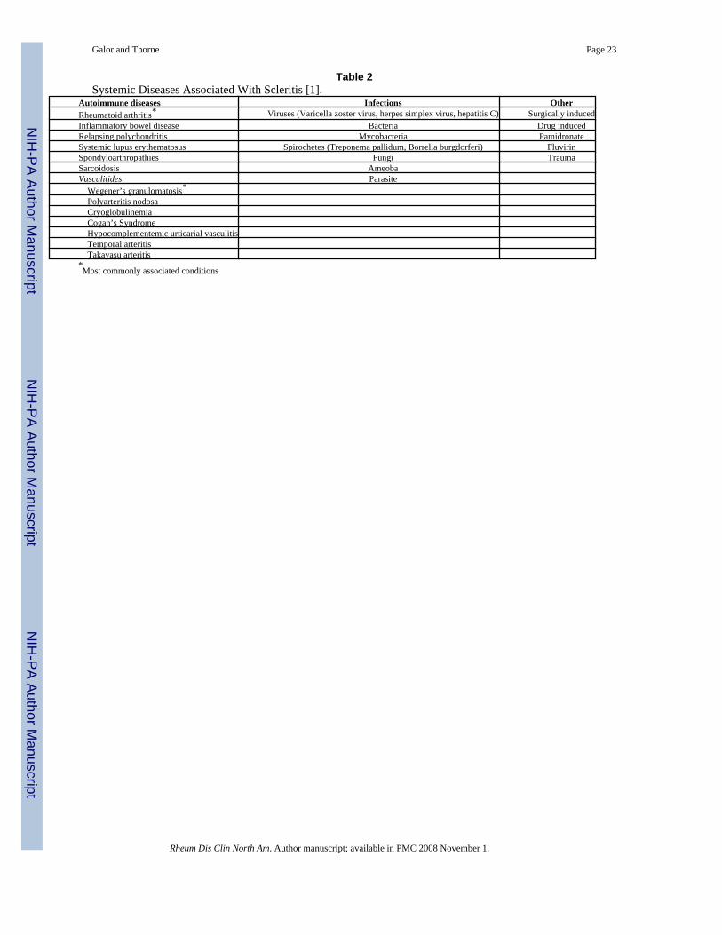
Systemic Conditions Associated With ScleritisFifty percent of patients with scleritis are diagnosed with an associated systemic condition[3,18]. Disorders associated with scleritis can be divided three categories: inflammatory,infectious, and other (Table 2). Autoimmune conditions are found in approximately 40% ofpatients and infections in approximately 7% [3,18]. The most commonly associated rheumaticdiseases are RA (18-33%); systemic vasculitis (7-19%), of which Wegener’s granulomatosisis the most common; systemic lupus erythematosus (SLE)(4-7%); inflammatory bowel disease(4-7%), and relapsing polychondritis (3%) [3,17]. Less commonly associated conditionsinclude sarcoidosis [19,20], cryoglobulinemia [21], and hypocomplementemic urticarialvasculitis [22]. The most commonly associated infection is herpes zoster [18].
In patients with scleritis and an associated systemic condition, the underlying disease is alreadyknown in about 80% of patients at the time scleritis presents. In one study, approximately 15%of patients were diagnosed with a new systemic condition as a result of the initial evaluation,and an additional 8% developed a systemic disease during follow-up [18]. A systemic vasculitiswas more likely to be discovered by the initial diagnostic evaluation than were other rheumaticdiseases [18]. Among patients with no systemic diagnosis after initial evaluation, a newrheumatic disease was diagnosed at a rate of 4% per person-year [18]. Among patients withapparent scleritis who have a known underlying inflammatory condition, an importantconsideration is the exclusion of an infectious complication of immunosuppression.
Clinical, Radiologic, and Laboratory EvaluationEvaluation should begin with careful history, review of systems, and physical examination.Specific questions should be directed toward symptoms of systemic conditions commonlyassociated with scleritis including RA, Wegener’s granulomatosis, and inflammatory boweldisease. Infectious etiologies also should be considered and a history of trauma or surgicalinsult should be sought.
Routine testing typically includes a complete blood count and metabolic panel and a urinalysiswith microscopy (Table 3). In addition, serologic testing for antinuclear antibodies (ANA),antineutrophil cytoplasmic antibodies (ANCA), rheumatoid factor, and antibodies to cycliccitrullinated peptides (anti-CCP antibodies) should also be performed. Additional serologicassays designed to exclude inflammatory conditions (e.g., for complement levels or other typesof autoantibodies) may be useful if dictated by the presence of specific clinical features or bythe results of initial serologic testing.
Spirochetal infections should be excluded with a rapid plasma reagin (RPR) test, a fluorescenttreponemal antibody (FTA-ABS) assay, and serological testing for Lyme disease. Chestimaging should be performed with either a radiograph or computed tomographic (CT) scan.The latter is preferred if suspicion for Wegener’s granulomatosis is strong. Other potentialdirected tests may include a tuberculin skin test, sacroiliac joint radiographs (forspondyloarthropathy), a CT scan of the sinuses, serologies for viral hepatitis panel (hepatitisB and C). When an infectious form of scleritis is suspected, cultures and/or a scleral biopsymay be needed to secure the diagnosis.
If posterior scleritis is suspected, ultrasonography is useful in evaluating the posterior globefor scleral thickening and fluid in Tenon’s capsule (Figure 8). When fluid in Tenon’s capsule
Galor and Thorne Page 5
Rheum Dis Clin North Am. Author manuscript; available in PMC 2008 November 1.
NIH
-PA Author Manuscript
NIH
-PA Author Manuscript
NIH
-PA Author Manuscript
is seen in the plane of the optic nerve, the finding is called the “T-sign”. CT scan of the orbitsalso can be of use in posterior scleritis, showing thickening of the sclera or orbital inflammation.
Complications of ScleritisScleritis can be associated with significant eye morbidity [13]. Visual loss can result from anumber of complications including uveitis, cataracts, corneal melts (see PUK, below),glaucoma, and posterior segment disease. Specific complications of posterior scleritis caninclude optic disk edema, cystoid macular edema (swelling of the macula), exudative retinaldetachment, and choroidal folds (wrinkles in the choroid – the layer of the eye interposedbetween the retina and sclera). Other less common complications include scleral thinning andglobe rupture with minor trauma.
In a retrospective study of 172 patients, anterior uveitis was found in 42% of patients withscleritis, cataracts were detected in 17%, PUK in 14%, glaucoma in 13%, and posterior segmentdisease in 6% [17]. In that same study, 37% of patients experienced a decrease in vision dueto scleritis, defined as a loss of two or more lines of vision during follow up or a visual acuityof 20/80 or worse at presentation. Vision loss was more commonly seen in patient withnecrotizing scleritis (82%) [17]. Another retrospective study of 290 patients found that visionloss (defined as a permanent drop in Snellen acuity of two or more lines) occurred in 9% ofpatients with diffuse anterior scleritis, 26% of patients with nodular scleritis, 74% of those withnecrotizing disease, and 84% of those with posterior scleritis [14]. Vision loss (loss of 2 ormore Snellen lines from initial best corrected acuity) is much more likely in patients withposterior scleritis (31% [2]) or an underlying systemic disorder (27% [3]).
Treatment of ScleritisIn a retrospective study of 97 patients with scleritis, 30% required nonsteroidal anti-inflammatory drugs (NSAIDs) alone, 31% required oral glucocorticoids, and 26% requiredimmunosuppressive drugs in addition to glucocorticoids to control their disease [3]. Treatmentsvaried with the specific type of scleritis. Nodular anterior scleritis responded more often toNSAIDs (57%), whereas necrotizing scleritis often required systemic immunosuppressivedrugs (70%). Posterior scleritis was more often treated with oral glucocorticoids (83%) andoccasionally immunosuppressive drugs (17%) [3].
A step-ladder approach typically is utilized in the treatment of scleritis. The first line therapyin treating non-necrotizing scleritis that is not caused by an infection is an NSAID. Two drugsNSAIDs have been shown to be effective: flurbiprofen 100 mg three times daily andindomethacin 25-50 mg three times daily [3,13]. If one NSAID does not relieve the pain andinflammation, another may be tried.
Systemic glucocorticoids are used in three general settings: when NSAIDs prove ineffective;in cases of necrotizing anterior scleritis; and in cases of posterior scleritis. The usual startingdose is 1 mg/kg/day (maximum of 60 mg/daily) followed by a tapering schedule based onclinical response. Table 4 describes the typical tapering schedule used in our clinic. In patientswhose disease appears particularly aggressive, pulse methylprednisolone can be administeredintravenously at a dose of one gram/day for three days, following by the initiation of prednisone60 mg/day. The role of depot glucocorticoid treatment is controversial, given the potential riskfor scleral perforation with this therapy [24-26].
Immunosuppressive drug therapy is instituted under several clinical circumstances: fornecrotizing scleritis, which usually requires cyclophosphamide plus glucocorticoids from theoutset of treatment; when other types of scleritis are not controlled by one month of high dose
Galor and Thorne Page 6
Rheum Dis Clin North Am. Author manuscript; available in PMC 2008 November 1.
NIH
-PA Author Manuscript
NIH
-PA Author Manuscript
NIH
-PA Author Manuscript
glucocorticoids; when more than 10 mg/day of prednisone is required to maintain diseasecontrol; and when glucocorticoid-related adverse effects become an issue [3,27].
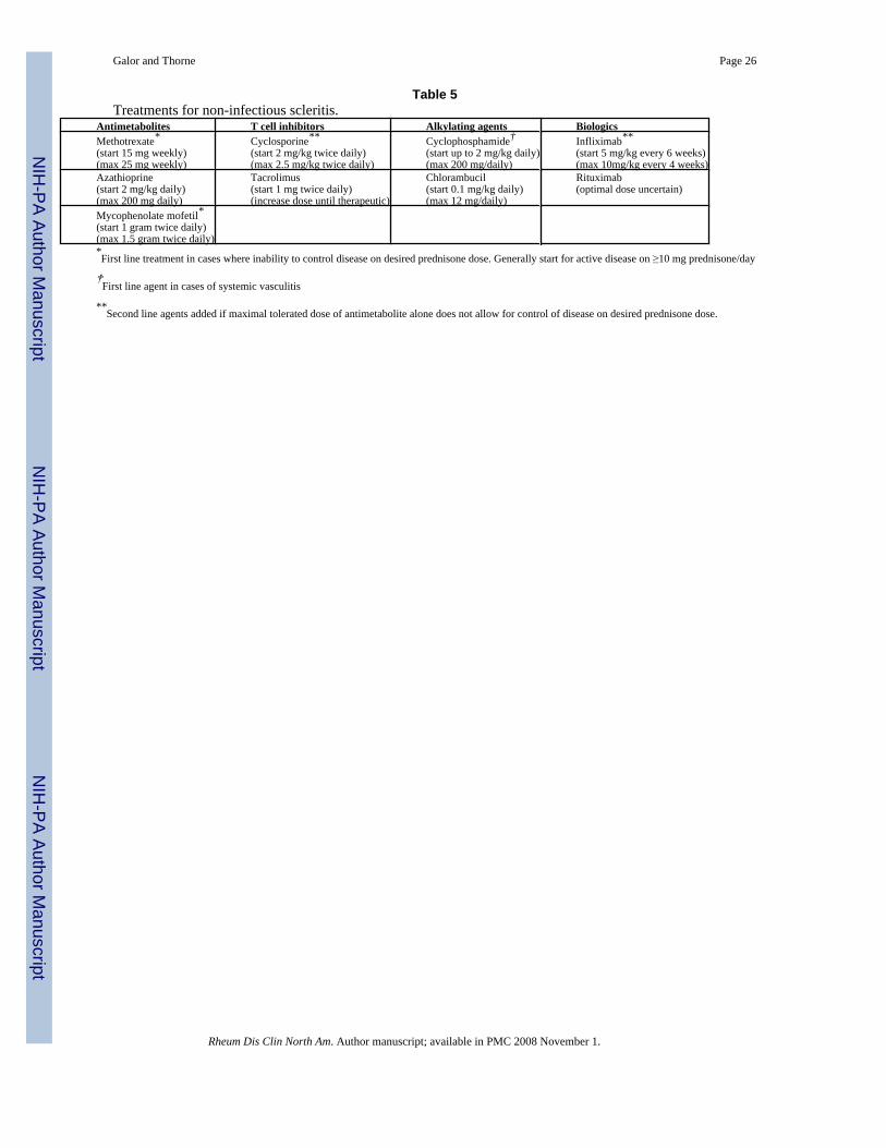
A variety of glucocorticoid sparing agents have been used in the treatment of scleritis (Table5). Cyclophosphamide (up to 2 mg/kg/day) is the drug of choice for patients with necrotizingscleritis and in patients with scleritis associated with a systemic vasculitis such as Wegener’sgranulomatosis [3]. The justification for use of an alkylating agent in such cases is the highrisk of progressive ocular damage, extraocular vasculitic lesions, and for death [28]. Forpatients with non-necrotizing scleritis who require a glucocorticoid-sparing agent, first linetreatment typically consists of methotrexate (up to 25 mg/week), azathioprine (up to 200 mg/day), or mycophenolate mofetil (1 gram twice daily). In a retrospective study that examinedthe clinical outcomes of 50 patients treated with these agents, 46% achieved disease quiescenceand were able to taper their prednisone to ≤ 10 mg/day of prednisone [Galor et al, unpublisheddata]. Depending on severity of disease, treatment typically is continued for one to two yearsafter control of inflammation.
Possible second-line agents for scleritis include calcineurin inhibitors (cyclosporine ortacrolimus), infliximab, or rituximab [29-33]. However, none of these agents has been studiedrigorously to date. In some cases of necrotizing anterior scleritis or scleromalacia perforans,surgical therapy is required to address extensive scleral thinning and avoid globe rupture.Scleral grafting surgery may be performed with donor sclera, periostium, or fascia lata.Simultaneous efforts to control the underlying inflammation with medical therapy are essentialwhen surgery is required.
The treatment of scleritis with immunosuppressive medications is fraught with the potentialfor treatment-related morbidity and mortality. Patients on high-dose glucocorticoids and otherimmunosuppressive agents require careful monitoring, with frequent clinic visits andbloodwork. Patients on cyclophosphamide should have a complete blood count checked notless often than every two weeks. Prophylaxis against Pneumocystis carinii (jiroveci)pneumonia (PCP) is also critical, particularly when the combination of glucocorticoids andanother immunosuppressive agent is used. One common approach to PCP prophylaxis is touse trimethoprim-sulfamethoxazole (one single-strength tablet daily). Evaluation for andprophylaxis against glucocorticoid-induced bone loss must also be considered.
Part 2: Peripheral ulcerative keratitisPeripheral ulcerative keratitis (PUK) is a form of ocular inflammation that involves the outerportions of the cornea and may be associated with systemic conditions such as RA, Wegener’sgranulomatosis, and other systemic conditions. The most severe complication of PUK, knownas corneal melt syndrome, is the progression of marginal corneal thinning to perforation of thecornea. Corneal melt syndromes can lead to the abrupt and permanent loss of vision in theinvolved eye.
Anatomy of the corneaThe cornea is a transparent, avascular structure that allows light into the eye and serves as theeye’s main focusing instrument (Figure 8). The peripheral cornea, the site of disease in PUK,is distinct from the central cornea both in its anatomic and physiologic characteristics [34].The peripheral cornea is near the capillary bed and partly derives its nutrients from the capillaryarcades [35]. Proximity to this vascular and lymphatic arcade may explain the peripherallocation of PUK, as inflammatory cells and mediators can gain access to this part of the corneamore readily.
Galor and Thorne Page 7
Rheum Dis Clin North Am. Author manuscript; available in PMC 2008 November 1.
NIH
-PA Author Manuscript
NIH
-PA Author Manuscript
NIH
-PA Author Manuscript
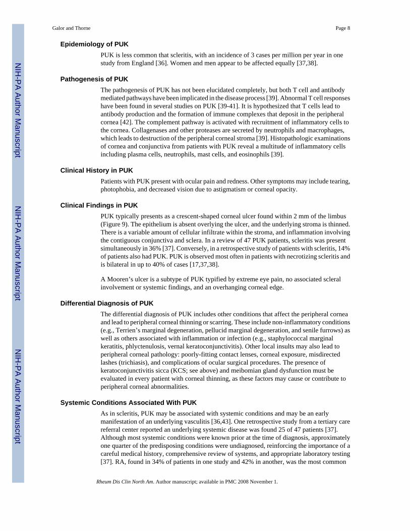
Epidemiology of PUKPUK is less common that scleritis, with an incidence of 3 cases per million per year in onestudy from England [36]. Women and men appear to be affected equally [37,38].
Pathogenesis of PUKThe pathogenesis of PUK has not been elucidated completely, but both T cell and antibodymediated pathways have been implicated in the disease process [39]. Abnormal T cell responseshave been found in several studies on PUK [39-41]. It is hypothesized that T cells lead toantibody production and the formation of immune complexes that deposit in the peripheralcornea [42]. The complement pathway is activated with recruitment of inflammatory cells tothe cornea. Collagenases and other proteases are secreted by neutrophils and macrophages,which leads to destruction of the peripheral corneal stroma [39]. Histopathologic examinationsof cornea and conjunctiva from patients with PUK reveal a multitude of inflammatory cellsincluding plasma cells, neutrophils, mast cells, and eosinophils [39].
Clinical History in PUKPatients with PUK present with ocular pain and redness. Other symptoms may include tearing,photophobia, and decreased vision due to astigmatism or corneal opacity.
Clinical Findings in PUKPUK typically presents as a crescent-shaped corneal ulcer found within 2 mm of the limbus(Figure 9). The epithelium is absent overlying the ulcer, and the underlying stroma is thinned.There is a variable amount of cellular infiltrate within the stroma, and inflammation involvingthe contiguous conjunctiva and sclera. In a review of 47 PUK patients, scleritis was presentsimultaneously in 36% [37]. Conversely, in a retrospective study of patients with scleritis, 14%of patients also had PUK. PUK is observed most often in patients with necrotizing scleritis andis bilateral in up to 40% of cases [17,37,38].
A Mooren’s ulcer is a subtype of PUK typified by extreme eye pain, no associated scleralinvolvement or systemic findings, and an overhanging corneal edge.
Differential Diagnosis of PUKThe differential diagnosis of PUK includes other conditions that affect the peripheral corneaand lead to peripheral corneal thinning or scarring. These include non-inflammatory conditions(e.g., Terrien’s marginal degeneration, pellucid marginal degeneration, and senile furrows) aswell as others associated with inflammation or infection (e.g., staphylococcal marginalkeratitis, phlyctenulosis, vernal keratoconjunctivitis). Other local insults may also lead toperipheral corneal pathology: poorly-fitting contact lenses, corneal exposure, misdirectedlashes (trichiasis), and complications of ocular surgical procedures. The presence ofkeratoconjunctivitis sicca (KCS; see above) and meibomian gland dysfunction must beevaluated in every patient with corneal thinning, as these factors may cause or contribute toperipheral corneal abnormalities.
Systemic Conditions Associated With PUKAs in scleritis, PUK may be associated with systemic conditions and may be an earlymanifestation of an underlying vasculitis [36,43]. One retrospective study from a tertiary carereferral center reported an underlying systemic disease was found 25 of 47 patients [37].Although most systemic conditions were known prior at the time of diagnosis, approximatelyone quarter of the predisposing conditions were undiagnosed, reinforcing the importance of acareful medical history, comprehensive review of systems, and appropriate laboratory testing[37]. RA, found in 34% of patients in one study and 42% in another, was the most common
Galor and Thorne Page 8
Rheum Dis Clin North Am. Author manuscript; available in PMC 2008 November 1.
NIH
-PA Author Manuscript
NIH
-PA Author Manuscript
NIH
-PA Author Manuscript
associated disease [37,38]. Other systemic conditions associated with PUK include the ANCA-associated vasculitides (Wegener’s granulomatosis, the Churg-Strauss syndrome, andmicroscopic polyangiitis), polyarteritis nodosa, relapsing polychondritis, and systemic lupuserythematosus [37,38].
In RA, corneal melt usually occurs in a patient with longstanding disease, the same type ofpatient prone to the development of rheumatoid vasculitis: nodular, erosive, rheumatoid factor-positive. In one study, there was a mean of 19.6 years between diagnosis of RA and PUK[36]. In Wegener’s granulomatosis and other forms of systemic vasculitis, corneal melt canoccur earlier in the disease course. Regardless of the underlying condition with which it isassociated, however, corneal melting can occur swiftly once inflammation begins within thecornea. Visual loss can ensue within days.
Ocular and systemic infections may also cause or be associated with PUK. Microbial pathogensimplicated in the etiology of PUK include bacteria (Staphylococcus and Streptococcusspecies), spirochetes (Treponema pallidum), Mycobacteria (tuberculosis), viruses (hepatitis C,herpes simplex virus, varicella zoster virus), acanthameoba, and fungi [39].
Laboratory Evaluation in PUKThe clinical, radiological, and laboratory evaluation in PUK is identical to that of scleritis (seeabove) (Table 3).
Complications of PUKPUK may be associated with significant eye and systemic morbidity. In one retrospectivereview of 24 patients with scleritis associated PUK, all 24 patients had impending cornealperforation, 16 (67%) had an associated anterior uveitis, and 20 (83%) had decreased vision,defined as a decrease in visual acuity of 2 or more Snellen lines at the end of follow up or visualacuity of 20/80 or worse at presentation [38]. In another retrospective review of 47 patientswith PUK, 34% had impending or frank corneal perforations (defined as peripheral cornealthinning of 75-100%), 47% required a tectonic graft procedure, 9% had an associated anterioruveitis, and 43% had visual acuity of 20/400 or worse at presentation [37]. In addition to thehigh risk of ocular damage from PUK, eye inflammation of this nature is a harbinger of activeinflammatory disease in other organ systems with major potential for morbidity and mortality[28].
Treatment of PUKThe treatment of PUK is determined by the severity of findings within the cornea and the extentof extra-ocular disease. Systemic glucocorticoids are the cornerstone of therapy for non-infectious PUK. Glucocorticoids are started at a dose of 1 mg/kg/day (maximum of 60 mgdaily), followed by a tapering schedule based on clinical response. As in scleritis, patients inwhom loss of vision is imminent should be treated with pulse methylprednisolone, 1 gram/dayfor three days. Topical glucocorticoids are not an appropriate therapy for PUK.
Alkylating agents may be used in conjunction with glucocorticoids in cases of PUK withimminent danger of corneal perforation and in cases associated with a systemic vasculitis.Because of the high likelihood of ocular morbidity resulting from ineffective treatment of PUK,the usual approach is to employ both cyclophosphamide (up to 2 mg/kg/day) in addition tohigh-dose glucocorticoids from the outset of therapy. As with scleritis, steroid-sparing agentsare also added when an initial course of high-dose glucocorticoids does not control the disease,when disease control cannot be maintained with less than 10 mg/day of prednisone, and whensignificant concern related to glucocorticoid-related adverse effects exists [27]. Methotrexate,
Galor and Thorne Page 9
Rheum Dis Clin North Am. Author manuscript; available in PMC 2008 November 1.
NIH
-PA Author Manuscript
NIH
-PA Author Manuscript
NIH
-PA Author Manuscript
azathioprine, mycophenolate mofetil, and the biologic agents are all employed in a fashionsimilar to their use in scleritis (Table 5).
The treatment of PUK with immunosuppressive medications is fraught with the potential fortreatment-related morbidity and mortality. Patients on high-dose glucocorticoids and otherimmunosuppressive agents require careful monitoring, with frequent clinic visits andbloodwork. Patients on cyclophosphamide should have a complete blood count checked notless often than every two weeks. Prophylaxis against Pneumocystis carinii (jiroveci)pneumonia (PCP) is also critical, particularly when the combination of glucocorticoids andanother immunosuppressive agent is used. One common approach to PCP prophylaxis is touse trimethoprim-sulfamethoxazole (one single-strength tablet daily). Evaluation for andprophylaxis against glucocorticoid-induced bone loss must also be considered.
Surgical resection of conjunctival tissue adjacent to PUK has been promoted as a means ofdecreasing access of inflammatory cells and factors to the peripheral cornea [37,44]. Thistreatment is controversial, as it is not uniformly agreed that resection alters the course ofdisease. Surgical management of PUK is used in cases of impending perforation to preserveglobe integrity. Several surgical options exist depending on the size of perforation. Theseinclude the use of a tissue adhesive bandage contact lens, a lamellar graft, and tectonic cornealgrafting.
A study of 34 patients with RA-associated PUK and/or necrotizing scleritis found decreasedmortality and ocular morbidity in patients treated with immunosuppressive medication [28].A lower mortality was seen (6% versus 53%) in patients managed with cyclophosphamide (12patients) or antimetabolite agents (5 patients) compared to those managed with oralglucocorticoids and NSAIDS alone. Progression of ocular disease occurred less often (0%versus 76%) in the former group, as well. Furthermore, no patient treated withimmunosuppressive agents developed extraocular vasculitis while on medication. Anotherretrospective review of 12 patients with RA-associated PUK found that 68% of patients hadstable or improved visual acuities after systemic treatment with cyclophosphamide ormethotrexate and surgical therapy (required in 63% of eyes) [45].
A retrospective study of 38 patients with PUK who underwent adjacent conjunctival biopsyfound vasculitis in 20 biopsies. Eighteen of the 20 patients were treated with chemotherapy;seventeen had no progression of ulceration. The two patients not treated with chemotherapystabilized with surgical management. Of 18 patients without vasculitis on biopsy, 11 weretreated with chemotherapy. All remained stable or improved with combination surgery andchemotherapy. Of the remaining 7 patients, 6 remained stable after conjunctival resection alone[37].
Acknowledgements
Grant Support: Supported by a grant EY-13707 (Dr. Thorne) from the National Eye Institute, Bethesda, Maryland,and by unrestricted funds form Research to Prevent Blindness (Dr. Galor).
References1. Okhravi N, Odufuwa B, McCluskey P, Lightman S. Scleritis. Surv Ophthalmol 2005;50(4):351–63.
[PubMed: 15967190]2. McCluskey PJ, Watson PG, Lightman S, Haybittle J, Restori M, Branley M. Posterior scleritis: clinical
features, systemic associations, and outcome in a large series of patients. Ophthalmology 1999;106(12):2380–6. [PubMed: 10599675]
3. Jabs DA, Mudun A, Dunn JP, Marsh MJ. Episcleritis and scleritis: clinical features and treatmentresults. Am J Ophthalmol 2000;130(4):469–76. [PubMed: 11024419]
Galor and Thorne Page 10
Rheum Dis Clin North Am. Author manuscript; available in PMC 2008 November 1.
NIH
-PA Author Manuscript
NIH
-PA Author Manuscript
NIH
-PA Author Manuscript
4. Joysey VC, Roger JH, Ashworth F, et al. Parallel studies of HLA antigens in patients with rheumaticheart disease and scleritis: comparisons with three control populations. J Rheumatol Suppl 1977;3:84–8. [PubMed: 266606]
5. Smith JR, Mackensen F, Rosenbaum JT. Therapy insight: scleritis and its relationship to systemicautoimmune disease. Nat Clin Pract Rheumatol 2007;3(4):219–26. [PubMed: 17396107]
6. Haynes BF, Fishman ML, Fauci AS, Wolff SM. The ocular manifestations of Wegener’sgranulomatosis. Fifteen years experience and review of the literature. Am J Med 1977;63(1):131–41.[PubMed: 327802]
7. Bullen CL, Liesegang TJ, McDonald TJ, DeRemee RA. Ocular complications of Wegener’sgranulomatosis. Ophthalmology 1983;90(3):279–90. [PubMed: 6602963]
8. Fong LP, Sainz de la Maza M, Rice BA, Kupferman AE, Foster CS. Immunopathology of scleritis.Ophthalmology 1991;98(4):472–9. [PubMed: 1828871]
9. Bernauer W, Buchi ER, Daicker B. Immunopathological findings in posterior scleritis. Int Ophthalmol1994;18(4):229–31. [PubMed: 7797388]
10. Bernauer W, Watson PG, Daicker B, Lightman S. Cells perpetuating the inflammatory response inscleritis. Br J Ophthalmol 1994;78(5):381–5. [PubMed: 8025072]
11. Riono WP, Hidayat AA, Rao NA. Scleritis: a clinicopathologic study of 55 cases. Ophthalmology1999;106(7):1328–33. [PubMed: 10406616]
12. Watson PG, Hayreh SS. Scleritis and episcleritis. Br J Ophthalmol 1976;60(3):163–91. [PubMed:1268179]
13. Schwam, B. Scleritis. In: Krachmer, J.; Mannis, M.; Holland, E., editors. Cornea and External Disease:Clinical Diagnosis and Management. 1. II. Saint Louis: Mosby; 1997. p. 1479-1491.
14. Tuft SJ, Watson PG. Progression of scleral disease. Ophthalmology 1991;98(4):467–71. [PubMed:2052300]
15. Hakin KN, Watson PG. Systemic associations of scleritis. Int Ophthalmol Clin 1991;31(3):111–29.[PubMed: 1679048]
16. Foster, C. The sclera. New York: Springer-Verlag; 1994.17. Sainz de la Maza M, Jabbur NS, Foster CS. Severity of scleritis and episcleritis. Ophthalmology
1994;101(2):389–96. [PubMed: 8115160]18. Akpek EK, Thorne JE, Qazi FA, Do DV, Jabs DA. Evaluation of patients with scleritis for systemic
disease. Ophthalmology 2004;111(3):501–6. [PubMed: 15019326]19. Dursun D, Akova YA, Bilezikci B. Scleritis associated with sarcoidosis. Ocul Immunol Inflamm
2004;12(2):143–8. [PubMed: 15512984]20. Qazi FA, Thorne JE, Jabs DA. Scleral nodule associated with sarcoidosis. Am J Ophthalmol 2003;136
(4):752–4. [PubMed: 14516826]21. Kedhar SR, Belair ML, Jun AS, Sulkowski M, Thorne JE. Scleritis and peripheral ulcerative keratitis
with hepatitis C virus-related cryoglobulinemia. Arch Ophthalmol 2007;125(6):852–3. [PubMed:17563007]
22. Thorne JE, Hernandez MI, Rencic A, Nousari HC. Severe scleritis and urticarial lesions. Am JOphthalmol 2002;134(6):932–4. [PubMed: 12470777]
23. Galor A, Thorne JE, Jabs DA. Rheumatic disease and scleritis. Ophthalmology 2007;114(6):1232.[PubMed: 17544785]
24. Albini TA, Zamir E, Read RW, Smith RE, See RF, Rao NA. Evaluation of subconjunctivaltriamcinolone for nonnecrotizing anterior scleritis. Ophthalmology 2005;112(10):1814–20.[PubMed: 16199269]
25. Zamir E, Read RW, Smith RE, Wang RC, Rao NA. A prospective evaluation of subconjunctivalinjection of triamcinolone acetonide for resistant anterior scleritis. Ophthalmology 2002;109(4):798–805. [PubMed: 11927443]discussion 805-7
26. Tu EY, Culbertson WW, Pflugfelder SC, Huang A, Chodosh JC. Therapy of nonnecrotizing anteriorscleritis with subconjunctival corticosteroid injection. Ophthalmology 1995;102(5):718–24.[PubMed: 7777270]
Galor and Thorne Page 11
Rheum Dis Clin North Am. Author manuscript; available in PMC 2008 November 1.
NIH
-PA Author Manuscript
NIH
-PA Author Manuscript
NIH
-PA Author Manuscript
27. Jabs DA, Nussenblatt RB, Rosenbaum JT. Standardization of uveitis nomenclature for reportingclinical data. Results of the First International Workshop. Am J Ophthalmol 2005;140(3):509–16.[PubMed: 16196117]
28. Foster CS, Forstot SL, Wilson LA. Mortality rate in rheumatoid arthritis patients developingnecrotizing scleritis or peripheral ulcerative keratitis. Effects of systemic immunosuppression.Ophthalmology 1984;91(10):1253–63. [PubMed: 6514289]
29. Murphy CC, Ayliffe WH, Booth A, Makanjuola D, Andrews PA, Jayne D. Tumor necrosis factoralpha blockade with infliximab for refractory uveitis and scleritis. Ophthalmology 2004;111(2):352–6. [PubMed: 15019389]
30. Galor A, Perez VL, Hammel JP, Lowder CY. Differential effectiveness of etanercept and infliximabin the treatment of ocular inflammation. Ophthalmology 2006;113(12):2317–23. [PubMed:16996615]
31. Ahmadi-Simab K, Lamprecht P, Nolle B, Ai M, Gross WL. Successful treatment of refractory anteriorscleritis in primary Sjogren’s syndrome with rituximab. Ann Rheum Dis 2005;64(7):1087–8.[PubMed: 15958765]
32. Minami R, Miyamura T, Watanabe H, Takahama S, Yamamoto M, Suematsu E. Successful treatmentof a patient with refractory Wegener’s granulomatosis by rituximab. Nihon Rinsho Meneki GakkaiKaishi 2007;30(2):133–8. [PubMed: 17473516]
33. Cheung CM, Murray PI, Savage CO. Successful treatment of Wegener’s granulomatosis associatedscleritis with rituximab. Br J Ophthalmol 2005;89(11):1542. [PubMed: 16234479]
34. Abelson M, Ousler G, Plummer A. An In-depth Look at ‘Our Two Corneas’. Review ofOphthalmology 2005 Aug 1;2005 12
35. Freeman RD. Oxygen consumption by the component layers of the cornea. J Physiol 1972;225(1):15–32. [PubMed: 4679699]
36. McKibbin M, Isaacs JD, Morrell AJ. Incidence of corneal melting in association with systemic diseasein the Yorkshire Region, 1995-7. Br J Ophthalmol 1999;83(8):941–3. [PubMed: 10413698]
37. Tauber J, Sainz de la Maza M, Hoang-Xuan T, Foster CS. An analysis of therapeutic decision makingregarding immunosuppressive chemotherapy for peripheral ulcerative keratitis. Cornea 1990;9(1):66–73. [PubMed: 2297997]
38. Sainz de la Maza M, Foster CS, Jabbur NS, Baltatzis S. Ocular characteristics and disease associationsin scleritis-associated peripheral keratopathy. Arch Ophthalmol 2002;120(1):15–9. [PubMed:11786052]
39. Dana M, Qian Y, Hamrah P. Twenty-five-year panorama of corneal immunology: emerging conceptsin the immunopathogenesis of microbial keratitis, peripheral ulcerative keratitis, and cornealtransplant rejection. Cornea 2000;19(5):625–43. [PubMed: 11009315]
40. Foster C, Kenyon K, Greiner J. The immunopathology of Mooren’s ulcer. Am J Ophthalmol1979;88:149–59. [PubMed: 382859]
41. Mondino B, Brown S, Rabin B. Cellular immunity in Mooren’s ulcer. Am J Ophthalmol 1978;85:788–91. [PubMed: 354388]
42. Mondino B. Inflammatory diseases of the peripheral cornea. Ophthalmology 1988;95:463–72.[PubMed: 3050690]
43. Ladas JG, Mondino BJ. Systemic disorders associated with peripheral corneal ulceration. Curr OpinOphthalmol 2000;11(6):468–71. [PubMed: 11141643]
44. Messmer EM, Foster CS. Vasculitic peripheral ulcerative keratitis. Surv Ophthalmol 1999;43(5):379–96. [PubMed: 10340557]
45. Messmer EM, Foster CS. Destructive corneal and scleral disease associated with rheumatoid arthritis.Medical and surgical management. Cornea 1995;14(4):408–17. [PubMed: 7671613]
Galor and Thorne Page 12
Rheum Dis Clin North Am. Author manuscript; available in PMC 2008 November 1.
NIH
-PA Author Manuscript
NIH
-PA Author Manuscript
NIH
-PA Author Manuscript
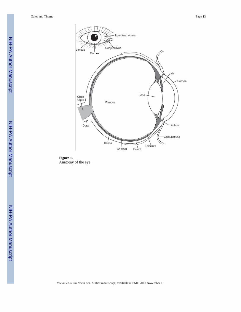
Figure 1.Anatomy of the eye
Galor and Thorne Page 13
Rheum Dis Clin North Am. Author manuscript; available in PMC 2008 November 1.
NIH
-PA Author Manuscript
NIH
-PA Author Manuscript
NIH
-PA Author Manuscript
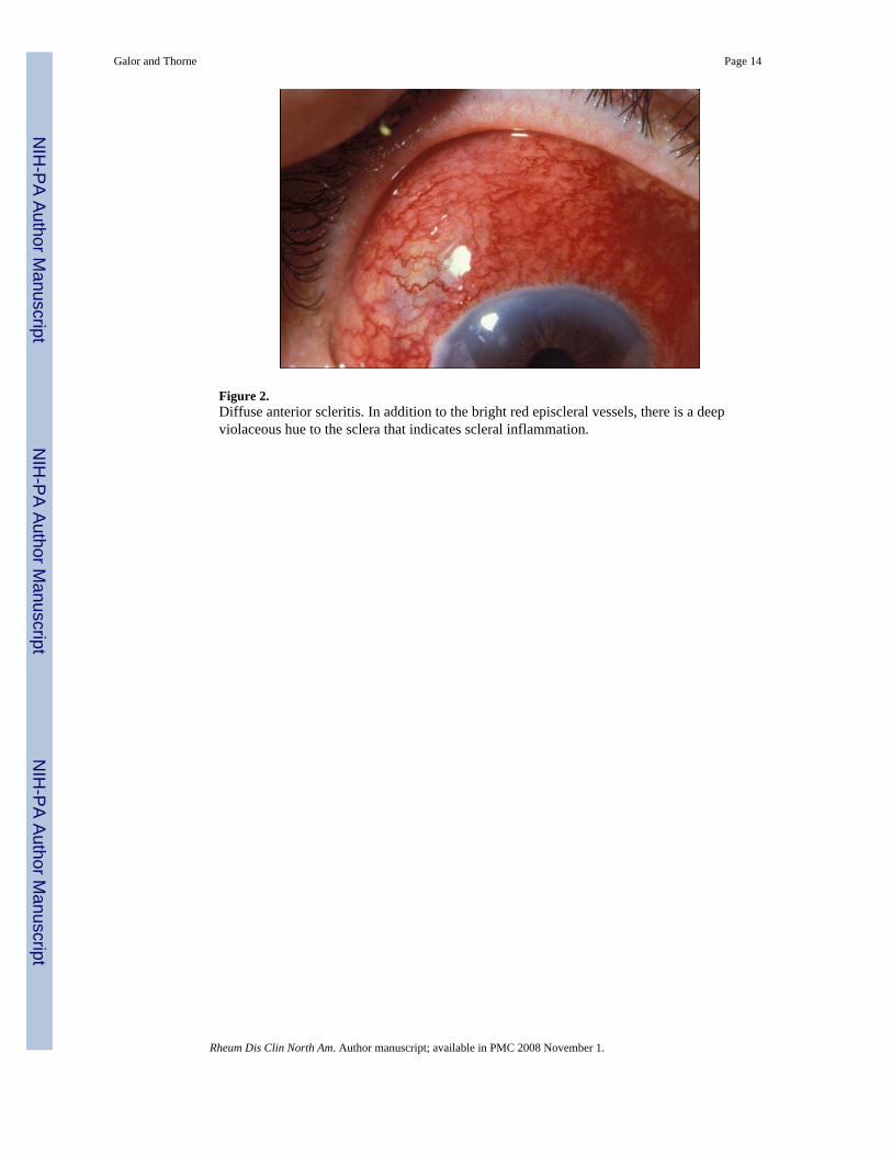
Figure 2.Diffuse anterior scleritis. In addition to the bright red episcleral vessels, there is a deepviolaceous hue to the sclera that indicates scleral inflammation.
Galor and Thorne Page 14
Rheum Dis Clin North Am. Author manuscript; available in PMC 2008 November 1.
NIH
-PA Author Manuscript
NIH
-PA Author Manuscript
NIH
-PA Author Manuscript
Figure 3.Active (left) and resolved (right) diffuse anterior scleritis. Resolution of diffuse anteriorscleritis in a 50 year-old man with Wegener’s granulomatosis after treatment with prednisoneand cyclophosphamide. Resolution of the scleral inflammation has left a bluish gray hue thatrepresents rearrangement of the collagen fibers in the sclera.
Galor and Thorne Page 15
Rheum Dis Clin North Am. Author manuscript; available in PMC 2008 November 1.
NIH
-PA Author Manuscript
NIH
-PA Author Manuscript
NIH
-PA Author Manuscript
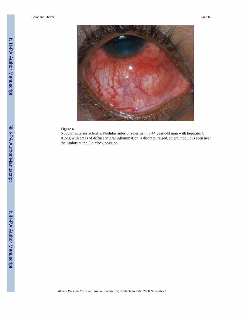
Figure 4.Nodular anterior scleritis. Nodular anterior scleritis in a 44 year-old man with hepatitis C.Along with areas of diffuse scleral inflammation, a discrete, raised, scleral nodule is seen nearthe limbus at the 5 o’clock position.
Galor and Thorne Page 16
Rheum Dis Clin North Am. Author manuscript; available in PMC 2008 November 1.
NIH
-PA Author Manuscript
NIH
-PA Author Manuscript
NIH
-PA Author Manuscript
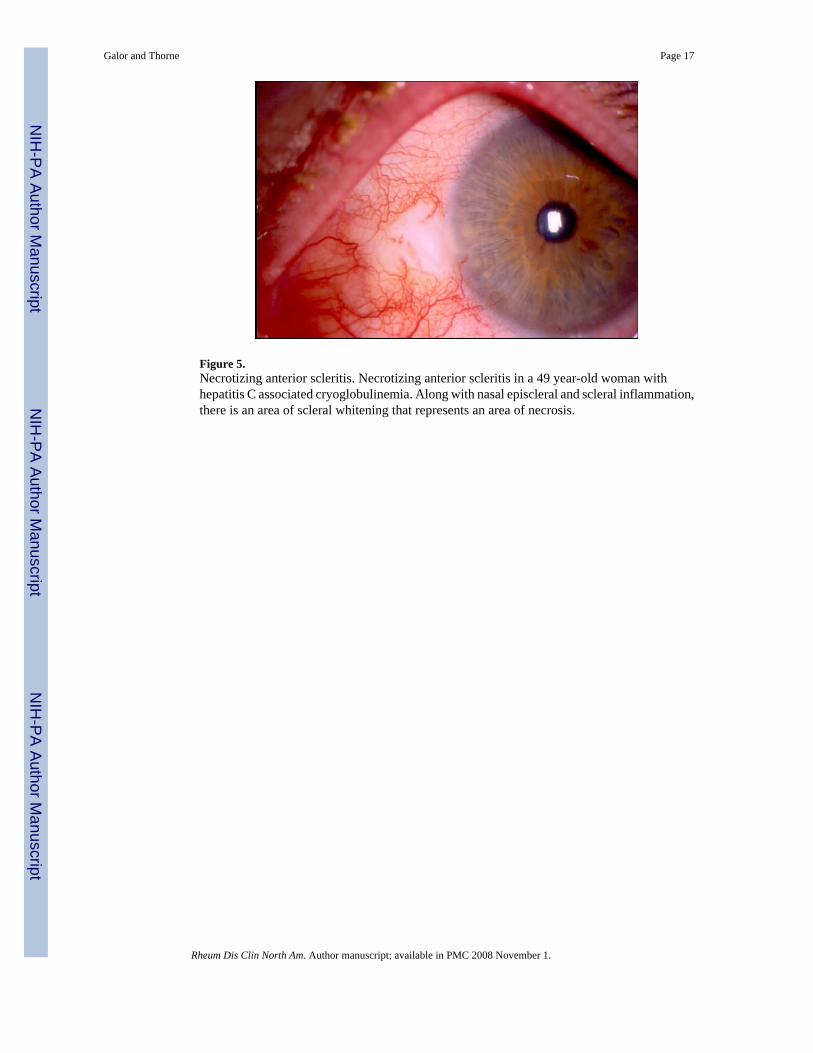
Figure 5.Necrotizing anterior scleritis. Necrotizing anterior scleritis in a 49 year-old woman withhepatitis C associated cryoglobulinemia. Along with nasal episcleral and scleral inflammation,there is an area of scleral whitening that represents an area of necrosis.
Galor and Thorne Page 17
Rheum Dis Clin North Am. Author manuscript; available in PMC 2008 November 1.
NIH
-PA Author Manuscript
NIH
-PA Author Manuscript
NIH
-PA Author Manuscript
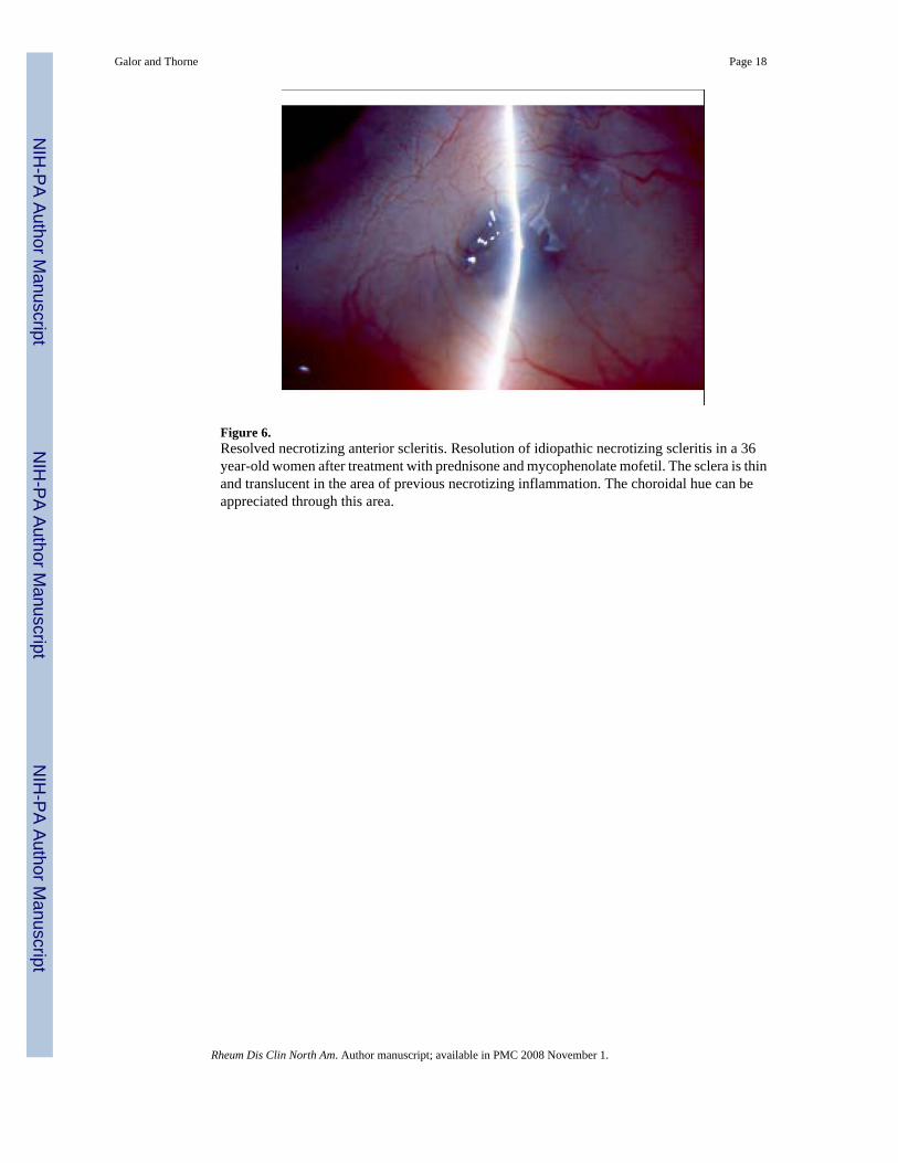
Figure 6.Resolved necrotizing anterior scleritis. Resolution of idiopathic necrotizing scleritis in a 36year-old women after treatment with prednisone and mycophenolate mofetil. The sclera is thinand translucent in the area of previous necrotizing inflammation. The choroidal hue can beappreciated through this area.
Galor and Thorne Page 18
Rheum Dis Clin North Am. Author manuscript; available in PMC 2008 November 1.
NIH
-PA Author Manuscript
NIH
-PA Author Manuscript
NIH
-PA Author Manuscript
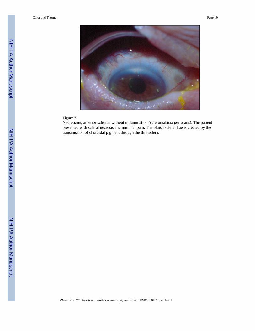
Figure 7.Necrotizing anterior scleritis without inflammation (scleromalacia perforans). The patientpresented with scleral necrosis and minimal pain. The bluish scleral hue is created by thetransmission of choroidal pigment through the thin sclera.
Galor and Thorne Page 19
Rheum Dis Clin North Am. Author manuscript; available in PMC 2008 November 1.
NIH
-PA Author Manuscript
NIH
-PA Author Manuscript
NIH
-PA Author Manuscript
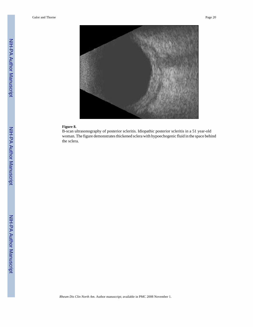
Figure 8.B-scan ultrasonography of posterior scleritis. Idiopathic posterior scleritis in a 51 year-oldwoman. The figure demonstrates thickened sclera with hypoechogenic fluid in the space behindthe sclera.
Galor and Thorne Page 20
Rheum Dis Clin North Am. Author manuscript; available in PMC 2008 November 1.
NIH
-PA Author Manuscript
NIH
-PA Author Manuscript
NIH
-PA Author Manuscript
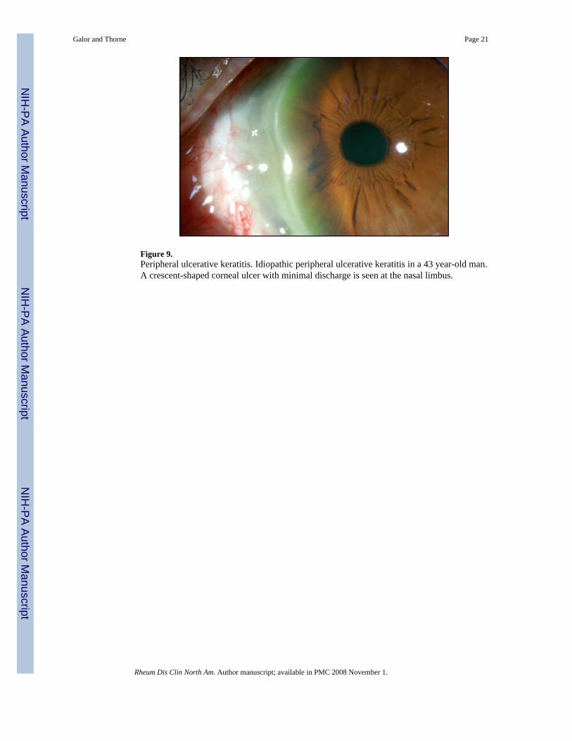
Figure 9.Peripheral ulcerative keratitis. Idiopathic peripheral ulcerative keratitis in a 43 year-old man.A crescent-shaped corneal ulcer with minimal discharge is seen at the nasal limbus.
Galor and Thorne Page 21
Rheum Dis Clin North Am. Author manuscript; available in PMC 2008 November 1.
NIH
-PA Author Manuscript
NIH
-PA Author Manuscript
NIH
-PA Author Manuscript

NIH
-PA Author Manuscript
NIH
-PA Author Manuscript
NIH
-PA Author Manuscript
Galor and Thorne Page 22
Table 1Differential diagnosis of scleritis.• Episcleritis
• Conjunctivitis
- Bacterial
- Viral
- Allergic
• Blepharitis
• Keratoconjunctivitis sicca
• Anterior uveitis
Rheum Dis Clin North Am. Author manuscript; available in PMC 2008 November 1.
NIH
-PA Author Manuscript
NIH
-PA Author Manuscript
NIH
-PA Author Manuscript
Galor and Thorne Page 23
Table 2Systemic Diseases Associated With Scleritis [1].
Autoimmune diseases Infections OtherRheumatoid arthritis* Viruses (Varicella zoster virus, herpes simplex virus, hepatitis C) Surgically inducedInflammatory bowel disease Bacteria Drug inducedRelapsing polychondritis Mycobacteria PamidronateSystemic lupus erythematosus Spirochetes (Treponema pallidum, Borrelia burgdorferi) FluvirinSpondyloarthropathies Fungi TraumaSarcoidosis AmeobaVasculitides Parasite Wegener’s granulomatosis* Polyarteritis nodosa Cryoglobulinemia Cogan’s Syndrome Hypocomplementemic urticarial vasculitis Temporal arteritis Takayasu arteritis*Most commonly associated conditions
Rheum Dis Clin North Am. Author manuscript; available in PMC 2008 November 1.

NIH
-PA Author Manuscript
NIH
-PA Author Manuscript
NIH
-PA Author Manuscript
Galor and Thorne Page 24
Table 3Clinical, Radiologic, and Laboratory Evaluation for Patients with Scleritis.
Standard Directed based on history and physical examinationComplete blood count Tuberculin skin testComplete metabolic panel Sacroiliac joint x-raysUrinalysis with microscopic analysis Sinus imagingAntineutrophil cytoplasmic antibody assayAntinuclear antibody assayRheumatoid factorAnti-cyclic citrullinated peptide antibodies
Viral hepatitis panel
Chest radiograph IgE levelsRapid plasma reagin Gastrointestinal evaluationFluorescent treponemal antibody absorption (FTA/Abs) assay Cultures for bacteria, virus, fungiLyme antibody Scleral biopsy
Rheum Dis Clin North Am. Author manuscript; available in PMC 2008 November 1.

NIH
-PA Author Manuscript
NIH
-PA Author Manuscript
NIH
-PA Author Manuscript
Galor and Thorne Page 25
Table 4Typical prednisone taper.
Dose Duration1 mg/kg (maximum 60 mg/day) 1 month40-60 mg/day Taper by 10 mg/week20-40 mg/day Taper by 5 mg/week10-20 mg/day Taper by 2.5 mg/week0-10 mg/day Taper by 1-2.5 mg/week
Rheum Dis Clin North Am. Author manuscript; available in PMC 2008 November 1.
NIH
-PA Author Manuscript
NIH
-PA Author Manuscript
NIH
-PA Author Manuscript
Galor and Thorne Page 26
Table 5Treatments for non-infectious scleritis.
Antimetabolites T cell inhibitors Alkylating agents BiologicsMethotrexate*(start 15 mg weekly)(max 25 mg weekly)
Cyclosporine**(start 2 mg/kg twice daily)(max 2.5 mg/kg twice daily)
Cyclophosphamide†(start up to 2 mg/kg daily)(max 200 mg/daily)
Infliximab**(start 5 mg/kg every 6 weeks)(max 10mg/kg every 4 weeks)
Azathioprine(start 2 mg/kg daily)(max 200 mg daily)
Tacrolimus(start 1 mg twice daily)(increase dose until therapeutic)
Chlorambucil(start 0.1 mg/kg daily)(max 12 mg/daily)
Rituximab(optimal dose uncertain)
Mycophenolate mofetil*(start 1 gram twice daily)(max 1.5 gram twice daily)*First line treatment in cases where inability to control disease on desired prednisone dose. Generally start for active disease on ≥10 mg prednisone/day
†First line agent in cases of systemic vasculitis
**Second line agents added if maximal tolerated dose of antimetabolite alone does not allow for control of disease on desired prednisone dose.
Rheum Dis Clin North Am. Author manuscript; available in PMC 2008 November 1.


![Understanding the relationship between diabetes, retinopathy, …Paper] uveitis scleritis and glycaemia.pdf · diffuse/nodular). It has been shown to cause vision loss (a permanent](https://static.fdocuments.us/doc/165x107/5ca0773788c99350178d3ca3/understanding-the-relationship-between-diabetes-retinopathy-paper-uveitis-scleritis.jpg)