NIH Public Access by Src or MAPK inhibition Cancer … · by Src or MAPK inhibition Zhen...
17
Cardiac glycosides inhibit p53 synthesis by a mechanism relieved by Src or MAPK inhibition Zhen Wang 1,*,† , Min Zheng 1,*,¶ , Zhichuan Li 2 , Ruiguo Li 1 , Lijun Jia 1 , Xiufang Xiong 1 , Noel Southall 3 , Shaomeng Wang 4 , Menghang Xia 3 , Christopher P. Austin 3 , Wei Zheng 3 , Zijian Xie 2 , and Yi Sun 1,# 1 Division of Radiation and Cancer Biology, Department of Radiation Oncology, University of Michigan Comprehensive Cancer Center, 4424B Medical Science 1, 1301 Catherine Street, Ann Arbor, MI 48109 2 Department of Physiology and Pharmacology, University of Toledo, Toledo, OH 43614 3 NIH Chemical Genomics Center, Bethesda, MD 20892 4 Department of Internal Medicine, University of Michigan Comprehensive Cancer Center, 4424B Medical Science 1, 1301 Catherine Street, Ann Arbor, MI 48109 Abstract p53 is regulated at the multiple levels. We report here that p53 in multiple lines of human cancer cells is down-regulated by cardiac glycoside drugs, digoxin or ouabain, the potent inhibitors of Na + /K + -ATPase. These drugs reduced the basal levels of p53 protein at nanomolar concentrations in a dose-, time- and cancer cell line-dependent manner, but independent of p53 status of wild type (wt) or mutant. The drugs also reduced the levels of p53 induced by its activators as well as p53 transfected into human cancer cells, regardless of its status. Interestingly, the drugs had no effect on endogenous p53 in two immortalized human cell lines. Mechanistically, p53 reduction did not occur at the mRNA levels, but at the protein levels, as a result of reduced protein synthesis rather than enhanced degradation. The cellular sensitivity to drug-induced p53 reduction was not associated with the levels of α subunits of Na + /K + -ATPase in different cell lines. While lowering extracellular K + did not reduce p53 as did ouabain and digoxin, it did potentiate both digoxin and ouabain-induced p53 reduction in sensitive lines. Finally, p53 reduction appears to be triggered by activation of Src/ MAPK signaling pathways upon drug binding to the Na + /K + -ATPase and can be completely blocked by the inhibitors of Src or MEK. This is the first report that cardiac glycoside drugs, by initiating the Src/MAPK signaling pathways, reduce the p53 levels via inhibition of p53 protein synthesis. The drugs may be useful in the treatment of human cancers with a gain-of-function p53 mutation. Keywords p53; Src/MAPK signaling pathways; cardiac glycosides; Na + /K + ATPase #Corresponding to Yi Sun, Department of Radiation Oncology, University of Michigan, 4424B Medical Science 1, 1301 Catherine Street, Ann Arbor, MI 48109, USA. Tel 734-615-1989, Fax 734-763-1581, [email protected]. * These authors contributed equally to the work. † Current address: Institute of Medicinal Biotechnology, Peking Union Medical College and Chinese Academy of Medical Sciences, Beijing 100050, China. ¶ Current address: Institute of Infectious Diseases, The First Affiliated Hospital, School of Medicine, Zhejiang University; State Key Laboratory for Diagnosis and Treatment of Infectious Diseases, 79 Qingchun Road, Hangzhou, Zhejiang, 310003. NIH Public Access Author Manuscript Cancer Res. Author manuscript; available in PMC 2010 August 15. Published in final edited form as: Cancer Res. 2009 August 15; 69(16): 6556–6564. doi:10.1158/0008-5472.CAN-09-0891. NIH-PA Author Manuscript NIH-PA Author Manuscript NIH-PA Author Manuscript
Transcript of NIH Public Access by Src or MAPK inhibition Cancer … · by Src or MAPK inhibition Zhen...
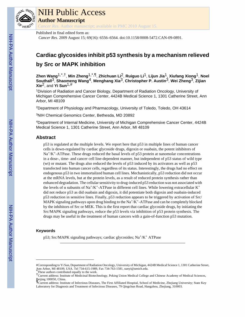
Cardiac glycosides inhibit p53 synthesis by a mechanism relievedby Src or MAPK inhibition
Zhen Wang1,*,†, Min Zheng1,*,¶, Zhichuan Li2, Ruiguo Li1, Lijun Jia1, Xiufang Xiong1, NoelSouthall3, Shaomeng Wang4, Menghang Xia3, Christopher P. Austin3, Wei Zheng3, ZijianXie2, and Yi Sun1,#1Division of Radiation and Cancer Biology, Department of Radiation Oncology, University ofMichigan Comprehensive Cancer Center, 4424B Medical Science 1, 1301 Catherine Street, AnnArbor, MI 481092Department of Physiology and Pharmacology, University of Toledo, Toledo, OH 436143NIH Chemical Genomics Center, Bethesda, MD 208924Department of Internal Medicine, University of Michigan Comprehensive Cancer Center, 4424BMedical Science 1, 1301 Catherine Street, Ann Arbor, MI 48109
Abstractp53 is regulated at the multiple levels. We report here that p53 in multiple lines of human cancercells is down-regulated by cardiac glycoside drugs, digoxin or ouabain, the potent inhibitors ofNa+/K+-ATPase. These drugs reduced the basal levels of p53 protein at nanomolar concentrationsin a dose-, time- and cancer cell line-dependent manner, but independent of p53 status of wild type(wt) or mutant. The drugs also reduced the levels of p53 induced by its activators as well as p53transfected into human cancer cells, regardless of its status. Interestingly, the drugs had no effect onendogenous p53 in two immortalized human cell lines. Mechanistically, p53 reduction did not occurat the mRNA levels, but at the protein levels, as a result of reduced protein synthesis rather thanenhanced degradation. The cellular sensitivity to drug-induced p53 reduction was not associated withthe levels of α subunits of Na+/K+-ATPase in different cell lines. While lowering extracellular K+
did not reduce p53 as did ouabain and digoxin, it did potentiate both digoxin and ouabain-inducedp53 reduction in sensitive lines. Finally, p53 reduction appears to be triggered by activation of Src/MAPK signaling pathways upon drug binding to the Na+/K+-ATPase and can be completely blockedby the inhibitors of Src or MEK. This is the first report that cardiac glycoside drugs, by initiating theSrc/MAPK signaling pathways, reduce the p53 levels via inhibition of p53 protein synthesis. Thedrugs may be useful in the treatment of human cancers with a gain-of-function p53 mutation.
Keywordsp53; Src/MAPK signaling pathways; cardiac glycosides; Na+/K+ ATPase
#Corresponding to Yi Sun, Department of Radiation Oncology, University of Michigan, 4424B Medical Science 1, 1301 Catherine Street,Ann Arbor, MI 48109, USA. Tel 734-615-1989, Fax 734-763-1581, [email protected].*These authors contributed equally to the work.†Current address: Institute of Medicinal Biotechnology, Peking Union Medical College and Chinese Academy of Medical Sciences,Beijing 100050, China.¶Current address: Institute of Infectious Diseases, The First Affiliated Hospital, School of Medicine, Zhejiang University; State KeyLaboratory for Diagnosis and Treatment of Infectious Diseases, 79 Qingchun Road, Hangzhou, Zhejiang, 310003.
NIH Public AccessAuthor ManuscriptCancer Res. Author manuscript; available in PMC 2010 August 15.
Published in final edited form as:Cancer Res. 2009 August 15; 69(16): 6556–6564. doi:10.1158/0008-5472.CAN-09-0891.
NIH
-PA Author Manuscript
NIH
-PA Author Manuscript
NIH
-PA Author Manuscript

Introductionp53 prevents tumor formation through transcriptional dependent and independent mechanisms.Transcriptional dependent mechanism is mainly mediated by p53 upregulation of itsdownstream targets. Upon activation by a variety of stimuli, p53 induces the expression of pro-arrest genes, such as p21, Gadd45 or 14-3-3σ to induce growth arrest or of pro-apoptotic genes,such as PUMA, PIG-3 or DR5 to induce apoptosis (1). Through a direct binding to mitochondriaand modulating BH3 family pro-apoptotic proteins, such as Bax, p53 can also regulateapoptosis in a transcriptional independent manner (2,3). Thus, p53 acts as a guardian of thegenome by inducing growth arrest to allow cells to repair the damage or apoptosis, if the damageis too severe and irreparable.
Since p53 plays a pivotal role in controlling abnormal cell growth and is inactivated by pointmutations in more than 50% human cancers, p53 has been a central target for mechanism-driven cancer drug discovery (4,5). Significant progress has been made in past decade, leadingto identification and characterization of several unique classes of small molecules that modulatep53 (6,7). They can be categorized as follows: 1) The molecules that restore wild type p53from a mutant conformation, which include CP-31398 (8), PRIMA-1 (9) and ellipticine (10).2) The molecules that target Mdm2 to reactivate p53, including Mdm2 E3 ubiquitin ligaseinhibitor, HLI98 (11) and inhibitors that disrupt Mdm2-p53 binding, such as Nutlin (12), RITA(13), and MI-219 and its analogues (14–16). 3) The molecules that inhibits wild type p53,including pifithrin-α (17) and pifithrin-mu (18). And 4) the molecules that selectively degrademutant p53, including Hsp90-active agents such as geldanamycin (19) and histone deacetylaseinhibitors, such as trichostatin (20).
During the screening for small molecules that selectively kill mutant-p53 containing cancercells via a synthetic lethal mechanism (21), we serendipitously found that cardiac glycosidedrugs, digoxin and ouabain, reduced the p53 levels in a time and dose dependent manner insensitive cancer cell lines. The drug sensitivity to p53 reduction is cancer cell line dependent,but independent of p53 status of a wild type or mutants. Importantly, the drugs are completelyinactive in reducing wt p53 in normal “immortalized” cells. Mechanistically, the drug-inducedp53 decrease occurred not at the mRNA levels, but at the protein levels, as a result of reducedsynthesis, rather than enhanced degradation. The drug induced p53 reduction can be rescuedby the inhibitors of Src and MEK, suggesting an involvement of Src/MAPK signalingpathways, initiated upon the drug binding to Na+/K+-ATPase. Our study revealed a novelmechanism by which activation of Src/MAPK kinase pathway could eventually lead to p53elimination by inhibiting p53 protein synthesis.
Materials and MethodsCell culture and drug treatment
All cell lines used in this study, except those mentioned below, were maintained in Dulbecco’smodified Eagle’s medium (DMEM) supplemented with 10% (v/v) fetal bovine serum. H1355,HCT116 and MRC5 were maintained in RPMI 1640 Medium and McCoy’s 5A medium,respectively, containing 10% serum. NL20 cells were cultured in Ham's F12 medium asdescribed (22). For drug treatment, subconfluent cells were treated with DG or OU alone or incombination of PP2, PD098059, or LY294002 (Sigma).
For culturing cells in low K+ conditions, A549 or H1355 cells were cultured in 10% DMEMuntil the cell densities reached 80%. The medium was replaced by K+-free DMEMsupplemented with normal K+ concentration (5 mM) or low K+ concentrations (1 mM or 0.3mM). Na+ was added to the low K+ medium to maintain the equal ion concentrations (23).
Wang et al. Page 2
Cancer Res. Author manuscript; available in PMC 2010 August 15.
NIH
-PA Author Manuscript
NIH
-PA Author Manuscript
NIH
-PA Author Manuscript

Western blotting analysisThe assay was performed as described previously (16). The antibodies used are p53(Calbiochem, CA, 1:1000), p21 (BD, 1:1000), Mdm2 (Calbiochem, CA, 1:500), FAK, Src ortubulin (Santa Cruz Biotechnology, 1:1000), Src-pY418 (Invitrogen, CA, 1:1000); pERK1/2(Cell Signaling) and β-actin (Sigma, St. Louis, MO 1:5000).
Transfection and infectionH1299 cells were plated into six-well plate at 2 ×10⁵ cells per well and transfected the followingday with 1 µg of plasmid expressing either p53-wt or p53-mutants for 24 h prior to drugtreatment. For the infection of sh-RNA targeting FAK viruses (gift from Junlin-Guan), a totalof 109 pfu viruses were infected into 2 ×10⁶ A549 or H1355 cells per 100 mm dish for 72h.Cells were split, followed by exposure to drugs next day for 24 hrs. To silence endogenous wtp53, A549 or H460 cells were infected with a lenti-virus based siRNA construct, LT-p53-siRNA, as described (16).
ATPlite growth assay and IC50 determinationCells were seeded in 96 well white plates and treated with the drugs with a range of indicatedconcentrations for 24 hrs. The viability of the cells was then measured using a one-step ATPlitekit (Perkin Elmer). The results were calculated and plotted in Prism 4.0 (Graphpad) to generateIC50 curves (22).
Quantitative RT-PCRTotal RNA was isolated from cells post drug treatment, using a Trizol kit (Invitrogen, Carlsbad,CA), and subjected to quantitative RT-PCR analysis, using QuantiTect SYBR green RT-PCRkit (Qiagen). Briefly, 50 µl reaction mixture was used for each reaction, which contained 2xQuantiTect SYBR Green RT-PCR Master Mix, 0.5 µl QuantiTect RT mix, 0.5 µM primer mixand 0.5 µg RNA. Cycling program was set as the following: 50 °C 30 min for RT, 95 °C 15min for the PCR initial activation and 40 cycles of denaturation at 94 °C for 15 sec, annealingat 55 °C for 30 sec and extension at 72 °C for 30 sec. The sequences of p53 and GAPDH areas follows: hu-p53 F1□TCTGTGACTTGCACGTAC; hu-p53 R1:ATTTCCTTCCACTCGGAT. GAPDH-F1: GTTGCCATCAATGACCCCTT; GAPDH -R1:AGAGGCAGGGATGATGTTCT.
35S-Met metabolic labelingSubconfluent cells were treated with the drugs for various time points with last hr in methionineand cysteine-free DMEM, containing 5% dialyzed FCS and 50 µM MG132. Cells were thenlabeled with 100 µCi/ml of [35S]-methionine (MP Biochemicals) for 5 min, followed byimmunoprecipitation with anti-p53 antibody (Santa Cruz). Immunoprecipitates, along withwhole cell extract, were then subjected to SDS-PAGE and autoradiography. The steady-statelevels of p53 were measured by immunoprecipitated, followed by Western blotting, along withthe detection of total cellular proteins by Coomassie staining.
ResultsCardiac glycosides reduced the basal levels of p53 in lung cancer cell lines
During our confirmation of candidates identified from a chemical library screen for selectivekilling of cancer cells with mutant p53 via synthetic lethal mechanism (21), we serendipitouslyfound that cardiac glycosides, including digoxin (DG) and ouabain (OU) (Figure 1A forstructure) are able to reduce the p53 levels. As shown in Figure 1B, DG or OU at the nanomolarconcentrations reduced or eliminated the basal levels of p53 in two lung cancer lines, A549with a wild type (wt) p53 and H1355 with a mutant p53, with OU being more potent. DG
Wang et al. Page 3
Cancer Res. Author manuscript; available in PMC 2010 August 15.
NIH
-PA Author Manuscript
NIH
-PA Author Manuscript
NIH
-PA Author Manuscript

induced p53 reduction or elimination was the dose-dependent, but the p53 status independentin four lung cancer cell lines with either wt or mutant p53 status (Figure 1C). Furthermore, p53reduction is treatment-time dependent, starting to occur at 8 hrs with a complete eliminationseen by OU at 16-hrs post drug exposure (Figure 1D). Finally, cell line dependent, but p53status-independent p53 reduction or elimination by DG or OU can be extended to multiplehuman cancer cell lines. These include colon cancer lines, DLD1, but not HCT116, nor HT29;breast cancer lines, MCF7, but not MDA-MB231; all three head and neck squamous carcinomalines, but not three glioblastoma lines tested (Supplemental materials, Figure S1). Digoxigenin,another cardiac glycoside was also active, but 10-fold less potent in reducing p53 levels inmultiple human cancer lines (data not shown). Our results clearly demonstrated that p53 levelsin multiple human cancer cell lines are subjected to reduction or elimination by cardiacglycosides, DG or OU in a cell line-dependent, but p53 status-independent manner.
Cardiac glycosides blocked p53 induction and p53 upregulation of its target genes by p53activators
We next determined if DG or OU also reduced the levels of induced p53 and p53 transactivationof its target genes by known p53 activators. Three lung cancer cell lines were treated for 24 hrwith MI-219, an Mdm2 inhibitor, which activates p53 by disrupting Mdm2-p53 binding (14)or etoposide, a DNA damaging agent, known to activate p53 in wt p53-containing lung cancercells (16), alone or in combination with DG or OU, respectively. As shown in Figure 2A,MI-219 or etoposide treatment induced p53 levels as well as p53 target proteins, p21 or Mdm2(by MI-219 only) in two wt p53-containing lines, A549 and H460, but not in mutant p53-containing line, H1355 (lanes 1–3). Combinational treatment with either DG or OU reducedthe induced levels of p53 and completely eliminated p53 induction of its targets, p21 and Mdm2in A549 and H460 cells (lanes 4–9). The mutant p53 in H1355 was not subjected to inductionby two p53 activators, but was completely eliminated in combinational treatment (bottompanel).
p53 can be stabilized by hypoxia in wt p53 containing MCF7 cells by chemical hypoxic agents,cobalt chloride and desferrioxamine (24). We next determined if hypoxia-stabilized p53 is alsosubjected to reduction by DG or OU. As shown in Figure 2B, A 24 hr treatment of cobaltchloride stabilized p53 up to 2-fold in MCF7 cells. This stabilized p53 was reduced andeliminated by simultaneous treatment of DG or OU in a dose-dependent manner up to 300 nM(lanes 3–8). Along with p53 reduction, two p53 target proteins, p21 and Mdm2, which weredetectable at the basal levels due to a relatively higher level of p53, were also reduced andeliminated. Taken together, these results indicate that both cardiac glycosides also reduced thelevels of p53 and its target proteins upon p53 activation and stabilization. The results furthersuggest that drug-induced p53 reduction may be Mdm2-independent, since Mdm2 is free fromp53 binding upon MI-219 treatment (14).
Cardiac glycosides reduced the levels of transfected p53 in cancer cells, but had no effecton endogenous wt p53 in normal cells
We next determined if p53 upon over-expression in a p53-null H1299 cells would alsosubjected to DG or OU reduction. H1299 cells over-expressing a temperature sensitive p53mutant (codon 138) (25) were treated with DG for 24 hrs at 37°C (p53 adopts a mutantconformation) or 32°C (wt conformation). As shown in Figure 3A, DG treatment caused adose-dependent reduction of over-expressed p53, regardless of p53 status. Upon p53 reduction,p53 target genes, p21 and Mdm2, induced at 32°C when p53 is in a wild type status, were alsoreduced accordingly. Likewise, DG reduced the levels of p53 transiently transfected intoH1299 cells regardless of the p53 status of wild type or two mutants, R248W and R273H, mostfrequently found in human cancer (Figure 3B). However, the potency of drugs against over-expressed p53 was much reduced with much high drug doses up to 10 µM to achieve a moderate
Wang et al. Page 4
Cancer Res. Author manuscript; available in PMC 2010 August 15.
NIH
-PA Author Manuscript
NIH
-PA Author Manuscript
NIH
-PA Author Manuscript
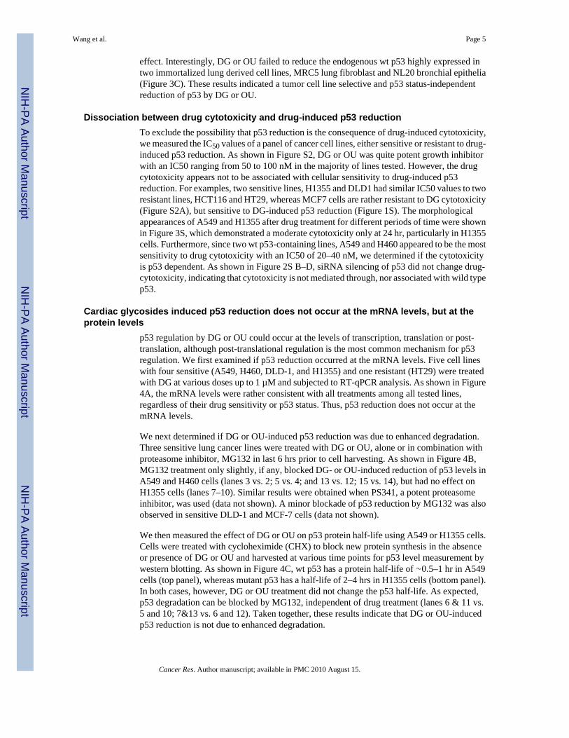
effect. Interestingly, DG or OU failed to reduce the endogenous wt p53 highly expressed intwo immortalized lung derived cell lines, MRC5 lung fibroblast and NL20 bronchial epithelia(Figure 3C). These results indicated a tumor cell line selective and p53 status-independentreduction of p53 by DG or OU.
Dissociation between drug cytotoxicity and drug-induced p53 reductionTo exclude the possibility that p53 reduction is the consequence of drug-induced cytotoxicity,we measured the IC50 values of a panel of cancer cell lines, either sensitive or resistant to drug-induced p53 reduction. As shown in Figure S2, DG or OU was quite potent growth inhibitorwith an IC50 ranging from 50 to 100 nM in the majority of lines tested. However, the drugcytotoxicity appears not to be associated with cellular sensitivity to drug-induced p53reduction. For examples, two sensitive lines, H1355 and DLD1 had similar IC50 values to tworesistant lines, HCT116 and HT29, whereas MCF7 cells are rather resistant to DG cytotoxicity(Figure S2A), but sensitive to DG-induced p53 reduction (Figure 1S). The morphologicalappearances of A549 and H1355 after drug treatment for different periods of time were shownin Figure 3S, which demonstrated a moderate cytotoxicity only at 24 hr, particularly in H1355cells. Furthermore, since two wt p53-containing lines, A549 and H460 appeared to be the mostsensitivity to drug cytotoxicity with an IC50 of 20–40 nM, we determined if the cytotoxicityis p53 dependent. As shown in Figure 2S B–D, siRNA silencing of p53 did not change drug-cytotoxicity, indicating that cytotoxicity is not mediated through, nor associated with wild typep53.
Cardiac glycosides induced p53 reduction does not occur at the mRNA levels, but at theprotein levels
p53 regulation by DG or OU could occur at the levels of transcription, translation or post-translation, although post-translational regulation is the most common mechanism for p53regulation. We first examined if p53 reduction occurred at the mRNA levels. Five cell lineswith four sensitive (A549, H460, DLD-1, and H1355) and one resistant (HT29) were treatedwith DG at various doses up to 1 µM and subjected to RT-qPCR analysis. As shown in Figure4A, the mRNA levels were rather consistent with all treatments among all tested lines,regardless of their drug sensitivity or p53 status. Thus, p53 reduction does not occur at themRNA levels.
We next determined if DG or OU-induced p53 reduction was due to enhanced degradation.Three sensitive lung cancer lines were treated with DG or OU, alone or in combination withproteasome inhibitor, MG132 in last 6 hrs prior to cell harvesting. As shown in Figure 4B,MG132 treatment only slightly, if any, blocked DG- or OU-induced reduction of p53 levels inA549 and H460 cells (lanes 3 vs. 2; 5 vs. 4; and 13 vs. 12; 15 vs. 14), but had no effect onH1355 cells (lanes 7–10). Similar results were obtained when PS341, a potent proteasomeinhibitor, was used (data not shown). A minor blockade of p53 reduction by MG132 was alsoobserved in sensitive DLD-1 and MCF-7 cells (data not shown).
We then measured the effect of DG or OU on p53 protein half-life using A549 or H1355 cells.Cells were treated with cycloheximide (CHX) to block new protein synthesis in the absenceor presence of DG or OU and harvested at various time points for p53 level measurement bywestern blotting. As shown in Figure 4C, wt p53 has a protein half-life of ∼0.5–1 hr in A549cells (top panel), whereas mutant p53 has a half-life of 2–4 hrs in H1355 cells (bottom panel).In both cases, however, DG or OU treatment did not change the p53 half-life. As expected,p53 degradation can be blocked by MG132, independent of drug treatment (lanes 6 & 11 vs.5 and 10; 7&13 vs. 6 and 12). Taken together, these results indicate that DG or OU-inducedp53 reduction is not due to enhanced degradation.
Wang et al. Page 5
Cancer Res. Author manuscript; available in PMC 2010 August 15.
NIH
-PA Author Manuscript
NIH
-PA Author Manuscript
NIH
-PA Author Manuscript

We finally determined if DG or OU could inhibit p53 protein synthesis. Two drug sensitivelines A549 and H1355 were used and newly synthesized p53 was measured by 35S-methioninelabeling in the presence of MG132 to block p53 degradation. As shown in Figure 4D, in bothlines DG or OU treatment induced a time-dependent inhibition of de novo p53 protein synthesiswith a complete elimination of p53 synthesis at 2.5 or 3 hrs post treatment, respectively (toppanel). The drugs also caused a time dependent inhibition of overall de novo protein synthesisbut to less extent (second panel with quantification data shown on the right). In contrast, thesame treatment did not change the steady-state levels of p53 (third panel), nor the total cellularproteins (bottom panel). The drug also inhibited de novo synthesis of p53 in an additionalsensitive line, MCF7 (data not shown). Thus, DG or OU-induced p53 reduction is not due toenhanced degradation, rather due to inhibited protein synthesis.
Cardiac glycosides-induced p53 reduction is independent of the levels of α subunits ofNa+/K+-ATPase, but is enhanced by lowering extracellular K+
The cardiac glycosides target for treatment of congestive heart failure is the Na+/K+-ATPase(26). We determine if the levels of Na+/K+-ATPase α subunits are associated with cellularsensitivity to OU-induced p53 reduction. As depicted in Figure 5A, Western blot analysesreveal that both OU-sensitive and resistant cells express α1 and α3. Apparently, the cellularsensitivity of OU-induced p53 reduction did not correlate with the basal levels of Na+/K+-ATPase in these cells. To test if inhibition of Na+/K+-ATPase by means other than cardiacglycosides is sufficient to reduce p53, we lowered extracellular K+ from 5 mM to 1 and 0.3mM (27), and measured for p53. As shown in Figure 5B, reduction of potassium concentrationhad no effect on the basal levels of p53 (lanes 1–3). Interestingly, DG- or OU-induced p53reduction was more pronounced progressively, consistent with the fact that loweringextracellular K+ increases DG- and OU-binding to the Na+/K+-ATPase. These results clearlyshowed that DG- or OU-induced p53 reduction is promoted by the binding of these drugs tothe Na+/K+-ATPase.
Cardiac glycosides reduced p53 levels via triggering and activating Src/MAPK signalingpathways
It has been proposed that the Na+/K+-ATPase is preassembled with its partners in caveolae;the binding of OU or DG to the pump activates the signalosome to transduce the signals viamultiple pathways, including Src, FAK, MAPK and PI-3K (28–30). We first determined if DGor OU treatment would activate Src or MAPK in sensitive A549 and H1355 cells. As shownin Figure 6A, a short treatment of cells with DG or OU for 5 or 15 min caused Src and MAPKactivation, as demonstrated by increased phosphorylation at the activation sites (pSrc-Y418and pERKs-T183/Y185). We then determined the effect of Src and FAK inhibition on drug-induced p53 reduction, since Src and FAK are two upstream molecules activated upon OU- orDG-pump binding (29). In both A549 and H1355 cells, a potent Src tyrosine kinase inhibitorPP2 (31) blocked the p53 reduction by DG or OU in a dose dependent manner (Figure 6B,lanes 3–5 vs. 1; 7–9 vs. 6 and 12–15 vs. 10; 17 vs. 16, respectively), while the inhibitor itselfhad no effect on the p53 level (lane 15 and data not shown). On the other hand, siRNA silencingof FAK in either A549 or H1355 cells had no effect on p53 reduction by DG or OU(Supplemental Figure S4). We, therefore, focused our attention on Src downstream pathways,particularly MAPK and PI3K pathways using specific inhibitors to determine if they blockedp53 reduction. Indeed, while PD098059, a MEK inhibitor that blocks MAPK pathway orLY294002, a PI3K inhibitor had no effect on the levels of p53 by drug itself (Figure 6C, lanes2&3 vs. 1 and lanes 8 vs. 7 and 14 vs. 13), both inhibitors were able to rescue the p53 reductionby DG in A549 cells (Figure 6C, lanes 5&6 vs. 4). Only MEK inhibitor but not PI3K inhibitor,partially rescued p53 reduction in H1355 cells (Figure 6C, 10–12 vs. 9 and 16 vs. 5). Takentogether, these results demonstrated that activation of SRC/MAPK/PI3K signaling pathwaystriggered by the binding of cardiac glycosides to the pump is responsible for p53 reduction.
Wang et al. Page 6
Cancer Res. Author manuscript; available in PMC 2010 August 15.
NIH
-PA Author Manuscript
NIH
-PA Author Manuscript
NIH
-PA Author Manuscript

While the SRC activation mediates p53 reduction in both cell lines, MAPK and PI3K areinvolved in A549 cells, whereas MAPK, but not PI3K, is partially involved in H1355 cells.
DiscussionCardiac glycosides are a class of natural products that have been used for medical purposessince ancient time. Three well-known cardiac glycosides, digoxin (DG), ouabain (OU) anddigitoxin were used for the treatment of congestive heart failure and atrial fibrillation viabinding and inhibiting Na+/K+ ATPase to increase intracellular calcium concentrations (28).In addition to benefit heart failure patients, the drugs were also found to be beneficial to breastcancer patients (32), and were associated with a lower risk for leukemia, lymphoma as well askidney and urinary tract cancer (33). Accumulated data in past few years have shown thatcardiac glycosides selectively inhibited proliferation and induced apoptosis and autophagy incancer cells, but not normal cells, suggesting their utility in anticancer therapy [(34) and forreview, see (28)]. Mechanistically, DG or OU inhibited catalytic activity of topoisomerase II(35) and stabilized DNA-topoisomerase II complexes to suppress growth. DG, OU or othercardiac glycosides up-regulated death receptor 4 and 5 to sensitize lung cancer cells to apoptosisinduced by Apo2L/TRAIL (36). DG or OU also remarkably inhibit protein synthesis ofHIF-1α to block HIF1 transcription factor activity and to inhibit tumor cell growth both invitro and in vivo (37). Furthermore, DG or OU could modulate signaling pathways of MAPK/AKT, PKC/AP-1, NF-κB, and reactive oxygen species to regulate cell growth and survival [forreviews, see (26,28)]. Finally, globe level reduction of protein synthesis upon drug exposure,as shown in our study (Fig. 4D), could also contribute to their cytotoxicity. However, the roleof p53 in the action of cardiac glycosides is totally unknown, although a recent study showedan observation that OU slightly reduced p53 level in a breast cancer line, MDA-MB-435swithout providing any mechanistic insight (38).
DG was identified in our chemical library screening for the drugs that selectively kill cancercells with mutant p53 via synthetic lethal mechanism (21). Subsequent analysis serendipitouslyfound that the drug effectively reduces p53 levels in a cell line-dependent, but p53 statusindependent manner. It is well-known that p53 is a short-lived protein whose expression wasregulated mainly at the post-translational levels, including phosphorylation, acetylation,ubiquitination, sumolyation and methylation (39–41). Recently, p53 is shown to be regulatedat the translational level on protein synthesis by ribosomal protein L26 (42), mTOR (43) orMdm2. Interestingly, Mdm2 could either stimulate p53 synthesis via a direct binding to p53mRNA (44) or inhibit p53 synthesis by promoting the degradation of L26 (45). We found thatthe drug-induced p53 reduction did not occur at the mRNA level, as demonstrated by RT-qPCRanalysis, but at the protein level. Failure to rescue drug-induced p53 reduction by proteasomeinhibitors and failure of drugs to shorten the p53 protein half-life indicate that p53 reductionby the drugs is not due to enhanced p53 degradation. The drug inhibition on metabolic labelingof newly synthesized p53 suggests that the change occur at the level of protein synthesis. Thus,DG and OU are potent inhibitors of p53 protein synthesis.
What is then the mechanism by which DG or OU inhibits the de novo p53 protein synthesis?Although the details are still unknown at the present time, it appears to involve Src/MAPKsignaling pathways, triggered and activated by OU/DG binding to Na+/K+ ATPase, since 1)OU or DG activates Src and MAPK, 2) their inhibitors are able to abrogate the OU/DG-inducedp53 reduction, whereas 3) inhibition of Na+/K+-ATPase activity by lowering extracellularK+ has no effect. Thus, a simplest explanation for these observations is that upon DG or OUbinding to Na+/K+-ATPase, Src/MAPK signaling pathways are activated particularly in drug-sensitive cancer cells, leading to activation of their down-stream effectors, eventually theinhibition of p53 protein synthesis. Future effort will be directed 1) to characterize which typeof p53 synthesis; cap-dependent and/or cap-independent, also known as internal ribosome entry
Wang et al. Page 7
Cancer Res. Author manuscript; available in PMC 2010 August 15.
NIH
-PA Author Manuscript
NIH
-PA Author Manuscript
NIH
-PA Author Manuscript

site (IRES) element dependent (46), is actually inhibited by cardiac glycosides and 2) toelucidate the mechanism of action.
Another interesting observation reported here is that cardiac glycosides, DG and OU, are potentcytotoxic agents in a panel of tumor cell lines with IC50s ranging from 50 to 100 nM. However,the cytotoxicity is tumor cell line dependent, but independent of wild type p53, dissociatingcancer cell killing from wild type p53 elimination. Although wild type p53 induces growtharrest and apoptosis in most cases (1,6,7), and acts as a survival protein in some particular cases[for review, see (47)], it is unlikely that wild type p53 plays any significant role in DG or OU-induced cell killing in few cancer cell lines tested. Nevertheless, the fact that cardiac glycosidesare inactive against wild type p53 in normal cells, but potently active in elimination of mutantp53 in some cancer cells, suggests that these drugs could have utility in the treatment of humancancer harboring a gain-of-function p53 mutant (48,49).
Supplementary MaterialRefer to Web version on PubMed Central for supplementary material.
AcknowledgementsWe would like to thank Dr. Jun-Lin Guan at the University of Michigan for providing us the Ad-sh-RNA virus silencingFAK as well as FAK antibody (50). This work was supported by the NCI grants (CA111554 and CA118762) andDOD concept Award (W81XWH-08-1-0539) to YS.
References1. Vogelstein B, Lane D, Levine AJ. Surfing the p53 network. Nature 2000;408:307–310. [PubMed:
11099028]2. Chipuk JE, Kuwana T, Bouchier-Hayes L, et al. Direct activation of Bax by p53 mediates mitochondrial
membrane permeabilization and apoptosis. Science 2004;303:1010–1014. [PubMed: 14963330]3. Mihara M, Erster S, Zaika A, et al. p53 has a direct apoptogenic role at the mitochondria. Mol Cell
2003;11:577–590. [PubMed: 12667443]4. Bouchet BP, de Fromentel CC, Puisieux A, Galmarini CM. p53 as a target for anti-cancer drug
development. Crit Rev Oncol Hematol 2006;58:190–207. [PubMed: 16690321]5. Sun Y. p53 and its downstream proteins as molecular targets of cancer. Mol Carcinog 2006;45:409–
415. [PubMed: 16652354]6. Bassett EA, Wang W, Rastinejad F, El-Deiry WS. Structural and functional basis for therapeutic
modulation of p53 signaling. Clin Cancer Res 2008;14:6376–6386. [PubMed: 18927276]7. Vazquez A, Bond EE, Levine AJ, Bond GL. The genetics of the p53 pathway, apoptosis and cancer
therapy. Nat Rev Drug Discov 2008;7:979–987. [PubMed: 19043449]8. Foster BA, Coffey HA, Morin MJ, Rastinejad F. Pharmacological rescue of mutant p53 conformation
and function. Science 1999;286:2507–2510. [PubMed: 10617466]9. Bykov VJ, Issaeva N, Shilov A, et al. Restoration of the tumor suppressor function to mutant p53 by
a low- molecular-weight compound. Nat Med 2002;8:282–288. [PubMed: 11875500]10. Peng Y, Li C, Chen L, Sebti S, Chen J. Rescue of mutant p53 transcription function by ellipticine.
Oncogene 2003;22:4478–4487. [PubMed: 12881704]11. Yang Y, Ludwig RL, Jensen JP, et al. Small molecule inhibitors of HDM2 ubiquitin ligase activity
stabilize and activate p53 in cells. Cancer Cell 2005;7:547–559. [PubMed: 15950904]12. Vassilev LT, Vu BT, Graves B, et al. In vivo activation of the p53 pathway by small-molecule
antagonists of MDM2. Science 2004;303:844–848. [PubMed: 14704432]13. Issaeva N, Bozko P, Enge M, et al. Small molecule RITA binds to p53, blocks p53-HDM-2 interaction
and activates p53 function in tumors. Nat Med 2004;10:1321–1328. [PubMed: 15558054]14. Shangary S, Qin D, McEachern D, Liu M, Miller RS, Qiu S, Nikolovska-Coleska Z, Ding K, Wang
G, Chen J, Bernard D, Zhang J, Lu Y, Gu Q, Shah RB, Pienta KJ, Ling X, Kang K, Guo M, Sun Y,
Wang et al. Page 8
Cancer Res. Author manuscript; available in PMC 2010 August 15.
NIH
-PA Author Manuscript
NIH
-PA Author Manuscript
NIH
-PA Author Manuscript

Yang D, Wang S. Temporal activation of p53 by a specific MDM2 inhibitor is selectively toxic totumors and leads to complete tumor growth inhibition PNAS. 2008;105:3933–3938.
15. Shangary S, Ding K, Qiu S, et al. Reactivation of p53 by a specific MDM2 antagonist (MI-43) leadsto p21-mediated cell cycle arrest and selective cell death in colon cancer. Mol Cancer Ther2008;7:1533–1542. [PubMed: 18566224]
16. Sun SH, Zheng M, Ding K, Wang S, Sun Y. A small molecule that disrupts Mdm2-p53 bindingactivates p53, induces apoptosis, and sensitizes lung cancer cells to chemotherapy. Cancer Biol Ther2008;7:845–852. [PubMed: 18340116]
17. Komarov PG, Komarova EA, Kondratov RV, et al. A chemical inhibitor of p53 that protects micefrom the side effects of cancer therapy. Science 1999;285:1733–1737. [PubMed: 10481009]
18. Strom E, Sathe S, Komarov PG, et al. Small-molecule inhibitor of p53 binding to mitochondriaprotects mice from gamma radiation. Nat Chem Biol 2006;2:474–479. [PubMed: 16862141]
19. Blagosklonny MV, Toretsky J, Neckers L. Geldanamycin selectively destabilizes andconformationally alters mutated p53. Oncogene 1995;11:933–939. [PubMed: 7675452]
20. Blagosklonny MV, Trostel S, Kayastha G, et al. Depletion of mutant p53 and cytotoxicity of histonedeacetylase inhibitors. Cancer Res 2005;65:7386–7392. [PubMed: 16103091]
21. Kaelin WG. The concept of synthetic lethality in the context of anticancer therapy. Nat Rev Cancer2005;5:689–698. [PubMed: 16110319]
22. Zheng M, Morgan-Lappe SE, Yang J, et al. Growth inhibition and radiosensitization of glioblastomaand lung cancer cells by small interfering RNA silencing of tumor necrosis factor receptor-associatedfactor 2. Cancer Res 2008;68:7570–7578. [PubMed: 18794145]
23. Bowen JW, McDonough A. Pretranslational regulation of Na-K-ATPase in cultured canine kidneycells by low K+ Am J Physiol 1987;252:C179–C189. [PubMed: 3030119]
24. An WG, Kanekal M, Simon MC, Maltepe E, Blagosklonny MV, Neckers LM. Stabilization of wild-type p53 by hypoxia-inducible factor 1alpha. Nature 1998;392:405–408. [PubMed: 9537326]
25. Robinson M, Jiang P, Cui J, Li J, Wang Y, Swaroop M, Madore S, Lawrence TS, Sun Y. GlobalGenechip profiling to identify genes responsive to p53-induced growth arrest and apoptosis in humanlung carcinomas. Cancer Biol Therapy 2003;2:406–415.
26. Schoner W, Scheiner-Bobis G. Endogenous and exogenous cardiac glycosides: their roles inhypertension, salt metabolism, and cell growth. Am J Physiol Cell Physiol 2007;293:C509–C536.[PubMed: 17494630]
27. Tepperman K, Millette LA, Johnson CL, Jewell-Motz EA, Lingrel JB, Wallick ET. Mutationalanalysis of Glu-327 of Na(+)-K(+)-ATPase reveals stimulation of 86Rb+ uptake by external K+ AmJ Physiol 1997;273:C2065–C2079. [PubMed: 9435514]
28. Newman RA, Yang P, Pawlus AD, Block KI. Cardiac glycosides as novel cancer therapeutic agents.Mol Interv 2008;8:36–49. [PubMed: 18332483]
29. Xie Z, Cai T. Na+-K+--ATPase-mediated signal transduction: from protein interaction to cellularfunction. Mol Interv 2003;3:157–168. [PubMed: 14993422]
30. Liang M, Cai T, Tian J, Qu W, Xie ZJ. Functional characterization of Src-interacting Na/K-ATPaseusing RNA interference assay. J Biol Chem 2006;281:19709–19719. [PubMed: 16698801]
31. Hanke JH, Gardner JP, Dow RL, et al. Discovery of a novel, potent, and Src familyselective tyrosinekinase inhibitor Study of Lck- and FynT-dependent T cell activation. J Biol Chem 1996;271:695–701. [PubMed: 8557675]
32. Stenkvist B, Bengtsson E, Eriksson O, Holmquist J, Nordin B, Westman-Naeser S. Cardiac glycosidesand breast cancer. Lancet 1979;1:563. [PubMed: 85158]
33. Haux J, Klepp O, Spigset O, Tretli S. Digitoxin medication and cancer; case control and internal dose-response studies. BMC Cancer 2001;1:11. [PubMed: 11532201]
34. Newman RA, Kondo Y, Yokoyama T, et al. Autophagic cell death of human pancreatic tumor cellsmediated by oleandrin, a lipid-soluble cardiac glycoside. Integr Cancer Ther 2007;6:354–364.[PubMed: 18048883]
35. Bielawski K, Winnicka K, Bielawska A. Inhibition of DNA topoisomerases I and II, and growthinhibition of breast cancer MCF-7 cells by ouabain, digoxin and proscillaridin A. Biol Pharm Bull2006;29:1493–1497. [PubMed: 16819197]
Wang et al. Page 9
Cancer Res. Author manuscript; available in PMC 2010 August 15.
NIH
-PA Author Manuscript
NIH
-PA Author Manuscript
NIH
-PA Author Manuscript

36. Frese S, Frese-Schaper M, Andres AC, Miescher D, Zumkehr B, Schmid RA. Cardiac glycosidesinitiate Apo2L/TRAIL-induced apoptosis in non-small cell lung cancer cells by up-regulation ofdeath receptors 4 and 5. Cancer Res 2006;66:5867–5874. [PubMed: 16740726]
37. Zhang H, Qian DZ, Tan YS, et al. Digoxin and other cardiac glycosides inhibit HIF-1alpha synthesisand block tumor growth. Proc Natl Acad Sci U S A 2008;105:19579–19586. [PubMed: 19020076]
38. Kometiani P, Liu L, Askari A. Digitalis-induced signaling by Na+/K+-ATPase in human breast cancercells. Mol Pharmacol 2005;67:929–936. [PubMed: 15602003]
39. Bode AM, Dong Z. Post-translational modification of p53 in tumorigenesis. Nat Rev Cancer2004;4:793–805. [PubMed: 15510160]
40. Brooks CL, Gu W. Ubiquitination, phosphorylation and acetylation: the molecular basis for p53regulation. Curr Opin Cell Biol 2003;15:164–171. [PubMed: 12648672]
41. Scoumanne A, Chen X. Protein methylation: a new mechanism of p53 tumor suppressor regulation.Histol Histopathol 2008;23:1143–1149. [PubMed: 18581285]
42. Takagi M, Absalon MJ, McLure KG, Kastan MB. Regulation of p53 translation and induction afterDNA damage by ribosomal protein L26 and nucleolin. Cell 2005;123:49–63. [PubMed: 16213212]
43. Lee CH, Inoki K, Karbowniczek M, et al. Constitutive mTOR activation in TSC mutants sensitizescells to energy starvation and genomic damage via p53. EMBO J 2007;26:4812–4823. [PubMed:17962806]
44. Candeias MM, Malbert-Colas L, Powell DJ, et al. p53 mRNA controls p53 activity by managingMdm2 functions. Nat Cell Biol. 2008PubMed ID18690233
45. Ofir-Rosenfeld Y, Boggs K, Michael D, Kastan MB, Oren M. Mdm2 regulates p53 mRNA translationthrough inhibitory interactions with ribosomal protein L26. Mol Cell 2008;32:180–189. [PubMed:18951086]
46. Halaby MJ, Yang DQ. p53 translational control: a new facet of p53 regulation and its implication fortumorigenesis and cancer therapeutics. Gene 2007;395:1–7. [PubMed: 17395405]
47. Janicke RU, Sohn D, Schulze-Osthoff K. The dark side of a tumor suppressor: anti-apoptotic p53.Cell Death Differ 2008;15:959–976. [PubMed: 18356920]
48. Di Agostino S, Strano S, Emiliozzi V, et al. Gain of function of mutant p53: the mutant p53/NF-Yprotein complex reveals an aberrant transcriptional mechanism of cell cycle regulation. Cancer Cell2006;10:191–202. [PubMed: 16959611]
49. Song H, Hollstein M, Xu Y. p53 gain-of-function cancer mutants induce genetic instability byinactivating ATM. Nat Cell Biol 2007;9:573–580. [PubMed: 17417627]
50. Peng X, Wu X, Druso JE, et al. Cardiac developmental defects and eccentric right ventricularhypertrophy in cardiomyocyte focal adhesion kinase (FAK) conditional knockout mice. Proc NatlAcad Sci U S A 2008;105:6638–6643. [PubMed: 18448675]
Wang et al. Page 10
Cancer Res. Author manuscript; available in PMC 2010 August 15.
NIH
-PA Author Manuscript
NIH
-PA Author Manuscript
NIH
-PA Author Manuscript

Figure 1. Cardiac glycosides, digoxin (DG) and ouabain (OU) reduces the basal p53 levels in lungcancer cell lines: Dose- and time-dependent: (A)Structure of DG and OU. (B) DG or OU reduces p53 levels: A549 and H1355 lung cancercells were treated with DG or OU at 100 nM for 24 hrs and subjected to western blotting. (C)Does dependent reduction of p53 levels by DG: Four lung cancer lines were treated with DGat 30 or 100 nM for 24 hrs and subjected to western blotting. (D) Time dependent reductionof p53 levels by DG or OU: H460 and H1355 cells were treated with DG or OU at 100 nMfor indicated periods of time, followed by western blotting.
Wang et al. Page 11
Cancer Res. Author manuscript; available in PMC 2010 August 15.
NIH
-PA Author Manuscript
NIH
-PA Author Manuscript
NIH
-PA Author Manuscript

Figure 2. DG or OU reduces the levels of induced or stabilized p53 and blocks p53 transactivationof its targets(A) Reduction of induced p53: Lung cancer cells with either wt p53 or mutant p53 weretreated with p53 activator, MI219 or etoposide (Etop) alone or in combination of DG or OU.(B) Reduction of stabilized p53: MCF7 cells were treated with CoCl2 (0.5 mM) alone or incombination with DG or OU for 24 hrs. Cell lysates were prepared for western blotting.
Wang et al. Page 12
Cancer Res. Author manuscript; available in PMC 2010 August 15.
NIH
-PA Author Manuscript
NIH
-PA Author Manuscript
NIH
-PA Author Manuscript

Figure 3. DG or OU selectively reduces the p53 levels in cancer cells, but not in immortalized normalcells: (A) p53 status-independent reduction of stably over-expressed p53A stable clone of H1299 lung cancer cells (endogenous p53-null), over-expressing atemperature sensitive p53 mutant (H1299-p53ts-A138V), was treated with variousconcentrations of DG, as indicated while grown at 37°C (adapting a mutant p53 conformation)or 32°C (adapting a wt p53 conformation). The levels of p53 and its two target proteins, p21and Mdm2 were measured by western blotting. (B) p53 status-independent reduction oftransiently over-expressed p53: p53-null H1299 cells were transiently transfected with wtp53 or two p53 mutants, most frequently found in human cancer (p53-R248W and p53-R273H). Cells were subsequently treated with the drugs for 24 hrs, followed by westernblotting. (C) Lack of p53 reduction by DG or OU in two immortalized lung-derivedcells: Lung fibroblast, MRC5 and bronchial epithelial NL20 cells were treated with DG or OUfor 24 hrs, followed by western blotting.
Wang et al. Page 13
Cancer Res. Author manuscript; available in PMC 2010 August 15.
NIH
-PA Author Manuscript
NIH
-PA Author Manuscript
NIH
-PA Author Manuscript

Figure 4. Reduction of p53 by DG or OU does not occur at the mRNA levels, but at the proteinlevels: (A) Lack of p53 mRNA changes upon DG treatmentFive cancer cell lines were treated with DG for 24 hrs, followed by RNA isolation and qRT-PCR analysis. The relative p53 mRNA levels were plotted. (B). Proteasome inhibitor failedto rescue p53 reduction: Cells were treated with DG or OU for 24 hrs, alone or in combinationwith MG132 (added in last 6 hrs), followed by western blotting. (C) DG or OU did not shortenp53 protein half-life: Cells were treated with cycloheximide (CHX) to block new proteinsynthesis in the absence or presence of DG (100 nM) or OU (30 nM). Cells were harvested atvarious time points and subjected to western blot analysis. (D). DG or OU inhibited de novop53 synthesis: Cells were treated with DG or OU for indicated periods of time with MG132
Wang et al. Page 14
Cancer Res. Author manuscript; available in PMC 2010 August 15.
NIH
-PA Author Manuscript
NIH
-PA Author Manuscript
NIH
-PA Author Manuscript

addition at the last hour. Cells were pulse-labeled with 35S-methionine for 5 min, followed byp53 immunoprecipitation and autoradiography (top panel). Total 35S-methionine incorporationinto cellular proteins was assessed by autoradiography (second panel). The steady-state levelsof p53 was measured by p53 immunoprecipitation, followed by Western blotting (third panel),whereas the total amounts of cellular proteins were measured by Coomassie blue staining(bottom panel).
Wang et al. Page 15
Cancer Res. Author manuscript; available in PMC 2010 August 15.
NIH
-PA Author Manuscript
NIH
-PA Author Manuscript
NIH
-PA Author Manuscript

Figure 5. Effect of Na+/K+-ATPase on p53 reduction by DG or OU: (A). The levels of α subunitsof the pump did not correlate with cellular sensitivity to p53 reductionTwo sensitive and two resistant colon and breast cancer cell lines were treated with OU andthe levels of α1 and α3 subunits were measured with western blotting. pSrc/pERK:phosphorylated form of Src/Erk; tSrc/tERK (total Src/ERK). (B). Lowering the pump activityenhanced p53 reduction: Cells were cultured in regular medium containing 5 mM potassiumor special media with potassium concentration reduced to 1 or 0.3 mM. Cells were left untreatedor treated with DG or OU for 24 hrs, followed by western blotting. Note that two bands (p53and p47) were visualized in A549 cells after prolonged electrophoresis, but both are subjectedto reduction by drugs.
Wang et al. Page 16
Cancer Res. Author manuscript; available in PMC 2010 August 15.
NIH
-PA Author Manuscript
NIH
-PA Author Manuscript
NIH
-PA Author Manuscript
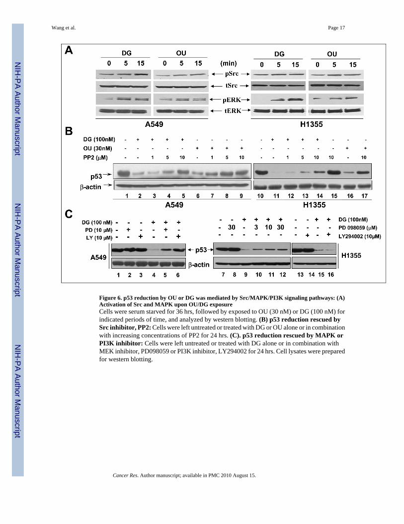
Figure 6. p53 reduction by OU or DG was mediated by Src/MAPK/PI3K signaling pathways: (A)Activation of Src and MAPK upon OU/DG exposureCells were serum starved for 36 hrs, followed by exposed to OU (30 nM) or DG (100 nM) forindicated periods of time, and analyzed by western blotting. (B) p53 reduction rescued bySrc inhibitor, PP2: Cells were left untreated or treated with DG or OU alone or in combinationwith increasing concentrations of PP2 for 24 hrs. (C). p53 reduction rescued by MAPK orPI3K inhibitor: Cells were left untreated or treated with DG alone or in combination withMEK inhibitor, PD098059 or PI3K inhibitor, LY294002 for 24 hrs. Cell lysates were preparedfor western blotting.
Wang et al. Page 17
Cancer Res. Author manuscript; available in PMC 2010 August 15.
NIH
-PA Author Manuscript
NIH
-PA Author Manuscript
NIH
-PA Author Manuscript