
NIH Public Access activity in Cell Drosophila 10021,...
22
A conserved F-box–regulatory complex controls proteasome activity in Drosophila Maya Bader 1 , Sigi Benjamin 1 , Orly L. Wapinski 1 , David M. Smith 2 , Alfred L. Goldberg 2 , and Hermann Steller 1 1 Howard Hughes Medical Institute, The Rockefeller University, 1230 York Avenue, NewYork, NY 10021, USA 2 Department of Cell Biology, Harvard Medical School, 240 Longwood Avenue, Boston, MA 02115, USA Summary The Ubiquitin-Proteasome System catalyzes the degradation of intracellular proteins. Although ubiquitination of proteins determines their stabilities, there is growing evidence that proteasome function is also regulated. We report the functional characterization of a conserved proteasomal regulatory complex. We identified DmPI31 as a binding partner of the F-box protein Nutcracker, a component of an SCF ubiquitin ligase (E3) required for caspase activation during sperm differentiation in Drosophila. DmPI31 binds Nutcracker via a conserved mechanism that is also used by mammalian FBXO7 and PI31. Nutcracker promotes DmPI31 stability, which is necessary for caspase activation, proteasome function and sperm differentiation. DmPI31 can activate 26S proteasomes in vitro, and increasing DmPI31 levels suppresses defects caused by diminished proteasome activity in vivo. Furthermore, loss of DmPI31 function causes lethality, cell-cycle abnormalities and defects in protein degradation, demonstrating that DmPI31 is physiologically required for normal proteasome activity. Introduction The Ubiquitin-Proteasome System (UPS) is the major system for selective degradation of proteins in eukaryotic cells, and is implicated in the regulation of most cellular processes, including cell-cycle control, transcription, signal transduction, inflammation and cell viability (Finley, 2009; Glickman and Ciechanover, 2002). Protein substrates are targeted for degradation by a set of enzymes that attach multiple molecules of the small protein ubiquitin (Ub). These include the Ub-activating enzymes (E1s), Ub-conjugating enzymes (E2s), and a variety of Ub-protein ligases (E3s), which are responsible for substrate recognition and specificity (Glickman and Ciechanover, 2002). The 26S proteasome is a large protease complex composed of two major components: the core or catalytic particle (CP or 20S), a cylindrical particle within which polypeptides are hydrolyzed to small peptides. The 20S proteasome is associated with one or two regulatory particles (RP or 19S) that bind the ubiquitinated substrate, unfold it and translocate the © 2011 Elsevier Inc. All rights reserved. Corresponding author: Hermann Steller [email protected], +1-212-327-7075. Publisher's Disclaimer: This is a PDF file of an unedited manuscript that has been accepted for publication. As a service to our customers we are providing this early version of the manuscript. The manuscript will undergo copyediting, typesetting, and review of the resulting proof before it is published in its final citable form. Please note that during the production process errors may be discovered which could affect the content, and all legal disclaimers that apply to the journal pertain. NIH Public Access Author Manuscript Cell. Author manuscript; available in PMC 2011 October 29. Published in final edited form as: Cell. 2011 April 29; 145(3): 371–382. doi:10.1016/j.cell.2011.03.021. NIH-PA Author Manuscript NIH-PA Author Manuscript NIH-PA Author Manuscript
Transcript of NIH Public Access activity in Cell Drosophila 10021,...

A conserved F-box–regulatory complex controls proteasomeactivity in Drosophila
Maya Bader1, Sigi Benjamin1, Orly L. Wapinski1, David M. Smith2, Alfred L. Goldberg2, andHermann Steller1
1 Howard Hughes Medical Institute, The Rockefeller University, 1230 York Avenue, NewYork, NY10021, USA2 Department of Cell Biology, Harvard Medical School, 240 Longwood Avenue, Boston, MA02115, USA
SummaryThe Ubiquitin-Proteasome System catalyzes the degradation of intracellular proteins. Althoughubiquitination of proteins determines their stabilities, there is growing evidence that proteasomefunction is also regulated. We report the functional characterization of a conserved proteasomalregulatory complex. We identified DmPI31 as a binding partner of the F-box protein Nutcracker, acomponent of an SCF ubiquitin ligase (E3) required for caspase activation during spermdifferentiation in Drosophila. DmPI31 binds Nutcracker via a conserved mechanism that is alsoused by mammalian FBXO7 and PI31. Nutcracker promotes DmPI31 stability, which is necessaryfor caspase activation, proteasome function and sperm differentiation. DmPI31 can activate 26Sproteasomes in vitro, and increasing DmPI31 levels suppresses defects caused by diminishedproteasome activity in vivo. Furthermore, loss of DmPI31 function causes lethality, cell-cycleabnormalities and defects in protein degradation, demonstrating that DmPI31 is physiologicallyrequired for normal proteasome activity.
IntroductionThe Ubiquitin-Proteasome System (UPS) is the major system for selective degradation ofproteins in eukaryotic cells, and is implicated in the regulation of most cellular processes,including cell-cycle control, transcription, signal transduction, inflammation and cellviability (Finley, 2009; Glickman and Ciechanover, 2002). Protein substrates are targetedfor degradation by a set of enzymes that attach multiple molecules of the small proteinubiquitin (Ub). These include the Ub-activating enzymes (E1s), Ub-conjugating enzymes(E2s), and a variety of Ub-protein ligases (E3s), which are responsible for substraterecognition and specificity (Glickman and Ciechanover, 2002).
The 26S proteasome is a large protease complex composed of two major components: thecore or catalytic particle (CP or 20S), a cylindrical particle within which polypeptides arehydrolyzed to small peptides. The 20S proteasome is associated with one or two regulatoryparticles (RP or 19S) that bind the ubiquitinated substrate, unfold it and translocate the
© 2011 Elsevier Inc. All rights reserved.Corresponding author: Hermann Steller [email protected], +1-212-327-7075.Publisher's Disclaimer: This is a PDF file of an unedited manuscript that has been accepted for publication. As a service to ourcustomers we are providing this early version of the manuscript. The manuscript will undergo copyediting, typesetting, and review ofthe resulting proof before it is published in its final citable form. Please note that during the production process errors may bediscovered which could affect the content, and all legal disclaimers that apply to the journal pertain.
NIH Public AccessAuthor ManuscriptCell. Author manuscript; available in PMC 2011 October 29.
Published in final edited form as:Cell. 2011 April 29; 145(3): 371–382. doi:10.1016/j.cell.2011.03.021.
NIH
-PA Author Manuscript
NIH
-PA Author Manuscript
NIH
-PA Author Manuscript

extended polypeptide through the narrow entry channel of the CP (Coux et al., 1996;Demartino and Gillette, 2007; Finley, 2009). Recent studies have identified many looselyassociated proteins of this structure, which may function as regulators or co-factors, but theprecise activities of most of these proteins remains elusive (Besche et al., 2009; Finley,2009).
An added level of proteasomal regulation is the replacement of the 19S RP with alternativeactivator complexes, such as REGαβ (PA28αβ or 11S), REGδ (PA28δ) and Blm10 (PA200)(Ma et al., 1992; Masson et al., 2001; Tanahashi et al., 1997; Ustrell et al., 2002). Thesecomplexes can bind to either or both end of the 20S particle, and enhance the entry anddegradation of small peptide substrates (Stadtmueller and Hill; Whitby et al., 2000). Giventhe variety and complexity of these regulatory components, and the growing number ofproteasome associated proteins being identified, it is clear that proteasomes are much morecomplicated, more diverse and highly regulated structures than previously thought(Demartino and Gillette, 2007).
Here we describe the identification and functional characterization of a novel proteasomeregulatory complex that controls caspase activation and spermatogenesis in Drosophila.Caspases are the key executioners of apoptosis, but during terminal sperm differentiationthey are utilized for an apoptosis-like process that eliminates excess organelles andcytoplasm (Arama et al., 2003). Non-lethal caspase activity is also used in other examples ofcellular remodeling, such as neuronal pruning (Kuo et al., 2006; Nikolaev et al., 2009;Williams et al., 2006). Neuronal remodeling is important for normal development, but hasalso been implicated in neurodegenerative diseases (Nikolaev et al., 2009; Raff et al., 2002;Tai and Schuman, 2008). Therefore, insights into cellular remodeling and the proteases thatmediate it will likely contribute to a better understanding of these diseases.
From a screen for genes that control caspase activation during terminal spermdifferentiation, we isolated the F-box protein Nutcracker, the substrate-binding unit of anSCF Ub-ligase complex (Bader et al., 2010). Mutations in nutcracker abrogate caspaseactivity and prevent the progression of cellular remodeling, resulting in sterility. Here wedescribe the identification and characterization of a Nutcracker-binding partner, DmPI31,discovered in a proteomic screen for Nutcracker interactors. We show that DmPI31 stabilityis promoted by Nutcracker, and that they functionally cooperate to activate caspases anddrive sperm differentiation. Furthermore, we find that DmPI31, which was originallydescribed as a proteasome inhibitor in mammalian systems, can activate purified 26Sproteasomes in vitro (Chu-Ping et al., 1992; McCutchen-Maloney et al., 2000; Zaiss et al.,1999; Zaiss et al., 2002). Elevated levels of DmP31 can suppress phenotypes caused byreduced proteasome activity in vivo. Finally, loss-of-function mutations in DmPI31 arelethal, demonstrating that this protein has an essential physiological function. DmPI31mutants accumulate poly-ubiquitinated proteins and display cell-cycle abnormalities,suggesting that DmPI31 is physiologically required for normal proteasome activity in vivo.These findings reveal a conserved mode of proteasome regulation.
ResultsProteomic screen to identify Nutcracker interacting proteins
Nutcracker is a Drosophila F-box protein previously identified in a screen for mutants thatfail to activate caspases during spermatid differentiation (Bader et al., 2010). To furtherunderstand its role in caspase activation and spermatogenesis, we sought to identifyinteracting partners by co-immunoprecipitation (co-IP) of Nutcracker followed byidentification of associated proteins using mass-spectrometry (Figure 1A).
Bader et al. Page 2
Cell. Author manuscript; available in PMC 2011 October 29.
NIH
-PA Author Manuscript
NIH
-PA Author Manuscript
NIH
-PA Author Manuscript

A protein-A(PrA) tagged version of full-length Nutcracker (PrA-ntc) was expressed in bothDrosophila testes and S2 cells. After co-IP, the interacting proteins were identified usingMS/MS mass-spectrometry (Figures 1B and S1A). This revealed a group of proteinsbelonging to the UPS, primarily proteasome subunits (alpha1 and alpha7). Co-IPexperiments confirmed that Nutcracker can associate with proteasomes in vivo (Figure 1C).In addition, we identified a putative proteasome inhibitor, DmPI31 (CG8979), as a bindingpartner of PrA-ntc in both S2 cells and testes (Figure 1B&D and S1A). The interactionbetween Nutcracker and DmPI31 appears to be direct since both proteins can bind eachother in a yeast two-hybrid assay (Giot et al., 2003). Next, we generated an antibody againstrecombinant DmPI31 and confirmed that this interaction occurs in vivo (Figure 1D). Wechose to focus on DmPI31 because of its potential role in regulating the proteasome.
DmPI31-Nutcracker interaction depends on the conserved FP domainWe previously showed that the F-box domain of Nutcracker is important for binding Cullin1and SkpA (Bader et al., 2010). To determine if the F-box domain is also required forNutcracker-DmPI31 binding, we performed co-IP experiments using testes expressing eitherthe original PrA-ntc, or a truncated version that lacks the F-box domain (PrA-ntcΔF). Co-IPof both PrA-ntc and PrA-ntcΔF resulted in enrichment of DmPI31 (Figure 1D). Thissuggests that the interaction between Nutcracker and DmPI31 is independent of the F-boxdomain.
To further investigate the nature of Nutcracker-DmPI31 interaction, we looked at a related,structurally defined association between the mammalian F-box protein FBXO7 and PI31(Kirk et al., 2008). Nutcracker has some homology with FBXO7 and they share a criticalconserved valine (Figure S1B) that is required for FBXO7-PI31 binding. We mutated thisvaline in Nutcracker (V>E) to examine its significance in the Nutcracker-DmPI31interaction. As shown in Figure 1E, significantly less endogenous DmPI31 was bound to themutant Nutcracker protein than the wild type form. These results show that the conserved FPdomain is essential for interaction with DmPI31.
DmPI31 is highly expressed in the testes and is localized to Individualization ComplexesDmPI31 is the Drosophila homologue of mammalian PI31 proteins, which have been foundto inhibit the activity of purified 20S proteasomes (Chu-Ping et al., 1992; McCutchen-Maloney et al., 2000; Zaiss et al., 1999). The Drosophila homologue shares over 45%homology with these proteins (Figure 2A). DmPI31 mRNA is expressed throughout theDrosophila lifecycle, but drops substantially in the adult female (Arbeitman et al., 2002;Gauhar, 2008). Semi-quantitative RT-PCR experiments demonstrated that mRNA levels inadult females are equivalent to those of son-of-oskar males, which lack germ cells andfunctioning testes. This suggests that the majority of adult mRNA resides in testes (Figure2B).
In order to determine the expression and intracellular localization of DmPI31, testes werestained with the DmPI31 antibody. At the onset of terminal sperm differentiation, in aprocess called “individualization”, actin-rich cones form around the nuclei to create theIndividualization Complex (IC) (Figure 2C and 2D) (Fuller, 1993). As the IC moves, mostof the cytoplasm and excess organelles are expelled in an inflated structure called the cysticbulge (CB). Nutcracker protein is highly localized around the IC’s actin cones in the CB(Figure 2E and (Bader et al., 2010)). DmPI31 is distributed in a very similar pattern (Figure2E and 2F), and both proteins can co-localize upon expression in cultured cells (Figure S2).This suggests that DmPI31 and Nutcracker localize to the same cellular compartment duringindividualization, consistent with their physical interaction in vivo.
Bader et al. Page 3
Cell. Author manuscript; available in PMC 2011 October 29.
NIH
-PA Author Manuscript
NIH
-PA Author Manuscript
NIH
-PA Author Manuscript
We also generated a mCherry-DmPI31 fusion protein driven by the dmPI31 endogenouspromoter. This reporter displayed a similar subcellular distribution as the antibody staining,indicating that the fusion protein is properly localized (Figure 2G). Furthermore, mCherrry-DmPI31 was able to rescue the lethality of dmPI31 mutants (see below). Therefore, thisfusion protein can substitute for endogenous DmPI31 protein and appears to be a faithfulreporter for DmPI31 localization and behavior.
Nutcracker controls DmPI31 protein levelsSince the Nutcracker-DmPI31 interaction is independent of the F-box domain, we askedwhether DmPI31 might be a substrate of the Nutcracker-containing SCF ubiquitin ligase. IfDmPI31 is a substrate for Nutcracker-mediated degradation, we would have expectedDmPI31 to accumulate in the mutants. In contrast, DmPI31 levels were greatly reduced intestes of both Nutcracker-null (nutcracker07259) or truncated F-box (nutcrackerms771)mutants (Figure 3A) (Bader et al., 2010). Instead, smaller molecular weight products weredetected. The full-length DmPI31 band was fully restored in nutcrackerms771−/− flies thatexpress wild type Nutcracker (nutcracker “rescue” transgenes: hsp83-nutcracker). Thisindicates that DmPI31 is cleaved in a nutcracker mutant background, suggesting thatNutcracker positively regulates DmPI31 protein stability. Also, since a F-box-truncatedform of Nutcracker (nutcrackerms771−/−) still destabilizes DmPI31, it appears that the F-boxdomain is important for stabilizing DmPI31. Finally, we investigated whether the cleavageof DmPI31 in a nutcracker mutant background depends on caspases or proteasome activityand found no evidence for an involvement of these proteases (Figure S3A).
The amino-terminal region of PI31 is structured and contains the FP domain, while thecarboxy-terminal end, which contains proline-rich sequences, is unstructured (Kirk et al.,2008; McCutchen-Maloney et al., 2000) (Figure 2A). We therefore investigated whether thecleaved form of DmPI31 seen in lysates of nutcracker mutants is the result of randomproteolysis, or cleavage of a specific portion of the protein. For this purpose, we expressedmCherry-DmPI31 in a nutcracker771−/− mutant background and probed with an anti-mCherry antibody. If cleavage of DmPI31 were near the C-terminus, the resulting bandwould decrease by approximately 10kD. On the other hand, if the cleavage were near the N-terminus, we would expect the molecular weight to decrease by 20KD (see diagram inFigure 3B). The size of mCherry-DmPI31 in a nutcracker mutant background decreased by10KD. Since mCherry-DmPI31 is biologically active, we assume that the cleavage site is notaffected by the presence of the mCherry tag and conclude that the C-terminus of DmPI31 istruncated in a nutcracker mutant background. The C-terminus of mammalian PI31 isrequired for proteasome interaction, so loss of nutcracker function is predicted to inactivateDmPI31 (McCutchen-Maloney et al., 2000).
Next we investigated if the interaction between DmPI31 and Nutcracker is lost upontruncation of the DmPI31 C-terminal domain. We repeated the co-IP experiment, but thistime we expressed PrA-ntc in the testes of the nutcrackerms771−/− mutant flies (Figure 3C).PrA-ntc was able to IP both the full length and cleaved forms of DmPI31, indicating that thecleaved form is folded and capable of interacting with Nutcracker. This suggests that thephenotypes observed in nutcracker mutants result from the cleavage of DmPI31, and notsimply from the inability of both proteins to interact.
Nutcracker and DmPI31 act together to activate caspases and control sperm differentiationWe asked if the interaction between the two proteins is important for their biological activityin vivo. For this purpose, we mutated the FP domain of Nutcracker and tested whether atransgene carrying this mutation can rescue nutcrackerms771−/−. Although both constructswere expressed at similar levels (Figure S3B), hsp83-nutcrackerV-E transgenenic flies were
Bader et al. Page 4
Cell. Author manuscript; available in PMC 2011 October 29.
NIH
-PA Author Manuscript
NIH
-PA Author Manuscript
NIH
-PA Author Manuscript

unable to rescue the nutcrackerms771−/− sterility phenotype (Table 1). When we stainedtestes from these males with the caspase3 antibody, we noticed that some staining wasrestored, demonstrating that the mutated construct was partially functional (data not shown).However, it appears that full Nutcracker activity requires binding to DmPI31.
We then investigated if the inability of the mutated rescue construct to restore fertility iscaused by persistent DmPI31 cleavage. We found that hsp83-nutcrackerV-E;nutcrackerms771−/− still displayed DmPI31 cleavage, albeit less than in the originalnutcrackerms771−/− homozygote mutant line (Figure 3D). This indicates that the interactionbetween Nutcracker and DmPI31 is important for Nutcracker’s role in stabilizing DmPI31,and that this stabilization is crucial for proper sperm differentiation.
If Nutcracker functions through DmPI31 stabilization to promote caspase activation andsperm differentiation, it may be possible to at least partially compensate for the loss ofNutcracker function by expressing DmPI31. We over-expressed DmPI31 in anutcrackerms771−/− mutant background and observed partial restoration of caspase staining(Figure 3E). This suggests that Nutcracker activates caspases, at least in part, by stabilizingDmPI31. According to this interpretation, Nutcracker is dispensable for caspase activation ifsufficient amounts of full-length DmPI31 are expressed in spermatids. Taken together, ourresults demonstrate that Nutcracker and DmPI31 need to physically interact for their fullbiological activities.
DmPI31 can stimulate 26S proteasomes in vitroPI31 proteins were identified as inhibitors of the catalytic particle of the proteasome basedon the ability of recombinant bovine PI31 to inhibit purified 20S proteasomes (Chu-Ping etal., 1992; McCutchen-Maloney et al., 2000; Zaiss et al., 1999). Subsequent studies alsodemonstrated PI31’s ability to compete with 19S and 11S regulatory particles which resultedin a net inhibitory effect (McCutchen-Maloney et al., 2000). In order to understand themolecular role of Drosophila PI31 in regulating spermatogenesis, we investigated its effecton the proteasome’s peptidase activity. Recombinant DmPI31 was purified from E. coli andadded to purified mammalian 20S proteasomes. As expected, DmPI31 inhibited 20S activity(Figure 4A). Surprisingly, when purified DmPI31 was added to mammalian 26Sproteasomes, its peptidase activity was enhanced up to 3-fold (Figure 4A). Thus, when the20S CP is associated with the 19S RP (and possibly various other interacting proteins),DmPI31 enhances proteasome function, in contrast to the inhibitory effect on purified 20SCP.
The capacity of 26S and 20S proteasomes to degrade small peptides is determined bywhether the gate for its substrate-entry channel is in its open conformation, and in the 26Sparticle, this entry is regulated by the ATPases in the 19S (Smith et al., 2005). Such an effecton gating could be mediated by DmPI31’s C-terminus Hydrophobic-Tyrosin-X (HbYX)motif that in 19S ATPases allows interaction with the proteasome and induces gate opening(Rabl et al., 2008; Smith et al., 2007). Consistently, we found that mutant DmPI31 proteinlacking the HbYX motif could still activate the 26S, but with a reduced efficiency (Figure4B). This suggests that while the HbYX domain is important for proteasome activation, itmay not be the only domain that mediates activation and binding. The difference in DmPI31effect on the 20S vs. 26S suggests that other domains in DmPI31 may bind to the 19Sregulatory subunit as well. Collectively, the in vitro data indicate that DmPI31, despite itsoriginal designation as a proteasome inhibitor, can function as an activator of the 26Sproteasome, which is the dominant form in vivo (Besche et al., 2009).
Bader et al. Page 5
Cell. Author manuscript; available in PMC 2011 October 29.
NIH
-PA Author Manuscript
NIH
-PA Author Manuscript
NIH
-PA Author Manuscript

Ectopic expression of DmPI31 increases proteasome activity in vivoWe conducted a series of genetic studies to determine whether DmPI31 can affectproteasome activity in vivo. First we analyzed whether expression of DmPI31 had an effecton phenotypes caused by impaired proteasome function. Targeted expression of thedominant temperature sensitive proteasome alleles UAS-DTS5 and UAS-DTS7 with GMR-Gal4 at 29°C causes a small, rough eye phenotype (Belote and Fortier, 2002) (Figure 5B and5C). Co-expression of DmPI31 with these constructs suppressed this phenotype (Figure 5D).This appears to be a direct effect on boosting proteasome activity because expression ofDmPI31 did not increase the expression of proteasome subunits (Figure 5E). Conversely,reducing DmPI31 function significantly enhanced the phenotype of the DTS alleles (Figure5F–5J). Whereas expression of either DTS allele at 25°C caused no detectable eyephenotype, down-regulation of DmPI31 via RNAi caused a strong eye phenotype in thebackground of both DTS alleles (Figure 5F–5J). At 29°C, this allelic combination results inorganismal lethality (Figure 5K–5N). These results show that DmPI31 can modifyproteasome activity in vivo and suggests that DmPI31 functions as a proteasome activator.
DmPI31 is physiologically required for proteasome functionTo determine the physiological function of DmPI31, we generated loss-of-functionmutations in this gene by homologous recombination (Gong and Golic, 2003). dmPI31mutants are recessively lethal and die at the late third instar larval/early pupal transition, adevelopmental stage where many cell cycle mutants are lethal (Gatti and Baker, 1989; Orr-Weaver, 1994). Mutant larvae also developed melanotic tumors, were slower to develop andsmaller. All these phenotypes were rescued by transgenic expression of dmPI31 (usingUASt-HA-PI31 transgenic flies under the control of the tubulin-Gal4, tubulin Gal80ts
promoters at 23°C), demonstrating that the mutant phenotypes are indeed caused by loss ofdmPI31 function. On the other hand, high-level over-expression of DmPI31 caused lethality(Table S1). This indicates that cells are highly sensitive to the amounts of PI31, since bothelimination of the gene and over-expression are detrimental and cause lethality. We alsogenerated transgenic fly lines to ectopically express various DmPI31 mutants for structure/function studies (Table S1). Consistent with our previous results, truncation of the C-terminal region of DmPI31 abrogated the ability of DmPI31 to stimulate proteasomeactivity.
Amongst the many cellular processes regulated by the UPS is cell cycle progression. Wetherefore examined whether dmPI31 mutants have cell cycle defects, focusing on the malegerm line. By expressing DmPI31 transgenically in somatic cells we were able to rescueorganismal lethality and generate viable “mosaic” adults that lack dmPI31 function in germcells because germ cells use a different tubulin promoter than somatic cells (Hoyle et al.,1995; Kemphues et al., 1980). When the testes of these flies were dissected they appearedirregular and exhibited a meiotic arrest phenotype, characterized by the abundance ofunderdeveloped 16 cell stage cysts, which fail to undergo proper meiosis (Lin et al., 1996).Antibody staining against DmPI31 confirmed the absence of protein in both the germ-linecells (GSCs) and primary spermatocytes (Figure 6A).
Next, we stained testes with cell cycle markers. BrdU staining at the tip of the testes closestto the hub was similar to the wild-type, indicating that mitosis of germ-line cells proceedednormally (Figure 6B). In contrast, when we stained with the phospho-histone3 (PH3)antibody, which labels dividing nuclei, we did not detect cysts in the 32-cell stage,suggesting that these cells are not undergoing meiosis (Figure 6C). Furthermore, stainingwith the Vasa antibody, which is specific for germ cell progenitors, showed that loss ofDmPI31 function prevents germ cell differentiation and leads to persistence of progenitoridentity (Jin et al., 2005) (Figure 6D). Moreover, we saw that CyclinB, a protein important
Bader et al. Page 6
Cell. Author manuscript; available in PMC 2011 October 29.
NIH
-PA Author Manuscript
NIH
-PA Author Manuscript
NIH
-PA Author Manuscript
for both mitotic and meiotic cell cycles, that is degraded by the proteasome during the finalstages of meiosis, also persisted in these cells (Fuller, 1998) (Figure 6E and S6). Theseresults are consistent with the idea that loss of DmPI31 impairs proteasome function and thatDmPI31 is a proteasome activator.
To further test whether dmPI31 mutants have reduced proteasome activity, we stained testeswith the FK2 antibody, which detects Ub-conjugated proteins and can therefore be used tovisualize poly-ubiquitinated, non-degraded proteins that accumulate when 26S proteasomefunction is impaired (Fujimuro et al., 1997; Haas et al., 2007). In wild type cells, FK2staining was diffuse in the cytoplasm and strong in the nucleus of meiotic cells (Figure 6F).In contrast, in dmPI31 mutant cells, punctate staining was seen in most cells, and in somecases in one or two nuclei of each cyst. This pattern of staining suggests that proteasomeactivity is defective, and that Ub-conjugates accumulate as intracellular aggregates. Takentogether, our results show that DmPI31 is required for proper protein degradation and cellcycle progression, supporting a role of this protein as a proteasome activator.
DiscussionA conserved proteasome regulatory complex is required for cellular remodeling
We identified and characterized a proteasome regulatory complex that combines two distinctcomponents of the UPS – an F-box protein, which usually catalyzes ubiquitin-conjugation toa set of substrates, and a proteasome regulator, which promotes substrate hydrolysis by theproteasome. Although several studies have reported the physical association between certainUb-ligases and the 26S proteasome, we now demonstrate a clear functional significance ofthis interaction in vivo (Demartino and Gillette, 2007; Finley, 2009). In particular, we showthat the F-box protein Nutcracker and the proteasome regulator DmPI31 function together asa complex to stimulate proteasome activity during normal development. This elevatedproteasome activity is required for non-apoptotic caspase activation and spermdifferentiation in Drosophila. The interaction of DmPI31 and Nutcracker is mediatedthrough their FP domain, a conserved domain found in mammalian homologues (Kirk et al.,2008). Therefore, it is possible that a similar mode of proteasomal regulation operates inmammals.
Nutcracker controls caspase activation and sperm individualization by promoting DmPI31stability
We initially considered that DmPI31 is a substrate of the Nutcracker-containing SCF E3-ligase complex. However, DmPI31 protein does not accumulate in nutcracker mutants, aswould be expected if it was a degradation substrate. Instead, a nutcracker mutant that carriesa stable but truncated F-box domain results in the cleavage and truncation of DmPI31.Hence, the Nutcracker F-box domain is required for the stability of full-length DmPI31 andthe proteasome-activating activity of this protein (Figure 7). Furthermore, stabilization ofDmPI31 by Nutcracker is required for normal caspase activation, sperm differentiation andmale fertility. First, loss of Nutcracker function causes defects in caspase activation andmale sterility (Bader et al., 2010). Second, a nutcracker transgene with reduced ability tobind DmPI31 failed to protect DmPI31 from cleavage and did not rescue nutcrackermutants. This suggests that binding of Nutcracker to DmPI31 is critically important topositively regulate DmPI31 by stabilizing this protein. Additionally, over-expression ofDmPI31 in a nutcracker mutant background was sufficient for caspase activation (Fig. 3E).This indicates that physical interaction with DmPI31 is important for Nutcracker activity,and that Nutcracker acts upstream of DmPI31 to control its stability and function. Sincegeneral over-expression of DmPI31 is lethal, it appears that the levels of active (full-length)DmPI31 need to be carefully regulated.
Bader et al. Page 7
Cell. Author manuscript; available in PMC 2011 October 29.
NIH
-PA Author Manuscript
NIH
-PA Author Manuscript
NIH
-PA Author Manuscript
DmPI31 is a proteasome activator in vivoContrary to the original identification of PI31 as a proteasome inhibitor (PI), thephysiological function of DmPI31 is to stimulate proteasome activity in vivo. Thisstimulatory effect of PI31 on proteasomes appears to be direct because DmPI31 can alsoactivate purified 26S proteasomes in vitro. The ability of DmPI31 to enhance thedegradation of standard tetrapeptide substrates implies a role in promoting the opening ofthe substrate-entry channel, a property exhibited by other proteasome activating complexes(Rabl et al., 2008; Smith et al., 2007). In support of this idea, DmPI31’s C-terminal HbYXmotif, which resembles the proteasome-interacting, gate-opening domains on theproteasome-regulatory ATPases, is also important for DmPI31 activity (Rabl et al., 2008;Smith et al., 2007).
The physiological role of DmPI31 as an essential proteasome activator is supported by thephenotypes of dmPI31 loss-of-function mutants. Loss of dmPI31 function causes lethality,indicating that this gene has a vital function. Furthermore, germ cells lacking dmPI31 fail toundergo meiosis and maintain stem cell identity. These phenotypes are consistent withdefects in protein degradation (Alessandrini et al., 1997; Fuller, 1998). Accordingly, weobserved that poly-ubiquitinated proteins accumulate in dmPI31 mutant germ-cells, provingstrong evidence that DmPI31 is required for normal proteasome activity in vivo. Finally, amodest elevation of DmPI31 protein was able to suppress phenotypes caused by reducedproteasome activity, indicating that DmPI31 can boost proteasome activity in vivo. Takentogether, all these results argue that DmPI31 plays an important physiological role as aproteasome activator.
Our findings may have important general implications for the role of regulated proteolysisduring cellular remodeling. Sperm differentiation involves a major reduction in cell volumeand can be viewed, from a cell biological standpoint, as ‘programmed cell atrophy’ (Aramaet al., 2006; Bader et al., 2010; Fabrizio et al., 1998; Noguchi and Miller, 2003; Zhong andBelote, 2007). Similar dramatic morphological changes and organelle breakdown also occurduring the differentiation of other cell types, for example during neuronal pruning (Lecker etal., 2006; Ventadour and Attaix, 2006). Consequently, it is possible that proteasomes arepositively regulated in these cases as well. Furthermore, intracellular proteolysis mediatedby both proteasomes and caspases is associated with various pathologies that involve celland tissue wasting, including neural degeneration and myopathies (Lecker et al., 2006; Raffet al., 2002; Saxena and Caroni, 2007). Therefore, insights into the regulation of proteindegradation are highly relevant for a wide range of human diseases. In the future, it will bein interesting to investigate if a mechanism similar to what we describe here controlsproteasome activity in mammals, and whether it contributes to human diseases that areassociated with excessive protein turnover.
Experimental ProceduresFly strains
yw flies were used as wild-type controls. The Zuker mutant Z3-4692 (ms771) was obtainedfrom C.S. Zuker; the son-of-oskar osk[301]/TM3 and osk[CE4]/TM3 lines from R.Lehmann; w1118;70FLP,70ISceI and w1118;70FLP lines from L.B. Vosshall; tubulin-Gal4from HD. Ryoo; UAS-DTS5 and UAS-DTS7 were obtained from J. Belote; the deficiencylines Df(2R)BSC199 and Df(2R)ED2247 and the PBac insertion PBac{WH}CG10855f07259
from the Bloomington Stock Center, as were tubulin-Gal80ts and actin-Gal4. DmPI31 RNAilines were obtained from the Vienna Drosophila RNAi Center (VDRC).
Bader et al. Page 8
Cell. Author manuscript; available in PMC 2011 October 29.
NIH
-PA Author Manuscript
NIH
-PA Author Manuscript
NIH
-PA Author Manuscript

ImmunoprecipitationsTestes-IP was conducted as in (Bader et al., 2010). For S2 IP, cells were co-transfected withActin-Gal4 and different UAS constructs (Fugene6, Roche) and left for 36–48 hrs. Cellswere washed twice in ice-cold PBS, lysed in 200μl ice-cold 1% NP-40 in PBS, left on icefor 15 minutes, then spun at 14,000 rpm for 15 minutes at 4°C. The IP was then conductedsimilarly to the testes IP, except the washes were done with ice-cold PBS. 0.5–1mg of total-protein was used for IP-Western, and 7–10mg for mass-spectrometry identification.
Mass spectrometry analysisAfter co-IP, proteins were resolved by SDS-PAGE (4%–12% gradient) and stained withCoomassie Blue (GelCode Blue; Pierce). Visible bands were excised and subjected totrypsin digestion. The resulting peptides were extracted, and proteins were identified bymass spectrometry at the Rockefeller University Proteomics Resource Center. Peptidessequences were analyzed using the MASCOT search engine(http://www.matrixscience.com/).
Western Blot AnalysisWestern blot analysis was conducted as in (Bader et al., 2010) with anti-DmPI31[sera],1:1000, anti-HA, 1:5000 (Roche), anti-alpha7, 1:200 (Biomol), mCherry 1:1000 (clontech),CyclinB (1:1000) Santa Cruz.
Antibody generation and tissue stainingCleaved effector caspase antibody staining of testes was as in (Bader et al., 2010). Anti-DmPI31 was created in guinea-pigs with full-length recombinant DmPI31 (CocalicoBiologicals, Inc.), and staining was as described in (Hime et al., 1996) with serum diluted1:250. Staining of whole mount testes and BrdU incorporation followed (Beall et al., 2002),except CyclinB staining was as in (Baker and Fuller, 2007). The following antibodies anddilutions were used: mouse anti-BrdU-FITC 1:100 (BD-Pharmingen), rabbit anti-Vasa 1:100(a gift from R. Lehmann), mouse anti-CyclinB clone F2F4 1:10 (DSHB) and mouse FK21:100 (Stressgen).
Proteasome activity assayTo measure proteasome activity, the fluorogenic peptide substrate Suc-LLVY-amc (EnzoLife Sciences, maintained in DMSO) was used at a final concentration of 100 μM in reactionbuffer (50mM Tris, 5% glycerol). Hydrolysis of suc-LLVY-amc was monitored at λex 380nm and λem 440 nm. All reactions were conducted in a 96-well plate and read on aSpectraMax M2 micro-plate reader (Molecular Devices). For experiments on 20Sproteasomes, 0.05–0.15μg of purified Bovine 20S was used per 100μl reaction, at 37°C. Forexperiments on 26S proteasomes, 0.05μg of purified Bovine 26S was used per 100 μlreaction, in the same buffer plus 10mM mgCl and 100μM ATP, at 37°C. K(apparent) wascalculated with the standard ligand binding equation using sigmaplot (www.sigmaplot.com)with DmPI31 concentration at saturation as maximal affinity.
Supplementary MaterialRefer to Web version on PubMed Central for supplementary material.
AcknowledgmentsWe are grateful to C. Zucker, R. Lehmann, HD, Ryoo, L. Vosshall and J. Belote for fly strains. R. Lehmanngenerously shared the Vasa antibody. B. Chait and M. Sekedat were a tremendous help in setting up the proteomicscreen. E. Arama devised the original male-sterile screen. Members of the Vosshall lab consulted on generating
Bader et al. Page 9
Cell. Author manuscript; available in PMC 2011 October 29.
NIH
-PA Author Manuscript
NIH
-PA Author Manuscript
NIH
-PA Author Manuscript

homologous recombination mutants and provided materials. K. Sobczyk participated in the proteasome-activityexperiments. We thank M. Garcia-Fernandez and M. Pratt for critically reading this manuscript. O. Wapinski wassupported by The Rockefeller University SURF Program. H. Steller is an investigator of the Howard HughesMedical Institute. This work was supported by NIH grant RO1GM60124 to HS.
ReferencesAlessandrini A, Chiaur DS, Pagano M. Regulation of the cyclin-dependent kinase inhibitor p27 by
degradation and phosphorylation. Leukemia. 1997; 11:342–345. [PubMed: 9067571]Arama E, Agapite J, Steller H. Caspase activity and a specific cytochrome C are required for sperm
differentiation in Drosophila. Dev Cell. 2003; 4:687–697. [PubMed: 12737804]Arama E, Bader M, Srivastava M, Bergmann A, Steller H. The two Drosophila cytochrome C proteins
can function in both respiration and caspase activation. EMBO J. 2006; 25:232–243. [PubMed:16362035]
Arbeitman MN, Furlong EE, Imam F, Johnson E, Null BH, Baker BS, Krasnow MA, Scott MP, DavisRW, White KP. Gene expression during the life cycle of Drosophila melanogaster. Science. 2002;297:2270–2275. [PubMed: 12351791]
Bader M, Arama E, Steller H. A novel F-box protein is required for caspase activation during cellularremodeling in Drosophila. Development. 2010; 137:1679–1688. [PubMed: 20392747]
Baker CC, Fuller MT. Translational control of meiotic cell cycle progression and spermatiddifferentiation in male germ cells by a novel eIF4G homolog. Development. 2007; 134:2863–2869.[PubMed: 17611220]
Beall EL, Manak JR, Zhou S, Bell M, Lipsick JS, Botchan MR. Role for a Drosophila Myb-containingprotein complex in site-specific DNA replication. Nature. 2002; 420:833–837. [PubMed: 12490953]
Belote JM, Fortier E. Targeted expression of dominant negative proteasome mutants in Drosophilamelanogaster. Genesis. 2002; 34:80–82. [PubMed: 12324954]
Besche HC, Haas W, Gygi SP, Goldberg AL. Isolation of mammalian 26S proteasomes and p97/VCPcomplexes using the ubiquitin-like domain from HHR23B reveals novel proteasome-associatedproteins. Biochemistry. 2009; 48:2538–2549. [PubMed: 19182904]
Chu-Ping M, Slaughter CA, DeMartino GN. Purification and characterization of a protein inhibitor ofthe 20S proteasome (macropain). Biochim Biophys Acta. 1992; 1119:303–311. [PubMed:1312359]
Coux O, Tanaka K, Goldberg AL. Structure and functions of the 20S and 26S proteasomes. Annu RevBiochem. 1996; 65:801–847. [PubMed: 8811196]
Demartino GN, Gillette TG. Proteasomes: machines for all reasons. Cell. 2007; 129:659–662.[PubMed: 17512401]
Fabrizio JJ, Hime G, Lemmon SK, Bazinet C. Genetic dissection of sperm individualization inDrosophila melanogaster. Development. 1998; 125:1833–1843. [PubMed: 9550716]
Finley D. Recognition and processing of ubiquitin-protein conjugates by the proteasome. Annu RevBiochem. 2009; 78:477–513. [PubMed: 19489727]
Fujimuro M, Sawada H, Yokosawa H. Dynamics of ubiquitin conjugation during heat-shock responserevealed by using a monoclonal antibody specific to multi-ubiquitin chains. Eur J Biochem. 1997;249:427–433. [PubMed: 9370350]
Fuller, MT. The Development of Drosophila melanogaster. Cold Spring Harbor, NY: Cold SpringHarbor Laboratory Press; 1993.
Fuller MT. Genetic control of cell proliferation and differentiation in Drosophila spermatogenesis.Semin Cell Dev Biol. 1998; 9:433–444. [PubMed: 9813190]
Gatti M, Baker BS. Genes controlling essential cell-cycle functions in Drosophila melanogaster. GenesDev. 1989; 3:438–453. [PubMed: 2498166]
Gauhar, Z.; Ghanim, M.; Herreman, T.; Lambert, JD.; Li, TR.; Mason, C.; Rifkin, S.; Sun, L.; White,KP.; Costello, JC.; Andrews, JR. Drosophila melanogaster life-cycle gene expression dataset andmicroarray normalisation protocols. 2008.
Bader et al. Page 10
Cell. Author manuscript; available in PMC 2011 October 29.
NIH
-PA Author Manuscript
NIH
-PA Author Manuscript
NIH
-PA Author Manuscript

Giot L, Bader JS, Brouwer C, Chaudhuri A, Kuang B, Li Y, Hao YL, Ooi CE, Godwin B, Vitols E, etal. A protein interaction map of Drosophila melanogaster. Science. 2003; 302:1727–1736.[PubMed: 14605208]
Glickman MH, Ciechanover A. The ubiquitin-proteasome proteolytic pathway: destruction for the sakeof construction. Physiol Rev. 2002; 82:373–428. [PubMed: 11917093]
Gong WJ, Golic KG. Ends-out, or replacement, gene targeting in Drosophila. Proc Natl Acad SciUSA. 2003; 100:2556–2561. [PubMed: 12589026]
Haas KF, Woodruff E 3rd, Broadie K. Proteasome function is required to maintain muscle cellulararchitecture. Biol Cell. 2007; 99:615–626. [PubMed: 17523916]
Hime GR, Brill JA, Fuller MT. Assembly of ring canals in the male germ line from structuralcomponents of the contractile ring. J Cell Sci. 1996; 109(Pt 12):2779–2788. [PubMed: 9013326]
Hoyle HD, Hutchens JA, Turner FR, Raff EC. Regulation of beta-tubulin function and expression inDrosophila spermatogenesis. Dev Genet. 1995; 16:148–170. [PubMed: 7736665]
Jin Z, Homola EM, Goldbach P, Choi Y, Brill JA, Campbell SD. Drosophila Myt1 is a Cdk1 inhibitorykinase that regulates multiple aspects of cell cycle behavior during gametogenesis. Development.2005; 132:4075–4085. [PubMed: 16107480]
Kemphues KJ, Raff EC, Raff RA, Kaufman TC. Mutation in a testis-specific beta-tubulin inDrosophila: analysis of its effects on meiosis and map location of the gene. Cell. 1980; 21:445–451. [PubMed: 6773669]
Kirk R, Laman H, Knowles PP, Murray-Rust J, Lomonosov M, Meziane elK, McDonald NQ.Structure of a conserved dimerization domain within the F-box protein Fbxo7 and the PI31proteasome inhibitor. J Biol Chem. 2008; 283:22325–22335. [PubMed: 18495667]
Kuo CT, Zhu S, Younger S, Jan LY, Jan YN. Identification of E2/E3 ubiquitinating enzymes andcaspase activity regulating Drosophila sensory neuron dendrite pruning. Neuron. 2006; 51:283–290. [PubMed: 16880123]
Lecker SH, Goldberg AL, Mitch WE. Protein degradation by the ubiquitin-proteasome pathway innormal and disease states. J Am Soc Nephrol. 2006; 17:1807–1819. [PubMed: 16738015]
Lin TY, Viswanathan S, Wood C, Wilson PG, Wolf N, Fuller MT. Coordinate developmental controlof the meiotic cell cycle and spermatid differentiation in Drosophila males. Development. 1996;122:1331–1341. [PubMed: 8620860]
Ma CP, Slaughter CA, DeMartino GN. Identification, purification, and characterization of a proteinactivator (PA28) of the 20 S proteasome (macropain). J Biol Chem. 1992; 267:10515–10523.[PubMed: 1587832]
Masson P, Andersson O, Petersen UM, Young P. Identification and characterization of a Drosophilanuclear proteasome regulator. A homolog of human 11 S REGgamma (PA28gamma). J BiolChem. 2001; 276:1383–1390. [PubMed: 11027688]
McCutchen-Maloney SL, Matsuda K, Shimbara N, Binns DD, Tanaka K, Slaughter CA, DeMartinoGN. cDNA cloning, expression, and functional characterization of PI31, a proline-rich inhibitor ofthe proteasome. J Biol Chem. 2000; 275:18557–18565. [PubMed: 10764772]
Muro I, Berry DL, Huh JR, Chen CH, Huang H, Yoo SJ, Guo M, Baehrecke, Nikolaev A, McLaughlinT, O’Leary DD, Tessier-Lavigne M. APP binds DR6 to trigger axon pruning and neuron death viadistinct caspases. Nature. 2009; 457:981–989. [PubMed: 19225519]
Noguchi T, Miller KG. A role for actin dynamics in individualization during spermatogenesis inDrosophila melanogaster. Development. 2003; 130:1805–1816. [PubMed: 12642486]
Orr-Weaver TL. Developmental modification of the Drosophila cell cycle. Trends Genet. 1994;10:321–327. [PubMed: 7974746]
Rabl J, Smith DM, Yu Y, Chang SC, Goldberg AL, Cheng Y. Mechanism of gate opening in the 20Sproteasome by the proteasomal ATPases. Mol Cell. 2008; 30:360–368. [PubMed: 18471981]
Raff MC, Whitmore AV, Finn JT. Axonal self-destruction and neurodegeneration. Science. 2002;296:868–871. [PubMed: 11988563]
Saxena S, Caroni P. Mechanisms of axon degeneration: from development to disease. Prog Neurobiol.2007; 83:174–191. [PubMed: 17822833]
Bader et al. Page 11
Cell. Author manuscript; available in PMC 2011 October 29.
NIH
-PA Author Manuscript
NIH
-PA Author Manuscript
NIH
-PA Author Manuscript

Smith DM, Chang SC, Park S, Finley D, Cheng Y, Goldberg AL. Docking of the proteasomalATPases’ carboxyl termini in the 20S proteasome’s alpha ring opens the gate for substrate entry.Mol Cell. 2007; 27:731–744. [PubMed: 17803938]
Smith DM, Kafri G, Cheng Y, Ng D, Walz T, Goldberg AL. ATP binding to PAN or the 26S ATPasescauses association with the 20S proteasome, gate opening, and translocation of unfolded proteins.Mol Cell. 2005; 20:687–698. [PubMed: 16337593]
Stadtmueller BM, Hill CP. Proteasome activators. Mol Cell. 41:8–19. [PubMed: 21211719]Tai HC, Schuman EM. Ubiquitin, the proteasome and protein degradation in neuronal function and
dysfunction. Nat Rev Neurosci. 2008; 9:826–838. [PubMed: 18931696]Tanahashi N, Yokota K, Ahn JY, Chung CH, Fujiwara T, Takahashi E, DeMartino GN, Slaughter CA,
Toyonaga T, Yamamura K, et al. Molecular properties of the proteasome activator PA28 familyproteins and gamma-interferon regulation. Genes Cells. 1997; 2:195–211. [PubMed: 9189757]
Ustrell V, Hoffman L, Pratt G, Rechsteiner M. PA200, a nuclear proteasome activator involved inDNA repair. Embo J. 2002; 21:3516–3525. [PubMed: 12093752]
Ventadour S, Attaix D. Mechanisms of skeletal muscle atrophy. Curr Opin Rheumatol. 2006; 18:631–635. [PubMed: 17053511]
Whitby FG, Masters EI, Kramer L, Knowlton JR, Yao Y, Wang CC, Hill CP. Structural basis for theactivation of 20S proteasomes by 11S regulators. Nature. 2000; 408:115–120. [PubMed:11081519]
Williams DW, Kondo S, Krzyzanowska A, Hiromi Y, Truman JW. Local caspase activity directsengulfment of dendrites during pruning. Nat Neurosci. 2006; 9:1234–1236. [PubMed: 16980964]
Zaiss DM, Standera S, Holzhutter H, Kloetzel P, Sijts AJ. The proteasome inhibitor PI31 competeswith PA28 for binding to 20S proteasomes. FEBS Lett. 1999; 457:333–338. [PubMed: 10471803]
Zaiss DM, Standera S, Kloetzel PM, Sijts AJ. PI31 is a modulator of proteasome formation andantigen processing. Proc Natl Acad Sci USA. 2002; 99:14344–14349. [PubMed: 12374861]
Zhong L, Belote JM. The testis-specific proteasome subunit Prosalpha6T of D. melanogaster isrequired for individualization and nuclear maturation during spermatogenesis. Development. 2007;134:3517–3525. [PubMed: 17728345]
Bader et al. Page 12
Cell. Author manuscript; available in PMC 2011 October 29.
NIH
-PA Author Manuscript
NIH
-PA Author Manuscript
NIH
-PA Author Manuscript

Figure 1. Proteomic screen for Nutcracker interacting proteins[A] Scheme for testis-specific interactors of Nutcracker; PrA-nutcracker (PrA-ntc) wasspecifically expressed in testes with the Don-Juan (DJ) promoter. Testes were dissected,lysed, and incubated with IgG beads. Interacting complexes were eluted off the beads, andidentified by either mass-spectrometry or Western-blot analysis. [B] SDS-PAGE commassieblots of Nutcracker interactors from cell lysate. PrA-ntc or PrA-ntc&DeltaF were expressedin S2 cells and used to make lysates for co-immunoprecipitation (co-IP) assays. Non-PrAexpressing cells were used as a negative control. An arrow marks the DmPI31 band. Seealso Figure S1. [C] PrA-ntc is associated with proteasomes in vivo. PrA-ntc was expressedin testes and used for co-IP assays. Probing with an antibody against the proteasome subunitalpha7 shows that PrA-ntc forms a complex with proteasome proteins in vivo. [D] TheNutcracker-DmPI31 interaction is not dependent on the F-box domain. PrA-ntc or PrA-ntcΔF were expressed in testes and used to make lysates for co-IP assays. Non-PrAexpressing wild-type testes (yw) were used as a negative control. DmPI31 binding wasdetected by Western-blot analysis using a DmPI31 antibody. [E] Nutcracker-DmPI31binding is mediated by a specialized domain. A conserved valine in Nutcarcker thatmediates the interaction between the human PI31 and the F-box protein FBXO7 wasmutated to investigate its importance for DmPI31-Nutcracker binding. PrA-ntc or PrA-ntcV-E were expressed in S2 cells and lysates were used for co-IP. Non-PrA expressing cells wereused as a negative control.
Bader et al. Page 13
Cell. Author manuscript; available in PMC 2011 October 29.
NIH
-PA Author Manuscript
NIH
-PA Author Manuscript
NIH
-PA Author Manuscript

Figure 2. DmPI31 is localized to Individualization Complexes[A] Multiple alignments of DmPI31 and three mammalian homologues (bovine, mouse, andhuman). DmPI31 shares overall 45% homology with the mammalian proteins,corresponding to 29% identity and 16% similarity. In particular, the DX7H and YXLXYmotifs, which are important for the PI31 structure, are highly conserved (labeled in yellowand green, respectively). Highlighted in blue are amino acids that mediate PI31-FBXO7binding (Kirk et al., 2008). In pink is the Hydrophobic-Tyrosine-X (HbYX) motif. Allaccession numbers are listed in the M&M. [B] DmPI31 mRNA is abundant in adult testes.Semi-quantitative RT-PCR of dmPI31 mRNA transcripts taken from whole-body adultfemales, wild-type males or son-of-oskar males, which lack germ-cells. These are alsocompared to mRNA transcripts from wild-type testes or son-of-oskar testes. Beta-tubulinprimers were used as control for total mRNA concentrations. Shown is cycle 25, 5 cyclesbefore saturation. [C] Schematic diagram of spermatid individualization. An actin-basedindividualization complex (IC, red) forms around the elongated nuclei of 64 spermatids(gray) that are connected by cytoplasmic bridges. As the IC moves, cytoplasm (green) andorganelles are collected in the cystic bulge (CB) and eventually discarded in the waste bag(WB), generating individual sperm devoid of most cytoplasm and organelles. [D] Caspasestaining of individualizing cysts. Wild-type cysts were stained with DAPI (nuclei, blue),phalloidin (IC, red) and anti-active-caspase-3 (cytoplasm, green). [E–G] DmPI31 andNutcracker localize to the same sub-cellular region during individualization. [E] Nutcrackerstaining during individualization as detected with an antibody that specifically recognizesthis protein (Bader et al.). Nutcracker staining was seen at the base of the nuclei when theactin cones form around it (inset), and in a circular pattern around the cones in the CB(asterisks). [F] DmPI31 antibody staining. Like Nutcracker, DmPI31 protein is detected atthe base of the elongated nuclei (inset), and co-localizes in a circular pattern with the actincones (asterisks). Colocalization was also seen in cultured cells (Figure S2) [G] mCherry-DmPI31 fusion protein localization during individualization. This fusion protein isexpressed under the control of dmPI31 endogenous promoter and localized in a patternvirtually identical to DmPI31 antibody staining. mCherry-DmPI31 is seen in the combinedcytoplasm of each cyst and at the base of fully elongated nuclei (inset), co-localizing withthe actin cones as they move down the cyst (asterisks).
Bader et al. Page 14
Cell. Author manuscript; available in PMC 2011 October 29.
NIH
-PA Author Manuscript
NIH
-PA Author Manuscript
NIH
-PA Author Manuscript

Figure 3. Nutcracker controls caspase activation and sperm individualization by promotingDmPI31 stability[A] Mutations in nutcracker affect DmPI31 stability. Testes lysates of the indicatedgenotypes were used to detect steady-state DmPI31 protein levels. The cleaved form ofDmPI31 found in nutcracker mutants is indicated by an arrow. The lowest molecular weightband is unspecific and serves as loading control. yw (wild type), ms771+/− (nutcrackerms771
heterozygote), ms771−/− (nutcrackerms771 homozygote), ms771(−/−) Rescue,(nutcrackerms771;hsp83-nutcracker), ms07259 (nutcracker07259 homozygote). [B] Mutationsin nutcracker result in the C-terminal cleavage of DmPI31. Total lysates from either wild-type or nutcrackerms771−/− homozygote flies expressing mCherry-DmPI31 were used todetect the size of the fusion protein after cleavage. The diagram explains the expectedmolecular weights, which differ depending on whether the cleavage is N- or C-terminal. Theobserved cleavage fragment was ~50KD, and clearly not ~37KD. This indicates that the C-terminal domain of DmPI31 is truncated in nutcracker mutants. [C] The cleaved form ofDmPI31 can physically associate with Nutcracker. PrA-ntc was expressed in testes of eithernutcrackerms771+/− or nutcrackerms771−/− background and used to make lysates for co-IP.The cleaved form of DmPI31 found in nutcracker homozygote mutants is indicated by anarrow. Non-PrA expressing (yw) testes were used as a negative control. [D] DmPI31stability depends on Nutcracker binding. Testes lysates from nutcrackerms771−/−
homozygote mutants expressing the indicated rescue constructs were used to detect steady-state DmPI31 protein levels. hsp83-nutcracker and hsp83-nutcrackerV-E rescue constructs
Bader et al. Page 15
Cell. Author manuscript; available in PMC 2011 October 29.
NIH
-PA Author Manuscript
NIH
-PA Author Manuscript
NIH
-PA Author Manuscript

are labeled wt and V-E respectively. The cleaved form of DmPI31 found in nutcrackermutants is indicated by an arrow. This cleavage is not dependent on caspase or proteasomeprotease activity (Figure S3A). The lowest molecular weight band is unspecific and servesas loading control. [E] Over-expression of DmPI31 is sufficient for caspase activation in theabsence of nutcracker function. Active caspase-3 staining is shown in green and actinfilaments (phalloidin staining) in red. Over-expression of DmPI31 in wild-type testes has nodetectable effect on caspase-3 staining (left panel). Whereas nutcrackerms771−/− mutanttestes lack caspase-3 staining (middle panel), expression of DmPI31 restores readilydetectable caspase activity (right panel). [Table 1] Summary of nutcracker mutant rescueexperiments. nutcrackerms771−/− (ms771−/) phenotypes rescued by ectopic expression ofeither wild-type nutcracker (nutcracker-WT), a nutcracker construct containing a mutationthat prevents binding to DmPI31 (nutcrackerV-E), or a DmPI31 construct (PI31). A controlfor equal expression is shown in Figure S3B.
Bader et al. Page 16
Cell. Author manuscript; available in PMC 2011 October 29.
NIH
-PA Author Manuscript
NIH
-PA Author Manuscript
NIH
-PA Author Manuscript

Figure 4. DmPI31 can stimulate proteasome activity in vitro[A–B] In vitro proteasome activity assays using Suc-LLVY-AMC substrate. Rates areplotted relative to that of the control sample lacking DmPI31. Each experiment was repeatedseveral times with similar results. Error bars represent the standard deviation from threeindependent readings. [A] DmPI31 is an inhibitor of 20S but an activator of the 26Sproteasomes. Increasing concentrations of purified DmPI31 were used and the effect on theactivity of purified bovine 20S or 26S proteasomes was monitored by rate of fluorogenicsubstrate hydrolysis. 0.15μg 20S or 0.05μg 26S were used per reaction (rxn). [B] DmPI31lacking the HbYX motif displays reduced ability to activate mammalian 26S proteasomes.These experiments were preformed as in [A] with 26S proteasomes. The K(apparent) wascalculated by using the concentration of DmPI31 or DmPI31-HbYX at saturation.
Bader et al. Page 17
Cell. Author manuscript; available in PMC 2011 October 29.
NIH
-PA Author Manuscript
NIH
-PA Author Manuscript
NIH
-PA Author Manuscript
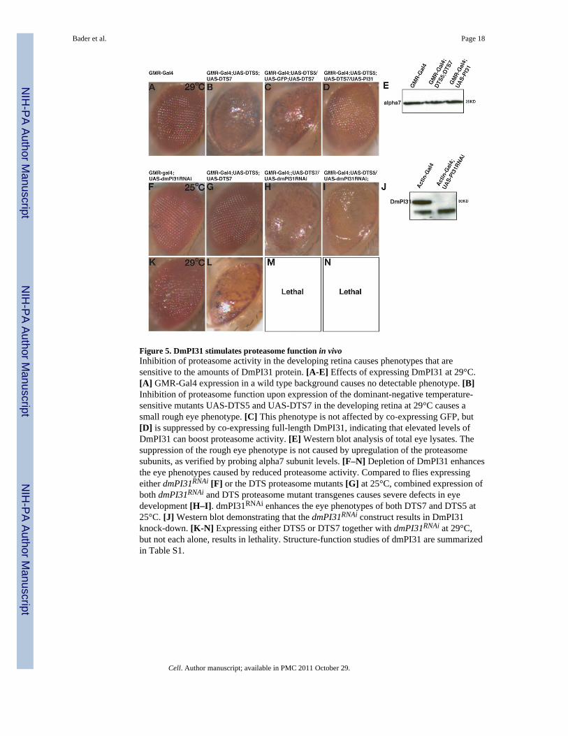
Figure 5. DmPI31 stimulates proteasome function in vivoInhibition of proteasome activity in the developing retina causes phenotypes that aresensitive to the amounts of DmPI31 protein. [A-E] Effects of expressing DmPI31 at 29°C.[A] GMR-Gal4 expression in a wild type background causes no detectable phenotype. [B]Inhibition of proteasome function upon expression of the dominant-negative temperature-sensitive mutants UAS-DTS5 and UAS-DTS7 in the developing retina at 29°C causes asmall rough eye phenotype. [C] This phenotype is not affected by co-expressing GFP, but[D] is suppressed by co-expressing full-length DmPI31, indicating that elevated levels ofDmPI31 can boost proteasome activity. [E] Western blot analysis of total eye lysates. Thesuppression of the rough eye phenotype is not caused by upregulation of the proteasomesubunits, as verified by probing alpha7 subunit levels. [F–N] Depletion of DmPI31 enhancesthe eye phenotypes caused by reduced proteasome activity. Compared to flies expressingeither dmPI31RNAi [F] or the DTS proteasome mutants [G] at 25°C, combined expression ofboth dmPI31RNAi and DTS proteasome mutant transgenes causes severe defects in eyedevelopment [H–I]. dmPI31RNAi enhances the eye phenotypes of both DTS7 and DTS5 at25°C. [J] Western blot demonstrating that the dmPI31RNAi construct results in DmPI31knock-down. [K-N] Expressing either DTS5 or DTS7 together with dmPI31RNAi at 29°C,but not each alone, results in lethality. Structure-function studies of dmPI31 are summarizedin Table S1.
Bader et al. Page 18
Cell. Author manuscript; available in PMC 2011 October 29.
NIH
-PA Author Manuscript
NIH
-PA Author Manuscript
NIH
-PA Author Manuscript

Figure 6. DmPI31 has an essential physiological function and is required for normal proteasomeactivity in vivo[A–F] dmPI31 mutants are lethal, but transgenic expression of DmPI31 in the soma rescuesthis lethality and permits the recovery of adult flies that lack DmPI31 in germ cells. Theseflies display defects in the cell-cycle and protein degradation. The left panel depicts wild-type testes, while the right depicts dmPI31 mutants that have been rescued to adulthood. [A–A′] Distribution of DmPI31 protein in the testis. Testes were stained with and antibodystaining towards DmPI31 (green). The wild type testis displays staining in germ-line stemcells (GSCs) and primary spermatocytes. This staining is lost in dmPI31 mutant testes. Theinset depicts a larger magnification of the apical tip, where these cells reside. [B–B′] BrdU
Bader et al. Page 19
Cell. Author manuscript; available in PMC 2011 October 29.
NIH
-PA Author Manuscript
NIH
-PA Author Manuscript
NIH
-PA Author Manuscript

incorporation assay to detect dividing GSCs. Anti-BrdU staining (green) labeling S-phasecells is detected at the apical tip of the wild type testis. A similar labeling is detected in thedmPI31 mutant testis, indicating that mitotic divisions are not disrupted. Nuclei are stainedwith DAPI (blue). [C–C′] Phospho-histone3 (PH3) antibody staining (green), which marksmeiotic divisions. The PH3 antibody stains dividing nuclei, and therefore detects cells thatare undergoing either mitosis or meiosis. Compared to wild-type testis, which displaystaining in the nuclei of 32 cell cysts, no staining is detected in cysts of dmPI31 mutanttestis, suggesting that meiosis is stunted. An arrow points to a 32-cell cyst (enlarged in inset)where PH3 staining is detected in wild type. [D–D′] Anti-Vasa antibody staining (green).This antibody is specific for germ cell progenitors. During normal differentiation, Vasastaining is strongest in GSCs and primary spermatocytes, and disappears when cellsapproach meiosis. In contrast, dmPI31 mutant testes contain cysts with persistent Vasastaining, indicating that these cells maintain progenitor identity and fail to differentiate. [E–E′] CyclinB staining. CyclinB is normally detected in 16 cell stage cysts and disappears justbefore meiosis completes. In dmPI31 mutant cells, CyclinB persists in 16-cell stage cysts,indicating that the degradation of this protein does not occur normally. See also Figure S4.[F–F′] FK2 staining of ubiquitin-conjugated proteins. FK2 detects the abundance of poly-ubiquitnated proteins and is thus a detector of proteasome activity. In normal germ-line cells(a pre-meiotic cyst is circled), FK2 staining is diffuse, and prominent staining is detected indifferentiating nuclei (asterisks in separate cyst). In contrast, most dmPI31 mutant cellsdisplay punctate staining (arrows point to poly-ubiquitnated protein clusters), indicatingaccumulation of non-degraded poly-ubiquitinated proteins.
Bader et al. Page 20
Cell. Author manuscript; available in PMC 2011 October 29.
NIH
-PA Author Manuscript
NIH
-PA Author Manuscript
NIH
-PA Author Manuscript

Figure 7. Model for proteasome regulation by the Nutcracker-DmPI31 complexDuring their terminal differentiation, spermatids undergo a severe reduction in cell volume.This process, termed “individualization”, requires proteasome activity (Zhong and Belote,2007). Both Nutcracker and DmPI31 are required for normal proteasome activity in thetestis (Bader et al., 2010). The binding of Nutcracker stabilizes DmPI31 by protecting thecarboxy-terminal region of DmPI31 from cleavage. The C-terminal domain of DmPI31 isnecessary for both binding to proteasomes and to stimulate their activity (McCutchen-Maloney et al., 2000). This domain contains a HbYX motif that has been implicated inproteasome gate opening (Smith et al., 2007). The formation and binding of the DmPI31-F-box regulatory complex stimulates proteasome activity and promotes caspase activation andspermatid differentiation. Since the function of Nutcracker is restricted to the testis, whereasDmPI31 has an essential function in somatic cells, it is likely that other factors contribute tothe regulation of DmPI31.
Bader et al. Page 21
Cell. Author manuscript; available in PMC 2011 October 29.
NIH
-PA Author Manuscript
NIH
-PA Author Manuscript
NIH
-PA Author Manuscript

NIH
-PA Author Manuscript
NIH
-PA Author Manuscript
NIH
-PA Author Manuscript
Bader et al. Page 22
Table 1
Genotype Fertility Caspase3 staining dmPI31 cleavage Morphology
Wild-type (yw) 100% yes no normal
ms771(−/−) 0% no yes no IC formed, failure to individualize
ms771(−/−) + nutcracker-WT 100% yes no normal
ms771(−/−) + nutcracker-V-E 0% yes partial incomplete individualization
ms771(−/−) + PI31 0% yes no IC formed failure to individualize
Cell. Author manuscript; available in PMC 2011 October 29.