NIH Common Fund HuBMAP/SCAP Mini Workshop · NIH Common Fund . HuBMAP / SCAP Mini Workshop ....
95
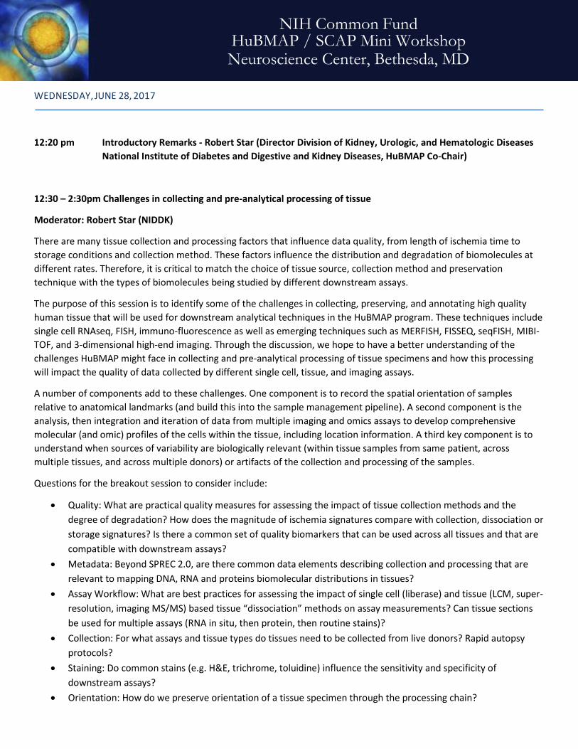
NIH Common Fund HuBMAP / SCAP Mini Workshop Neuroscience Center, Bethesda, MD WEDNESDAY, JUNE 28, 2017 12:20 pm Introductory Remarks - Robert Star (Director Division of Kidney, Urologic, and Hematologic Diseases National Institute of Diabetes and Digestive and Kidney Diseases, HuBMAP Co-Chair) 12:30 – 2:30pm Challenges in collecting and pre-analytical processing of tissue Moderator: Robert Star (NIDDK) There are many tissue collection and processing factors that influence data quality, from length of ischemia time to storage conditions and collection method. These factors influence the distribution and degradation of biomolecules at different rates. Therefore, it is critical to match the choice of tissue source, collection method and preservation technique with the types of biomolecules being studied by different downstream assays. The purpose of this session is to identify some of the challenges in collecting, preserving, and annotating high quality human tissue that will be used for downstream analytical techniques in the HuBMAP program. These techniques include single cell RNAseq, FISH, immuno-fluorescence as well as emerging techniques such as MERFISH, FISSEQ, seqFISH, MIBI- TOF, and 3-dimensional high-end imaging. Through the discussion, we hope to have a better understanding of the challenges HuBMAP might face in collecting and pre-analytical processing of tissue specimens and how this processing will impact the quality of data collected by different single cell, tissue, and imaging assays. A number of components add to these challenges. One component is to record the spatial orientation of samples relative to anatomical landmarks (and build this into the sample management pipeline). A second component is the analysis, then integration and iteration of data from multiple imaging and omics assays to develop comprehensive molecular (and omic) profiles of the cells within the tissue, including location information. A third key component is to understand when sources of variability are biologically relevant (within tissue samples from same patient, across multiple tissues, and across multiple donors) or artifacts of the collection and processing of the samples. Questions for the breakout session to consider include: • Quality: What are practical quality measures for assessing the impact of tissue collection methods and the degree of degradation? How does the magnitude of ischemia signatures compare with collection, dissociation or storage signatures? Is there a common set of quality biomarkers that can be used across all tissues and that are compatible with downstream assays? • Metadata: Beyond SPREC 2.0, are there common data elements describing collection and processing that are relevant to mapping DNA, RNA and proteins biomolecular distributions in tissues? • Assay Workflow: What are best practices for assessing the impact of single cell (liberase) and tissue (LCM, super- resolution, imaging MS/MS) based tissue “dissociation” methods on assay measurements? Can tissue sections be used for multiple assays (RNA in situ, then protein, then routine stains)? • Collection: For what assays and tissue types do tissues need to be collected from live donors? Rapid autopsy protocols? • Staining: Do common stains (e.g. H&E, trichrome, toluidine) influence the sensitivity and specificity of downstream assays? • Orientation: How do we preserve orientation of a tissue specimen through the processing chain?
Transcript of NIH Common Fund HuBMAP/SCAP Mini Workshop · NIH Common Fund . HuBMAP / SCAP Mini Workshop ....
NIH Common Fund HuBMAP / SCAP Mini Workshop Neuroscience Center, Bethesda, MD
WEDNESDAY, JUNE 28, 2017
12:20 pm Introductory Remarks - Robert Star (Director Division of Kidney, Urologic, and Hematologic Diseases National Institute of Diabetes and Digestive and Kidney Diseases, HuBMAP Co-Chair)
12:30 – 2:30pm Challenges in collecting and pre-analytical processing of tissue
Moderator: Robert Star (NIDDK)
There are many tissue collection and processing factors that influence data quality, from length of ischemia time to storage conditions and collection method. These factors influence the distribution and degradation of biomolecules at different rates. Therefore, it is critical to match the choice of tissue source, collection method and preservation technique with the types of biomolecules being studied by different downstream assays.
The purpose of this session is to identify some of the challenges in collecting, preserving, and annotating high quality human tissue that will be used for downstream analytical techniques in the HuBMAP program. These techniques include single cell RNAseq, FISH, immuno-fluorescence as well as emerging techniques such as MERFISH, FISSEQ, seqFISH, MIBI-TOF, and 3-dimensional high-end imaging. Through the discussion, we hope to have a better understanding of the challenges HuBMAP might face in collecting and pre-analytical processing of tissue specimens and how this processing will impact the quality of data collected by different single cell, tissue, and imaging assays.
A number of components add to these challenges. One component is to record the spatial orientation of samples relative to anatomical landmarks (and build this into the sample management pipeline). A second component is the analysis, then integration and iteration of data from multiple imaging and omics assays to develop comprehensive molecular (and omic) profiles of the cells within the tissue, including location information. A third key component is to understand when sources of variability are biologically relevant (within tissue samples from same patient, across multiple tissues, and across multiple donors) or artifacts of the collection and processing of the samples.
Questions for the breakout session to consider include:
• Quality: What are practical quality measures for assessing the impact of tissue collection methods and the degree of degradation? How does the magnitude of ischemia signatures compare with collection, dissociation or storage signatures? Is there a common set of quality biomarkers that can be used across all tissues and that are compatible with downstream assays?
• Metadata: Beyond SPREC 2.0, are there common data elements describing collection and processing that are relevant to mapping DNA, RNA and proteins biomolecular distributions in tissues?
• Assay Workflow: What are best practices for assessing the impact of single cell (liberase) and tissue (LCM, super-resolution, imaging MS/MS) based tissue “dissociation” methods on assay measurements? Can tissue sections be used for multiple assays (RNA in situ, then protein, then routine stains)?
• Collection: For what assays and tissue types do tissues need to be collected from live donors? Rapid autopsy protocols?
• Staining: Do common stains (e.g. H&E, trichrome, toluidine) influence the sensitivity and specificity of downstream assays?
• Orientation: How do we preserve orientation of a tissue specimen through the processing chain?

• Fixing, clearing and embedding: Are there tissue stabilization techniques that can be used before or during collection? For current and emerging fixatives/preservatives of excised tissue, which biomolecular species do they preserve with good fidelity (not only nucleic acids and proteins, but how effective are these techniques at preserving metabolites or carbohydrates), what compatibility issues are there with different tissue types, cell types, dissociation techniques and assays? What are some of the challenges associated with clearing techniques?
• Sectioning: What are tissue-specific considerations in preparing tissue sections? How does the choice of tissue size and format influence ischemia and preservation timing and in term the quality of the tissue for different downstream assays?
• End-users: What format, quantity, and quality level is needed for: RNAseq, DropSeq, MERFISH / FISSEQ / seqFISH, immuno-florescence, MIBI-TOF and CyTOF approaches?
2:30 – 3:00pm Break
3:00 – 5:00 pm Data Analysis, Standards, and Benchmarks for Single Cell Analysis
Moderator: Junhyong Kim (University of Pennsylvania)
Because of the difficulty of obtaining measurements at the single cell scale, the field has been driven by technological advances, including various RNA/DNA sequencing technologies, high-resolution proteomics and metabolomics, multiplexing strategies, cell handling technologies, etc. Despite these technological advances, single cell measurements remain difficult and is fundamentally challenged by the fact that the units of measurement, each cell, has no replication. It has been extremely difficult to assess the efficiency of measurements, establish benchmarks or controls, agree on protocols for data analysis, and coherently define standards for reporting experiments and data analysis. An especially important challenge is placing single cell data in their organismal context, including spatial coordinates.
Questions for this breakout session to consider include:
• Is there benchmark data to compare new experimental or computational methods? • How do we establish material standards such as specific cells or spike-in RNA? • What metadata about calibration is important to know? • What information is important to collect about the sample and its preparation? • How can we work together with manufacturers to build standards into their methods? • Does an ontology need to be established for single cell analysis? • How can we associate single cells to tissue orientation information? More generally, how can data be organized
from the single cell scale to whole organism scale? • What are the common data elements between imaging and sequencing assays? Is there a common header we
can use for all data, similar to FITS or DICOM?
5:00 pm Closing Remarks

Suggested background reading for these breakouts:
• Unhale, S. A., Skubitz, A. P., Solomon, R., & Hubel, A. (2012). Stabilization of tissue specimens for pathological examination and biomedical research. Biopreservation and biobanking, 10(6), 493-500. [http://online.liebertpub.com/doi/abs/10.1089/bio.2012.0031]
• Hubel, A., Spindler, R., & Skubitz, A. P. (2014). Storage of human biospecimens: selection of the optimal storage temperature. Biopreservation and biobanking, 12(3), 165-175. [http://online.liebertpub.com/doi/abs/10.1089/bio.2013.0084]
• Hubel, A., Aksan, A., Skubitz, A. P., Wendt, C., & Zhong, X. (2011). State of the art in preservation of fluid biospecimens. Biopreservation and biobanking, 9(3), 237-244. [http://online.liebertpub.com/doi/abs/10.1089/bio.2010.0034]
• Chung, Cho H, Hewitt SM (2016). The paraffin-embedded RNA metric (PERM) for RNA isolated from formalin-fixed, paraffin-embedded tissue. Biotechniques. May 1;60(5):239-44 [http://www.biotechniques.com/BiotechniquesJournal/2016/May/The-paraffin-embedded-RNA-metric-PERM-for-RNA-isolated-from-formalin-fixed-paraffin-embedded-tissue/biotechniques-364401.html]
• Carithers, L. J., Ardlie, K., Barcus, M., Branton, P. A., Britton, A., Buia, S. A., ... & Guan, P. (2015). A novel approach to high-quality postmortem tissue procurement: the GTEx project. Biopreservation and biobanking, 13(5), 311-319. [http://online.liebertpub.com/doi/full/10.1089/bio.2015.0032]

NIH Common Fund
Single Cell Analysis Program (SCAP)
Human BioMolecular Atlas Program (HuBMAP)
Tissue Acquisition and Metadata Standards Workshop
Robert A. Star, MDNIDDK
https://commonfund.nih.gov/
DisclosureMy laboratory is involved in research to improve pre-analytic processing steps after tissue biopsy. I am an inventor on a provisional patent application for a tissue transfer device that reduces tissue damage after biopsy.I am an inventor on a provisional patent application for a chemically engineered fixative (BE70G) that does not contain formaldehyde, and improves molecular analyses.If NIH successfully commercializes the inventions, I may receive royalty payments using standard NIH formulas.

NIH HuBMAP Working GroupCo-Chairs:Gary Gibbons, M.D. (NHLBI)Roderic Pettigrew, Ph.D., M.D. (NIBIB)Robert Star, M.D. (NIDDK)
Working Group Coordinators: Zorina Galis, Ph.D. (NHLBI)Deborah K. Hoshizaki, Ph.D. (NIDDK)
Common Fund Program Leader:Richard Conroy. Ph.D., M.B.A. (OD)
Members:David Balasundaram, Ph.D.(CSR)Jenna Baker, Ph.D.(NIDDK)Andrea Beckel-Mitchener, Ph.D. (NIMH)Francesca Bosetti, Pharm. D., Ph.D. (NINDS)Katarzyna Bourcier, Ph.D. (NIAID)Robert Carter, M.D. (NIAMS)Tony Casco (OD)Elizabeth Church, Ph.D. (NIAID)Jennifer Couch, Ph.D. (NCI)Sarah Dunsmore, Ph.D. (NIGMS)
Joseph G. Gindhart, Ph.D. (NIGMS)Patricia Greenwel, Ph.D. (NIDDK)Jill Heemskerk, Ph.D. (NIBIB)Shannon Hughes, Ph.D. (NCI)Halonna Kelly, Ph.D. (NIAID)J. Randy Knowlton, Ph.D. (NCI)Lillian S. Kuo, Ph.D. (NIAID)Jerry Li, Ph.D. (NCI)Sara Lin, Ph.D. (NHLBI)Margaret Ochocinska, Ph.D. (NHLBI)Aaron Pawlyk, Ph.D. (NIDDK)Ajay Pillai, Ph.D. (NHGRI)Ipolia Ramadan, Ph.D. (NINDS)Krystyna Rys-Sikora, Ph.D. (NIDDK)John Satterlee, Ph.D. (NIDA)Tonya Scott (OD)Salvatore Sechi, Ph.D. (NIDDK)Kentner Singleton, Ph.D. (NIAID)Jessica Smith, Ph.D.(OD)Pothur Srinivas, Ph.D. (NHLBI)Reiko Toyama, Ph.D. (NICHD)José M. Velázquez, Ph.D. (NIA)Yong Yao, Ph.D. (NIMH)
https://commonfund.nih.gov/HuBMAP/index

HuBMAP: Background• In past, cells classified by structure, function, location in
tissue, histologic staining• Opportunity:
– Massively parallel single cell analysis (genomics) assays– Computational algorithms to find types, sub-types, states,
transitions; – Imaging at growing scale and resolution
• New paradigm: Classify cells and tissue components based on molecular omic profile
• Critical questions (for example)– How do cells vary within structure; within tissue; across tissues,
systems, and organs (vasculature, supportive cells). – Are there undiscovered sub-compartments, rare cells– How do cells interact (ligands, receptors)– How do cells influence health and disease
https://commonfund.nih.gov/HuBMAP/index
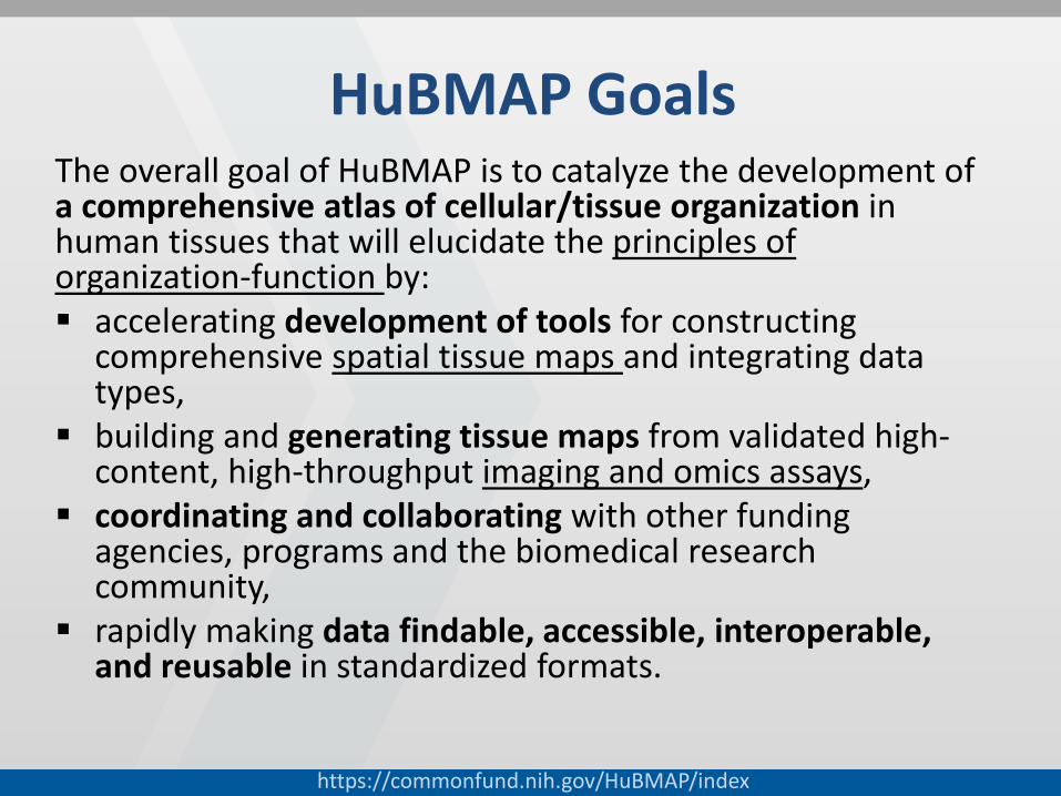
HuBMAP GoalsThe overall goal of HuBMAP is to catalyze the development of a comprehensive atlas of cellular/tissue organization in human tissues that will elucidate the principles of organization-function by: accelerating development of tools for constructing
comprehensive spatial tissue maps and integrating data types,
building and generating tissue maps from validated high-content, high-throughput imaging and omics assays,
coordinating and collaborating with other funding agencies, programs and the biomedical research community,
rapidly making data findable, accessible, interoperable, and reusable in standardized formats.

HuBMAP VisionIf successful, this program will lead to a data resource like “Google Maps” for tissues in the human bodythat will give rise to new insights into inter-individual variation and tissue changes across the lifespan, and serve as baseline for understanding disease.

What is a tissue atlas?Kidney pathology of the future
Analyze single cells/tissue to find tissue markers (cells, and interstitial areas between cells)then paint cells, structures, cell trajectory (healthy, injured, repair, regen), activated pathways
Understand heterogeneity between regions, neighboring cellsEven better, use 3D imaging to better see interstitium, glomerulus
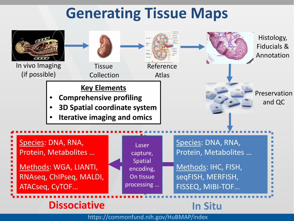
Generating Tissue Maps
Dissociative In Situ
Species: DNA, RNA, Protein, Metabolites …
Methods: WGA, LIANTI, RNAseq, ChIPseq, MALDI, ATACseq, CyTOF…
Laser capture,Spatial
encoding, On tissue
processing …
Species: DNA, RNA, Protein, Metabolites …
Methods: IHC, FISH, seqFISH, MERFISH, FISSEQ, MIBI-TOF…
In vivo Imaging (if possible)
Tissue Collection
ReferenceAtlas
Histology, Fiducials & Annotation
Key Elements• Comprehensive profiling• 3D Spatial coordinate system• Iterative imaging and omics
Preservationand QC
https://commonfund.nih.gov/HuBMAP/index
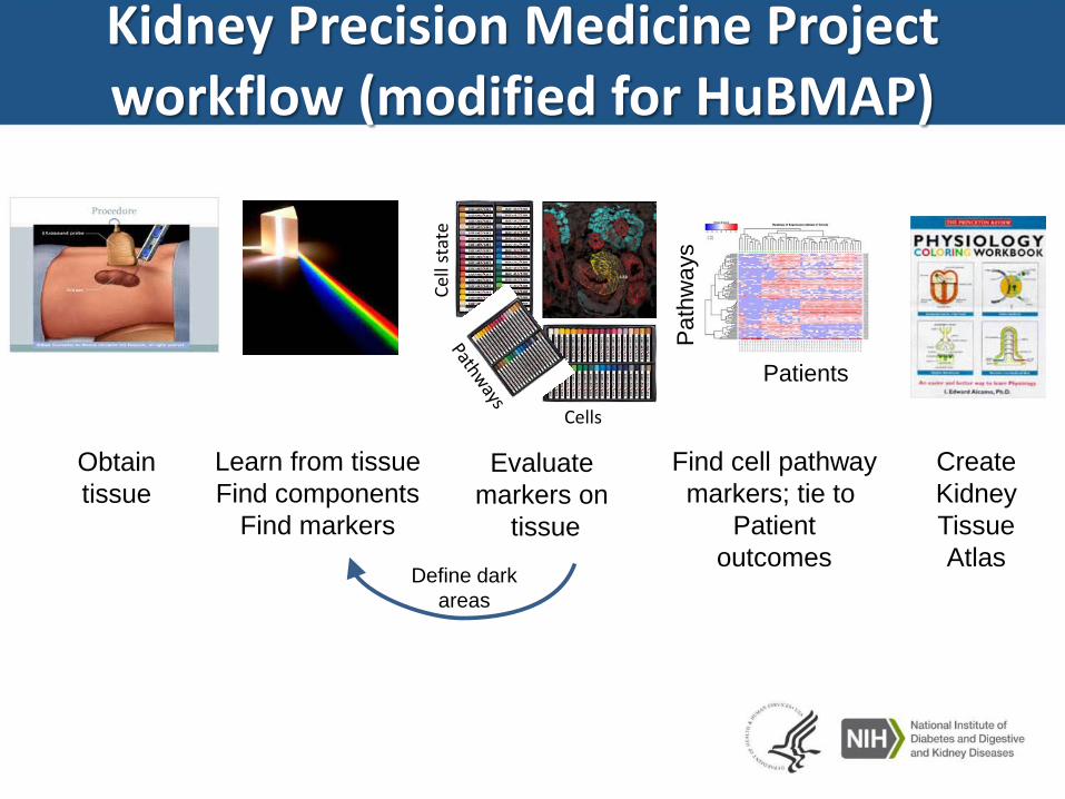
Kidney Precision Medicine Projectworkflow (modified for HuBMAP)
Obtaintissue
Learn from tissueFind components
Find markers
Evaluatemarkers on
tissue
CellsCe
ll st
ate
Create KidneyTissue Atlas
Define darkareas
Find cell pathwaymarkers; tie to
Patientoutcomes
Patients
Pat
hway
s

Identifying Key Areas in a Human BioMolecular Atlas
Planning WorkshopJune 15, 2016
Areas with challenges and opportunities for investment by the NIH:
1. Sourcing high quality tissue from multiple organ sites
2. Processing and preserving tissue for multiple imaging and omics assays
3. Quality control, validation and variation in data generation
4. Data coordination across multiple acquisition techniques
5. Annotation, curation and archiving of the data
6. Browsing, visualizing and searching the data
7. Building statistical and analytic techniques and models for nonlinear analysis of highly multidimensional data
8. Community engagement
Challenges in collecting and pre-analytical processing of tissue
Data analysis, standards, and benchmarks for single cell analysis

https://commonfund.nih.gov/HuBMAP/index
Questions?
What’s next?• FOAs this Fall• Program rolled out in phases• Mini workshop June 28, 2017
– Pre-analytic processing– Metadata
• Single Cell Analysis Investigators Meeting, June 29-30, 2017


Challenges in collecting and pre-analytical processing of
tissue
Robert A. Star, MDNIDDK
https://commonfund.nih.gov/
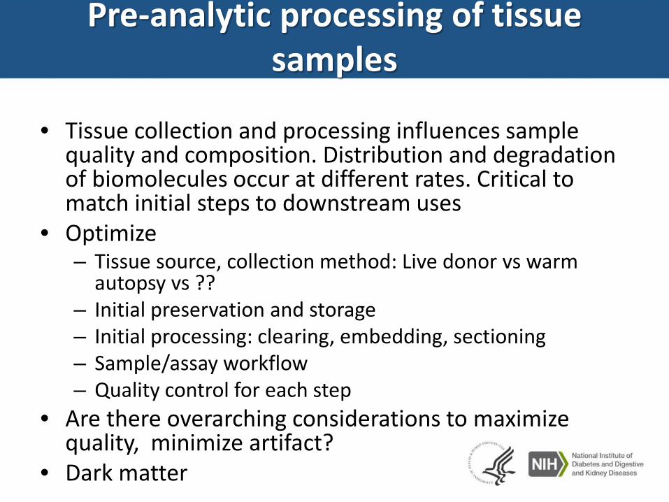
Pre-analytic processing of tissue samples
• Tissue collection and processing influences sample quality and composition. Distribution and degradation of biomolecules occur at different rates. Critical to match initial steps to downstream uses
• Optimize– Tissue source, collection method: Live donor vs warm
autopsy vs ??– Initial preservation and storage– Initial processing: clearing, embedding, sectioning– Sample/assay workflow– Quality control for each step
• Are there overarching considerations to maximize quality, minimize artifact?
• Dark matter
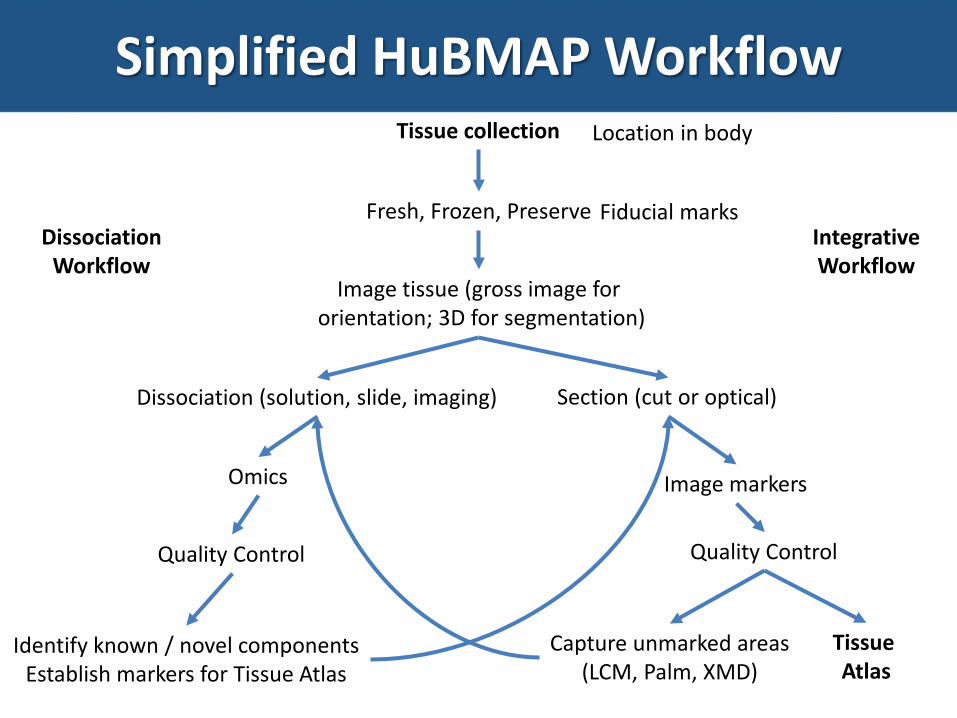
Simplified HuBMAP WorkflowTissue collection
Fresh, Frozen, Preserve
Image tissue (gross image fororientation; 3D for segmentation)
Section (cut or optical)Dissociation (solution, slide, imaging)
Image markers
Capture unmarked areas(LCM, Palm, XMD)
Quality Control
Location in body
Fiducial marks
Omics
Tissue Atlas
Identify known / novel componentsEstablish markers for Tissue Atlas
Quality Control
DissociationWorkflow
IntegrativeWorkflow

Many areas for ImprovementTissue collection
Fresh, Frozen, Preserve
Image tissue (gross image fororientation; 3D for segmentation)
Section (cut or optical)Dissociation (solution, slide, imaging)
Image markers
Capture unmarked areas(LCM, Palm, XMD)
Quality Control
Location in body
Fiducial marks
Omics
Tissue Atlas
Identify known / novel componentsEstablish markers for Tissue Atlas
Quality Control
DissociationWorkflow
IntegrativeWorkflow

Examples of dark matterTissue preservation / fixation step• Formalin bad for RNA, some proteins (‘antigen retrieval’)• Frozen bad for histology• Fresh tissue is fragile• Need better preservation steps that match preservation to
intended downstream uses (fit for context)
Single cell dissociation step• Digestion (37oC) leaves mRNA signature• Selective for mobile inflammatory cells• Not many cells analyzed• Very few structural (organ) cells analyzed• Need less destructive dissociation technologies

How to make espresso
How avoid sour or bitter espresso?

Grid, volume, temperature influence extraction, and taste. Need optimization process
Bottom line: difficult, need balance multiple factors

Innovation: Look for alternative
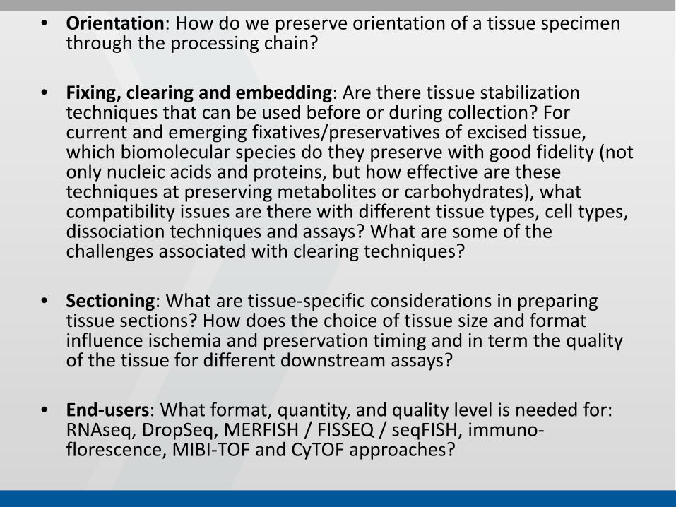
• Orientation: How do we preserve orientation of a tissue specimen through the processing chain?
• Fixing, clearing and embedding: Are there tissue stabilization techniques that can be used before or during collection? For current and emerging fixatives/preservatives of excised tissue, which biomolecular species do they preserve with good fidelity (not only nucleic acids and proteins, but how effective are these techniques at preserving metabolites or carbohydrates), what compatibility issues are there with different tissue types, cell types, dissociation techniques and assays? What are some of the challenges associated with clearing techniques?
• Sectioning: What are tissue-specific considerations in preparing tissue sections? How does the choice of tissue size and format influence ischemia and preservation timing and in term the quality of the tissue for different downstream assays?
• End-users: What format, quantity, and quality level is needed for: RNAseq, DropSeq, MERFISH / FISSEQ / seqFISH, immuno-florescence, MIBI-TOF and CyTOF approaches?

• Quality: What are practical quality measures for assessing the impact of tissue collection methods and the degree of degradation? How does the magnitude of ischemia signatures compare with collection, dissociation or storage signatures? Is there a common set of quality biomarkers that can be used across all tissues and that are compatible with downstream assays?
• Metadata: Beyond SPREC 2.0, are there common data elements describing collection and processing that are relevant to mapping DNA, RNA and proteins biomolecular distributions in tissues?
• Assay Workflow: What are best practices for assessing the impact of single cell (liberase) and tissue (LCM, super-resolution, imaging MS/MS) based tissue “dissociation” methods on assay measurements? Can tissue sections be used for multiple assays (RNA in situ, then protein, then routine stains)?
• Collection: For what assays and tissue types do tissues need to be collected from live donors? Rapid autopsy protocols?
• Staining: Do common stains (e.g. H&E, trichrome, toluidine) influence the sensitivity and specificity of downstream assays?

Why are we here?
• What is working, working well?• What are the weak links in chain?• What tools, techniques are needed before
going into production phase?

Advancing the preservation of tissue biospecimens
AllisonHubel,PhDBiopreservationCoreResource(BioCoR)Univer
sityofMinnesota
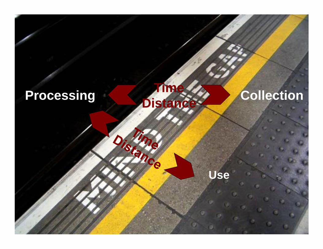
Processing
Processing
Use
Collection Time Distance

BioCoR Resources
Research
Service
Education

InTechOpen.com
BioCoR Service • PDX models have
complex work flow • Viability must be
maintained along this workflow
BioCoR tasks: • Develop short term
storage solution suitable for resected tumors
• Develop effective cryopreservation protocols for xenografts
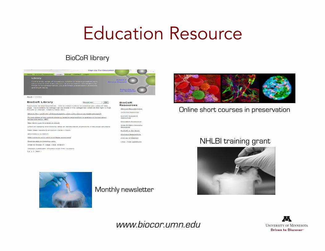
Education Resource BioCoR library
Online short courses in preservation
NHLBI training grant
Monthly newsletter
www.biocor.umn.edu

BioCoR Research Why do tissues respond poorly (compared to cells isolated
from the same tissue)?
Freezing response of isolated iPS cells (Imaged at -50oC)
1 °C/min
Ice
Area/µm2 104.8 105.8 71.6 94.5 114.7 81.5 111.8 138.5
Aic-s 0.15 0.07 0 0 0 0 0.12 0.16
3 °C/min
Ice
Area/µm2 113.2 74.6 160.2 129.0 117.8 118.2 132.1 119.8
Aic-s 0.08 0.26 0.37 0.42 0.15 0.24 0.33 0
10 °C/min
Ice
Area/µm2 148.3 159.1 223.6 128.6 252.6 189.1 157.7 132.3
Aic-s 0.11 0.49 0.67 0.43 0.65 0.46 0.20 0.29
Scale bar: 3 µm

BioCoR Research
1 °C/min
3 °C/min
10 °C/min
C1-1 C1-2
C3-1 C3-2
C10-1 C10-2
• We can interrogate • small aggregates (3-5
cells) • Full sized colonies
• A wide range of signals can be detected
• Water (liquid or ice) • DMSO • Cryoprotective agents • Proteins • DNA
• These signals can help answer the central questions

Five to ten years from now…
• Dispel the myth of the ‘cold black box’ • Improve and disseminate preservation protocols • Improve preservation of tissue

Acknowledgements Collaborators: Students:
Katie Pollock Peter Dosa Guanglin Yu Dave McKenna Rui Li
Jane Vanderkooi Yan Rou Yap Elizabeth Moy
Chia Hsing Pi Marissa Koran
Funding: R21EB016247 R25HL128372


Disclosures• “Employee” Of The US Federal Government
– Inventor On Multiple Technologies For Which The Intellectual Property Is Assigned To The Federal Government
• For Those Inventions That Are Licensed, I Am Eligible To Receive Royalties, As Stipulated In US Code, Title 15, Chapter 63.
• Editor-in-Chief, Journal of Histochemistry & Cytochemistry
• Chair-holder, Subcommittee on Immunohistochemical Assays, Clinical & Laboratory Standards Institute
• Food & Drug Administration, Center For Devises & Radiological Health – Consultant, Hematology & Pathology Devices Panel

Quality Is EverythingQuality Remains Subjective
• Tissue Quality– Histology– Proteins– Nucleic Acids
• Clinical Data– Complete– Detailed– Defined

Development of A TissueHandling Protocol
• Historic Perspective– Histology & Protein – FFPE Small Study- Frozen– Nucleic Acids – Frozen Large Study – FFPE
• Recommended Strategy– Fit-For-Purpose Define Goals
Physiology Is BiologySpecimen Preservation Is Chemistry

Chemical Preservation• Formalin
• PaxGene• Ethanol• ……….
• Lack Of Scientific Underpinning• Two Broad Classes
– Acid/Aldehyde Fixatives– Coagulative Fixatives
• Underlying Critical Factors– Ischemia– Size– Time Temp?– Impregnation– Storage
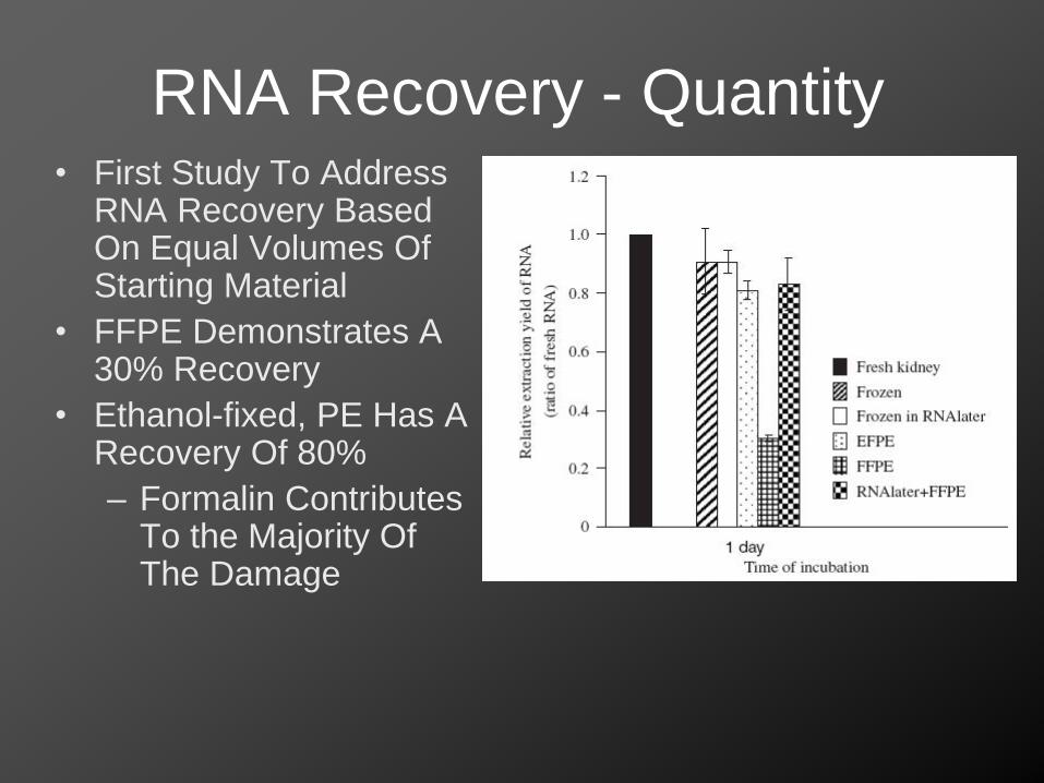
RNA Recovery - Quantity• First Study To Address
RNA Recovery Based On Equal Volumes Of Starting Material
• FFPE Demonstrates A 30% Recovery
• Ethanol-fixed, PE Has A Recovery Of 80%– Formalin Contributes
To the Majority Of The Damage

RNA Recovery From Tissue
0
1
5' UTR Exon 1 Exon2 Exon3 3' UTR poly A
Frozen AnticipatedModel 1Model 2Model 3
AAAA
AAAA
TTTT
RNA In FFPE -Revised Results Of RT-PCR
AAAA
TTTT
RNA In FFPE -AnticipatedRNA In Frozen Tissue
?

Quantitative Amplification Based On RNA Source & Primer Location
5’ ORF Middle of ORF 3’ UTR
Region of GAPDH gene
Rel
ativ
e ex
pres
sion
leve
l of G
APDH
gene
(rela
tive
to3’
UTR
regi
on o
f fre
sh ra
t kid
ney;
log 2
)
FreshRT37oCRNAlaterFFFFPE
0.0000
0.0001
0.0010
0.0100
1.0000
10.0000
0.1000
Same Quantity of Starting RNA
Random Prime cDNA

Tissue Collection, Handing & Processing
• No Such Thing As “Standard Protocol”• Multiple Steps, Multiple Parameters

Effect Of Fixation Time On RNA Quality
200
500
1000
2000
4000
6000
bp
28S
18S
Froz
en K
idne
y
Mar
ker
0 12 24 36 48 72
Fixation time (h)
Fluo
resc
ence
2
3
4
5
1
0
Time (sec)
25 30 35 40 45 50 55 60 65
0 h
24 h12 h
48 h36 h
72 h
B
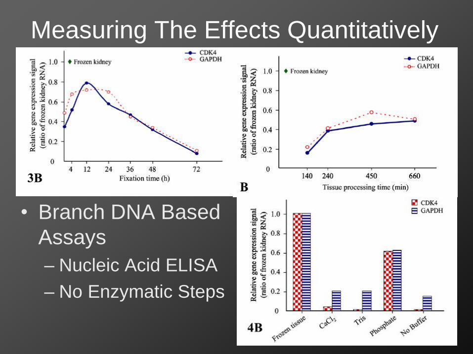
Measuring The Effects Quantitatively
• Branch DNA Based Assays– Nucleic Acid ELISA– No Enzymatic Steps
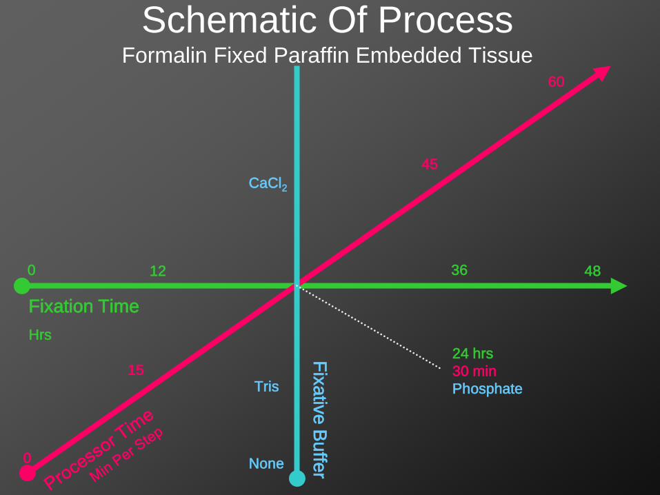
Schematic Of ProcessFormalin Fixed Paraffin Embedded Tissue
Fixation TimeHrs
Fixative Buffer0
15
60
0 12 36 48
CaCl2
None
Tris
24 hrs30 minPhosphate
45

Revised Model Of Chemical Fixation• Tissue Hypoxia & Switching To Glycolysis -
“Drowning”– RNA stores consumed, Alterations in Phospho-
Proteome• Infiltration & Inhibition Of Glycolysis &
Oxidative Phosphorylation – Halting Of Most Biologic Process
• Chemical Reactions Crosslinking Proteins and Nucleic Acids– Halting Of Remaining Enzymatic Activity

DNA
RNA
Protein (WB)

Chemical Fixation• Aldehyde Fixatives Are Two-Step Fixatives
– Coagulative– Acid/Aldehyde Crosslinking - Degradation
• Alcohol Fixatives Are Single-Step Fixatives– Coagulative– No Acid-Base Degradative Chemistry
• Coagulative Fixatives Are More Stable & Result In Improved Biomolecular Analytes

Experimental Pathology LaboratoryLaboratory of Pathology, Center for Cancer Research, National Cancer Institute, National Institutes of Health
Joon-Yong Chung Food & Drug AdministrationCenter For Device & Radiologic HealthHanbyoul Cho
Candice Perry NEPTUNE / CureGN Jeffrey Hansen
Kris Ylaya Former Laboratory Members
Russell Bandle Till Braunschweig Petra LenzMikiko Takikita Kant Matsuda Ran Xie Catherine Conway
Philip Song Haru Kitano Chel-Hun Choi
Victoria Burton Reginald Williams
Kimberly Tuttle Yvonne GathrightLangston Lim Jennifer Martinez


Challenges in collecting and pre-analytical processing of tissue:
the human arterial wall
Chiara Giannarelli, MD, PhDAssistant Professor of Medicine, CardiologyAssistant Professor of Genetics and Genomics SciencesIcahn School of Medicine at Mount Sinai
NIH Common FundHuBMAP / SCAP Mini WorkshopJune 28, 2017Neuroscience Center, Bethesda, MD
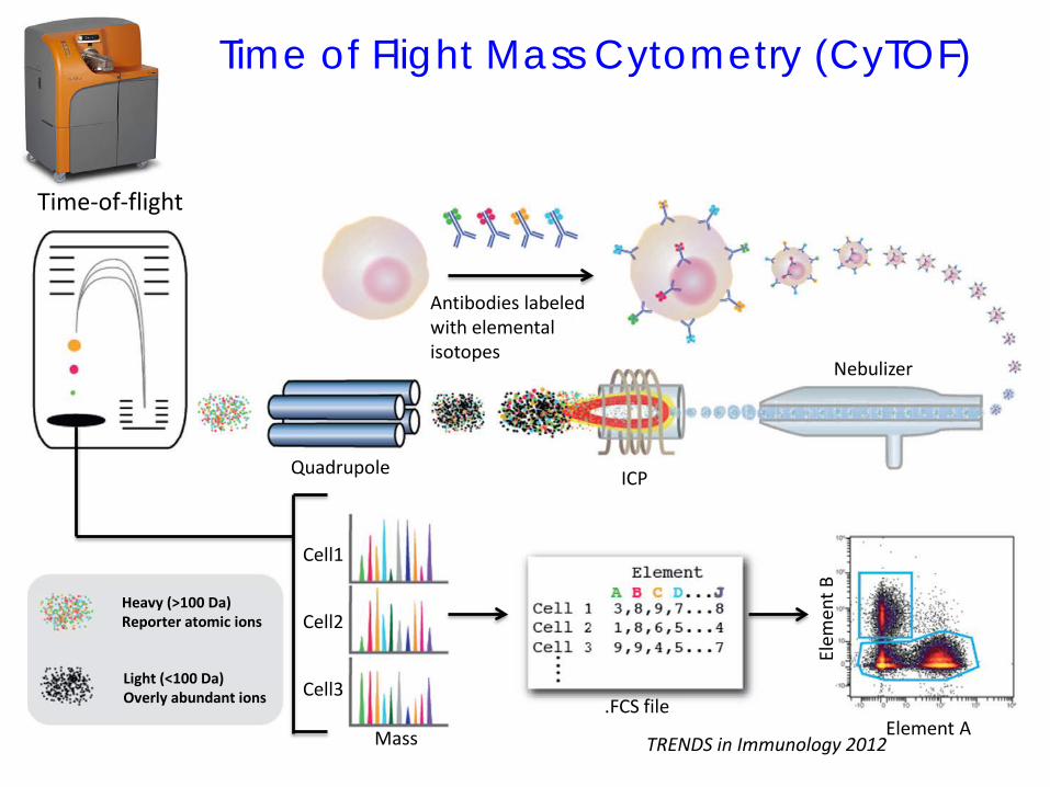
Time of Flight Mass Cytometry (CyTOF)
Time-of-flight
Antibodies labeled with elemental isotopes
Nebulizer
ICPQuadrupole
Cell1
Cell2
Cell3
Heavy (>100 Da)Reporter atomic ions
Light (<100 Da)Overly abundant ions .FCS file
Mass Element A
Elem
ent B
TRENDS in Immunology 2012

Experimental PipelineCyTOF Single-Cell Analysis
Barcoding
Cell isolation
Atherosclerotic Tissue
BloodHigh-Dimensional Single-Cell Analysis
Across Tissues (CyTOF)
Lai, Cytometry 2015.Amir, Nature Biotechnology 2013
viSNE tissue vs. PBMCs

Multiplexed IHC for Validation and Tissue Discrimination
26
2513

Experimental Workflow: challenges
?
In the lab
90% of the challenges
1. Alteration of surface markers2. Alteration of functional state3. Incomplete or too harsh digestion4. Minimize the digestion time: live cell recovery5. Tissue debris (collagen, elastin, calcium deposits)6. Blood contamination
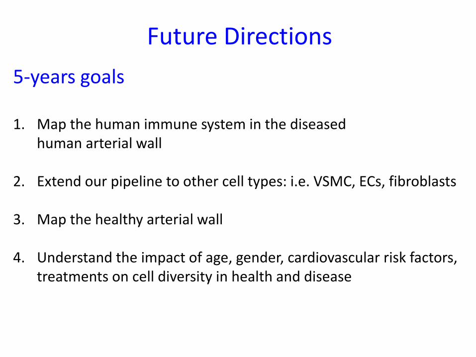
Future Directions5-years goals
1. Map the human immune system in the diseased human arterial wall
2. Extend our pipeline to other cell types: i.e. VSMC, ECs, fibroblasts
3. Map the healthy arterial wall
4. Understand the impact of age, gender, cardiovascular risk factors, treatments on cell diversity in health and disease

Future Directions
10-years goalsTo identify of tissue-specific immune and other cell type variations to
provide new mechanist insights for the rational design of immunotherapies in atherosclerosis and to preserve vascular health

Schematic representation of the computational workflow for the repositioning approach used to identify candidate drugs targeting RGN42.
Drug A
NIH LINCS Program
LIBRARY OF INTEGRATED NETWORK-BASED CELLULAR SIGNATURES
We are here!!!
R21TR001739
Network-driven drug repositioning approaches to treat CAD

Future Directions
10-years goalsTo identify of tissue-specific immune and other cell type variations to
provide new mechanist insights for the rational design of immunotherapies in atherosclerosis and to preserve vascular health
To integrate non-invasive imaging modalities for precision diagnosis and personalized treatments

Systems Biology of Human Atherosclerotic Arterial Wall
Protein Networks
Imaging
Gene networks
Cellular Networks
Drs. Fayad and Calcagno-TMII

How to build an ideal future state
1. Standardized SOP across different lab
2. Data sharing policy to build a human atlas
3. Establish collaborative multidisciplinary environment for investigators
4. Rigorous collection of health and disease information for each individual
5. Interdisciplinary working groups (bioinformatics, biology, medical background)

Thank you !

Acknowledgements Giannarelli LabNayaab KhanPauline MuryPeik Sean ChongRoza ShamailovaChristian Pina
Mount Sinai PriSMMiriam MeradAdeeb RahamnEl-ad David AmirSeunghee Kim-SchulzeAleksey ChudnovoskiySasha GnaticRomain Remark
Vascular Surgery Peter FariesNeurosurgeryJ MoccoAhmed J. Awad
BioinformaticsAvi Maya’anSimon KoplevNick Fernandez
Genetics Dept.Johan BjorkegrenJoel Dudley
Oxford UniversityClaudia MonacoThanks!
K23HL111339R03HL135289
R21TR001739

Optical Microscopy and Analysis Laboratory
Stephen J. Locketthttps://confocal.cancer.gov/cores/optical-microscopy-and-analysis-laboratory
The Frederick National Laboratory is a Federally Funded Research and Development Center operated by Leidos Biomedical Research, Inc., for the National Cancer InstituteDEPARTMENT OF HEALTH AND HUMAN SERVICES • National Institutes of Health • National Cancer Institute
Hair follicle
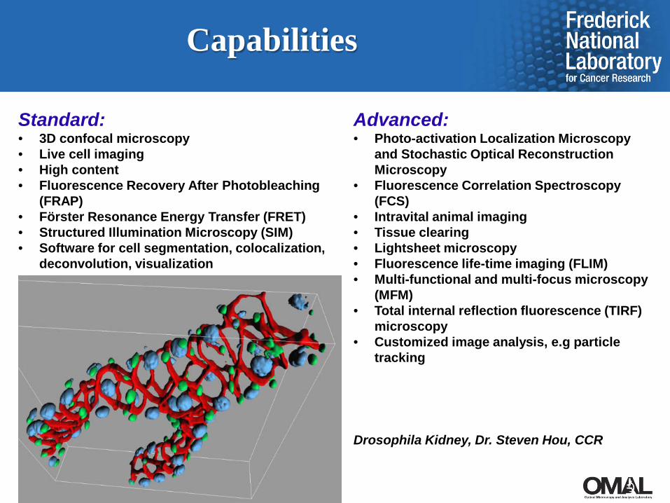
Capabilities
Standard:• 3D confocal microscopy• Live cell imaging• High content• Fluorescence Recovery After Photobleaching
(FRAP)• Förster Resonance Energy Transfer (FRET)• Structured Illumination Microscopy (SIM)• Software for cell segmentation, colocalization,
deconvolution, visualization
Advanced:• Photo-activation Localization Microscopy
and Stochastic Optical Reconstruction Microscopy
• Fluorescence Correlation Spectroscopy (FCS)
• Intravital animal imaging• Tissue clearing• Lightsheet microscopy• Fluorescence life-time imaging (FLIM)• Multi-functional and multi-focus microscopy
(MFM)• Total internal reflection fluorescence (TIRF)
microscopy• Customized image analysis, e.g particle
tracking
Drosophila Kidney, Dr. Steven Hou, CCR
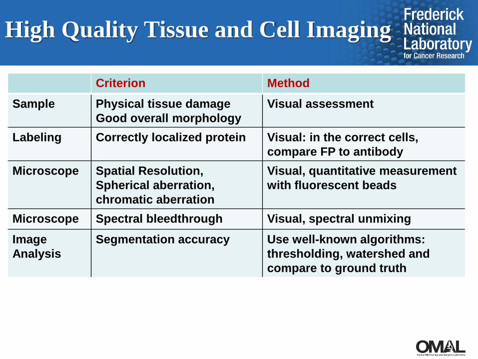
High Quality Tissue and Cell Imaging
Hair follicle
Criterion MethodSample Physical tissue damage
Good overall morphologyVisual assessment
Labeling Correctly localized protein Visual: in the correct cells, compare FP to antibody
Microscope Spatial Resolution,Spherical aberration, chromatic aberration
Visual, quantitative measurement with fluorescent beads
Microscope Spectral bleedthrough Visual, spectral unmixingImage Analysis
Segmentation accuracy Use well-known algorithms: thresholding, watershed and compare to ground truth

2D and 3D Cell and Nucleus Segmentation
Hair follicle
Basic Research: Some interaction generally OK
Drug screening: Automatic. Some degree of error can be tolerated
Pathology: Mixture of automatic and interactive. Inherent sample heterogeneity.
EMT panel on mouse xenograft of human gastric cell line, MKN45. In collaboration with Dr. Robert Kinders, Leidos / DCTD

Algorithms for 3D Segmentation
Hair follicle
Graph cut segmentation:
- One click per nucleus- Plus correction points- Finds optimal surface
Nandy et at, IEEE Selected Topics in Signal Processing Special Issue on Advanced Signal Processing in Microscopy and Cell Imaging. 2016

3D Segmentation Results
Hair follicle
Automatic segmentation can be trusted
Volume rendering of low density of nuclei
Volume rendering of high density of nuclei
Need semi-interactive segmentation

3D Ground Truth for Validation
1) Segment actual 3D images as accurately as possible.2) Treat the segmented image as the “perfect” image.3) Artificially reintroduce the distortions of 3D microscopy: 3D PSF and noise, then use
this image to test segmentation methods
Simulated image
Ground truth segmentation Graph-cut segmentation
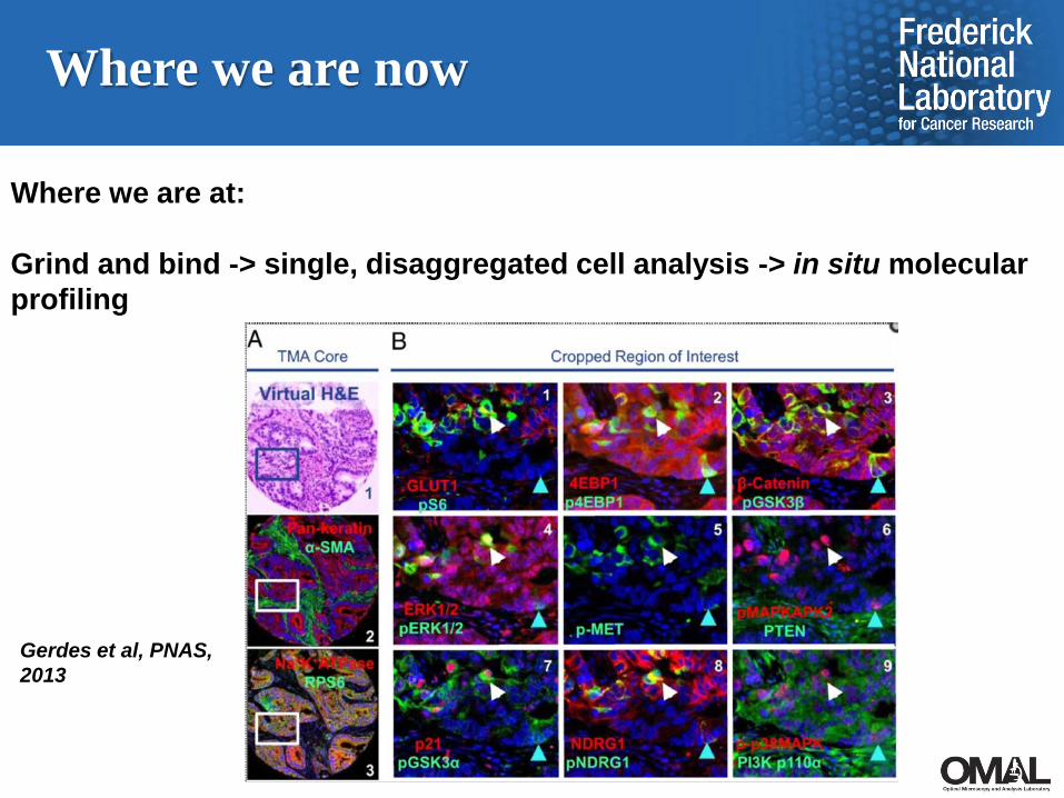
Where we are now
Where we are at:
Grind and bind -> single, disaggregated cell analysis -> in situ molecular profiling
Gerdes et al, PNAS, 2013

Five Years from Now
In situ molecular profiling -> tissue / cell / nucleus structure -> phenotype
SOPs for: - tissue collection- staining- clearing- 3D image acquisition- 3D image analysis- visual representation of results
) e
190 µm
Image acquired with two photon (2P3D microscopy of TDE cleared tissu
2017 HuBMAP Mini-workshop:Data Analysis, Standards, and
Benchmarks for Single Cell Analysis
Junhyong KimUniversity of Pennsylvania

Questions
• Is there benchmark data to compare new experimental or computational methods?
• How do we establish material standards such as specific cells or spike-in RNA?
• What metadata about calibration is important to know?
• What information is important to collect about the sample and its preparation?

Questions
• How can we work together with manufacturers to build standards into their methods?
• Does an ontology need to be established for single cell analysis?
• How can we associate single cells to tissue orientation information? More generally, how can data be organized from the single cell scale to whole organism scale?
• What are the common data elements between imaging and sequencing assays? Is there a common header we can use for all data, similar to FITS or DICOM?

Agenda:
• Overview (3:00-3:15)• Breakout sessions (3:20-4:00)• Summary of breakout sessions and synthesis (4:10-
5:00)

Breakout Sessions
• Material Standards and Benchmarks• Calibration, QC, and Instruments• Experimental Designs, Ontologies, and Metadata• Data Integration, Scale Alignment, and Data
Analysis

Material Standards and Benchmarks
• Standard Cells?• Control RNA?• Compendium Data as Benchmarks?

ERCC probes: 20 levels spanning 106 range

Calibration, QC, and Instruments
• Can there be an instrument calibration protocol?• Quality Control protocol?• Commercial instruments: standards and
comparisons?

40-fold
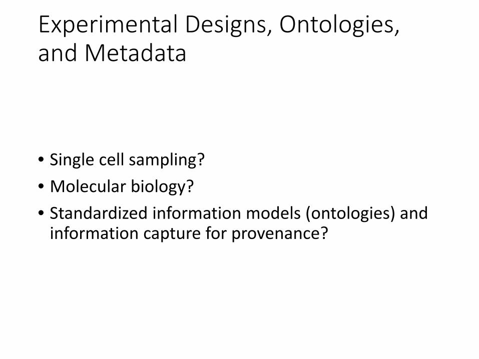
Experimental Designs, Ontologies, and Metadata
• Single cell sampling?• Molecular biology?• Standardized information models (ontologies) and
information capture for provenance?
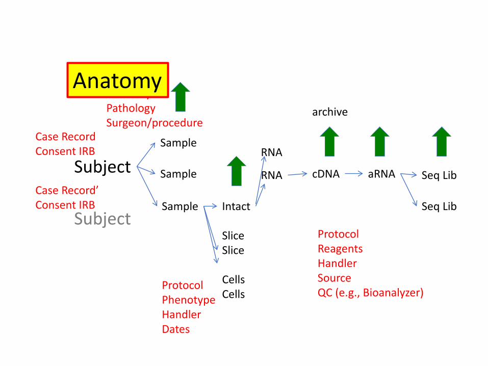
Subject
Subject
Case RecordConsent IRB
Case Record’Consent IRB
AnatomyPathologySurgeon/procedure
Sample
Sample
Sample Intact
SliceSlice
CellsCells
ProtocolPhenotypeHandlerDates
ProtocolReagentsHandlerSourceQC (e.g., Bioanalyzer)
cDNA aRNA Seq Lib
Seq Lib
archive
RNA
RNA
Anatomy

Sampling Variance Associated with RNA sequencing
Single Cell:~0.2-1 pg
mRNA= 1x10⁵ –5x10⁵ 3kb molecules
RNA amplification: ~10ng-100ng5x109 ~5x10¹⁰
300-500bp molecules
Library:Input 10ng
PCR amplification
5x10¹²
molecules
Load:4pM
2x10¹⁰
molecules
Read:104-107 spots
x104-105 x10-2104 x10-2 x10⁻⁴-10-7

Data Integration, Scale Alignment, and Data Analysis
• What are the common data elements between imaging and sequencing assays?
• How do we integrate information from different modalities?
• What are the signal to noise characteristics of various single cell platforms?
• How do we align information from single cell scales, to tissues, to organs, to whole bodies?

Replicate variance as a function of expression levelsLo
g of
obs
erve
d va
rianc
e
P=10-9 P = 0.05

Scale Alignment

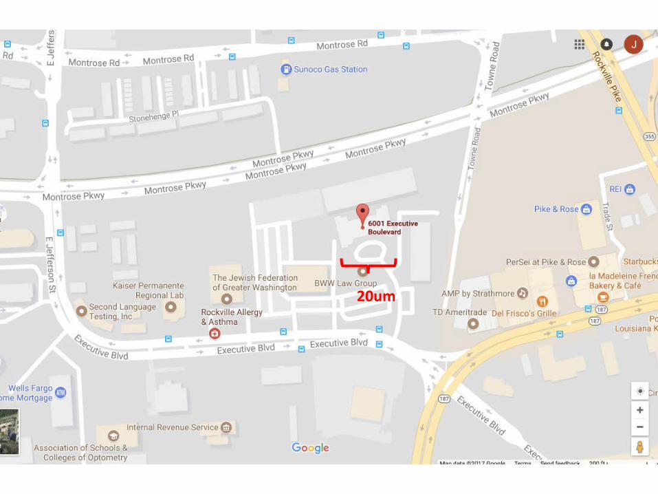
20um

105 scale

Breakout Sessions
• Material Standards and Benchmarks• Calibration, QC, and Instruments• Experimental Designs, Ontologies, and Metadata• Data Integration, Scale Alignment, and Data
Analysis
http://discourse.singlecellbiology.org