Nida Nosher 08030614-050 Topic: Amino acids Coding Codons Bs 7 th Presented to: Miss Aqsad.
25
Nida Nosher 08030614-050 Topic: Amino acids Coding Codons Bs 7 th Presented to: Miss Aqsad
-
Upload
frederica-sharp -
Category
Documents
-
view
219 -
download
1
Transcript of Nida Nosher 08030614-050 Topic: Amino acids Coding Codons Bs 7 th Presented to: Miss Aqsad.
Nida Nosher08030614-050
Topic: Amino acids Coding CodonsBs 7th
Presented to: Miss Aqsad

PROTEIN SYNTHESIS
TRANSLATION

The Genetic Code
The nucleotide sequence of mRNA contains three letter codons that specify all of the 20 amino acids found in proteins plus a signal to terminate protein synthesis
The order that the codons appear in the mRNA (5’ - 3’) directly dictates the order of the amino acids in the polypeptide chain of the protein (N - C termini)

Amino Acid DNA Base Triplets M-RNA Codons T-RNA Anticodons
alanine CGA, CGG, CGT, CGC GCU, GCC, GCA, GCG CGA, CGG, CGU, CGC
arginine GCA, GCG, GCT, GCC
TCT, TCCCGU, CGC, CGA, CGG
AGA, AGGGCA, GCG, GCU, GCC
UCU, UCC
asparagine TTA, TTG AAU, AAC UUA, UUG
aspartate CTA, CTG GAU, GAC CUA, CUG
cysteine ACA, ACG UGU, UGC ACA, ACG
glutamate CTT, CTC GAA, GAG CUU, CUC
glutamine GTT, GTC CAA, CAG GUU, GUC
glycine CCA, CCG, CCT, CCC GGU, GGC, GGA, GGG CCA, CCG, CCU, CCC
histidine GTA, GTG CAU, CAC GUA, GUG
isoleucine TAA, TAG, TAT AUU, AUC, AUA UAA, UAG, UAU
leucine AAT, AAC, GAA, GAG
GAT, GACUUA, UUG, CUU, CUC
CUA, CUGAAU, AAC, GAA, GAG
GAU, GAC
lysine TTT, TTC AAA, AAG UUU, UUC
methionine TAC AUG UAC
phenylalanine AAA, AAG UUU, UUC AAA, AAG
proline GGA, GGG, GGT, GGC CCU, CCC, CCA, CCG GGA, GGG, GGU, GGC
serine AGA, AGG, AGT, AGC
TCA, TCGUCU, UCC, UCA, UCG
AGU, AGCAGA, AGG, AGU, AGC
UCA, UCG
stop ATG, ATT, ACT UAA, UAG, UGA AUG, AUU, ACU
threonine TGA, TGG, TGT, TGC ACU, ACC, ACA, ACG UGA, UGG, UGU, UGC
tryptophan ACC UGG ACC
tyrosine ATA, ATG UAU, UAC AUA, AUG
valine CAA, CAG, CAT, CAC GUU, GUC, GUA, GUG CAA, CAG, CAU, CAC

Genetic code can be read in 3 ways depending upon where you start!
+1 frameshift
+2 frameshift
The genetic information encoded in each reading frame is different
DIF
FE
RE
NT
RE
AD
ING
FR
AM
ES
OF
m
RN
A T
HE
SA
ME
SE
QU
EN
CE
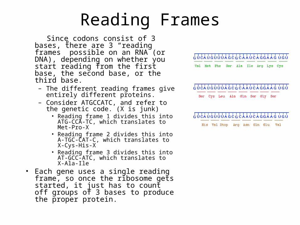
Reading Frames Since codons consist of 3 bases, there
are 3 “reading frames” possible on an RNA (or DNA), depending on whether you start reading from the first base, the second base, or the third base.– The different reading frames give
entirely different proteins.– Consider ATGCCATC, and refer to the
genetic code. (X is junk)• Reading frame 1 divides this into ATG-
CCA-TC, which translates to Met-Pro-X• Reading frame 2 divides this into A-TGC-
CAT-C, which translates to X-Cys-His-X• Reading frame 3 divides this into AT-
GCC-ATC, which translates to X-Ala-Ile • Each gene uses a single reading
frame, so once the ribosome gets started, it just has to count off groups of 3 bases to produce the proper protein.
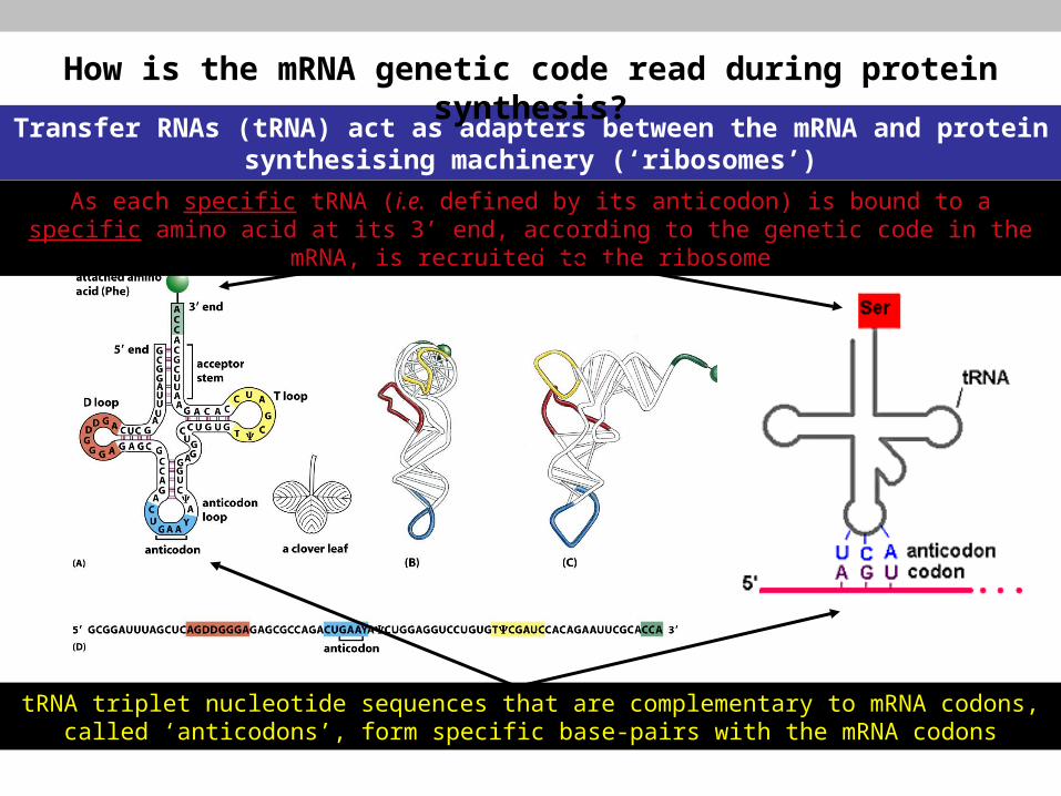
Transfer RNAs (tRNA) act as adapters between the mRNA and protein synthesising machinery (‘ribosomes’)
How is the mRNA genetic code read during protein synthesis?
tRNA triplet nucleotide sequences that are complementary to mRNA codons, called ‘anticodons’, form specific base-pairs with the mRNA codons
As each specific tRNA (i.e. defined by its anticodon) is bound to a specific amino acid at its 3’ end, according to the genetic code in the mRNA, is recruited to the ribosome
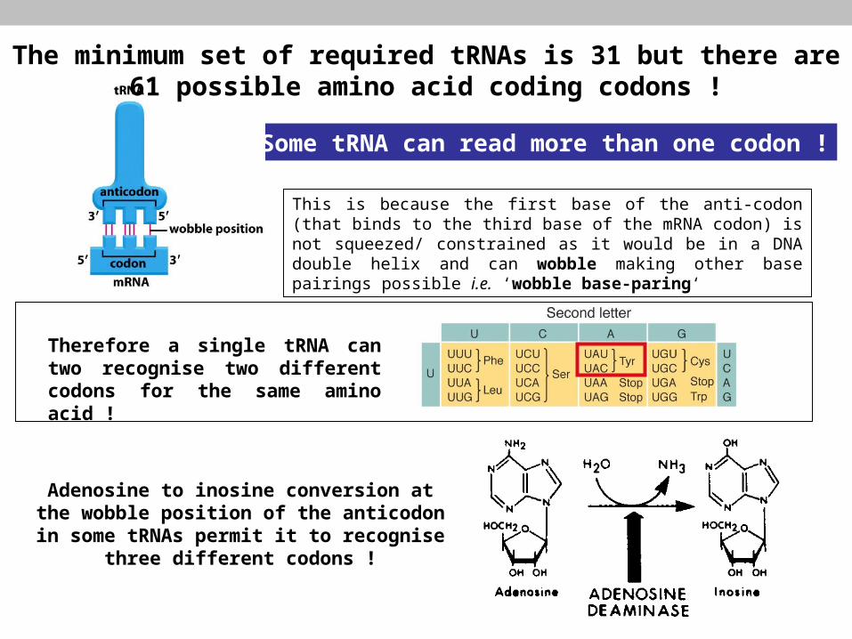
Some tRNA can read more than one codon !
Adenosine to inosine conversion at the wobble position of the anticodon in some
tRNAs permit it to recognise three different codons !
This is because the first base of the anti-codon (that binds to the third base of the mRNA codon) is not squeezed/ constrained as it would be in a DNA double helix and can wobble making other base pairings possible i.e. ‘wobble base-paring‘
The minimum set of required tRNAs is 31 but there are 61 possible amino acid coding codons !
Therefore a single tRNA can two recognise two different codons for the same amino acid !

Figure 6-58 Molecular Biology of the Cell (© Garland Science 2008)
Attachment of amino-acids to tRNAs (‘Charging’)
Each tRNA is charged by a specific enzymes that recognise both the tRNA and the amino acid - called ‘aminoacyl tRNA synthetases‘
e.g. tryptophanyl tRNA synthetase
Charging is a two step process
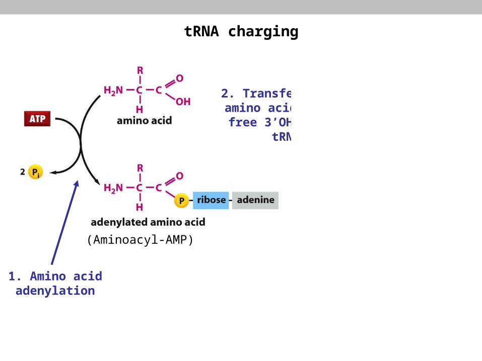
(Aminoacyl-AMP)
tRNA charging
Uncharged tRNA
Charged tRNA
2. Transfer of the amino acid to the free 3’OH of
the tRNA
1. Amino acid adenylation
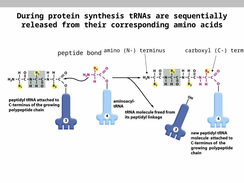
amino (N-) terminus carboxyl (C-) terminuspeptide bond
During protein synthesis tRNAs are sequentially released from their corresponding amino acids
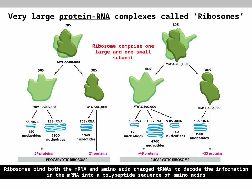
Very large protein-RNA complexes called ‘Ribosomes’
Ribosome comprise one large and one small subunit
Ribosomes bind both the mRNA and amino acid charged tRNAs to decode the information in the mRNA into a polypeptide sequence of amino acids

Prokaryotic 16S rRNA
Ribosomal RNA (‘rRNA’) critical to ribosome function
rRNAs:
• 2/3 of the molecular weight for ribosome (prokaryotes)
• form complex and defined secondary structure
• originally thought to have structural role, now known to required for most of the ribosome’s functions
• X-ray crystallography show no proteins are proximal to catalytic site to participate in peptide bond formation
• 23S rRNA (prokaryotes) acts as a ‘peptidyl transferase’ ribozyme
• sequence mutagenesis studies of 23S rRNA show its function is to correctly position the incoming charged tRNA to allow spontaneous formation of the peptide bond

Figure 6-64 Molecular Biology of the Cell (© Garland Science 2008)
3D ribosomal structure (70S prokaryotic)
The interface between large & small s/u’s form a groove for mRNA binding and three tRNA binding sites: A (acceptor), P (peptide) & E (exit)

Prokaryotic ribosomes
nnnnnnAGGAGGUnnnnnnnAUGnnnnnnn UCCUCCA
Shine-Delgarno sequence
start codon
16S rRNA base-pairing leads to small ribosomal s/u recognition, large s/u
recruitment and formation of the ‘70S initiation complex’
Correctly identifying the translation start-point in mRNA
Translation always starts at an AUG codon (coding for methionine) called the ‘start codon’
How does the ribosome
recognise the correct AUG as
the start codon ?
7bp
Shine-Delgarno sequence
Enables translation of polycistronic mRNAs
N-Formyl methionine charged tRNA is then recruited into the P-site ready for translation to start
mRNA
Various ‘initiation
factors (IFs)’ participate in this
process
Variations in the S-D sequence can effect translation initiation efficiency

The elongation phase of translation is essentially similar in prokaryotes
and eukaryotes involving a repetition of a series of steps
Elongation phase of translation
Charged tRNA enters A-site.
Specificity dictated by
codon-anticodon base-pairing
New peptide bond formation
(between adjacent amino acids in P
& A-sites)
Ribosome ‘translocates’ along mRNA to
next codon
Bound tRNAs move to next site (A-P or P-E)
As next charged tRNA enters A-site the E-site occupant
departs the ribosome
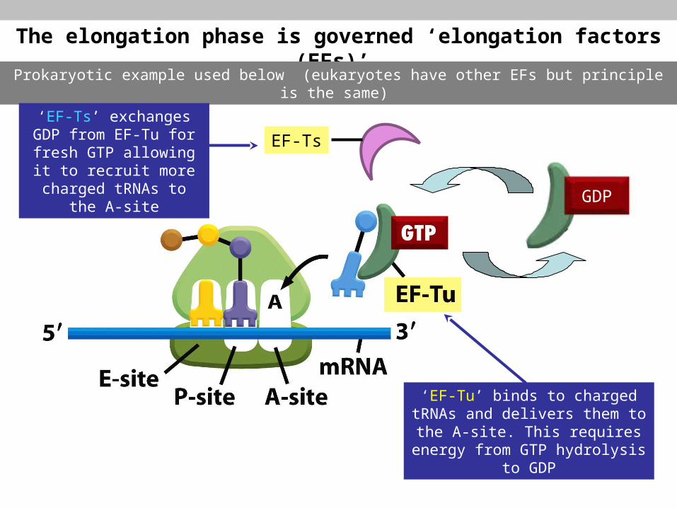
The elongation phase is governed ‘elongation factors (EFs)’
Prokaryotic example used below (eukaryotes have other EFs but principle is the same)
‘EF-Tu’ binds to charged tRNAs and delivers them to the A-site. This requires energy from GTP
hydrolysis to GDP
GDP
EF-Ts
‘EF-Ts’ exchanges GDP from EF-Tu for fresh GTP allowing it to recruit more charged tRNAs to the A-
site
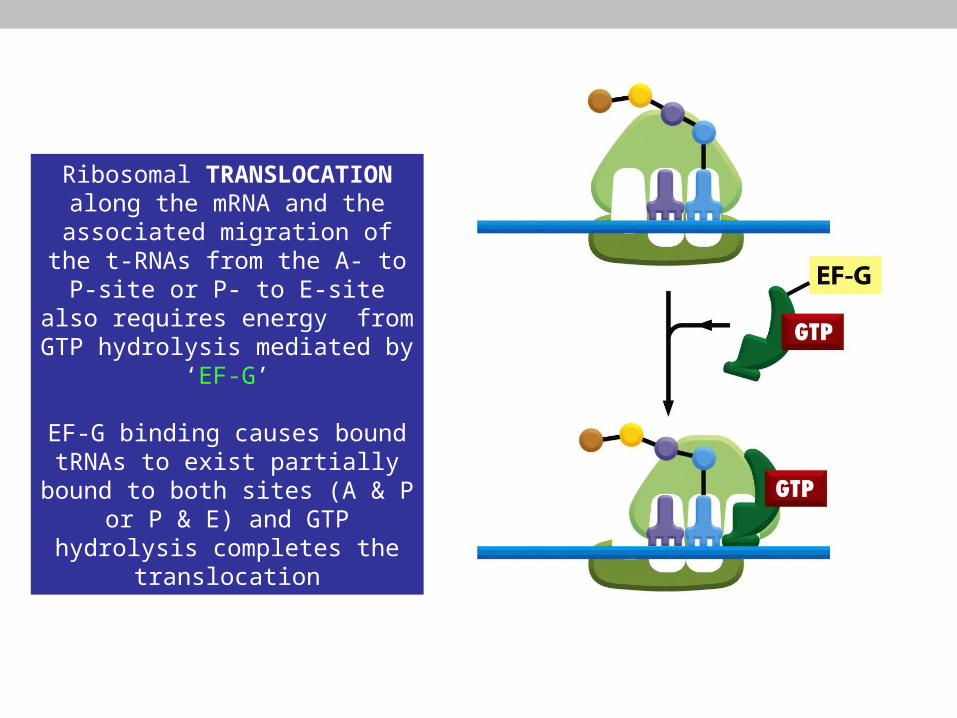
Ribosomal TRANSLOCATION along the mRNA and the
associated migration of the t-RNAs from the A- to P-site or P- to E-site
also requires energy from GTP hydrolysis mediated by ‘EF-G’
EF-G binding causes bound tRNAs to exist partially bound to both sites
(A & P or P & E) and GTP hydrolysis completes the
translocation
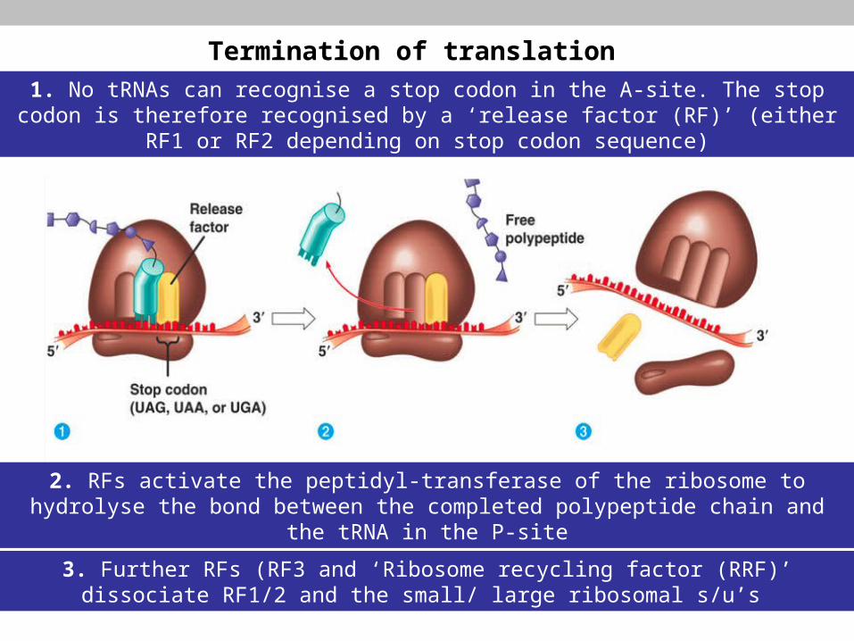
1. No tRNAs can recognise a stop codon in the A-site. The stop codon is therefore recognised by a ‘release factor (RF)’ (either RF1 or RF2 depending on stop codon
sequence)
Termination of translation
2. RFs activate the peptidyl-transferase of the ribosome to hydrolyse the bond between the completed polypeptide chain and the tRNA in the P-site
3. Further RFs (RF3 and ‘Ribosome recycling factor (RRF)’ dissociate RF1/2 and the small/ large ribosomal s/u’s
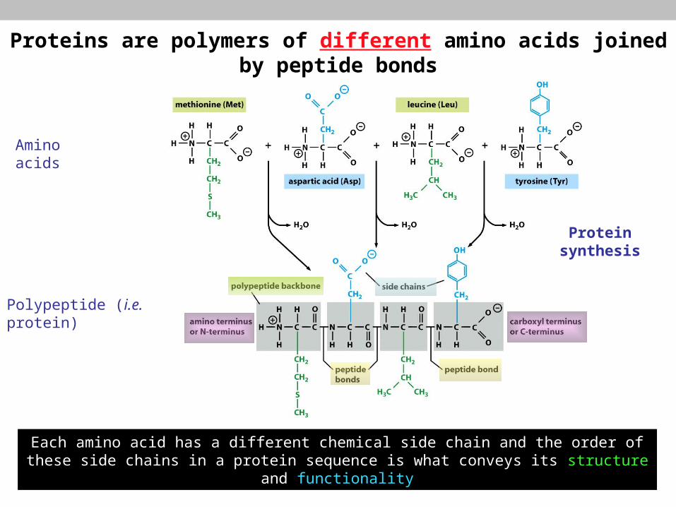
Proteins are polymers of different amino acids joined by peptide bonds
Each amino acid has a different chemical side chain and the order of these side chains in a protein sequence is what conveys its structure and functionality
Amino acids
Polypeptide (i.e. protein)
Protein synthesis
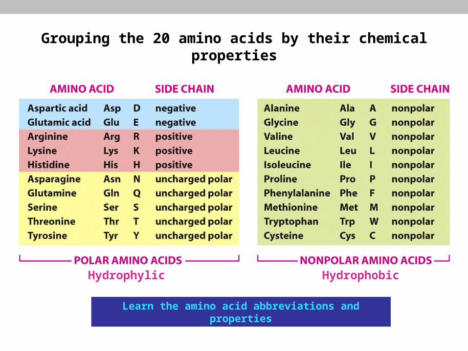
Hydrophylic Hydrophobic
Grouping the 20 amino acids by their chemical properties
Learn the amino acid abbreviations and properties

e.g. Heat Shock Protein 70 (Hsp70)
The expression of Hsps (heat shock proteins) increases as temperature increases because folded proteins are more likely to unfold/ denature at higher temperatures
1. Hsp70-ATP able to loosely bind hydrophobic patches of amino acids as they emerge from the
ribosome
2. Peptide binding induces intrinsic ATPase activity in
HSP70
3. Hsp70-ADP tightly associates with unfolded protein and
protects it from aggregating
4. Nucleotide exchange factors eventually replace the ADP with
ATP and HSP70 releases the unfolded protein
5. Protein spontaneously folds into correct confirmation
6. A small percentage of protein incorrectly folds
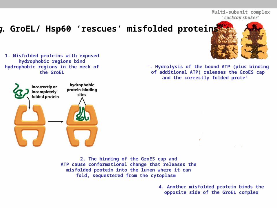
e.g. GroEL/ Hsp60 ‘rescues’ misfolded proteins
1. Misfolded proteins with exposed hydrophobic regions bind hydrophobic
regions in the neck of the GroEL
Multi-subunit complex ‘cocktail shaker’
2. The binding of the GroES cap andATP cause conformational change that releases the misfolded protein into the lumen where it can fold, sequestered from the
cytoplasm
3. Hydrolysis of the bound ATP (plus binding of additional ATP) releases the GroES cap and the
correctly folded protein
4. Another misfolded protein binds the opposite side of the GroEL complex
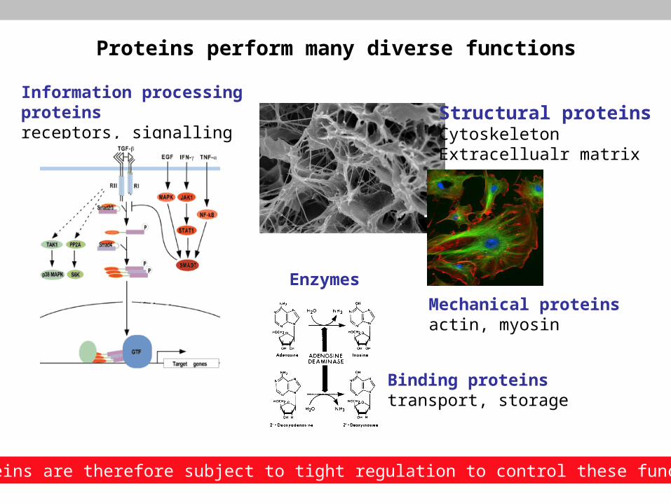
Structural proteinsCytoskeletonExtracellualr matrix
Mechanical proteinsactin, myosin
Enzymes
Binding proteinstransport, storage
Information processing proteinsreceptors, signalling
Proteins perform many diverse functions
Proteins are therefore subject to tight regulation to control these functions

Any Question?